Article

A Preliminary Investigation into the Degradation of Asbestos Fibres in Soils, Rocks and Building Materials Associated with Naturally Occurring Biofilms

1
School of Future Environments, Auckland University of Technology, Auckland 1142, New Zealand
2
Environmental Innovation Centre, Auckland 1025, New Zealand
3
Applied Molecular Solutions Research Centre, Unitec Institute of Technology, Auckland 1025, New Zealand
4
Interdepartmental Centre for Studies on Asbestos and Other Toxic Particulates, University of Torino, 10125 Torino, Italy
5
Auckland Museum Tāmaki Paenga Hira, Auckland 1010, New Zealand
*
Authors to whom correspondence should be addressed.
Minerals 2024, 14(1), 106; https://doi.org/10.3390/min14010106
Submission received: 22 November 2023 / Revised: 9 January 2024 / Accepted: 16 January 2024 / Published: 19 January 2024

Abstract

:
Bioremediation utilizes living organisms such as plants, microbes and their enzymatic products to reduce toxicity in xenobiotic compounds. Microbial-mediated bioremediation is cost effective and sustainable and in situ application is easily implemented. Either naturally occurring metabolic activity can be utilized during bioremediation for the degradation, transformation or accumulation of substances, or microbial augmentation with non-native species can be exploited. Despite the perceived low potential for the biological degradation of some recalcitrant compounds, successful steps towards bioremediation have been made, including with asbestos minerals, which are prevalent in building stock (created prior to the year 2000) in New Zealand. Evidence of the in situ biodegradation of asbestos fibres was investigated in samples taken from a retired asbestos mine, asbestos-contaminated soils and biofilm or lichen-covered asbestos-containing building materials. Microbial diversity within the biofilms to be associated with the asbestos-containing samples was investigated using internal transcribed spacer and 16S DNA amplicon sequencing, supplemented with isolation and culturing on agar plates. A range of fungal and bacterial species were found, including some known to produce siderophores. Changes to fibre structure and morphology were analysed using Transmission Electron Microscopy and Energy-Dispersive X-ray Spectroscopy. Chrysotile fibrils from asbestos-containing material (ACMs), asbestos-containing soils, and asbestos incorporated into lichen material showed signs of amorphisation and dissolution across their length, which could be related to biological activity.

1. Introduction

Modern landfills were designed to safeguard human health from hazards associated with municipal solid waste [1]. Whilst providing public health protection in the short-term, landfills can still pose a long-term risk made worse by environmental and human-derived change to land-use and the climate (Wallis et al., 2020) [2]. Composting, incineration and recycling are among the available alternatives for waste disposal, but typically, these are only suitable for non-hazardous and/or organic substances. Hazardous and inorganic waste is often considered untreatable.
One such persistent, inorganic waste stream is derived from asbestos, which is a globally ubiquitous hazard within the built environment. Asbestos comprises six types of elongate mineral particles (EMPs) corresponding to specific dimensional and morphological criteria [3]. Responsible for health issues including pleural abnormalities, bronchogenic carcinomas, asbestosis and malignant mesothelioma (MM), asbestos has been well re-searched [4,5,6]. Despite this, significant exposure continues to occur throughout the world, potentially affecting 125 million people in the workplace [7]. Although the majority of diagnosed MM cases over the last 50 years have been attributed to occupational exposure, a significant number of cases have arisen via other pathways, including para-occupational, domestic and environmental exposure [8].
Asbestos is commonly combined within a building material matrix, such as concrete, and is rarely disposed of in its raw (unbonded) fibrous form [9]. This greatly increases the volume going to landfill, which is estimated (using a conservative average asbestos concentration of 5% within the building product matrix) to be around 4 billion tonnes [9]. However, the volumes of contaminated soil that must be disposed of, plus the associated contaminated waste, are likely to exceed this estimate because safety limits for asbestos in soils can be as low as 10 mg/kg [10]. Currently, there is no long-term unified approach to address increasing waste volumes or the legacy of multiple sites of marked (or unmarked) contaminated land.
Although physico-chemical treatments have been developed to treat asbestos waste, high energy requirements and health and safety issues [11], combined with potentially high soil volumes, have presented significant limitations. Research has shown that microorganisms, such as fungi and lichens can, to some extent, degrade asbestos fibres, scavenging energy for microbial metabolism and extracting cations [12,13,14]. Observations that fungi and lichens spontaneously develop on asbestos rich substrates (Figure 1 and Figure 2), such as serpentinite rocks [15], suggest that this might be a viable option for bioremediation.
A growing body of research has investigated the mechanisms by which this process could work. The production of iron-chelating siderophores and/or organic acids by fungi can complex mineral constituents of rocks, including different forms of asbestos [14,16,17]. For example, Daghino et al. [18] reported that Fusarium oxysporum extracted iron from asbestos fibres, and later found that some fungal species isolated from chrysotile mines removed iron from the fibres through the production of siderophores [16]. In controlled laboratory studies, cultures of the fungi Fusarium oxysporum and Verticillium leptobactrum, which had been isolated from serpentinic rocks, were found to extract iron from asbestos fibres [18,19]. Further experiments demonstrated the chelating activity of exudates from some fungi (and lichens) leads to the subsequent modification of the chemical composition of chrysotile fibres in vitro. These modifications affected chemical reactivity and structure, potentially altering toxicity. More recently, Mohanty et al. [17] performed experiments on crushed chrysotile fibres with environmentally realistic concentrations of three different organic acids and fungal and bacterial siderophores. Both the bacterial and fungal siderophores significantly reduced iron on the surface of and within the chrysotile fibres, but the organic acids did not. These studies have been trialled on pure asbestos materials; however, it is less clear how effective these iron-removing processes would be when asbestos fibres are dispersed in soil (or in ACM).
This paper presents the results of a preliminary study of asbestos fibre integrity in samples where asbestos fibres are dispersed in soil, embedded in biofilm-coated building materials, incorporated into lichen tissue or in a raw state from an asbestos mine.

2. Materials and Methods

2.1. Asbestos Identification and Biofilm Sampling Procedures

Ten samples were collected for analysis, from a natural asbestos deposit contaminated soils and asbestos-containing materials, including roofing, cladding and fragments of lichens from asbestos roofing samples (Table 1). In this paper, we will use the mineralogical term “fibre” when referring to minerals with highly elongated morphology developed during growth. The term “fibril” will be used when single crystal asbestos minerals can be distinguished.

2.1.1. Natural Deposits

Samples of biofilms and soil were taken following approved health and safety protocols (Section 2.5) during a site visit to a disused asbestos mine in Kahurangi National Park, South Island, New Zealand (Lat 41.123107/Long 172.699993, Figure 1 and Figure 2). Rocks bearing asbestos fibres at the mine site were identified visually (Figure 3A), and most of these were found to have a black-brown or orange biofilm growing on them (Figure 3B). Research was carried out with the support of indigenous tribal authorities (Mana Whenua ki Mōhua (Ngāti Rārua, NgātiTama and Te Ātiawa), and with permission from the New Zealand Department of Conservation (permit # 93501-GEO).
Biofilms were moistened with sterile water to make sampling easier and to prevent fibres from becoming airborne. Sterile cotton swabs were rubbed across the biofilm surface to collect microbial material. These swabs were placed in separate labelled plastic bags and stored at 4 °C for less than 36 h. Swabs for DNA extraction were then frozen at −80 °C, and swabs for culturing were kept refrigerated. In addition, small samples of asbestos-containing rock were collected for later analysis. All samples were double-bagged and stored in a third bag.
Sub-samples of the raw fibres were sent to IANZ (International Accreditation New Zealand)-accredited Focus Analytics Laboratories for formal analysis of fibre type and also couriered to the University of Torino (Università di Torino, UNITO), Italy, for further structural and morphological analysis.

2.1.2. Asbestos-Containing Materials

Externally located asbestos-containing materials were sampled and swabbed from pre-identified sites by licensed removalists whose clients had agreed to the use of their samples for further testing. The swabs were frozen for later DNA extraction, refrigerated for later culturing or further analysed within 24 h of sampling within a negative pressure unit (NPU, WAYSAFE® SA1000 Sample Analysis and Preparation Safety Enclosure, Ashton-under-Lyne, UK). Two lichen samples from ACM were carefully peeled off their substrate and identified within the NPU. The remaining samples of ACM or lichen material were furnaced at 400 °C, to remove any organic content at the Focus Analytics Laboratory from which another sub-sample (at least 10 g of material) was transferred to facilities at UNITO for fibre characterisation.

2.1.3. Asbestos Contaminated Soils

Asbestos-contaminated soil samples were taken from pre-identified sites by licensed removalists whose clients had agreed to the use of their samples for further testing. A small sub-sample of 0.25 g was extracted within the NPU and frozen within 24 h for later DNA extraction. Once a sub-sample had been taken, the remaining soil sample was dried (at 110 °C) and then furnaced (at 400 °C) to remove organic fibres at the Focus Analytics Laboratory, from which another sub-sample (at least 10 g of material) was transferred to UNITO for fibre characterization.

2.2. Asbestos Fibre Identification

All samples (bulk materials and soil) were formally analysed for asbestos fibres by Focus Analytics, according to Standard AS4964 [20].

2.3. Asbestos Fibre Characterisation

To preserve the original morphology and the crystallinity degree as much as possible, the samples for preliminary observation were hand ground in an agate mortar for one minute, and then transferred to a TEM copper grid. A thorough investigation of samples showed that a 1 min hand grinding was not sufficient to observe the mineral particles in sufficiently high numbers and detail. Subsequently, all samples were hand-ground for 10 min, using an agate mortar and pestle. This procedure did not affect the crystal structure or the morphology of the fibres. The suspension containing the ground particles was transferred onto carbon-coated copper grids. Once the grids were loaded on a double-tilt sample holder, the holder was left within the column for 30 min before the exposure to the electron beam was begun to reduce beam damage [21,22,23]. An electron beam damage test was performed on each sample since chrysotile is particularly beam-sensitive and recognizable morphological transformations can be induced at this stage. A TEM CM12 operating (Philips, Amsterdam, The Netherlands) at 120 kV was used to collect dimensional, structural, morphological and chemical data from the samples. Pictures were taken at 10,000× to clearly distinguish bundles and detect particle regions with morphological modifications. The qualitative chemical composition of each observed fibril was confirmed through a semi-quantitative Energy-Dispersive X-ray Spectroscopy spot analysis. Selected Area Electron Diffraction was performed to confirm the identity of the particles. The photographic film exposed within the TEM CM12 was developed and fixed in a dark room, scanned and digitalised. The measurement was performed on the converted digital images using ImageJ-FIJI (Version 2.9.0) [24]. Results are summarized in Table 1.

2.4. Identification of Organisms Found in Association with Asbestos-Containing Materials

Three different methods were used to identify organisms associated with the asbestos samples. Lichens were visually identified (by DB). DNA was extracted from samples of biofilms from asbestos-containing rock, asbestos-containing building materials and asbestos contaminated soils using either DNeasy® Plant Mini Kits (Qiagen, Germantown, MD, USA) or NucleoSpin® (Machery-Dagel, Düren, Germany) Soil Kits, followed by internal transcribed spacer and 16S amplicon DNA sequencing to identify bacteria and fungi. A subset of these biofilm samples was used to isolate and culture fungi and identify resulting pure cultures using DNA barcoding.

2.4.1. DNA Extraction and 16S and ITS Amplicon Sequencing

DNA extractions were carried out inside a negative pressure unit, following safety protocols for the handling of asbestos-containing materials as recommended by the Health and Safety Executive [25]. DNA was extracted from swabs collected from biofilms from asbestos-containing building materials (ACM) and asbestos-containing rocks in asbestos mine tailings (Mine) using DNeasy® Plant Mini Kits (Qiagen). The cotton tips of the swabs were cut off and incubated in AP1 Buffer for three hours at 65 °C. The extraction was then carried out following the manufacturer’s instructions, with DNA being eluted into 50 µL of AE buffer. DNA was not extracted from the soil samples examined in this paper. To identify the diversity of fungi and bacteria present within each sample, a 500–600 bp section of the internal transcribed spacer (ITS) region was amplified via PCR using the primers ITS1F, 5′-CTT GGT CAT TTA GAG GAA GTA A and ITS2 5′-GCT GCG TTC TTC ATC GAT GC [26], and a 290–295 bp section of the bacterial 16S ribosomal RNA region was amplified using the primers 515F, 5′-GTG YCA GCM GCC GCG GTA A, and 806R, 5′-GG ACT ACN VGG GTW TCT AAT [27]. PCRs were performed in 25 µL reactions containing 13 µL of ultra-pure water, 0.5 µL of each primer (10 µM), 10 µL of Platinum Green Hot Start DNA Polymerase (Thermofisher Scientific, Waltham, MA, USA) and 1 µL of DNA template (10 ng/µL). For the ITS region, DNA was denatured at 95 °C for 2 min, followed by 40 cycles of 95 °C for 30 s, 48 °C for 30 s and 72 °C for 1 min, with a final extension period of 10 min at 72 °C. For 16S, DNA was denatured at 94 °C for 3 min, followed by 35 cycles of denaturation at 94 °C for 45 s, annealing at 50 °C for 60 s and extension at 72 °C for 90 s, with a final extension period at 72 °C for 10 min. PCR products were run on a 2% agarose gel and visualized using an Uvidoc HD6 (Uvitec, Cambridge, UK) to confirm that amplification was successful. ITS and 16S PCR amplicons from each sample were pooled, purified using AMPure XP magnetic beads (Beckman Coulter, Indianapolis, IN, USA) and eluted into ultrapure water prior to Illumina MiSeq sequencing at Massey Genome Service (Massey University, Palmerston North, New Zealand).
Illumina MiSeq 300 bp paired-end reads were processed for quality using a standard Illumina sequence analysis pipeline. Reads were trimmed to their longest contiguous segment for which quality scores were less than a quality cutoff (set at 0.01), using the dynamictrim application from the SolexaQA++ software package (http://solexaqa.sourceforge.net/, accessed on 26 January 2023) with further processing performed in R v4.2.2. ITS, and 16S reads were demultiplexed using the function filterFastq in the ShortRead [28] and fastqPairedFilter packages in dada2 Version 1.0.3 [29]. Sequences were then processed further following the dada2 amplicon sequencing workflow. Primer sequences were removed using Cutadapt v.4.1, and paired-end reads were merged and chimeric sequences removed using the BimeraDenovo function. Amplicon sequence variants’ (ASVs) identities were assigned using the National Center for Biotechnology Information (NCBI) BLASTn algorithm. Identification at the species level was made if there was a match of 99% or above to one or more reference sequences for that species, with no other close matches. Where two or more species were a 100% match, the sequence was assigned an identification at the genus level. Where there were no close matches to one species, but multiple matches (>95%) to species in different genera, identification was assigned at the family level.

2.4.2. Culturing of Fungi and DNA Barcoding

Swabs were streaked over three agar plates in succession (Potato dextrose agar with chloramphenicol, Fort Richard, Auckland, New Zealand) in an NPU to isolate and culture fungi. Cultures were grown for one to two weeks at 21 °C before any morphologically distinct colonies were sub-cultured in the NPU to isolate pure cultures of each fungal species. Following this, DNA was extracted using a Qiagen DNeasy Plant Mini Kit, following the manufacturer’s instructions. The ribosomal DNA internal transcribed spacer region (ITS) was amplified and sequenced using primers ITS1f [30] and ITS4 [31]. PCR products were purified using the Qiagen MinElute PCR purification kit. Sequencing was performed by the Massey Genome Service (Massey University, Palmerston North, New Zealand) using an Applied Biosystems model 3730 automated capillary DNA sequencer, using the same primers used for PCR. DNA sequences were assembled, edited and aligned using Geneious® 11.0.5 software and identified using the National Center for Biotechnology Information (NCBI) BLASTn algorithm.

2.5. Health and Safety

In NZ, there are no standard health and safety guidelines available for asbestos analysis that lie outside of procedures covered by IANZ accreditation (for asbestos laboratories offering asbestos sample identification). Therefore, comprehensive sampling and analytical procedures were created based on the British Occupational Hygiene Society (BOHS) courses IP402 Surveying and Sampling Strategies for Asbestos in Buildings, IP404 Air Monitoring, Clearance Inspections and Reoccupation Following the Removal of Asbestos and IP405 Management of Asbestos in Buildings and IANZ Chemical Testing Programme [32].

3. Results

Six soil samples were found to contain asbestos bundles and were suitable for analysis. Locating asbestos bundles in contaminated soil samples can be problematic because (a) their distribution is heterogeneous and (b) permission was required from site owners to formally identify the presence of the fibres, which was not always provided. Samples of ACM and from the chrysotile mine were also found to contain asbestos. Table 1 shows sample type, sample size and asbestos fibre type (s) identified. Where amosite was found, it was 1% of the total fibre sample.

3.1. Asbestos Characterisation and Fibril Modification

Samples of asbestos-containing materials (including amosite and chrysotile), chrysotile bound in serpentinite rocks and free particles of chrysotile in soil were analysed by the Department of Earth Sciences, University of Torino (UNITO), Italy. The samples included roofing material (Super Six), shed flashing and cladding for which there were clearly visible areas of lichen colonisation.

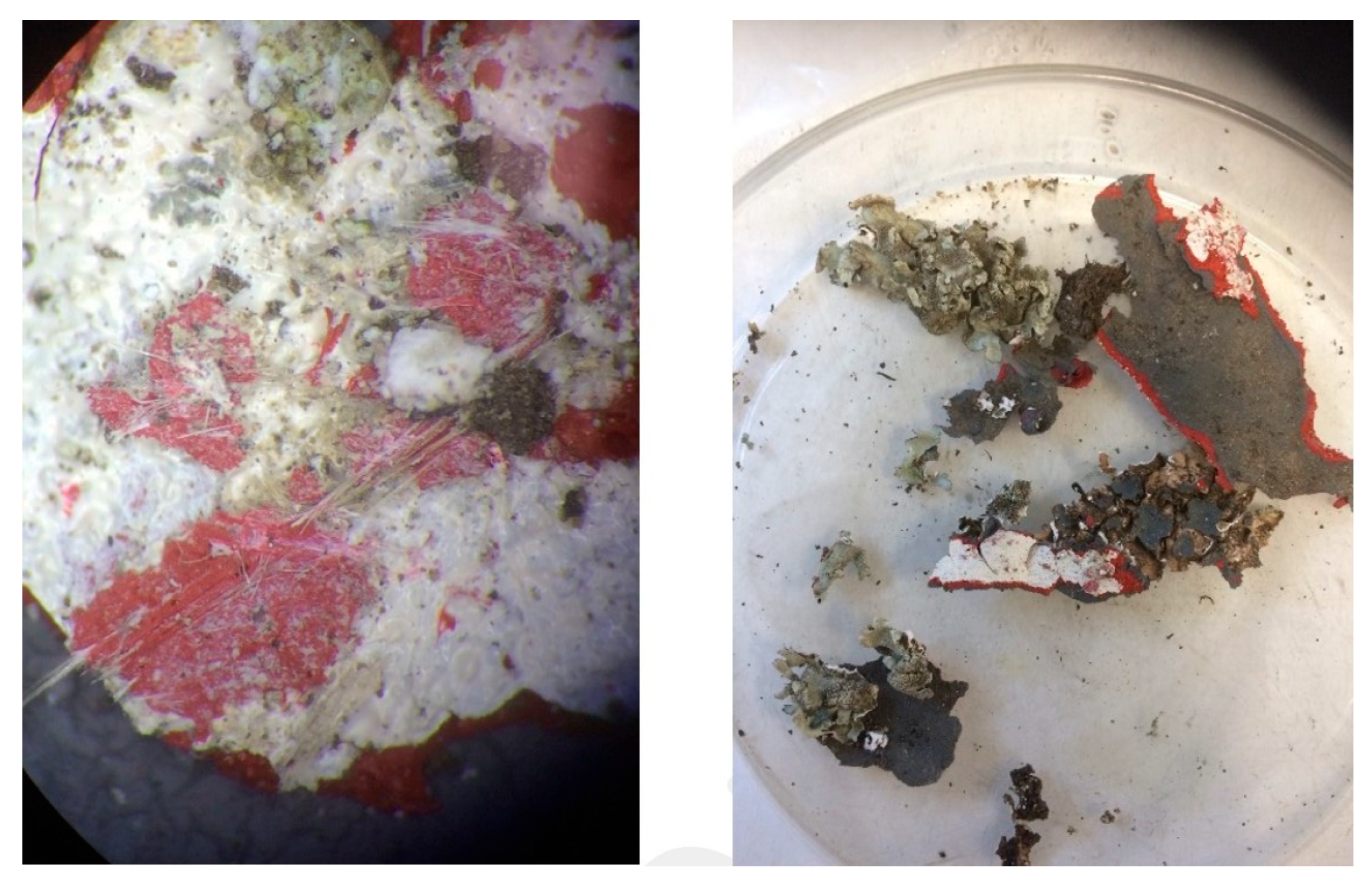
3.1.1. Lichen Material from ACM Samples and ACM Samples

Bright-field TEM observations suggest that chrysotile fibrils in these samples of ACM and from within the lichen material, underwent partial modification. The fibril transformation consisted of surface (and possibly bulk) amorphization and evidence of dissolution at the fibril apex (Figure 4). Amorphization and morphological transformations can be induced by the electron beam during TEM observations; however, the formation of “bubble-like” structures along the fibril induced by the electron beam occurred during the beam damage testing, but did not occur during analytical observations. Furthermore, the amorphization induced by the beam damage testing did not allow for the observation of diffraction spots, while diffraction spots were recognizable during analysis.

3.1.2. Asbestos from Serpentinite Rocks

The serpentinite rocks samples were characterised by chrysotile fibrils only. No other asbestos or EMPs were found. The fibrils were usually collected in bundles with a parallel to nearly parallel arrangement (Figure 5—left panel). The fibrils were usually straight, but were occasionally visibly curved (Figure 5—right panel), and were generally unmodified in terms of morphology and crystallinity degree.

3.1.3. Asbestos Contaminated Soils

The soil samples were mostly composed of chrysotile fibrils, soil remains and amphibolic fibres. Fibrils were found both in disorganized bundles mixed with soil remains (Figure 6—left panel), as well as straight isolated fibrils (Figure 6—right panel). Fibrils were found to be partially modified in terms of morphology and crystallinity degree.

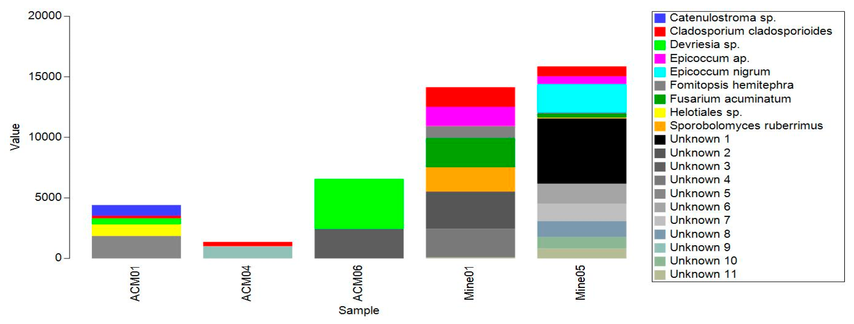
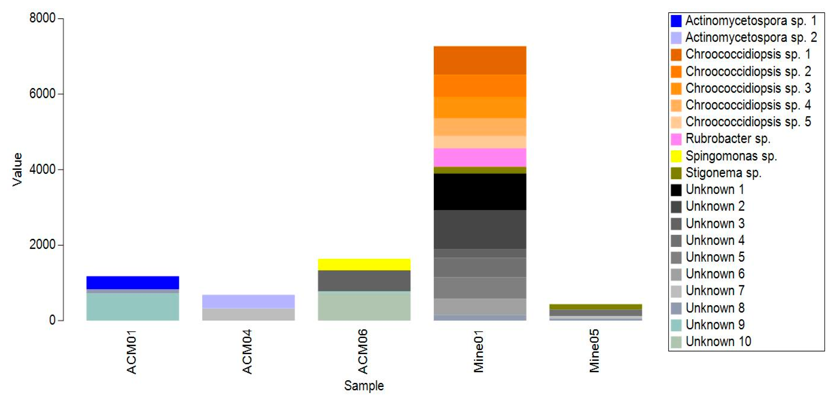
3.2. Microbial Identification

3.2.1. Lichen Material Taken from ACM Samples

Two of the samples contained lichen material removed from old Super Six asbestos-based roof material (Figure 7) and the lichen in both samples was identified as Xanthoparmelia scabrosa.

3.2.2. ACM Samples

A range of different fungi and bacteria were identified from ACM biofilms using Amplicon DNA sequencing (Figure 8 and Figure 9). Fungi included species of Catenulostroma, Cladosporium and Devriesia. Bacteria included species of Actinomycetospora and Sphingomonas. Around half of the fungal and bacterial sequences did not have close matches on Genbank. One sample from ACM (ACM06) was also inoculated on PDA plates, and three fungal species (Cladosporium cladosporioides, Leucosporidium scottii and Pseudopithomyces chartarum were successfully isolated and cultured.

3.2.3. Asbestos Bundles from Serpentinite Rock

Biofilms on samples from the asbestos mine were mainly made up of cyanobacteria and fungi (Figure 8 and Figure 9). Amplicon DNA sequencing indicated that the main fungi present were species of Cladosporium, Epicoccum, Fusarium, Fomitopsis and Sporobolomyces. The bacteria were mainly different isolates of the cyanobacteria Chroococcidiopsis and Stigonema, and the bacterium Rubrobacter. Around half of the fungi and bacteria detected did not have a close match on Genbank. One sample (Mine01) was also inoculated onto PDA plates, with Cladosporium cladosporioides, unknown species of Biscogniauxia and Crustomyces and unidentified isolates from the Pezizales and Hymenochaetales being successfully isolated and cultured.

3.2.4. Asbestos Contaminated Soils

None of the soil samples sent for asbestos particle analysis were successfully DNA sequenced or cultured.

4. Discussion

The finding that chrysotile fibrils from ACM, associated with biofilms, lichens growing on ACM and asbestos-contaminated soil, show signs of degradation is promising.
The modification of chrysotile fibrils that were found to be incorporated into the tissues of the lichen Xanthoparmelia scabrosa is consistent with previous research on the related species X. tinctina [12,13]. It was found that X. tinctina was able to selectively remove magnesium ions from chrysotile fibres, and further follow-up studies with the isolated mycobiont from X. tinctina found that it removed magnesium from the chrysotile fibrils through the production of oxalic acid. Another possibility is that microorganisms present within or under the lichen thallus may be responsible for fibre modification. Lichens are known to host complex microbiomes of bacteria and filamentous fungi and yeasts [33,34], some of which may produce siderophores and organic acids, and further research will be carried out to explore this.
ACM samples also showed signs of chrysotile amorphisation. Some of the organisms found in the biofilms growing on the ACM samples have been reported in the literature as being able to produce siderophores. For example, Leucosporidium scottii is known to produce siderophores in culture [35] and potentially chelate iron. Cladosporium cladosporioides has previously been isolated from serpentinite and was also found to produce iron-chelating siderophores (ferricrocin) in culture [16].
The two samples of chrysotile from the asbestos mine in Kahurangi National Park show little sign of modification, despite contact with biofilms that contained fungal species such as Cladosporium cladosporioides and Epicoccum nigrum that have previously been reported to produce siderophores [16,36]. The site receives a high level of rainfall annually [37], and is exposed to the sun, so if weathering was responsible for the modification of asbestos, then it should be found in these samples. One explanation is that the asbestos particles are continuously being eroded and are relatively newly exposed to both the weather and developing biofilms, and so not enough time has elapsed for the fibres to be modified. Another possibility is that fibres from the centre of bundles were analysed, and that they may be less exposed to weathering and bioweathering. The two samples had similar numbers of fungal ASVs, but one sample (Mine05) had far fewer cyanobacterial ASVs than the other sample (Mine01). This is most likely due to variability in biofilm composition and structure on the serpentinite rocks sampled from different locations within the site.
Asbestos-contaminated soil samples also showed indications of fibril amorphization. Unfortunately, no DNA sequence data were available for these particular samples, but other asbestos-contaminated soil samples from the same areas were sequenced by Doyle et al. [38], who identified a range of fungal and bacterial species as being present, including Cladosporium cladosporoides, Epicoccum nigrum, Fusarium oxysporum and Yarrowia lipolytica, all of which have been reported in the literature as being able to produce siderophores in vitro [16,36,39,40]. Previous research by Martino et al. [19] found that several soil fungi were able to remove iron from crocidolite fibres in vitro. The removal of magnesium and calcium ions by soil fungi and bacteria has also been identified as a mechanism for the degradation of chrysotile in vitro. Borges et al. [41] found that cultures of the fungus Aspergillus niger, the bacterium Acidithiobacillus thiooxidans and the acids they produce (gluconic, citric, oxalic and sulphuric) were able to degrade loose chrysotile fibrils, and ground asbestos cement in laboratory experiments. These results are a positive indication that it may be feasible to remediate asbestos-contaminated soils in situ.
The finding of chrysotile and amosite fibrils in two lichen samples from an ACM roof is concerning. The lichen material was initially attached to pieces of ACM roof found around a building. Two layers of paint physically separated the lichen and the ACM. This suggests that either the lichen was able to access asbestos fibres through two layers of paint, or that the painted roof surface had been disrupted in some way to expose friable amosite and chrysotile fibres that were subsequently incorporated by the lichen. Asbestos-containing building materials are often encapsulated with stabilizing paint products, but the performance of these is reduced if the product has a low binding effectiveness, or the roof surface has already deteriorated. In addition, visual evaluation of ACM for signs of deterioration can be misleading unless fibres are clearly visible on the surface [42].
Further research will focus on confirming whether the samples were subjected to bioweathering, chemical-physico weathering or a combination of both. This research will also seek to establish the likely timeframe over which in situ degradation may occur. The impact of weathering on the toxicological properties of the degraded asbestos will also require careful investigation. As toxicological comparisons between different mineral fibres (e.g., crocidolite versus chrysotile) are performed based on equal mass of fibres [43], it is important that any future toxicological tests made on degraded asbestos from trials accurately represent what would actually be deposited in the lung during inhalation.

5. Conclusions

Samples of asbestos fibres taken from soils, biofilm-covered ACM and from within lichens growing on ACM showed promising signs of degradation in those contexts, while raw fibres taken from an asbestos mine showed no indications of degradation. This degradation may be the result of chemical, physical or bioweathering and further research is required using more examples of in situ degradation. In addition, more advanced characterisation techniques such as low-dose electron microscopy and Electron Energy-Loss Spectroscopy should be used to further investigate fibril transformation.
Organisms sampled from ACM included Leucosporidium scottii and Cladosporium cladosporioides which have both been previously identified as producing siderophores. Those from raw asbestos fibres extracted from the asbestos mine were Cladosporium cladosporioides and Epicoccum nigrum. Although DNA sequence data were not available for the soil samples, previous research from the same set of sites isolated Cladosporium cladosporoides, Epicoccum nigrum, Fusarium oxysporum and Yarrowia lipolytica. Lichen material taken from ACM was found to contain asbestos fibres, indicating fibre liberation from the substrate. This requires further research to better control a potential exposure pathway to protect human health.
These preliminary investigations show promising signs for the in situ bioremediation of asbestos- gcontaminated soils, However, more research is required to establish degradation timeframes and the mechanisms responsible. It is also essential that degraded fibres are further investigated for associated changes in toxicological response.

Author Contributions

Conceptualization, T.-A.B., S.W., R.V. and D.B.; methodology, T.-A.B., R.V., E.B. and D.B.; formal analysis, R.V., E.B. and E.D.; investigation, D.B., E.D., P.d.L., R.V. and E.B.; resources, T.-A.B., S.W. and G.S.; data curation, E.D.; writing—original draft preparation, T.-A.B., S.W., G.S., R.V., E.B. and D.B.; writing—review and editing, T.-A.B., S.W., P.d.L., G.S., R.V., E.B. and D.B.; visualization, R.V., E.D. and D.B.; supervision, T.-A.B.; project administration, T.-A.B.; funding acquisition, T.-A.B. All authors have read and agreed to the published version of the manuscript.

Funding

Financial support for this project was received from the Waste Minimisation Fund (grant number 22917), which is administered by the Ministry for the Environment. The Ministry for the Environment does not necessarily endorse or support the content of this publication in any way.

Data Availability Statement

Raw reads of the biofilm sample ITS and 16S rRNA gene amplicons have been deposited in the NCBI Sequence Read Archive (SRA) and are available under the Project accession number PRJNA940522.

Acknowledgments

We would like to thank Mana Whenua ki Mōhua (Ngāti Rārua, Ngāti Tama and Te Ātiawa) for supporting our research and giving us permission to take samples from the disused asbestos mine in Kahurangi National Park. We would also like to thank the New Zealand Department of Conservation for granting a permit to do research in Kahurangi National Park, 4SIGHT Consulting and Forensic Specialists for collecting asbestos samples from Auckland, and Focus Analytics for confirming the presence of asbestos in collected samples. We thank Sarah Wells for assistance with data analysis.

Conflicts of Interest

The authors declare no conflicts of interest.

References

  1. Kumar, S.; Chiemchaisri, C.; Mudhoo, A. Bioreactor landfill technology in municipal solid waste treatment: An overview. Crit. Rev. Biotechnol. 2011, 31, 77–97. [Google Scholar] [CrossRef] [PubMed]
  2. Wallis, S.; Lemckert, C.; Hardy, R.; Berry, T.A. Disposal or Treatment: Future considerations for solid waste from the construction and demolition industry. In WIT Transactions on Ecology and the Environment; Wessex Waste Management: Southhampton, UK, 2020; Volume 247, pp. 171–183. [Google Scholar]
  3. National Institute for Occupational Safety and Health (NIOSH). Asbestos Fibers and Other Elongate Mineral Particles: State of the Science and Roadmap for Research, Revised Edn., Department of Health and Human Services, DHHS (NIOSH) Publication No. 2011–159, 2011, Current Intelligence Bulletin, 62, 1–159. Available online: https://www.cdc.gov/niosh/docs/2011-159/default.html (accessed on 16 November 2022).
  4. Jablonski, R.P.; Kim, S.J.; Cheresh, P.; Kamp, D.W. Insights into mineral fibre-induced lung epithelial cell toxicity and pulmonary fibrosis. In Mineral Fibres: Crystal Chemistry, Chemical-Physical Properties, Biological Interaction and Toxicity; Mineralogical Society: Washington, DC, USA, 2017; pp. 447–500. [Google Scholar]
  5. Vigliaturo, R.; Jamnik, M.; Dražić, G.; Podobnik, M.; Žnidarič, M.T.; Ventura, G.D.; Redhammer, G.J.; Žnidaršič, N.; Caserman, S.; Gieré, R. Nanoscale transformations of amphiboles within human alveolar epithelial cells. Sci. Rep. 2022, 12, 1782. [Google Scholar] [CrossRef]
  6. Lai, H.; Hu, C.; Qu, M.; Liu, X.; Xue, Y.; Xu, P.; Hao, D. Mesothelioma due to Workplace Exposure: A Comprehensive Bibliometric Analysis of Current Situation and Future Trends. Int. J. Environ. Res. Public Health 2023, 20, 2833. [Google Scholar] [CrossRef]
  7. World Health Organization (WHO). Chemical Safety and Health—Asbestos. Available online: https://www.who.int/teams/environment-climate-change-and-health/chemical-safety-and-health/health-impacts/chemicals/asbestos (accessed on 20 October 2020).
  8. Berry, T.-A.; Belluso, E.; Vigliaturo, R.; Giere, R.; Emmett, E.A.; Testa, J.R.; Steinhorn, G.; Wallis, S.L. Asbestos and Other Hazardous Fibrous Minerals: Potential Exposure Pathways and Associated Health Risks. Int. J. Environ. Res. Public Health 2022, 19, 4031. [Google Scholar] [CrossRef] [PubMed]
  9. Wallis, S.L.; Emmett, E.A.; Hardy, R.; Casper, B.B.; Blanchon, D.J.; Testa, J.R.; Mendes, C.W.; Gonneau, C.; Jerolmack, D.J.; Seiphoori, A.; et al. Challenging Global Waste Management—Bioremediation to Detoxify Asbestos. Front. Environ. Sci. 2020, 8, 20. [Google Scholar] [CrossRef] [PubMed]
  10. Government of Western Australia Department of Health. Guidelines for the Assessment Remediation and Management of Asbestos-Contaminated Sites in Western Australia; Government of Western Australia, Department of Health: Perth, Australia, 2009.
  11. Spasiano, D.; Pirozzi, F. Treatments of asbestos containing wastes. J. Environ. Manag. 2017, 204, 82–91. [Google Scholar] [CrossRef]
  12. Favero-Longo, S.E.; Turci, F.; Tomaits, M.; Castelli, D.; Bonfante, P.; Hochella, M.F.; Piervittori, R.; Fubini, B. Chrysotile asbestos is progressively converted into non-fibrous amorphous material by the chelating action of lichen metabolites. J. Environ. Monitor. 2005, 7, 764–766. [Google Scholar] [CrossRef]
  13. Favero-Longo, S.E.; Girlanda, M.; Honegger, R.; Fubini, B.; Pievittori, R. Interactions of sterile-cultured lichen forming ascomycetes with asbestos fibres. Mycol. Res. 2007, 111, 473–481. [Google Scholar] [CrossRef]
  14. Daghino, S.; Turci, F.; Tomatis, M.; Favier, A.; Perotto, S.; Douki, T.; Fubini, B. Soil fungi reduce the iron content and the DNA damaging effects of asbestos fibers. Environ. Sci. Technol. 2006, 40, 5793–5798. [Google Scholar] [CrossRef]
  15. Favero-Longo, S.E.; Siniscalco, C.; Piervittori, R. Plant and lichen colonization in an asbestos mine: Spontaneous bioattenuation limits air dispersion of fibres. Plant Biosyst. 2006, 140, 190–205. [Google Scholar] [CrossRef]
  16. Daghino, S.; Martino, E.; Vurro, E.; Tomatis, M.; Girlanda, M.; Fubini, B.; Perotto, S. Bioweathering of chrysotile by fungi isolated in ophiolitic sites. FEMS Microbiol. Lett. 2008, 285, 242–249. [Google Scholar] [CrossRef]
  17. Mohanty, S.K.; Gonneau, C.; Salamantipour, A.; Pietrofesa, R.A.; Casper, B.; Christofidou-Solomidou, M.; Willenbring, J.K. Siderophore-mediated iron removal from chrysotile: Implications for asbestos toxicity reduction and bioremediation. J. Hazard. Mater. 2018, 341, 290–296. [Google Scholar] [CrossRef] [PubMed]
  18. Daghino, S.; Martino, E.; Fengolio, I.; Tomatis, M.; Perotto, S.; Fubini, B. Inorganic materials and living organisms: Surface modifications and fungal responses to various asbestos forms. Chem.-Eur. J. 2005, 11, 5611–5618. [Google Scholar] [CrossRef] [PubMed]
  19. Martino, E.; Prandi, L.; Fenoglio, I.; Bonfante, P.; Perotto, S.; Fubini, B. Soil fungal hyphae bind and attack asbestos fibers. Angew. Chem. 2003, 115, 229–232. [Google Scholar] [CrossRef]
  20. AS 4964-2004; Method for the qualitative identification of asbestos in bulk samples. Standards Australia: Sydney, Australia, 2004.
  21. Rice, S.B.; Treacy, M.M.J.; Newsam, J.M. Shear faults in Lovelock ferrierite. Zeolites 1994, 14, 335–343. [Google Scholar] [CrossRef]
  22. Arletti, R.; Fantini, R.; Giacobbe, C.; Gieré, R.; Vezzalini, G.; Vigliaturo, R.; Quartieri, S. High-temperature behavior of natural fer-rierite: In-situ synchrotron X-ray powder diffraction study. Am. Mineral. 2018, 103, 1741–1748. [Google Scholar] [CrossRef] [PubMed]
  23. Giacobbe, C.; Wright, J.; Dejoie, C.; Tafforeau, P.; Berruyer, C.; Vigliaturo, R.; Gieré, R.; Gualtieri, A.F. Depicting the crystal structure of fibrous ferrierite from British Columbia using a combined synchrotron techniques approach. J. Appl. Cryst. 2019, 52, 1397–1408. [Google Scholar] [CrossRef]
  24. Schindelin, J.; Arganda-Carreras, I.; Frise, E.; Kaynig, V.; Longair, M.; Pietzsch, T.J.; Preibisch, S.; Rueden, C.; Saalfeld, S.; Schmid, B.; et al. Fiji: An open-source platform for biological-image analysis. Nat. Methods 2012, 9, 676–682. [Google Scholar] [CrossRef]
  25. HSE. Asbestos: The Analysts’ Guide for Sampling, Analysis and Clearance Procedures (HSG248), 2nd ed.; Health and Safety Executive: Sheffield, UK, 2021.
  26. Op De Beek, M.; Lieven, B.; Busschaert, P.; Declerck, S.; Vangronsveld, J.; Colpaert, J. Comparison and validation of some ITS primer pairs useful for funal metabarcoding studies. PLoS ONE 2014, 9, e97629. [Google Scholar]
  27. Walters, W.; Hyde, E.; Berg-Lyons, D.; Ackerman, G.; Humphrey, G.; Parada, A.; Gilbert, J.; Jansson, J.; Caporaso, J.; Fuhrman, J.; et al. Improved bacterial 16S rRNA gene (V4 and V4-5) and fungal internal transcribed spacer marker gen primers for microbial community surveys. mSystems 2015, 1, e00009-15. [Google Scholar] [CrossRef]
  28. Morgan, M.; Anders, S.; Lawrence, M.; Aboyoun, P.; Pagès, H.; Gentleman, R. ShortRead: A Bioconductor package for input, quality assessment and exploration of high-throughput sequence data. Bioinformatics 2009, 25, 2607–2608. [Google Scholar] [CrossRef]
  29. Callahan, B.J.; McMurdie, P.J.; Rosen, M.J.; Han, A.W.; Johnson, A.J.A.; Holmes, S.P. DADA2: High-resolution sample inference from Illumina amplicon data. Nat. Methods 2016, 13, 581–583. [Google Scholar] [CrossRef]
  30. Gardes, M.; Bruns, T.D. ITS primers with enhanced specificity for basidiomycetes—Application to the identification of mycorrhizae and rusts. Mol. Ecol. 1993, 2, 113–118. [Google Scholar] [CrossRef] [PubMed]
  31. White, T.J.; Bruns, T.D.; Lee, S.B.; Taylor, J.W. PCR Protocols: A Guide to Methods and Applications; Innis, M.A., Gelfand, D.H., Sninsky, J.J., White, T.J., Eds.; Academic Press: San Diego, CA, USA, 1990; pp. 315–322. [Google Scholar]
  32. International Accreditation New Zealand (IANZ). Asbestos-Surveying, Inspection, Sampling & Testing: Supplementary Criteria for Accreditation, AS LAB C2.3/AS IB C1.1, 3rd ed.; IANZ: Auckland, New Zealand, 2021.
  33. Bjelland, T.; Grube, M.; Hoem, S.; Jorgensen, S.L.; Daae, F.L.; Thorseth, I.H.; Øvreås, L. Microbial metacommunities in the lichen–rock habitat. Environ. Microbiol. Rep. 2011, 3, 434–442. [Google Scholar] [CrossRef]
  34. Spribille, T.; Tuovinen, V.; Resl, P.; Vanderpool, D.; Wolinski, H.; Aime, M.C.; Schneider, K.; Stabentheiner, E.; Toome-Heller, M.; Thor, G.; et al. Basidiomycete yeasts in the cortex of ascomycete macrolichens. Science 2016, 353, 488–492. [Google Scholar] [CrossRef] [PubMed]
  35. Vero, S.; Garmendia, G.; González, M.B.; Bentancur, O.; Wisniewski, M. Evaluation of yeasts obtained from Antarctic soil samples as biocontrol agents for the management of postharvest diseases of apple (Malus× domestica). FEMS Yeast Res. 2013, 13, 189–199. [Google Scholar] [CrossRef] [PubMed]
  36. Frederick, C.B.; Szaniszlo, P.J.; Vickrey, P.E.; Bentley, M.D.; Shive, W. Production and isolation of siderophores from the soil fungus Epicoccum purpurascens. Biochemistry 1981, 20, 2432–2436. [Google Scholar] [CrossRef]
  37. Macara, G.R. The Climate and Weather of Nelson and Tasman. NIWA Science and Technology Series; NIWA: Auckland, New Zealand, 2016; Volume 71, 40p. [Google Scholar]
  38. Doyle, E.; Blanchon, D.; Wells, S.; de Lange, P.; Lockhart, P.; Waipara, N.; Manefield, M.; Wallis, S.; Berry, T.A. Internal transcribed spacer and 16s amplicon sequencing identifies microbial species associated with asbestos in New Zealand. Genes 2023, 14, 729. [Google Scholar] [CrossRef]
  39. Ahmed, E.; Holmström, S.J. Siderophore production by microorganisms isolated from a podzol soil profile. Geomicrobiol. J. 2015, 32, 397–411. [Google Scholar] [CrossRef]
  40. El-Maraghy, S.S.; Tohamy, T.A.; Hussein, K.A. Expression of SidD gene and physiological characterization of the rhizosphere plant growth-promoting yeasts. Heliyon 2020, 6, e04384. [Google Scholar] [CrossRef]
  41. Borges, R.; Klaic, R.; Sanchez Farinas, C.; Ribeiro, C. Biological treatment of asbestos cement wastes by Aspergillus niger and Acidithiobacillus thiooxidans. Appl. Clay Sci. 2022, 216, 106375. [Google Scholar] [CrossRef]
  42. Obmiński, A.; Janeczek, J. The effectiveness of asbestos stabilizers during abrasion of asbestos-cement sheets. Constr. Build. Mater. 2020, 249, 118767. [Google Scholar] [CrossRef]
  43. Webber, J.S.; Blake, D.J.; Ward, T.J.; Pfau, J.C. Separation and Characterization of Respirable Amphibole Fibers from Libby, Montana. Inhal. Toxicol. 2008, 20, 733–740. [Google Scholar] [CrossRef] [PubMed]
Figure 1. Kahurangi National Park, New Zealand, location of disused asbestos mine.
Minerals 14 00106 g001
Figure 2. Close-up of serpentinite rock containing asbestos.
Minerals 14 00106 g002
Figure 3. (A) Chrysotile asbestos fibres clearly visible within serpentinite rock samples at Kahurangi National Park, (B) microbial swab taken from rock surface in the field.
Minerals 14 00106 g003
Figure 4. (A) Sample 3 (Batch 1- ACM with lichen growth). A chrysotile fibril with a partially visible inner channel (aligned with the yellow arrow). T. (B) Selected Area Electron Diffraction (SAED) showing both a crystalline diffraction pattern and diffused rings characteristics of amorphous material. a* indicates the generic crystallographic direction. (C) Details of the fibril in which possible amorphization and/or dissolution (red arrows) evidence might be observed.
Minerals 14 00106 g004
Figure 5. Straight chrysotile fibrils collected in bundles from sample S-10146-S1 (left panel). Well separated chrysotile fibrils from sample S-10146-S4 (right panel) occasionally showing extremely long curved fibrils (yellow arrow).
Minerals 14 00106 g005
Figure 6. Disordered chrysotile fibril bundles from sample Q-00036-S3 showing the presence of soil remains (dark rounded particles—(left panel)). Short and isolated chrysotile fibrils from sample Q-00036-S1 (right panel).
Minerals 14 00106 g006
Figure 7. Chrysotile asbestos fibres within a section of super six roofing which shows lichen growth (left panel); isolated section of lichen taken from roofing (right panel).
Minerals 14 00106 g007
Figure 8. Taxonomic identifications of the most abundant amplicon sequence variants (ASVs) from the ITS region. ACM = Asbestos-containing building materials; Mine = samples taken from asbestos bundles in a natural serpentinite deposit.
Minerals 14 00106 g008
Figure 9. Taxonomic identifications of the most abundant amplicon sequence variants (ASVs) from the 16S region. ACM = Asbestos-containing building materials; Mine = samples taken from asbestos bundles in a natural serpentinite deposit.
Minerals 14 00106 g009
Table 1. Laboratory analysis results from Focus Analytics and sample observations from UNITO.
TEM Sample IdentifierContentsIANZ AnalysisObservationsDNA Sample Code
NZ_A_01ACM Roof fragmentChrysotile/AmositePresence of cement/concrete-derived material on fibre surfaces/boundaries. Minor evidence of fibre surface modification.ACM01
NZ_A_04ACM Shed CladdingChrysotile/AmositePresence of cement/concrete-derived material on fibre surfaces/boundaries. Minor evidence of fibre surface modification.-
NZ_A_06Lichen material from ACMChrysotile/AmositePresence of cement/concrete-derived material on fibre surfaces/boundaries. Minor evidence of fibre surface amorphisation/modification.-
NZ_A_07Lichen material from ACMChrysotilePresence of cement/concrete-derived material on fibre surfaces/boundaries. Minor evidence of fibre surface amorphisation/modification.ACM04
Q-00036, S3-3Asbestos contaminated soilChrysotileMinor evidence of fibre surface amorphisation/modification. Al-based phases related to the soil present-
Q-00036, S1-020Asbestos contaminated soilChrysotileMinor evidence of fibre surface amorphisation/modification. Al-based phases related to the soil present-
Q-00036, S2-013Asbestos contaminated soilChrysotileMinor evidence of fibre surface amorphisation/modification. Al-based phases related to the soil present-
S-09884, S1-RS01ACM -Base cladding fragmentChrysolite/AmositePresence of cement/concrete-derived material on fibre surfaces/boundaries. Minor evidence of fibre surface amorphisation/modification.ACM06
S-10146-S1.1Chrysotile mine sampleChrysotilePristine fibres with some signs of surface modification in rare casesMine01
S-10146-S4.5Chrysotile mine sampleChrysotilePristine fibres with some signs of surface modification in rare casesMine05
Disclaimer/Publisher’s Note: The statements, opinions and data contained in all publications are solely those of the individual author(s) and contributor(s) and not of MDPI and/or the editor(s). MDPI and/or the editor(s) disclaim responsibility for any injury to people or property resulting from any ideas, methods, instructions or products referred to in the content.

