Article

Textural Study of Vesicles in Tagish Lake (C2-ung) Meteorite Fusion Crust: Constraints on Vesicle Formation during Their Entry into the Earth’s Atmosphere

1
Dipartimento di Scienze della Terra, Università degli Studi di Firenze, Via G. La Pira 4, 50121 Firenze, Italy
2
Istituto Nazionale di Astrofisica e Planetologia Spaziali, Via Fosso del Cavaliere 100, 00133 Roma, Italy
3
Dipartimento di Ingegneria Industriale, Università degli Studi di Firenze, Via di S. Marta 3, 50139 Firenze, Italy
4
Istituto di Geoscienze e Georisorse-CNR, Via Moruzzi 1, 56124 Pisa, Italy
*
Author to whom correspondence should be addressed.
Minerals 2024, 14(1), 99; https://doi.org/10.3390/min14010099
Submission received: 15 November 2023 / Revised: 15 January 2024 / Accepted: 15 January 2024 / Published: 16 January 2024

Abstract

:
Vesicles are characteristic structures within the outer layer of many stony meteorites’ fusion crusts. Although these features are well-developed in hydrated carbonaceous chondrites and some micrometeorites, their formation mechanism remains poorly understood. This study provides new insights into the understanding of physical vesiculation processes by presenting the results of vesicle size distribution (VSD)—i.e., a quantitative method for vesicle analysis—applied to the study of the Tagish Lake (C2-ung) meteorite fusion crust. Tagish Lake was chosen because it shows a scoriaceous texture and a significant number of vesicles (about 24,000 vesicles/mm2), thus allowing statistical analysis. Vesicles range from being spherical to irregular-shaped and from a few μm to ~70 μm (equivalent diameter) in size. Vesicle size distribution and cumulative number density analyses show a high nucleation event and a fractal distribution of the vesicle population, respectively. We suggest these features are due to disequilibrium degassing processes, which simultaneously produce continuous/accelerating vesicle nucleation and growth. Finally, possible analogies between the scoriaceous Tagish Lake fusion crust and the space-weathered “frothy layer” on the surface of Ryugu’s grains could be found in terms of vesicularity.

1. Introduction

Volatile component release (e.g., H2O, CO2) leads to vesicle formation in the outer part of stony meteorites’ fusion crusts when meteoroids enter the Earth’s atmosphere [1]. Heating events—induced by aerodynamic forces during the atmosphere passage [2]—cause rapid gas release from thermal-sensitive minerals, such as phyllosilicates (H2O-bearing), carbonates (CO2-developing), and sulfides (SO2-developing). The release of these gases represents the initial stage of vesicle formation [3,4,5].
Vesicles are characteristic structures within the outer layer of many hydrated carbonaceous chondrites’ (CCs’) fusion crusts, as shown in [1]. Still, they can be found in well-developed shapes in some micrometeorites [5] and references therein.
From a textural–mineralogical and chemical point of view, CC fusion crusts show an internal structure composed of different domains, the so-called “outer melted crust” and “thermally altered substrate” [1,3,6]. Vesicles are enriched in the outer melted crust, which experiences a high degree of partial melting and is richer in glass, refractory silicates, and oxides [1,3,6]. The thermally altered substrate is affected by thermal processes at a lower grade and thus is characterized by dehydrated cracks and the absence of vesicles, as shown in [3].
Several studies have investigated the nature of vesicles in micrometeorites to understand their formation and evolution [5,7,8,9,10]. For instance, numerical simulations, e.g., ref. [7] suggest vesicle migration due to the rotation processes affecting micrometeorites in their passage through the Earth’s atmosphere. Still, a relatively small body of literature examines their origin in meteorites’ fusion crusts, e.g., [11,12].
Experimental and investigative works [8,9,10] suggest the mechanism of vesicle formation as a progressive evolution from small cracks—induced by the decomposition of phyllosilicates—into vesicular structures.
An in-depth textural analysis of the vesicles in the Tagish Lake meteorite (C2-ung) fusion crust is presented to better understand the physical processes of their formation in meteorites. To achieve this goal, this study focuses on the exclusive analysis of the Tagish Lake meteorite because it is characterized by high vesicle density (ca. 24,000 vesicles/mm2), thus representing a statistical validation model.
Finally, since the chemistry and density of the Tagish Lake meteorite and the Ryugu asteroid material show similarities, analogies could be found in terms of vesicularity between the space-weathered “frothy layer” on the surface of Ryugu’s grains and the scoriaceous Tagish Lake fusion crust.

2. Materials and Methods

The Tagish Lake meteorite is a fall ungrouped CC (C2-ung) related to CI and CM chondrites [13], which is similar to the Ryugu asteroidal material [14]. Tagish Lake is characterized by a higher volatile content (e.g., water (~16 wt. %) and carbon (~5 wt. %)) compared to the other CC meteorites and presents the following mineralogy: phyllosilicate (~60 wt. %), pyrrhotite (~9 wt. %), magnetite (~8 wt. %), olivine (~8 wt. %), carbonate (~14 wt. %), and pentlandite (~1 wt. %) [13,15,16,17].
A small fragment (~2 mm) of Tagish Lake’s fusion crust was embedded in epoxy resin, polished with decreasing mesh size SiC paper up to mirror finishing, and then sputter-coated with a 30 nm thick carbon film to make it conductive.
The polished mount was imaged and mapped with Zeiss EVO MA15 (Carl Zeiss GmbH, Jena, Germany) equipped with a 10 mm2 Silicon Drift Detector (SDD) and OXFORD INCA 250 (Oxford Instruments, Tubney Woods, Oxforshire, UK), microanalysis system at the MEMA laboratory of the University of Firenze (Firenze, Italy). An accelerating voltage of 15 KeV and beam current of 10 nA were used to collect backscattered electrons (BSEs).
Vesicle size distribution (VSD) is a quantitative statistical method commonly applied to volcanic rocks to gain information about gas exsolution, expansion, nucleation, and growth during magmatic processes, as shown in [18] and references therein. This method uses the vesicle geometrical parameters, not the physical–chemical ones from which vesicles/crystals originated, such as melt composition, scale, or heating processes, as shown in [18,19,20,21].
Since VSD is based on a statistical approach, it is unsuitable for investigating vesicle formation in micrometeorites due to their low amount [7]. In this study, VSD was used to analyze the outer part of Tagish Lake’s well-developed fusion crust, composed of the outer melted crust and characterized by a significant number of vesicles (~24,000 vesicles/mm2). Vesicle size distribution was obtained from 2D analysis of the vesicle populations using electron backscatter (EBS) imaging and then corrected to 3D distribution using stereological correction methods. In particular, the image-processing program FIJI was used to separated vesicles from the rest of the components. The backscattered electron image was calibrated for its scale bar, and vesicles were pointed out using the command brightness/contrast (Figure 1A,B). The image was then binarized, and the vesicles were highlighted through the threshold command (Figure 1B). The inner part of the Tagish Lake fusion crust, which presented a complex system of fractures and low/absent vesicularity (i.e., the thermally altered substrate), and the small parts that were removed during the sample preparation (Figure 1C) were excluded from the VSD analysis. A graphic board was employed to remove the background noise and manually outline the vesicle margins (Figure 1D,E). This operation was performed using the overlay command, i.e., the original BSE image (Figure 1A) was overlayed to the vesicle image (Figure 1B) to delineate the vesicles from the other low-brightness components (Figure 1D,E). Vesicles were then highlighted, underlining the external perimeters without applying any corrections (e.g., joining broken menisci walls).
The physical parameters of each vesicle—length, width, and area– were obtained from the edited image using the FIJI program (Figure 1E). The length and width dimensions of the vesicle population were then input into the spreadsheet program, i.e., CSDslice developed by Morgan and Jerram in 2006 [22], which provides the appropriate shape parameters (X, Y, and Z) for 3D correction; see [21] and references therein. The length, width, and area of the vesicles obtained by editing the image (Figure 1E) and the X, Y, and Z shape parameters acquired with CSDslice were then input into the CSDcorrections program [23,24,25] for the stereological corrections. Subsequently, the area size distributions (number per area) were converted into the volume distributions (number per volume).

3. Results I

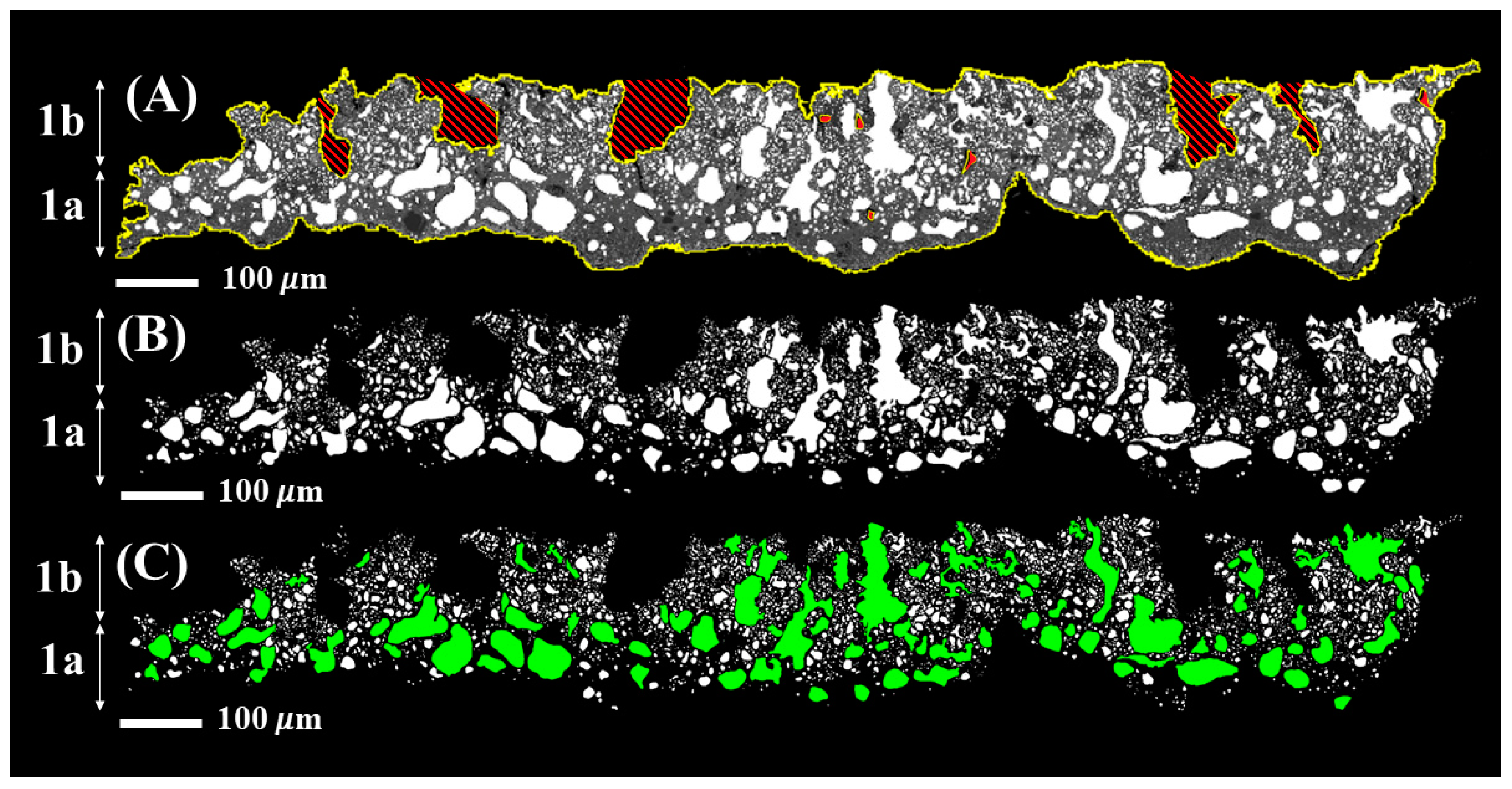
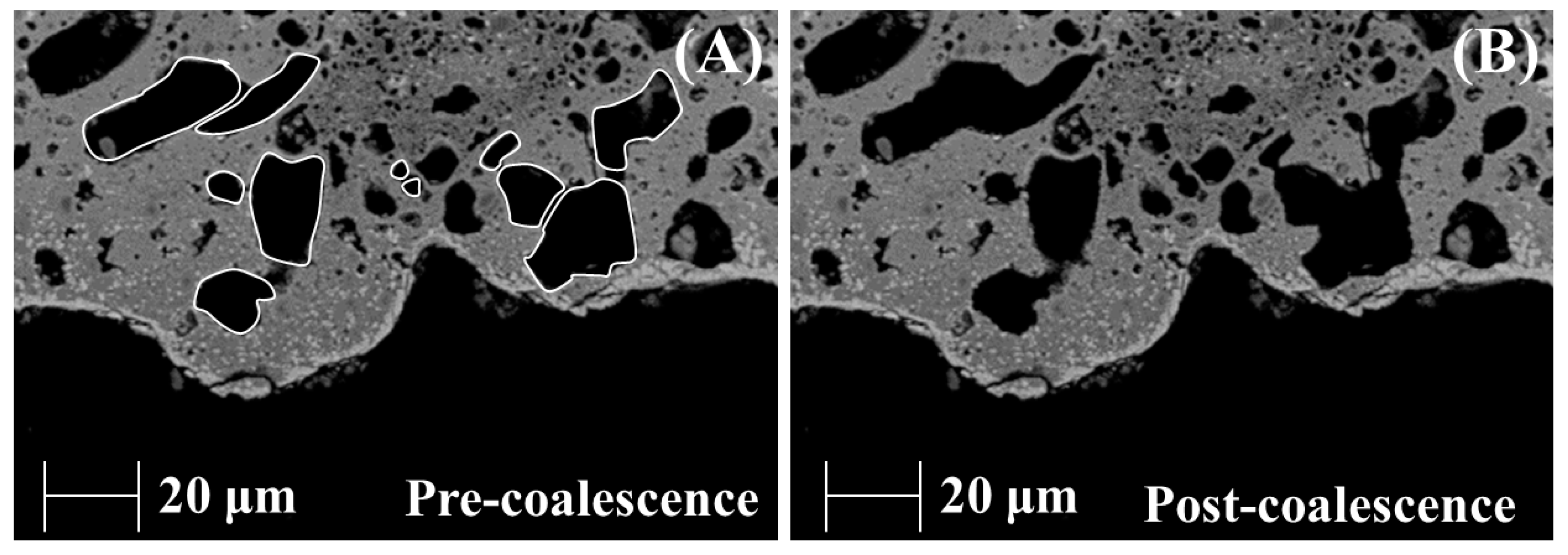
3.1. Vesicle Texture Analysis

The Tagish Lake fusion crust BSE image shows the presence of two distinct portions (Figure 2): (i) a pristine portion characterized by a fine-grained matrix displaying the typical texture of the Tagish Lake meteorite, as in [15], and (ii) a fusion crust portion, clearly identified by the presence of abundant fracture and vesicles, similar to the fusion crusts of other hydrated CC meteorites, as in [1,3].
A fusion crust can be further subdivided into different regions based on their texture and mineralogy, as shown in [1]. In this study, we identified two main areas (1 and 2) based on fractures and vesicle distribution (Figure 2C).
However, only the outer melted crust was considered for the VSD analysis because it is characterized by high vesicle content (Region 1 in Figure 2C). The thermally altered substrate (Region 2), where diverse cracks can be noted, was excluded from the VSD analysis because of its poor/absent vesicle content. Although the transition from Region 1 to Region 2 shows gradual changes in the number of the vesicles, Region 2 was distinguished from Region 1 by vesicle content (i.e., Region 1 vesicle-rich, Region 2 vesicle-poor/absent, as shown in Figure 1C and Figure 2C). Since the region borders were manually drawn, some vesicles might have been excluded from the VSD analysis due to being in Region 2. However, this effect is considered negligible due to the high number of identified vesicles.
Region 1 (~200 μm in thick) is characterized by a scoriaceous texture and a significant number of vesicles (~24,000 vesicles/mm2), as shown in Figure 3A,B. The vesicles present a wide range of variability in both shape and size (Figure 3). For example, shapes vary from circular to twisted in complex ways (from ~1 to 0.17 in aspect ratio, respectively), while their size goes from a few μm to ~70 μm (equivalent diameter). The smaller vesicles (<10 μm) range from spherical to elliptical forms, while the larger ones (>20 μm) present subspherical to complex convoluted shapes and are enriched in the outermost melted crust portion (Figure 3C). Based on the vesicle distribution, Region 1 can be further subdivided into two areas: Region 1a and 1b (Figure 3C). Region 1a is characterized by a low number of small vesicles (<20 μm) and a high number of larger ones (>20 μm) (Figure 3C), whereas Region 1b shows a high number of small vesicles (<20 μm) (Figure 3C). Region 2 is ~500 μm in thickness (range from ~750 to ~250 μm) and presents abundant cracks (Figure 2C). The latter, a typical texture for CC meteorite fusion crusts [1,3], has been interpreted as the decomposition of phyllosilicates and other thermal-sensitive minerals, such as sulfates and carbonates [1,3].

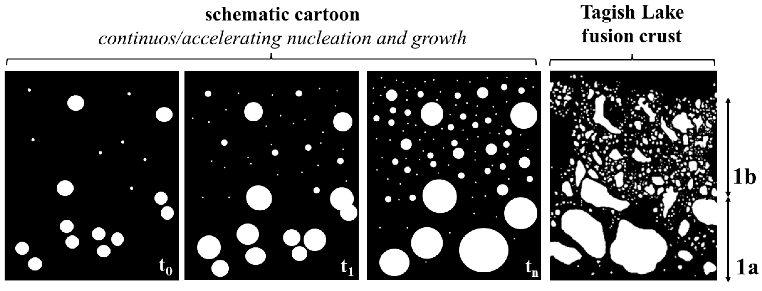
3.2. Vesicle Size Distribution (VSD)

Quantitative textural data are presented as size distributions [18] and provide significant insights into the nature of vesicles and their formation mechanisms. Here, the results obtained from CSDcorrection are reported and displayed according to four plots (Figure 4): (i) volume fraction (Vf) distribution (VFD); (ii) cumulative volume distributions (CVD); (iii) vesicle size distribution (VSD); and (iv) cumulative number density (CVSD).
Volume fraction distribution (Vf (%) vs. L plot, where L is the equivalent diameter of the vesicles expressed in μm) shows a polymodal distribution with dominant peaks (modes) at larger diameters (from ~60 μm to ~100 μm) and subordinate peaks at smaller (~10 μm) ones (Figure 4A). Cumulative volume distribution (cumulative Vf (%) vs. L (μm) plot) forms a curve with multiple slope ruptures, showing the polymodal nature of the vesicle populations (Figure 4B). Vesicle size distribution (ln(n) vs. L (μm) plot, where n is the vesicle number density per unit volume) provides insights into the nucleation and growth rate of the vesicle population, displaying a non-linear trend (Figure 4C). Cumulative number density (ln (Nv) > L vs. ln (L) plot, where Nv (>L) is the number of vesicles per volume with diameters greater than L) illustrates the vesicle number density and the bubble nucleation through time, presenting a “power-law” trend with a fractal distribution (fractal dimension (D) = 2.6, R2 = 0.99; Figure 4D).

4. Discussion

The distribution diagrams in Figure 4 and the textural images in Figure 1 and Figure 2 provide information about the vesicle formation processes, such as nucleation and growth. The polymodal distribution of VFD could be attributed to multiple events of bubble nucleation and growth and to bubble coalescence [26,27,28,29,30,31].
The complex convoluted forms of the vesicles can be interpreted as the result of coalescence processes, where one or more sub-spherical vesicles coalesce together, forming complex shapes (Figure 5).
Moreover, the polymodal distribution of VFD also suggests that most of the vesiculation occurred over a relatively restricted period [26], similar to the rapid formation of vesicles observed in scoriaceous micrometeorites [5]. The non-linear trend of VSD reflects a disequilibrium degassing of the nucleation and growth rate in the vesicle population [27]. Cumulative number density shows a fractal distribution that can be attributed to continuous/accelerating vesicle nucleation and growth processes [18] and references therein (Figure 4 and Figure 6). These processes are also highlighted by the vesicle distribution on the fusion crust, which shows a high number of nucleation events (Figure 3).
After nucleation, we suggest bubbles grow primarily due to expansion and coalescence processes (Figure 4 and Figure 6). Vesicle size distribution and CVSD trends exclude the vesicle growth for diffusion processes (Ostwald Ripening) in the Tagish Lake fusion crust due to their rapid formation, as observed in micrometeorites [5]. In this regard, as stated by Taylor et al. [8] and Suttle et al. [9], we believe that the Tagish Lake fusion crust experienced a progressive evolution from Region 2 (highly fractured) to Region 1 (highly vesiculated). Fractures formed due to rapid gas lost from phyllosilicates (in a brittle–ductile realm) and then evolved in vesicles (plastic–viscous realm) when the rise in temperature caused the melting of the mineral phases. Moreover, numerical simulations for vesicle formation in cosmic spherules suggest vesicle migration throughout the atmospheric passage [7]. Therefore, the possibility of vesicle migration during the Tagish Lake fusion crust formation cannot be excluded.
Although the Tagish Lake fusion crust results from the interaction between meteoroids and the Earth’s atmosphere, analogies could be suggested in terms of vesicularity between the Tagish Lake fusion crust and the processes occurring on the surface of Ryugu’s grains.
The micro-impacts occurring on asteroids like Ryugu are one of the causes of the space weathering on the surface of the asteroidal material. For example, high-velocity micrometeoroid bombardment may induce the amorphization and partial melting of the affected surface [32]. In this case, since vesiculation is primarily related to the mineralogy of the material and the process speed (very rapid for both fusion crust and shock melting), the vesicle features could be similar. As an illustration of this, analogies in the vesiculation could be found between the meteorite fusion crust and the “frothy layer” on the Ryugu grains [33].

5. Conclusions

An in-depth analysis of vesicles’ size distribution in a carbonaceous chondrite (Tagish Lake meteorite, C2-ung) fusion crust has been carried out using a quantitative approach. The vesicular portion of the Tagish Lake fusion crust shows a scoriaceous texture with a high number of vesicles (~24,000 vesicles/mm2). Vesicle shapes range from circular to twisted in a very complex way, and their size varies from a few μm to ~70 μm (equivalent diameter). Vesicle size distribution (VSD) and cumulative number density (CVSD) show a high nucleation event and a fractal distribution of the vesicle population, respectively. We suggest these features result from the disequilibrium degassing processes occurring simultaneously with continuous/accelerating nucleation and the growth of vesicles in a short period. After nucleation, we assume that bubbles grow mostly though expansion and coalescence processes.
In conclusion, this study presents the mechanism of vesicle nucleation and growth in the Tagish Lake meteorite fusion crust for the first time. This work shows that VSD is an effective method to investigate vesicle nucleation and growth in meteorite fusion crusts if the number of vesicles is statistically significant.

Author Contributions

Conceptualization, X.S., S.C. and G.P.; methodology, X.S., S.C. and G.P.; formal analysis, X.S., S.C. and G.P.; investigation, X.S., S.C. and G.P.; resource, E.P. and G.P.; data curation, X.S., S.C. and G.P.; writing—original draft, X.S., S.C. and G.P.; writing—review and editing, X.S., S.C. and G.P. All authors have read and agreed to the published version of the manuscript.

Funding

This research was funded by the Agenzia Spaziale Italiana project “Attività scientifiche per l’estensione della missione Hayabusa2 grant N.2022-12-HH-0” and the APC was fully waived by the Editor.

Data Availability Statement

Data are contained within the article.

Acknowledgments

Authors thank the anonymous reviewers for their valuable comments and suggestions. The authors also thank the “Centro di Servizi di Microscopia Elettronica e MicroAnalisi” (MEMA) and the “Laboratory of Molecular Magnetism” (LaMM) of the Università degli Studi di Firenze (Italy) for analysis and sample preparation.

Conflicts of Interest

The authors declare no conflicts of interest.

References

  1. Genge, M.J.; Grady, M.M. The fusion crusts of stony meteorites: Implications for the atmospheric reprocessing of extraterrestrial materials. Meteorit. Planet. Sci. 1999, 34, 341–356. [Google Scholar] [CrossRef]
  2. Dias, B.; Turchi, A.; Stern, E.C.; Magin, T.E. A model for meteoroid ablation including melting and vaporization. Icarus 2020, 345, 113710. [Google Scholar] [CrossRef]
  3. Genge, M.J.; Alesbrook, L.; Almeida, N.V.; Bates, H.C.; Bland, P.A.; Boyd, M.R.; Burchell, M.J.; Collins, G.S.; Cornwell, L.T.; Daly, L.; et al. The fusion crust of the Winchcombe meteorite: A preserved record of atmospheric entry processes. Meteorit. Planet. Sci. 2023; in press. [Google Scholar] [CrossRef]
  4. Llana-Funez, S.; Brodie, K.H.; Rutter, E.H.; Arkwrigt, J.C. Experimental dehydration kinetics of serpentinite using pore volumometry. J. Metamorph. Geol. 2007, 25, 423–438. [Google Scholar]
  5. Genge, M.J. An increased abundance of micrometeorites on Earth owing to vesicular parachutes. Geophys. Res. Lett. 2017, 44, 1679–1686. [Google Scholar] [CrossRef]
  6. Ramsdohr, P. Die schmelzkruste der meteorite. Earth Planet. Sci. Lett. 1967, 2, 593–598. [Google Scholar]
  7. Genge, M.J. Vesicle dynamics during the atmospheric entry heating of cosmic spherules. Meteorit. Planet. Sci. 2017, 52, 443–457. [Google Scholar]
  8. Taylor, S.; Jones, K.W.; Herzog, G.F.; Hornig, C.E. Tomography: A window on the role of sulfur in the structure of micrometeorites. Meteorit. Planet. Sci. 2011, 46, 1498–1509. [Google Scholar] [CrossRef]
  9. Suttle, M.D.; Genge, M.J.; Folco, L.; Van Ginneken, M.; Lin, Q.; Russell, S.S.; Najorka, J. The atmospheric entry of fine-grained micrometeorites: The role of volatile gases in heating and fragmentation. Meteorit. Planet. Sci. 2019, 54, 503–520. [Google Scholar] [CrossRef]
  10. Toppani, A.; Libourel, G.; Engrand, C.; Maurette, M. Experimental simulation of atmospheric entry of micrometeorites. Meteorit. Planet. Sci. 2001, 36, 1377–1396. [Google Scholar]
  11. Nicolau-Kuklińska, A.; Łosiak, A.I. Formation of Vesicles Within the Fusion Crust of Eucritic Meteorites. In Proceedings of the 79th Annual Meeting of the Meteoritical Society, Berlin, Germany, 7–12 August 2016; Volume 79, p. 6424. [Google Scholar]
  12. Scott, J.M.; Negri, M.; Faure, K.; Palmet, M.C.; Knaack, D.R.; Leybourne, M.I. Multi-zone fusion crust formation and classification of the 2004 Auckland meteorite (L6, S5, and W0). Meteorit. Planet. Sci. 2023, 58, 328–340. [Google Scholar] [CrossRef]
  13. Brown, P.G.; Hildebrand, A.R.; Zolensky, M.E.; Grady, M.; Clayton, R.N.; Mayeda, T.K.; Tagliaferri, E.; Spalding, R.; MacRae, N.D.; Hoffman, E.L.; et al. The fall, recovery, orbit, and composition of the Tagish Lake meteorite: A new type of carbonaceous chondrite. Science 2000, 290, 320–325. [Google Scholar] [CrossRef] [PubMed]
  14. Yokoyama, T.; Nagashima, K.; Nakai, I.; Young, E.D.; Abe, Y.; Aléon, J.; Alexander, C.M.O.; Amari, S.; Amelin, Y.; Bajo, K.I.; et al. Samples returned from the asteroid Ryugu are similar to Ivuna-type carbonaceous meteorites. Science 2022, 379, eabn7850. [Google Scholar]
  15. Zolensky, M.E.; Nakamura, K.; Gounelle, M.; Mikouchi, T.; Kasama, T.; Tachikawa, O.; Tonui, E. Mineralogy of Tagish Lake: An ungrouped type 2 carbonaceous chondrite. Meteorit. Planet. Sci. 2002, 37, 737–761. [Google Scholar] [CrossRef]
  16. Bland, P.A.; Cressey, G.; Menzies, O.N. Modal mineralogy of carbonaceous chondrites by X-ray diffraction and Mössbauer spectroscopy. Meteorit. Planet. Sci. 2004, 39, 3–16. [Google Scholar] [CrossRef]
  17. Gilmour, C.M.; Herd, C.D.K.; Cloutis, E.A.; Cuddy, M.; Mann, P. Water abundance in the Tagish Lake meteorite from TGA and IR spectroscopy: Evaluation of aqueous alteration. In Proceedings of the 47th Lunar and Planetary Science Conference, The Woodlands, TX, USA, 21–25 March 2016; p. 1765. [Google Scholar]
  18. Shea, T.; Houghton, B.F.; Gurioli, L.; Cashman, K.V.; Hammer, J.E.; Hobden, B.J. Textural studies of vesicles in volcanic rocks: An integrated methodology. J. Volcanol. Geotherm. Res. 2010, 190, 271–289. [Google Scholar]
  19. Larson, M.A.; Randolph, A.D. Size distribution analysis in continuous crystallization. In Crystallization from Solutions and Melts; Springer: Berlin/Heidelberg, Germany, 1969; Volume 65, pp. 1–13. [Google Scholar]
  20. Marsh, B.D. Crystal size distribution (CSD) in rocks and the kinetics and dynamics of crystallization: I. Theory. Contrib. Mineral. Petrol. 1988, 99, 277–291. [Google Scholar] [CrossRef]
  21. Cross, J.K.; Roberge, J.; Jerram, D.A. Constraining the degassing processes of Popocatépetl Volcano, Mexico: A vesicle size distribution and glass geochemistry study. J. Volcanol. Geotherm. Res. 2012, 225, 81–95. [Google Scholar]
  22. Morgan, D.; Jerram, D.A. On estimating crystal shape for crystal size distribution analysis. J. Volcanol. Geotherm. Res. 2006, 154, 1–7. [Google Scholar]
  23. Higgins, M.D. Measurement of crystal size distributions. Am. Mineral. 2000, 85, 1105–1116. [Google Scholar] [CrossRef]
  24. Higgins, M.D. Closure in crystal size distributions (CSD), verification of CSD calculations, and the significance of CSD fans. Am. Mineral. 2002, 87, 171–175. [Google Scholar] [CrossRef]
  25. Higgins, M.D.; Chandrasekharam, D. Nature of sub-volcanic magma chambers, Deccan province, India: Evidence from quantitative textural analysis of plagioclase megacrysts in the Giant Plagioclase Basalts. J. Petrol. 2007, 48, 885–900. [Google Scholar]
  26. Klug, C.; Cashman, K.V.; Bacon, C. Structure and physical characteristics of pumice from the climactic eruption of Mount Mazama (Crater Lake), Oregon. Bull. Volcanol. 2002, 64, 486–501. [Google Scholar]
  27. Houghton, B.F.; Hobden, B.J.; Cashman, K.V.; Wilson, C.J.N.; Smith, R.T. Large-scale interaction of lake water and rhyolitic magma during the 1.8 ka Taupo eruption, New Zealand. In Explosive Subaqueous Volcanism; Geophysical Monograph Series; American Geophysical Union: Washington, DC, USA, 2003; Volume 140, pp. 97–109. [Google Scholar]
  28. Blower, J.D.; Keating, J.P.; Mader, H.M.; Phillips, J.C. Inferring volcanic degassing processes from vesicle size distributions. Geophys. Res. Lett. 2001, 28, 347–350. [Google Scholar]
  29. Orsi, G.; Gallo, G.; Heiken, G.; Wohletz, K.; Yu, E.; Bonani, G. A comprehensive study of pumice formation and dispersal: The Cretaio Tephra of Ischia (Italy). J. Volcanol. Geotherm. Res. 1992, 53, 329–354. [Google Scholar]
  30. Klug, C.; Cashman, K.V. Permeability development in vesiculating magmas: Implications for fragmentation. Bull. Volcanol. 1996, 58, 87–100. [Google Scholar]
  31. Mangan, M.T.; Cashman, K.V. The structure of basaltic scoria and reticulite and inferences for vesiculation, foam formation, and fragmentation in lava fountains. J. Volcanol. Geotherm. Res. 1996, 73, 1–18. [Google Scholar] [CrossRef]
  32. Pieters, C.M.; Noble, S.K. Space weathering on airless bodies. J. Geophys. Res. Planets. 2016, 121, 1865–1884. [Google Scholar]
  33. Noguchi, T.; Matsumoto, T.; Miyake, A.; Igami, Y.; Haruta, M.; Saito, H.; Hata, S.; Seto, Y.; Miyahara, M.; Tomioka, N.; et al. A dehydrated space-weathered skin cloaking the hydrated interior of Ryugu. Nat. Astron. 2023, 7, 170–181. [Google Scholar] [CrossRef]
Figure 1. Backscattered electron image sequence of the Tagish Lake fusion crust edited using FIJI program. (A) Original BSE image of the outermost portion of the fusion crust. (B) Image elaborated upon using the FIJI program. (C) Selection of the region of interest for the VSD analysis. The portions removed during the sample preparation, identified by poorly delimitated edges (red perimeter) and the fractured region, were not included in the VSD analysis. (D) Manual image setting obtained using a graphic board by overlaying the original BSE image. (E) Image used for the VSD analysis.
Minerals 14 00099 g001
Figure 2. Backscattered electron images of the Tagish Lake fusion crust. (A) Panoramic BSE image of the Tagish Lake fragment showing pristine and fusion crust portions. (B) Zoomed-in images showing the outer fusion crust portion. (C) In the outer part of the fusion crust, three regions (1a, 1b, and 2) were identified, based on fractures and the vesicle size distribution. Region 1a is characterized by a low number of small (<20 μm) vesicles and a high number of larger (>20 μm) vesicles. On the contrary, region 1b is distinguished by a high number of small (<20 μm) vesicles. Finally, region 2 is characterized by abundant cracks.
Minerals 14 00099 g002
Figure 3. (A) Backscattered electron image (yellow perimeter) showing the fusion crust portion used for the vesicle size distribution (VSD) analysis. The red barred areas indicate the portions removed during the polishing of the sample and, thus, were not considered for the VSD analysis. Region 1 is characterized by ca. 24,000 vesicles/mm2—where mm2 is the area delimited by the yellow perimeter and then corrected by deleting the red barred portions. (B) Vesicle representation without the BSE image in the background. (C) Large vesicles (>20 μm) highlighted in green showing enrichment in the outermost portions of the fusion crust.
Minerals 14 00099 g003
Figure 4. Diagrams obtained from textural vesicle analysis. (A) Volume fraction (Vf) size distribution (VFD). (B) Cumulative volume fraction size distribution (CVD). (C) Vesicle size distributions (VSDs). (D) Cumulative number density (CVSD).
Minerals 14 00099 g004
Figure 5. Vesicles in the outer portion of the Tagish Lake fusion crust. (A) Hypothetical pre-coalescence configuration where vesicles were separated by menisci. (B) Present configuration where vesicles coalesce.
Minerals 14 00099 g005
Figure 6. Schematic cartoon illustrating the hypothetical mechanism for nucleation and growth (from time zero (t0) to time n (tn)) of vesicles compared to the original vesicle texture of the Tagish Lake fusion crust. Fast gas release leads to continuous/accelerating nucleation and growth bubble formation. After their nucleation, bubbles in Region 1a are dominated by growth processes (expansion and coalescence), whereas in Region 1b, bubbles are dominated by continued nucleation subordinate growth processes.
Minerals 14 00099 g006
Disclaimer/Publisher’s Note: The statements, opinions and data contained in all publications are solely those of the individual author(s) and contributor(s) and not of MDPI and/or the editor(s). MDPI and/or the editor(s) disclaim responsibility for any injury to people or property resulting from any ideas, methods, instructions or products referred to in the content.

