Article

Geobiological Cycling of Gold: From Fundamental Process Understanding to Exploration Solutions

1
Centre for Tectonics, Resources and Exploration (TRaX), School of Earth and Environmental Sciences, the University of Adelaide, North Terrace, Adelaide, SA 5005, Australia
2
CSIRO Land and Water, Environmental Biogeochemistry, PMB2, Glen Osmond, SA 5064, Australia
3
Department of Mineralogy, South Australian Museum, Adelaide, SA 5000, Australia
4
Institute for Biology/Mikrobiology, Martin-Luther-University Halle-Wittenberg, DE-06120 Halle, Germany
5
School of Earth Sciences, the University of Queensland, Brisbane, QLD 4072, Australia
*
Author to whom correspondence should be addressed.
Minerals 2013, 3(4), 367-394; https://doi.org/10.3390/min3040367
Submission received: 7 September 2013 / Revised: 16 October 2013 / Accepted: 21 October 2013 / Published: 4 November 2013
(This article belongs to the Special Issue Interactions between Microbes and Minerals)

Abstract

:
Microbial communities mediating gold cycling occur on gold grains from (sub)-tropical, (semi)-arid, temperate and subarctic environments. The majority of identified species comprising these biofilms are β-Proteobacteria. Some bacteria, e.g., Cupriavidus metallidurans, Delftia acidovorans and Salmonella typhimurium, have developed biochemical responses to deal with highly toxic gold complexes. These include gold specific sensing and efflux, co-utilization of resistance mechanisms for other metals, and excretion of gold-complex-reducing siderophores that ultimately catalyze the biomineralization of nano-particulate, spheroidal and/or bacteriomorphic gold. In turn, the toxicity of gold complexes fosters the development of specialized biofilms on gold grains, and hence the cycling of gold in surface environments. This was not reported on isoferroplatinum grains under most near-surface environments, due to the lower toxicity of mobile platinum complexes. The discovery of gold-specific microbial responses can now drive the development of geobiological exploration tools, e.g., gold bioindicators and biosensors. Bioindicators employ genetic markers from soils and groundwaters to provide information about gold mineralization processes, while biosensors will allow in-field analyses of gold concentrations in complex sampling media.

1. Introduction

Today, bacteria, archaea, fungi and algae play critical roles in driving the carbon-, nitrogen-, sulfur- and phosphorus-cycles as well as many metal cycles [1,2]. Metal cycles can be directly driven by microorganisms because they: (i) require metals as micro-nutrients for cell growth, (ii) are capable of obtaining metabolic energy from metal respiration or chemolithoautotrophic metal oxidation; and (iii) harbor extensive capabilities for (heavy) metal detoxification [1,3,4,5]. Microorganism also influence metal cycles indirectly: (i) due to their high metabolic rates they control geochemical parameters such as pH- and redox conditions, and (ii) through the formation, secretion, and/or decomposition of complexing ligands, e.g., low and high molecular weight organic acids, siderophores, exopolymers, cyanides and sulfur compounds, in soils, regolith materials, unconsolidated sediments, surface- and groundwaters [2,4]. Today we know that bacteria and archaea are ubiquitous in the deep subsurface down to several kilometers depth, where they occur in igneous, sedimentary and metamorphic rocks and appear to contribute to the formation of mineral deposits [6,7,8,9]. Consequently the cycling of major metals, e.g., Fe, Mn, Ca, Mg, K, Na, trace- and ultra-trace metals, e.g., Ag, Mo Cr, Cu, Ni, Pd, Se, W, V, U, Hg, C and Zn in Earth surface and in some crustal environments is thought to be controlled by microbial processes [2,5,10,11].
An example of a biogeochemical cycle of a metal, which until recently was considered to be inert, immobile and not biologically active under Earth surface conditions is that of gold (Figure 1; [11,12]). Until recently the occurrence of gold grains and -nuggets and the formation of secondary gold in surface environments were also considered to be mediated purely by abiogenic processes (e.g., [13]). According to Hough et al. [13] gold nuggets in surface environments are hypogene in origin. Their occurrence in surface environments was proposed to reflect the weathering of rocks hosting primary mineralization, and their distribution was linked to physical redistribution and mechanical accumulation in placer environments (e.g., [13]). Coatings of highly pure gold (up to 99.9 wt %) covering many gold grains collected from surface environments were explained by dealloying and chemical mobilization of the silver from the primary gold/silver alloys [13]. The formation of nano-phase gold (nano-particles and 200 nm sized, nano-particulate gold plates) in surface environments was attributed to the inorganic reduction of mobile gold complexes linked to evaporation, and the formation of evaporate minerals, e.g., barite and halloysite [14,15].
While these abiogenic processes likely play an important role in the surface cycling of gold, recent research has shown that microbiota are also involved in every step of a biogeochemical cycle of gold, from the formation of primary mineralization in deep subsurface-, hydro- and epithermal systems to its solubilization, dispersion and re-concentration as secondary gold in surface environments (Figure 1; [11,12]). In a recent study, Tomkins [16] suggested that microbial processes may have had a far greater influence on the formation of orogenic gold deposits as previously believed. His study indicated that interactions between the Earth’s evolving tectonic processes and biosphere may have driven changes in global geochemistry that established conditions more suitable for uptake of gold into sedimentary pyrite. For instance, for approximately 3.5 billion years sulfate-reducing bacteria (SRB), which are anaerobic, heterotrophic bacteria that reduce sulfate and thiosulfate to hydrogen sulfide (H2S) and release it as a by-product of metabolism, have been active. Modern SRB, e.g., Desulfovibrio spp., are able to reduce the thiosulfate from mobile gold thiosulfate complexes; this destabilizes gold in solution, which may then be precipitated intracellularly or incorporated into the newly forming sulfide minerals [17,18]. This allows for the formation of metallogenically enhanced sedimentary sequences, which are ideal source rocks for hydrothermal gold deposits. Enzymatically catalyzed precipitation of gold has also been observed in thermophilic and hyperthermophilic bacteria and archaea (e.g., Thermotoga maritime, Pyrobaculum islandicum), and their activity has led to the formation of gold- and silver-bearing sinters in New Zealand’s hot spring systems [19].
Figure 1. Understanding the geobiological cycle of gold, i.e., solubilization and transport, bioaccumulation, reductive biomineralization, and secondary gold formation helps mineral explorers to find new gold deposits and provide new ore-processing approaches (after [20]).
Minerals 03 00367 g001
Iron- and sulfur-oxidising bacteria (e.g., Acidithiobacillus ferrooxidans, A. thiooxidans) are known to break down gold-hosting sulfide minerals in zones of primary mineralization and release the associated gold in the process. These and other bacteria produce thiosulfate, which is known to contribute to gold mobility by forming stable, water-soluble complexes with gold [21]. Other microbial processes, e.g., the excretion of low molecular weight organic acids and cyanide, may drive gold solubilization in auriferous top- and rhizosphere soils. A characteristic of Group IB metals, e.g., gold, is their ability to strongly bind to organic matter, and gold was shown to readily form complexes with organic ligands [22,23]. The interaction of gold and organic matter involves mostly electron donor elements, e.g., N-, O- and in particular S-containing groups [11]. Cell walls of microorganisms contain large amounts of highly reactive thiol-containing groups mediating the sorption of metals [11]. This makes microorganisms a focus of an accelerated precipitation of gold in environmental systems compared to less reactive mineral surfaces [24]. Hence, a large number of studies using a range of environmentally relevant gold complexes have demonstrated the ability of many groups of microorganisms to rapidly passively accumulate gold complexes (e.g., [11] and references therein). A number of bacteria and archaea are also capable of actively catalyzing the precipitation of toxic gold(I/III) complexes [11]. Reductive precipitation of these complexes may improve survival rates of bacterial populations that are capable of (i) gaining metabolic energy by utilizing gold-complexing ligands (e.g., thiosulfate by A. ferrooxidans) and (ii) detoxifying the immediate cell environment by detecting, excreting and reducing gold complexes (e.g., Salmonella typhimurium, Plectonema boryanum and C. metallidurans CH34 [25,26,27]. C. metallidurans was detected in biofilms that form on gold grains from Australian sites located in moderate and wet tropical climatic zones, indicating that gold bioaccumulation may lead to gold biomineralization by forming secondary “bacteriomorphic” gold [28]. Formation of secondary octahedral gold crystals from gold(III)-chloride solution was also promoted by a cyanobacterium (P. boryanum) via an amorphous gold(I)-sulfide intermediate [26,29]. Secondary, bacteriomorphic gold is common in quartz pebble conglomerates deposits (QPC), such as Witwatersrand QPC, which is the world's largest gold deposit [30,31]. Here gold is commonly associated with bituminous organic matter of putative microbial origin. Falconer et al. [32] and Falconer and Craw [33] provided further evidence that geobiological processes play an important role for the formation of QPC-deposits by showing that carbonaceous mudstones within a QPC sequence in New Zealand contain fine gold grains of detrital origin as well as gold of secondary (authigenic) origin displaying bacteriomorphic and sheet-like morphologies. The porous, sheet-like authigenic gold is morphologically similar to gold associated with carbonaceous material in the Witwatersrand QPC. In addition, the authigenic sulfides from QPCs in New Zealand are compositionally similar to sulfides from Archaean QPC and their sulfur isotope ratios indicate biogenic origins [32,33].
Many of these concepts were introduced in a review by Reith et al. [11], but since then significant progress has been made in our fundamental understanding of geobiological gold cycling. Hence, the aim of this manuscript is to summarize this progress and highlight how this new knowledge provides new opportunities in helping mineral explorers to detect new gold deposits. The main questions that have been have addressed since 2007 are: (i) How widespread are gold-transforming biofilms on gold grains, and in particular do they extend to different climatic zones? (ii) Which organisms are present in the biofilms, and which are important for gold transformation? (iii) How do these organisms mediate gold transformation? (iv) Is gold cycling different to the cycling of other rare noble metals, such as platinum? (v) Are microbial communities resident in soils overlying gold deposits influenced by the underlying gold mineralization? (vi) Are other gold-containing secondary biominerals important for gold exploration? Moreover, (vii) how can this knowledge improve the development of biotechnological methods specific to mineral exploration?

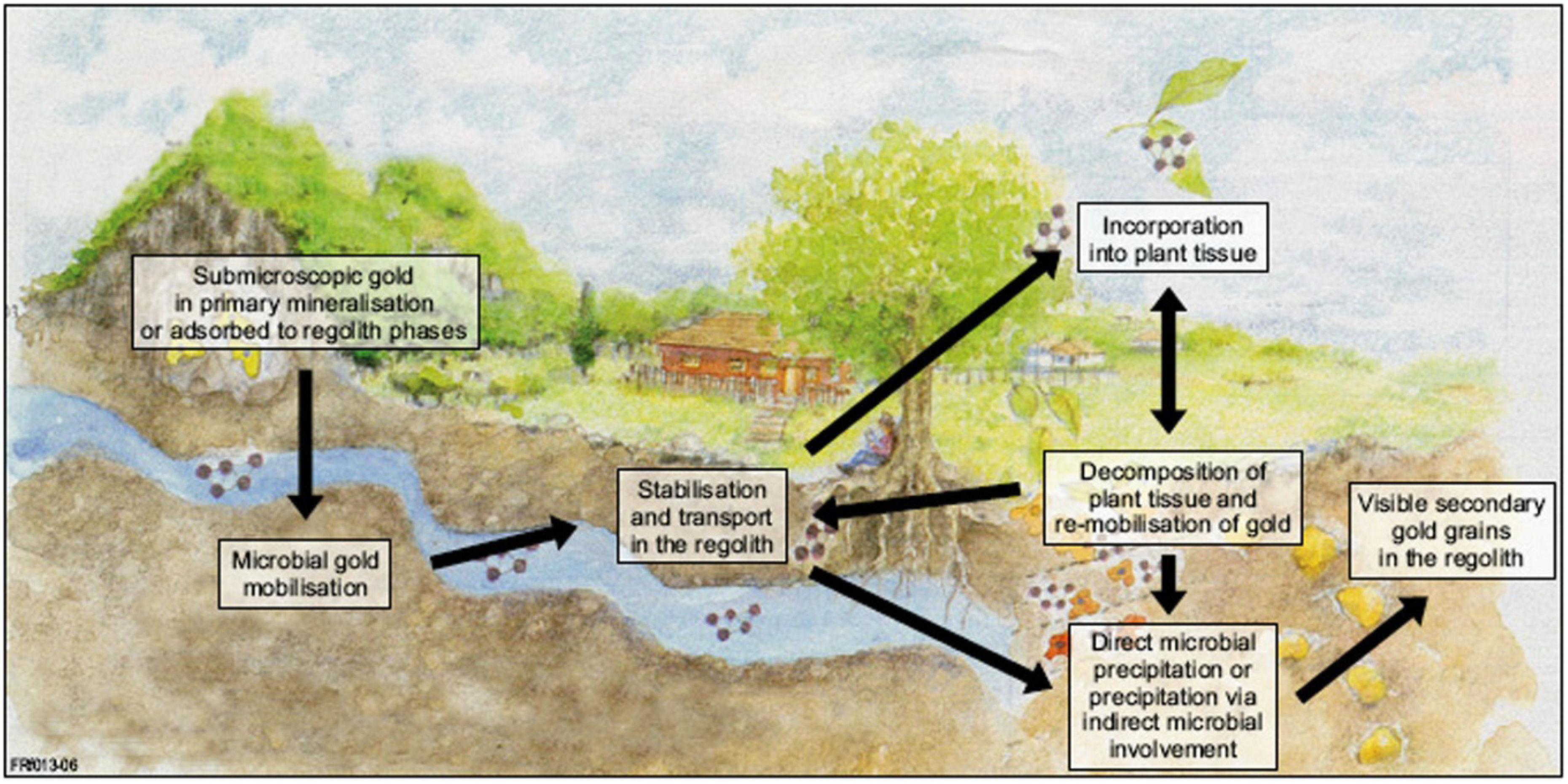
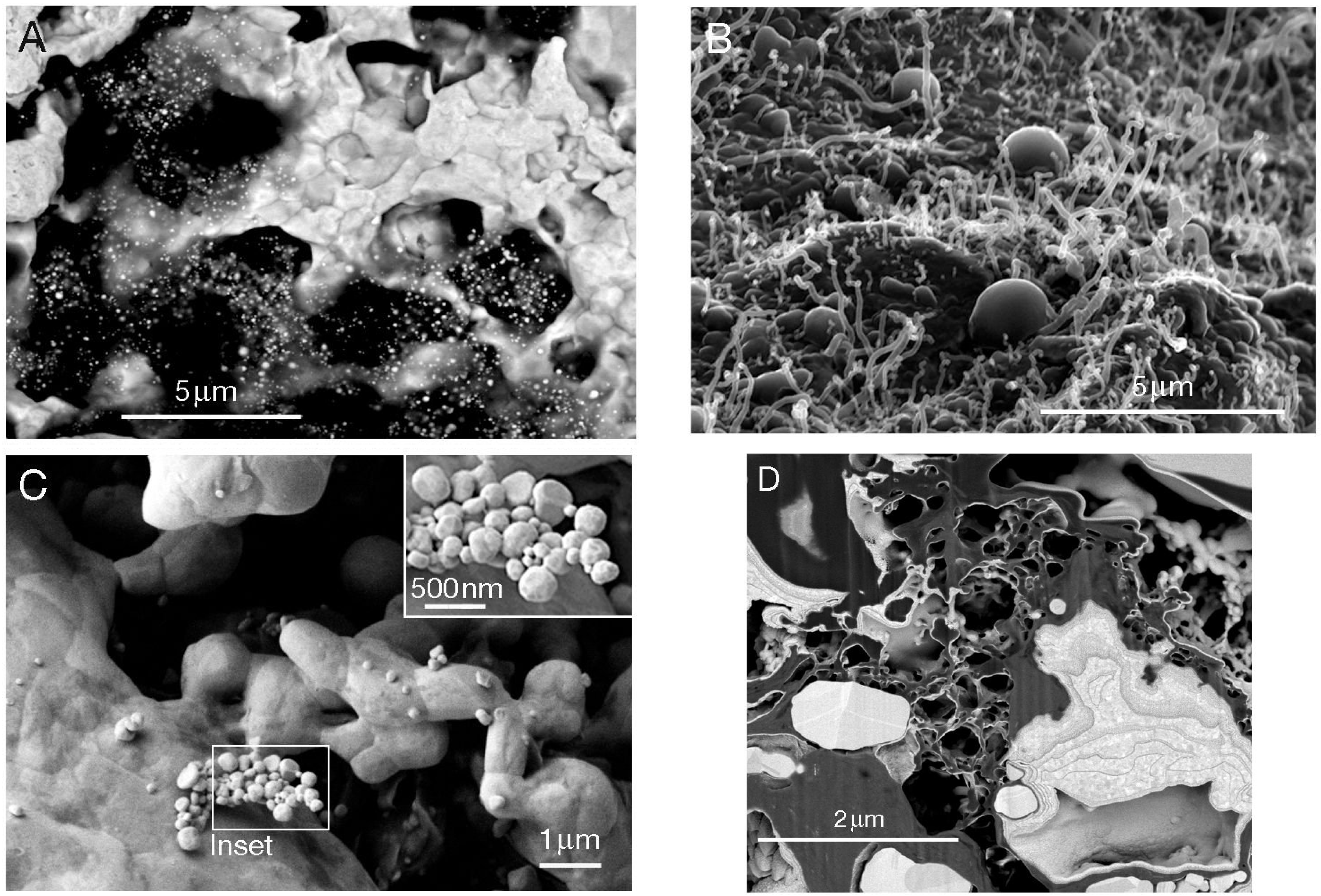
2. Biologically Mediated Transformation of Gold Grains

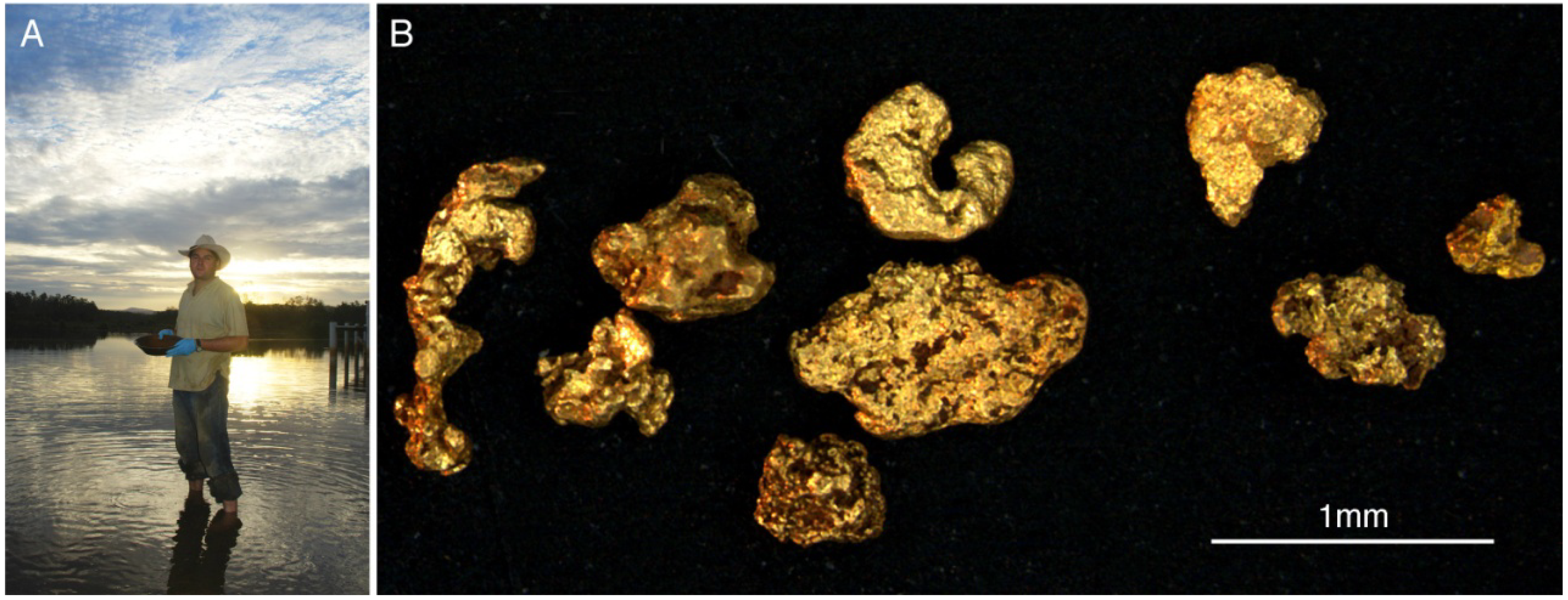
2.1. Wet Subtropical Environments

Gold grains from the Prophet Mine, Kilkivan, Queensland, Australia, were covered by a polymorphic, organic-inorganic layer that was up to 40 μm thick (Figure 2; [34]). It consisted of a bacterial biofilm containing gold nano-particles associated with extracellular polymeric substances as well as aggregates of secondary and bacteriomorphic gold (Figure 3C,D). The biofilm community comprised up to 11 taxonomic units, of which ten were identified as β-Proteobacteria. The dominant organisms, present on more than 90% of DNA-positive grains, were C. metallidurans, which also dominated grains from two other Australian sites, and D. acidovorans. Two types of bacteriomorphic gold were differentiated. Type 1 resulted from the re-precipitation of dissolved gold, and internal growth structures provide direct evidence for coarsening of the gold grains (Figure 3C,D). Type 2 is present at the contact between the polymorphic layer and the primary gold (Figure 3C). It consists of solid rounded forms into which crystal boundaries of underlying primary gold extend, and is the result of de-alloying and silver dissolution from the primary gold. This demonstrated that microbially driven dissolution, precipitation and aggregation contributes to the formation of bacteriomorphic gold and hence to the (trans)formation of gold grains in surface environments conditions. In addition, the microbially driven mobilization of coarse gold into nano-particles is likely to play a key role in mediating the mobility of gold in surface environments, because the release of nano-particulate gold upon biofilm disintegration would greatly enhance environmental mobility compared to gold complexes only.

2.2. Arid Environments

At arid sites, water and nutrient availability are limited and episodic, hence abiogenic evaporative mechanisms were thought to control the formation of secondary and especially nano-particulate gold [14,15]. To assess if geobiological processes play a role for the transformations of gold grains, grains were collected from eight arid sites in three Australian gold provinces, i.e., Lawlers (Yilgarn Craton, Western Australia), Tanami (Northern Territory), and Flinders Ranges (South Australia; [35]). Sites were chosen based on contrasting deposit styles, i.e., primary underground and epithermal deposits as well as secondary eluvial-, colluvial- and alluvial placers at increasing distances from primary mineralization. Gold grains from all surface environments displayed secondary transformation features, i.e., morphotypes indicative of gold and silver dissolution, gold aggregation and gold neoformation. The latter included spheroidal and bacteriomorphic gold, which increased in abundance with distance from source. Viable biofilms containing abundant gold nano-particles and spheroidal gold μ-crystals were detected on all grains from the Flinders Ranges (Figure 3B). Gold grains from the Lawlers and the Tanami provinces are covered by polymorphic layers containing abundant nano-particulate, spheroidal and bacteriomorphic gold. The polymorphic layers consisted of vermiform clays and organic matter, suggestive of remnant biofilms. These results demonstrate that biofilms capable of transforming gold grains will develop episodically on gold grains in arid environments, and that microbial processes play a critical role for the (trans)formation of gold grains and geochemical anomalies in arid environments.
Figure 2. (A) Gold panning in the Tomago River, New South Wales, Australia; and (B) light microscopic image of secondary gold grains collected by panning (after Reith et al. [34]).
Minerals 03 00367 g002
Figure 3. Biofilms, polymorphic layers, and associated gold particles from the surfaces of natural secondary gold grains. (A) Polymorphic layer containing gold nanoparticles on the surface of gold grains from New Zealand; (B) biofilms displaying nanowires on gold grains from the Flinders Ranges in South Australia; (C) spheroidal secondary gold forming on gold grains from Kilkivan, Queensland, Australia; and (D) nanocrystalline structure of neoformed gold replacing a bacterial cell from Kilkivan grains (after [24]).
Minerals 03 00367 g003

2.3. Temperate and Subarctic Environments

The link between surface morphologies of gold grains and supergene transformation processes, with a focus on the formation of secondary gold in temperate and subarctic environments was assessed [36]. Gold grains were collected from six localities across the South Island of New Zealand and one site in northern Finland. Deposit styles varied from eluvial-, alluvial-, and beach placer deposits in areas of moderate to very high levels of precipitation. Morphologies indicative of gold- and silver dissolution, e.g., grain boundary dissolution, were visible. In addition, abundant gold neoformation- and aggregation morphologies were observed on all grains. The latter included a variety of secondary gold morphotypes, in particular nano-particulate- and μ-crystalline forms as well as bacteriomorphic gold, sheet-gold and porous, branched gold networks. Pervasive dissolution features on grains from an outcropping quartz-vein system as well as extensive nano-particle formation on weathered quartz-vein- and placer grains from the west coast of New Zealand’s South Island, which is subject to very heavy orographic precipitation, suggest that these climatic conditions enhance the transformation of gold grains. At these sites, gold nano-particles were most abundant in the polymorphic layers and in soil materials associated with the grains. Nano-particulate gold was also highly abundant in carbonaceous, likely exopolymeric, coatings on gold grains from Orepuki Beach, suggesting that gold dissolution in seawater and microbial biomineralization are important contributors to gold grain transformations in beach placer deposits (Figure 3A). Gold grains from Lemmenjoki in subarctic northern Finland also showed considerable accumulations of nm- and μm-sized gold particles in polymorphic layers. This suggests that surface morphologies of gold grains from New Zealand and Finland are the result of supergene transformations occurring in current environments. As evaporation (as suggested by Hough et al. [13,15] for semi-arid environments) in unlikely to be a major process for the formation of nano-particulate gold in these high-rainfall environments, the abundance of gold nano-particles in New Zealand and Finland strongly suggests that geobiological processes are drivers of secondary gold formation and gold dispersion in temperate and subarctic environments.

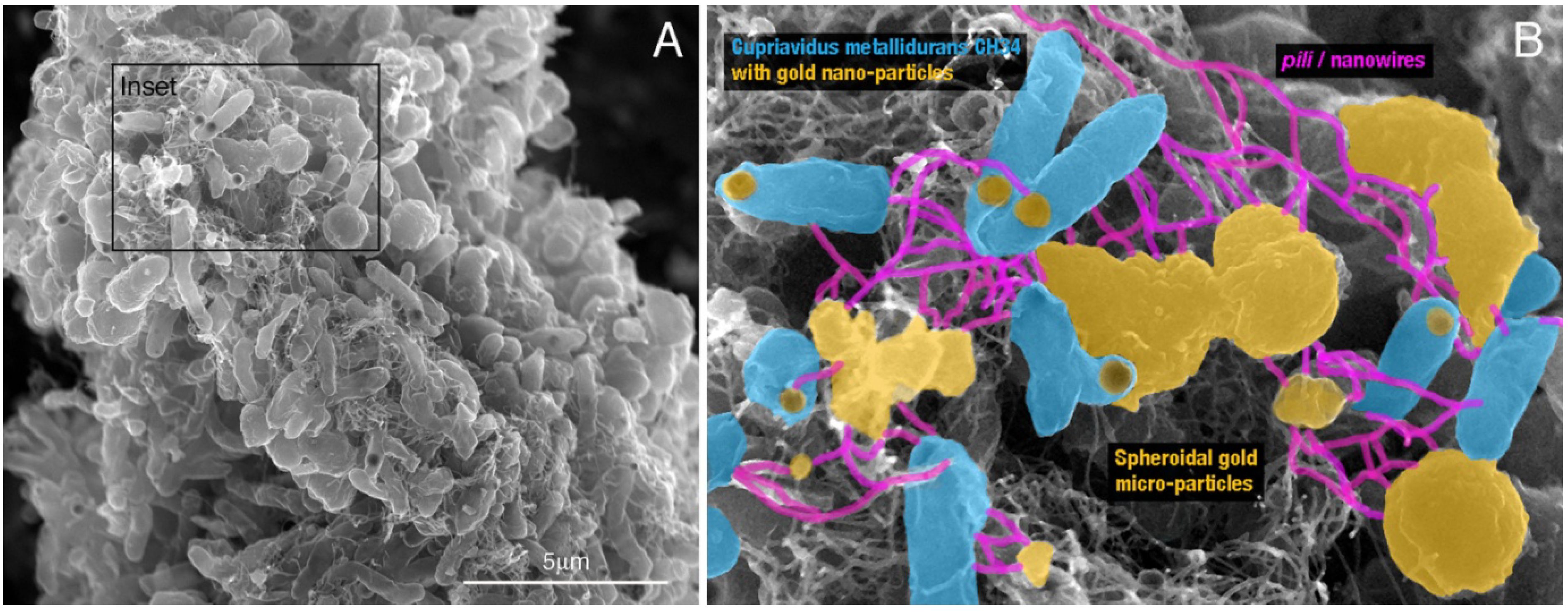
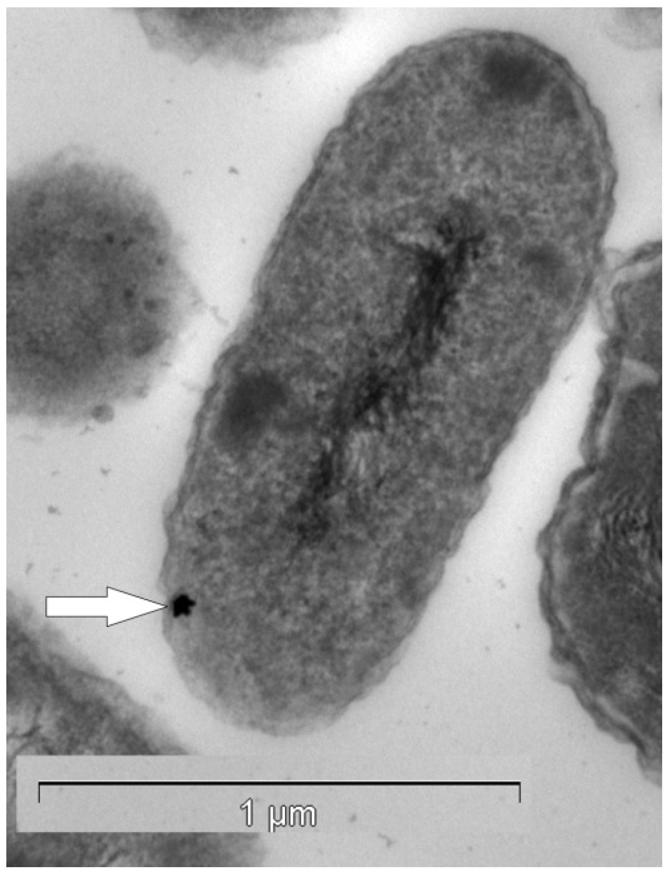
2.4. Laboratory Environments—Formation of Gold Biominerals by C. metallidurans

C. metallidurans CH34, a bacterium dominating biofilm communities on gold grains from three Australian sites, is capable of reductively precipitating toxic, aqueous gold(I/III)-complexes [27]. This suggests that this bacterium plays a fundamental role in the formation of highly pure (secondary) and bacteriomorphic gold in surface environments. To experimentally verify this assumption we assessed the formation of gold biominerals in quartz-sand-packed columns inoculated with C. metallidurans biofilms, amended periodically with gold(I)-thiosulfate and incubated for four months [24]. Formation of metallic gold particles was only observed in the presence of viable biofilms, but not in sterilized or abiotic controls. In experiments with viable biofilms, more than 99 wt % of the gold was retained in the column, compared to <30 wt % in sterilised and abiotic controls. Biomineralization of gold occurred via the formation of intra- and extracellular spherical nano-particles, which aggregated into spheroidal and framboidal micro-particles of up to 2 μm in diameter (Figure 4). Aggregates of gold formed around cells, eventually encapsulating and ultimately replacing them (Figure 4B). These particles were morphological analogues to gold particles commonly observed on natural gold grains. Bacterial cells were connected via exopolymer or nanowires to μm-sized, extracellular gold-aggregates, which would intuitively improve the flow of electrons through the biofilm (Figure 4B). This provided experimental verification for the importance of biofilms of C. metallidurans for the bacterial biomineralization in the formation of highly pure gold in surface environments.
Figure 4. Secondary electron micrographs of a C. metallidurans biofilm precipitating secondary gold in column experiments. (A) Overall biofilm morphology, individual cells and associated gold biominerals; and (B) association of individual cells with gold nano-particles and connections of cells to extracellular gold aggregates by putative nanowires (after [24]).
Minerals 03 00367 g004

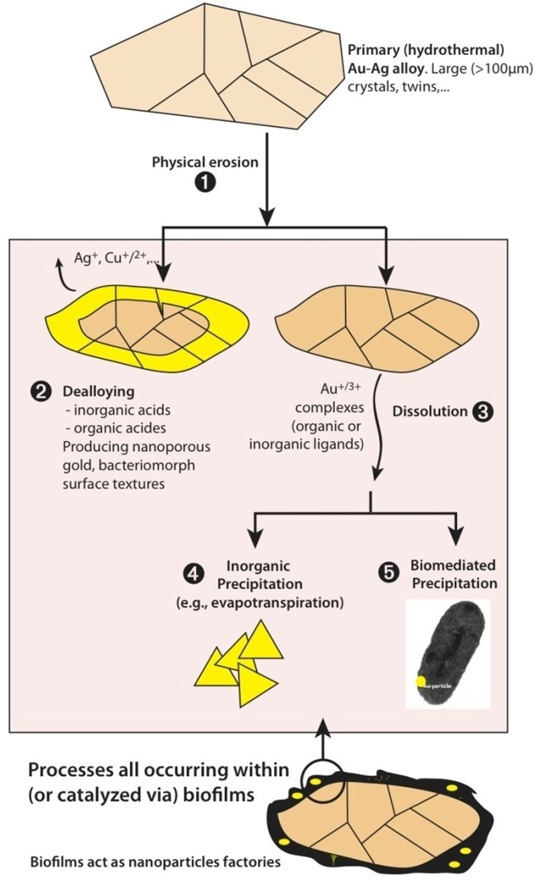
2.5. A Unified Model for Gold Grain Formation and Implications for Exploration

Our studies in combination with works of other authors led to the understanding that the detrital and aggregation models for gold grain and nugget formation need to be combined into a unified formation model that covers all alluvial and lateritic gold (Figure 5; e.g., [11,13,14,24,32,33,34,35,36,37,38,39]. Hence, the more than 100 year lasting debate concerning the formation of gold grains and nuggets in surface environments was finally resolved. In the unified model, primary gold grains and nuggets are brought to the surface through weathering and/or mechanical transport. Here they are subject to biotic and abiotic processes leading to the solubilization of primary gold and precipitation of secondary gold, thus forming the commonly observed grains/nuggets with a primary gold-silver alloy center and a secondary polymorphic layer of high purity gold (Figure 5). Biomineralization of mobile gold from solution/suspension enhances the growth of grains in surface environments, which may explain why secondary gold grains are often coarser than gold hosted in the associated primary mineralization. Weathering of primary sulfides can lead to the mobilization and dispersion of gold that was present as solid solution, to form pure secondary gold grains (Figure 5). The occurrence of bacteriomorphic gold (type 2), which is the result of biomediated de-alloying and silver mobilization, may explain bud-like gold morphologies where no secondary gold occurs (Figure 5). Most importantly, biofilms play a previously unrecognized role in promoting gold dispersion by acting as effective “nano-particle factories”. Since biofilms are subject to cycles of formation and destruction via desiccation or grazing by soil fauna, nano-particulate gold will be periodically released and dispersed in the environment via transport in soil solutions and groundwater (Figure 5). This continuous release of nano-particulate gold can explain the high mobility of gold in many near surface environments, which leads to the formation of secondary gold deposits [40] and the development of geochemical halos around buried mineralization even in transported cover [11,14].
Figure 5. Model of processes responsible for the (trans)formation of gold grains in supergene environments; bright yellow is high fineness gold (>95 wt %), orange is gold-silver alloy, black is biofilm (after [35]).
Minerals 03 00367 g005
Transported gold grains are commonly used in mineral exploration for gold to pinpoint a primary source [13]. For example, Chapman et al. [41] used a combination of composition and inclusion mineralogy in placer gold grains to assess their relationship to sources and styles of mineralization in the Yukon Territory, Canada. The placer gold inventory derived from this study led to the identification of the characteristics of populations of placer grains contributing to economically important placers. Knight et al. [42] studied the relationship between distance travelled in fluvial systems and gold grain shape and rimming. The roundness and flatness of gold grains increased rapidly over the first five kilometers of transport from the source. This, coupled with the observed development of gold-rich rims, allowed for the separation of gold grains into the classification of proximal and distal relative to a likely source [42]. However, Hough et al. [13] noted that the morphology of grains, and in particular those found in stream sediments, is not diagnostic for origin or distance, but rather provides a record of the supergene transformations the grains have undergone. Our studies confirmed these findings and provided a biogeochemical process for the initial and subsequent stages of gold grain transformations. In combination with geological and physicochemical conditions, this model can now provide a baseline for the interpretation of macro- and micro-morphologies and chemical compositions of gold grains.

3. Biochemistry of Gold Biomineralization

Unlike other heavy metals gold does not form free ions in aqueous solution under surface conditions, but occurs as metallic nano-particles (0), aurous (I) and auric (III) complexes [43]. Based on thermodynamic calculations and natural abundances of possible ligands, complexes with chloride, ammonium, thiosulfate, cyanide, amines and organic acids appear to be most important in surface solutions [11]. Similar to free heavy metal ions many environmentally relevant gold(I/III)-complexes are highly toxic to microbiota, with minimal inhibitory concentrations in a similar range than those of mercury, which is considered one of the most toxic elements [44,45]. One reason for the observed toxicity appears to be that gold complexes, which are readily taken up by cells, inhibit intracellular enzyme functioning [27]. Another reason may lie in the generation of oxidative stress following the formation of free radicals in the cells, which can lead to the damage of cellular DNA [46]. The formation of radicals is likely to occur during the reduction of gold(I/III)-complexes to metallic gold, as this reductive process involves an odd number of electrons per gold(I/III)-reduced, whereas commonly electron pairs are transferred in cells [46]. Hence, some bacteria have developed specific and unspecific intra- and extracellular mechanisms to deal toxic effects of mobile gold [25,27,46,47]. As a result, these mechanisms may control the formation of secondary gold in surface environments [27,34].

3.1. Cupriavidus metallidurans CH34—Putative Co-Utilization of Other Metal Resistance Systems for Gold Detoxification

C. metallidurans CH34 contains a variety of metal resistance factors that allow it to thrive in metal-contaminated environments [48,49]. The respective metal resistance determinants are located on the two native megaplasmids pMOL28 and pMOL30 and on chromosomal DNA, mainly on chromosome 2 [49,50]. C. metallidurans rapidly (<1 min) accumulates gold(I/III)-complexes from solution [37]. This rapid uptake leads to the formation of gold(I)-complexes bound to S-containing compounds, presumably proteins, in the cells. This suggested that this rapid uptake and reduction to from gold(III)- to gold(I)-complexes was pH depended and occurred passively due to the high affinity of gold-complexes to organic matter.
This was confirmed in a study by Kenney et al. [51], who examined the rapid sorption of aqueous gold(III)-hydroxide-chloride complexes by non-metabolizing bacterial cells. The experiments were conducted with gram positive, i.e., Bacillus subtilis, and gram negative species, i.e., Pseudomonas putida, suspended in a buffered 5 mg kg−1 gold(III)-(hydroxide)-chloride solution. Their results showed that both bacteria removed more than 85 wt % of the gold from solution after 2 h at pH values below 5. This lead to the formation of gold(I)-S cell wall complexes [52]. Further reduction to metallic gold particles was not observed, even after extended incubation periods [52]. This initial passive sorption is followed by uptake into the cytoplasm, which promotes gold toxicity [27,45].
Metabolically active metal-resistant bacteria, such as C. metallidurans, react to gold toxicity by inducing oxidative stress and metal resistances gene clusters (cop, cup, ars, mer) as well as gold-specific operon, gig, for gold-induced genes [27]. As a result, gold detoxification was mediated by a combination of efflux, reduction, and possibly methylation of gold-complexes, leading to the formation of gold(I)-C-compounds and metallic nano-particulate gold (Figure 6). The products of up-regulated gene clusters in C. metallidurans suggested that specific gold-handling systems could be involved in the uptake of gold complexes into the cytoplasm, the export of gold(I) back to the periplasm after reduction, and further chemical reduction to gold(0) in the periplasm [27]. However, the highly up-regulated gold specific Gig-system was also not directly involved in detoxifying gold complexes via uptake and reduction of gold(III) to gold(I) or gold(0). The two indigenous plasmids of C. metallidurans, which harbor several transition metal resistance determinants, were also not involved in resistance to gold(I/III)-complexes nor in their transformation to metallic nano-particles. However, the copABCD determinant on chromosome 2, which encodes periplasmic proteins involved in copper resistance, is required for gold resistance in C. metallidurans. This suggested that biomineralization of gold particles via the reduction of mobile gold(I/III)-complexes in C. metallidurans appeared to primarily occur in the periplasmic space by copper-handling systems [46].
Figure 6. Transmission electron micrograph of a C. metallidurans ultra-thin section containing a gold nano-particle (white arrow) in the periplasm (after Reith et al. [27]).
Minerals 03 00367 g006
At this stage no comprehensive pathway for the intracellular transport and bioreduction of gold in C. metallidurans has been reported. But our current understanding of the biochemistry of gold detoxification in C. metallidurans in combination with environmental consideration of metal abundances make a co-utilization of detoxification pathways for other metals plausible. Compared to gold, copper is abundant in the surface environments and displays higher chemical solubility and mobility, and hence bioavailability. While the average concentrations of copper in soils are 25 μg g−1, average gold concentrations lie in the range of a few ng g−1 [53]. For example, at Australian sites from which gold grains for DNA-fingerprinting of associated biofilms were obtained, ratios of copper to gold in soils lay between 25 and 9000 [54]. In areas where biogeochemical cycling of gold and copper has led to the formation of soil anomalies gold concentrations of up to 2 μg g−1 of gold were detected, whereas these soils can contain several hundred μg of copper g−1 of soil [55]. This suggests that organisms living in metal-rich environments are likely to encounter toxic effects from mobile copper rather than from mobile gold complexes. This is well reflected in genetic make-up of these bacteria, which harbor specific copper resistance systems used to regulate copper homeostasis [56]. In contrast, toxic concentrations of gold complexes might rarely be encountered; hence specific genetic gold resistance systems are likely to be energetically unfavorable, if other copper resistance and homeostasis systems can be co-utilized.

3.2. Salmonella typhimurium Serovar Typhimurium—A Gold Specific Detoxification Pathway

Salmonellae are gram-negative, facultatively anaerobic bacteria of the family Enterobacteriaceae, made up of non-spore-forming rods, usually motile with peritrichous flagella [57]. They can utilize citrate as a sole carbon source and generally ferment glucose but not sucrose or lactose. The genus Salmonella contains over 2000 sero-species and is one of the most important pathogens in the family Enterobacteriaceae [57]. Pontel et al. [47] have shown that S. enterica serovar Typhimurium employs a set of specific proteins that allows it to detect the presence of gold complexes in its environment and to mount the appropriate resistance response. The authors found that this set of proteins included a P-type ATPase, GolT, and a small cytoplasmic metal binding protein, GolB. Their expression is controlled by a MerR-like sensor, GolS, which is highly selective for gold complexes. To avoid toxic effects that occur even at very low concentrations, S. enterica uses a gold-specific MerR-type transcriptional regulators to detect the presence of these toxic ions, and control the expression of specific resistance factors. In contrast to the related copper sensor CueR, the gold-selective metalloregulatory proteins were able to distinguish gold(I) from copper(I) or silver(I). The authors further suggest that the presence of a selective gold sensory system for gold allows species harboring resident copper-homeostasis systems to eliminate the toxic gold complexes without affecting copper acquisition in gold rich environments. In addition, the authors suggest that this is achieved by finely tuning a single dithiolate metal coordination with conserved cysteine residues at the metal binding site of the proteins to lower the affinity for Cu(I) in comparison to the Cu-sensors, while maintaining or even increasing the affinity for Au(I). In another study they show that a GolS-controlled operon named gesABC, which codes for a CBA efflux system, is important for gold resistance. This Salmonella-specific gesCBA operon encoding an efflux pump is located next, but transcribed in the opposite direction to the golTS operon [25]. The demonstrated role of GesABC in gold resistance strongly supports a direct role of this CBA system in gold efflux [58]. CBA efflux systems are tripartite protein complexes that mediate the efflux of metal ions and xenobiotics from the cytoplasm, the inner membrane or the periplasm, across the outer membrane, and into the extracellular space [44,59]. The complex consists of an inner-membrane protein, i.e., the resistance–nodulation–cell division (RND) protein, which is the central component of this efflux system as it represents the active transport process and determines substrate specificity. The other two components of the complex are a periplasmic membrane-fusion protein (MFP) and an outer-membrane factor (OMF). During transport, the substrate which is initially bound to the RND protein is transferred to the OMF for export, with the MFP protein mediating the transfer of from the RND protein to the OMF [44,59].
In contrast to D. acidovorans and C. metallidurans, which have been detected on gold grains, a direct environmental connection has not yet been established. Given that Samonella spp. belong to the Enterobacteriaceae, which commonly occur inside the gut of higher organisms. It may be hypothesized that the genetic response developed to counter gold toxicity from gold complexes formed in animal guts, e.g., through the digestion of gold containing plant or animal matter.

3.3. Extracellular Compounds for Gold Detoxification—Delftia acidovorans and other Organisms

Another bacterium that was present on many gold grains from the Kilkivan site in Queensland is Delftia acidovorans. D. acidovorans (strain DSM 14801/SPH-1) is a non-halophilic aerobic gram-negative β-Proteobacterium. D. acidovorans cells are straight to slightly curved, possess a polar tuft of flagella and are typically non-pigmented [60]. This bacterium is commonly found in soil, mud and water, and has been isolated from soils as well as oil brine, crude oil and wastewater from the petroleum industry in Japan, the Netherlands, Great Britain, the USA, Spain and Sweden. D. acidovorans is able to decompose large numbers of complex organic chemicals, e.g., unsaturated carboxylic acids, aromatic acids and sterols [61]. Until recently little was known about its capabilities to detoxify metals, but in a recent study it was shown that it is capable of detoxifying gold complexes along a different pathway than C. metallidurans [62]. D. acidovorans excretes a secondary metabolite, i.e., a siderophore/metallophore, to alleviate gold(III) toxicity by forming extracellular gold particles [62]. Siderophores/metallophores are chelators with extremely strong affinity for ferric iron and are best known for their capacity to supply this essential nutrient to microorganisms in times of iron limitation [63]. Despite their preference for iron, they are also known to chelate a range of other metal ions, e.g., silver, aluminium, cadmium, cobalt, chromium, copper, mercury, manganese, nickel, lead, tin and zinc [63]. There is increasing evidence that these metals can also induce the production of siderophores in bacteria, thereby implying that siderophores might play an important role in heavy metal tolerance by reducing the extracellular concentration of bioavailable metals [63]. In what is the first example of a co-opted metallophore that protects its producer from toxic soluble gold, Johnston et al. [62] have shown that upon contact with the gold(III)-complexes D. acidovorans excretes the metallophore delftibactin. Delftibactin protects the bacterium toxic gold by promoting the biomineralization of extracellular metallic gold nano-particles; this was not observed in ΔdelG strain of the bacterium [62].
It is likely that other substances produced and excreted by microbiota, e.g., extracellular polysaccharides (EPS), may also limit the amount of toxic mobile gold reaching the cells. A number of recent studies with cyanobacteria, purple nonsulfur bacteria, diatoms and Stenotrophomonas sp. have shown that EPS as well as excreted proteins mediate the formation of extracellular nano-particulate gold [64,65,66]. In natural systems gold detoxification via extracellular substances appears to be particular advantageous to biofilm communities, because biofilms contain large amounts EPS and other extracellular substances. Recent research has shown that biofilms are less susceptible to metal toxicity compared to planktonic cells. For example, single-species biofilms of Burkholderia cepacia and E. coli have shown resistance to five times higher concentrations of silver nanoparticles and Ga3+, respectively, compared to planktonic cells. Biofilms of C. metallidurans displayed a similar behavior, while the minimum bactericidal concentrations for planktonic C. metallidurans with aqueous gold(I)-thiosulfate approximated 100 μM, biofilm communities remained viable after amendment with solutions containing more than 6 times this concentration.
This suggests that mixed biofilm communities containing C. metallidurans and D. acidovorans may be particularly effective in producing secondary and bacteriomorphic gold [34]. This is due to the intrinsic electrochemical affinity of complexed and particulate gold to gold surfaces, which is commonly known as the “nugget effect”. In this process, encrusted cells of C. metallidurans cells will act as nuclei for further aggregation of nano-particles formed as a result of interactions with EPS and siderophores/metallophores, e.g., delftibactin, in the biofilm. These gold aggregates around cells will keep growing via active and passive biomineralization and electrochemical aggregation, leading to abundant layers of secondary and bacteriomorphic gold detected on the grains from many sites.

4. Differences in Environmental Mobility of Gold- and Platinum

In spite of the similar chemical properties of gold and platinum, gold is highly mobile in surface environments, whereas platinum appears to be far less so. To assess if geomicrobial processes are likely to cause these differences, because the mobility of platinum and gold should differ little based on thermodynamic solubility alone, samples were collected in an environment where both metals occur under the same conditions [67]. Mineral- and groundwater samples were obtained from the Fifield platinum-gold field in New South Wales, Australia, where platinum and gold nuggets occur in a series of Tertiary eluvial and alluvial palaeo-placers. In particular, the μm-scale dispersion of gold and platinum within an extraordinary 10 mm-sized fragment of ferruginous palaeochannel material, which contained abundant native gold- and isoferroplatinum grains, was studied. Gold grains displayed complex secondary morphologies indicative of biogeochemical transformations, whereas isoferroplatinum grains appeared smooth and well-rounded and no signs of supergene transformation were reported. Gold grains were surrounded by a dusting of highly pure metallic gold particles (<10 nm to >10 μm in diameter), whereas no metallic platinum particles were detected (Figure 7). A search for ionic platinum was also unsuccessful. A series of biotic and abiotic incubation experiments was conducted to investigate the hypothesis that these differences in mobility are driven by interactions with microbiota. Biofilms consisting of metallophilic bacteria formed on ultraflat gold surfaces and caused significant surface transformations. In contrast, only subtle changes were observed on platinum surfaces incubated under similar conditions. MICs for gold complexes are more than an order of magnitude lower than those measured for platinum complexes in C. metallidurans cells. This higher cell-toxicity of mobile gold- compared to platinum-complexes can lead to toxic levels of mobile gold in the vicinity of gold grain surfaces. The elevated toxicity likely drives the formation of gold-detoxifying biofilms that catalyze the biomineralization of spheroidal nano-particulate and bacteriomorphic gold. In contrast, this does not occur on isoferroplatinum grains due to the lower toxicity of platinum. These results were consistent with microbial adaptation to element toxicity driving the cycling of precious metals in surface environments. This suggested that complex interactions of organisms in biofilms are responsible for solubilizing gold via the generation of organic acids, cyanide and transforming coarse gold into (nano)-particulate gold that is more reactive and available for mechanical transport, e.g., by organisms that graze on biofilms. In contrast, under most regolith conditions, the toxicity of platinum complexes around isoferroplatinum grains is not sufficient to drive an analogous response, which explains the contrast in gold and platinum mobility.
Figure 7. Synchrotron micro-X-ray fluorescence (S-μXRF) element of serial sections through the aggregate from Fifield using the Maia massively parallel X-ray detector (Australian Synchrotron, Melbourne, Australia), shown in the RGB (red-green-blue) color space. (A) RGB (gold-platinum-chromium) image of the deepest section. And (B) Detail RGB (gold-platinum-iron) image, showing particulate gold remobilization and embayments corresponding to weathered out olivines in the isoferroplatinum (after Brugger et al. [67]).
Minerals 03 00367 g007

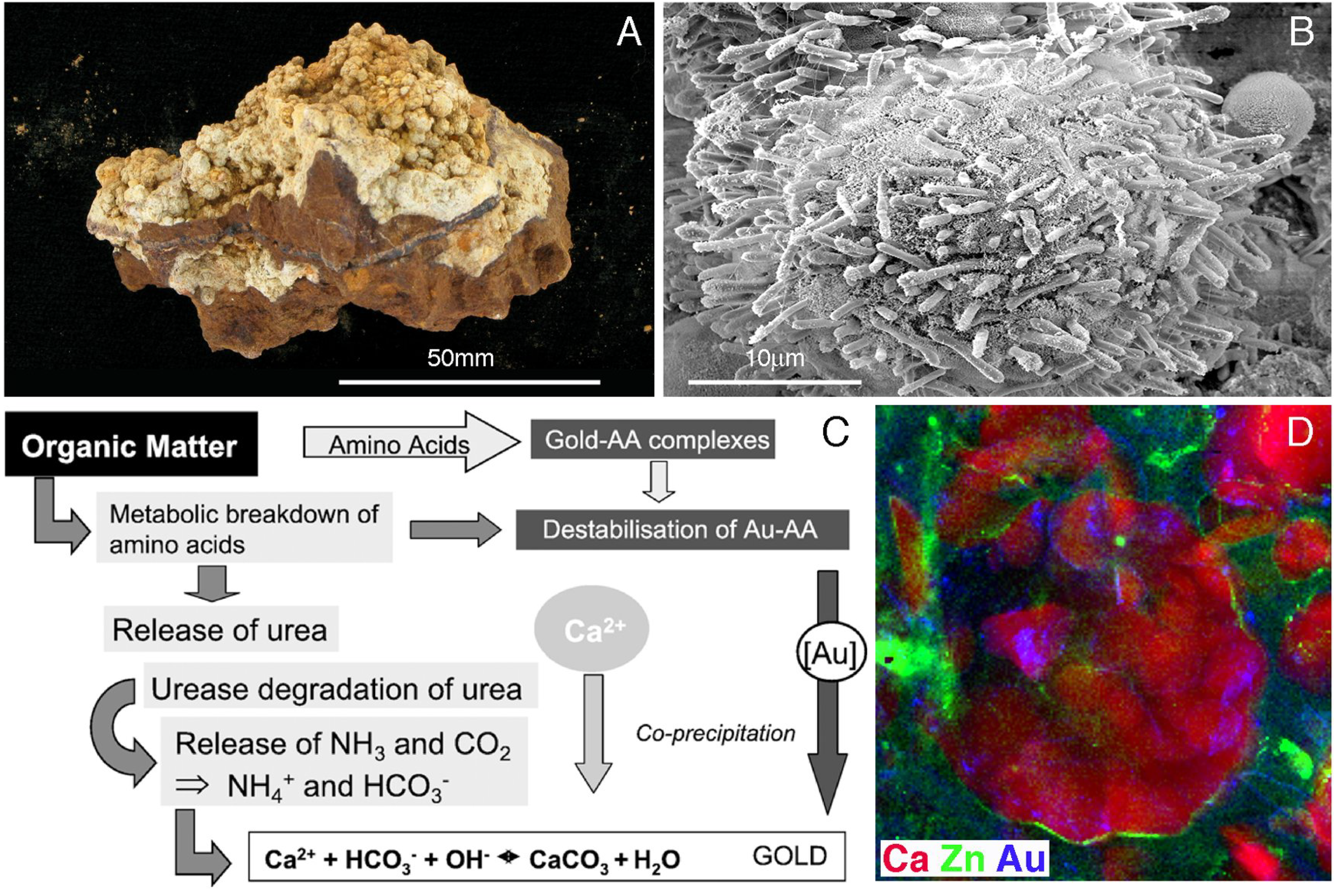
5. Geobiological Formation of Gold Anomalous Calcrete

Calcrete (pedogenic calcium carbonate) is an important sampling medium for geochemical gold exploration in semi-arid and arid regions of Australia, because it is widespread and easy to sample (Figure 8A; [68]). In addition, calcium shows a strong positive correlation with gold, but not with base metals, in calcrete overlying buried gold mineralization [68,69,70]. The formation of pedogenic carbonates has been ascribed to abiogenic as well as biogenically mediated precipitation, and both mechanisms have been observed in the natural environments. However, mass balance estimates by Castanier et al. [71] suggested that the heterotrophic microbially mediated carbonatogenesis accounts for most of the formation of these carbonates rather than abiotic processes. One of the most common heterotrophic processes leading to the formation of pedogenic carbonates is the degradation of urea (ureolysis), which is a product of the decomposition of purines and amino acids by microorganisms [72,73,74].
Figure 8. (A) Pedogenic carbonate forming on the surface of a weathered saprolitic rock from Ora Banda, Western Australia; (B) scanning electron micrographs showing the progressive calcification of bacterial cells in the enrichment culture experiments leading to the formation of spherical zoned biogenic calcium carbonates; (C) the coupled biogenic/abiogenic model for gold enrichment in calcrete; and (D) high resolution synchrotron μ-XRF element map of a spherical biogenic calcium carbonate crystal; RGB-overlay, calcium, red; zinc, blue, gold green (after [68,69,70]).
Minerals 03 00367 g008
Recent research has shown that the formation of gold-anomalous calcrete is biomediated through the activity of resident microorganisms, and is not simply the result of passive nucleation on inactive cells or evapotransporative processes as previously thought [11,37,68,70]. Calcified microfossils are highly abundant in calcrete from the Barns and other gold-prospects in Australia. These microfossils are morphological analogues of calcified cells and biofilms observed in incubation experiments conducted with active bacterial enrichment cultures from gold-anomalous calcareous sand (Figure 8B). Enriched cultures consisted of Bacillus and related Paenibacillus and Lysinibacillus. Calcium carbonates precipitated by this consortium consisted mostly of calcite, which is the main carbonate mineral constituting calcrete. Synchrotron micro-X-ray fluorescence (S-μXRF) mapping was used to assess the distribution of gold, zinc, calcium and other metals in calcium carbonates precipitated by active cultures. On a μm-scale the distribution of gold was heterogeneous in biogenic calcite and differed from base metal distributions, thus mimicking the spatial separation of these metals observed in calcrete (Figure 8D). The speciation of gold in these calcium carbonates, measured using micro-X-ray absorption near edge structure spectroscopy (μ-XANES), closely resembled that observed in environmental gold-anomalous calcrete. While metallic secondary gold was observed in gold “hotpots”, ionic gold was detected in the halo surrounding the “hotspot” [70]. In contrast, precipitates forming in the presence of dead bacterial cells or in abiogenic controls displayed different mineralogies as well as differences in gold distribution and speciation compared to calcrete. This indicated that active microbial processes combining biogenic calcium-carbonatogenesis with gold precipitation are likely to drive the formation of gold-anomalous calcrete.
Hence, a new model for gold anomalous calcrete formation was developed. In this model, gold is derived from underlying geologic sources, e.g., the hydrothermal deposits, where it is present in its free metallic state or associated with sulfide minerals, such as pyrite and arsenopyrite. In arid, surficial environments (down to 500 m below the land surface; [75]) chemolithoautotrophic iron- and sulfur-oxidizing bacteria, e.g., A. ferrooxidans and A. thiooxidans, and archaea, are known to form biofilms on metal sulfides (e.g., gold-bearing pyrites and arsenopyrites), and obtain metabolic energy by oxidizing these minerals [3,76]. During this process thiosulfate is produced, which in the presence of oxygen leads to gold oxidation and complexation [77]. Mobile gold complexes can then be taken up by plants and incorporated into plant tissue as nano- and micro-clusters of metallic gold or gold(I) bound to sulfur and nitrogen containing macromolecules. The subsequent decomposition of plant materials by microbiota leads to the formation of amino acids, cyanides and other organic compounds, which remobilize the plant-bound gold (Figure 8C). The decomposition of these gold complexing ligands by ureolytic microorganisms leads to the formation of urea resulting in the formation ammonium and carbon dioxide, hence inducing the physicochemical conditions required for calcium-carbonatogenesis and the co-precipitation of gold (Figure 8C). In addition, the bacteria may accumulate and biomineralise gold and act as nucleation sites for carbonate precipitation.
While it is apparent that microbial activities are at the heart of this model, abiogenic processes also play a role [68,69,70]. The dominance of the spore-forming bacteria at the Barns sites suggested a strong seasonal dependence of the microbial activity, most likely linked to the availability of water in the regolith profile. At times of higher water availability microbial decomposition of plant-derived organic matter leads to the formation of gold-bearing carbonates. Drying conditions lead to the passive sorption of aqueous gold complexes onto different regolith materials, especially organic matter, and evapotransporative processes promote the abiotic precipitation of additional calcium carbonates.

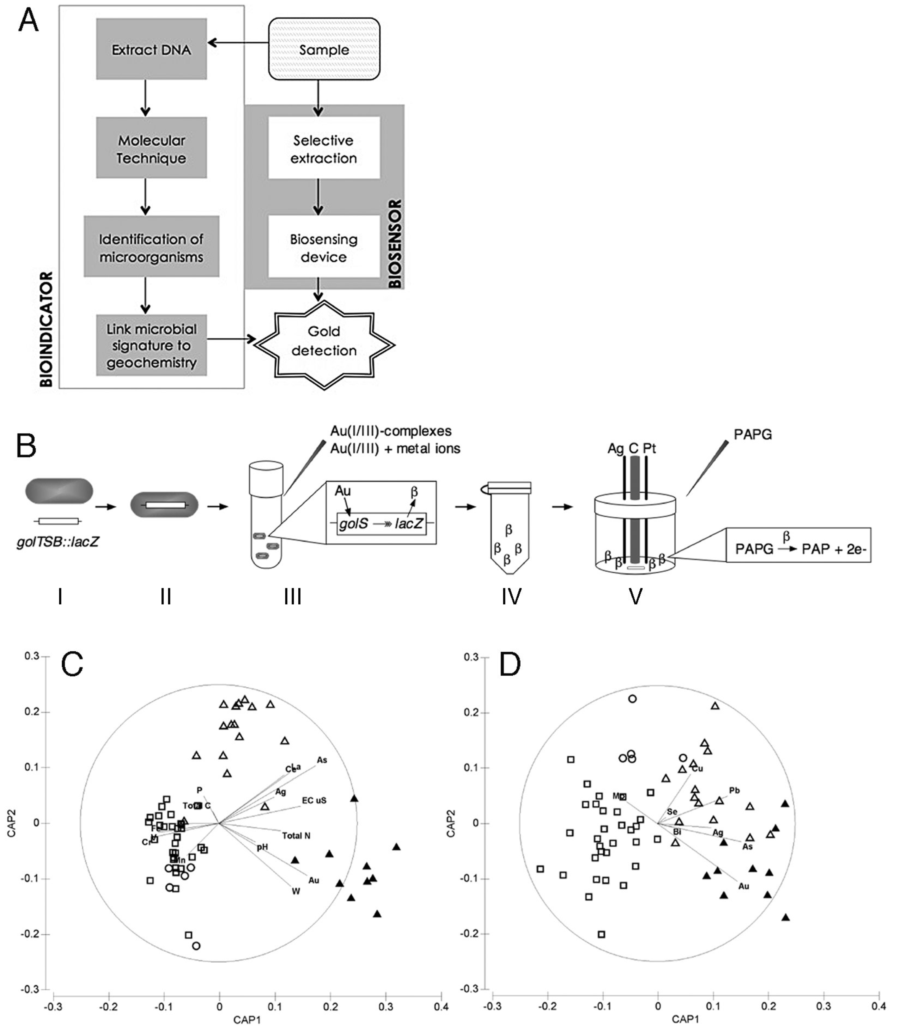
6. Exploration Geomicrobiology

The role of biological agents in gold mining operations is currently limited to the use of microorganisms in bioleaching and bioremediation. However, there are a number of ways in which biotechnology will be used in the near future to aid mineral exploration. The development of these biotechnologies was enabled by advances in the molecular-level understanding of role of microorganisms in the solubilization, dispersion and re-precipitation of gold. Based on this knowledge, the distribution of microbial species in soils overlying mineralization can be utilized to develop bioindicator systems (Figure 9A; [11,20,78,79]). In addition, the new in-depth knowledge of how microorganisms specifically interact with toxic gold complexes is being used to develop biosensors (Figure 9A,B).
Figure 9. (A) Distinction between biosensor and bioindicators [79]. (B) Construction and testing of a gold biosensor [20]: (I) The golTSB regulon is influenced by gold complexes in S. enterica serovar Typhimurium. (II) This golTSB::lacZ transcriptional fusion was introduced into E. coli. (III) Gold(I/III)-complexes or other metals were added and cells incubated. (IV) Cells were permeabilised for access to the β-galactosidase produced by the lacZ gene in the presence of gold. (V) Cells were transferred to an electrochemical cell, p-aminophenyl-β-D-galactopyranoside (PAPG) was added. The β-galactosidase cleaves PAPG to p-aminophenol (PAP), which is oxidized by the carbon electrode giving off electrons that are used to determine β-galactosidase activity. (C) and (D) results of a bioindicator study conducted with samples from the Old Pirate deposit, Australia [55]. Shown are ordination plots of the first two canonical axes produced by constrained canonical analysis of principal coordinates (CAP). In it 62 sites are compared based on geochemistry (C) and T-RFLP profiles of bacterial communities with vectors of correlations of pathfinder elements with CAP axes are overlain (D); symbols correspond to factors defining sampling sites: (▲) auriferous, erosional; (△) non-auriferous, erosional; (■) non-auriferous, collovial; (○) non-auriferous alluvial.
Minerals 03 00367 g009

6.1. Bioindicators

A central aim of exploration geomicrobiology is to understand the effect of microbial communities on metal turnover in geogenic environments (e.g., soils overlying mineral deposits), and in turn how community diversity and functioning are affected by these metals [4]. In recent years, modern molecular techniques have facilitated the generation of detailed inventories of microorganisms inhabiting different environments. Coupled with physico- and geochemical information this can give a complete biological and geochemical picture of a mineralized system [11,20]. Hence, bioindicator technologies can be developed, where the detection of certain microorganisms, microbial communities or functions indicates the presence of specific metals.
Techniques such as terminal-restriction fragment length polymorphism (T-RFLP), denaturing gradient gel electrophoresis (DGGE), thermal gradient gel electrophoresis (TGGE), and single strand conformation polymorphism (SSCP) allow for the high-throughput analysis of microbial communities. Recent developments in microarray analysis (PhyloChip and GeoChip) and DNA sequencing technologies have meant that microbial community profiles can be generated to cover a far greater depth of microbial species and functions than ever before [80]. For example, the PhyloChip is able to simultaneously detect more than 40,000 strains of microorganisms, while high-throughput sequencing methods theoretically can detect every single microorganism present in a sample.
In principle, there are two possible avenues for the application of microbial bioindicators [20]. These are: (i) comparison of microbial community profiles, known as “phylogenetic fingerprinting”; and, (ii) detection of specific genes, proteins and/or metabolites. Phylogenetic fingerprinting firstly involves identifying the microorganisms present in an environment. This information can then be used to link the presence of these microorganisms or combinations of microorganisms to environmental parameters. The detection of specific genes, proteins and/or metabolites is another very promising basis for the development of bioindicators [81]. Like antibiotic resistant genes, metal resistance genes are often located on plasmids, which are mobile genetic elements that are transferable between compatible microorganisms [82]. This means that two completely different microorganisms may have the same mechanisms of metal resistance. In contrast, phylogenetic fingerprinting relies on the assumption that the presence of microbial genera, species or strains (or their associations) can be specific to certain environmental conditions. If an indicator microorganism or set of microorganisms is not present it is assumed that these environmental conditions are not met. This may lead to the inadvertent dismissal of a positive site when in reality another species may have gained the ability to occupy that ecological niche, e.g., through the acquisition of a plasmid. Hence, the identification of genes and subsequently their proteins and metabolites may allow for more accurate profiling of an exploration site. There are a range of genes that are associated with the ability of microorganisms to resist and detoxify heavy metals, for example: sil genes for resistance to silver [83]; the czc, and cad genes for cadmium resistance [81]; chr, ncc and mer genes for resistance to chromium, nickel and mercury, respectively [84]. Other genes have been shown to be differentially abundant in the presence of metals, such as the gol and gig genes in the presence of gold [25,27]. The major advantages of using bioindicators as mineral exploration tools lie in: (i) the possibility that a multitude of different metals will be detectable simultaneously; and (ii) the ability of microorganisms to respond to very low concentrations of metals.
Research into the phylogenetic fingerprinting of anthropogenically heavy metal polluted soils is ongoing, and strong correlative links between the phylogenetic fingerprint of a given microbial community and the presence of metal contaminants have been established. However, few studies exist that have assessed microbial communities in geogenic metallomorphic environments with low to moderate metal contents, such as soils overlying buried mineralization [55,85]. Wakelin et al. [86,87] integrated several techniques to gain a better understanding of the composition and function of bacterial communities from a sulfidic mine tailing dump located 600 km northeast of Perth, Western Australia. Differences in bacterial communities were first assessed with DGGE and then members of the community were further identified using the PhyloChip. The results from this study showed that mobile phase elements, such as sulfur, zinc, chloride and aluminum, were the dominant drivers of bacterial community structures at this site. Using PhyloChip the authors were able to identify a greater level of species richness than had been reported at similar sites in the past [86,87]. In another study, 187 soil samples were collected from four naturally metallomorphic sites spanning three climatic zones across Australia to assess the links between soil microbial communities and geogenic factors, namely the underlying geology, the position in the landscape, i.e., regolith landform, and the presence of gold mineralization [54]. Field fresh soils and soils incubated with soluble gold(III)-complexes were analyzed using a polyphasic approach consisting of three-domain M-TRFLP combined with high-density phylogenetic (PhyloChip) and functional (GeoChip) microarrays. Geogenic factors were determined using geologic-, regolith- and soil-mapping criteria combined with geochemical analyses for 49 elements, pH and electrical conductivity in the soil samples. Multivariate statistical analyses showed the concentration of elements in soils was strongly linked to the underlying gold deposit and regolith landform (Figure 9C); note the example shown in Figure 9C,D relates to one of four case studies presented in Reith et al. [54], i.e., the study of 62 samples collected at the Old Pirate deposit in Northern Territory of Australia. Here, canonical correlation coefficients were high (δ1 0.91; δ2 0.70), and CA1 and CA2 accounted for 34.2% and 12.0% of variation in the bacterial dataset, respectively (Figure 9D). Minor elements were capable of explaining a significant proportion of the spread, e.g., gold-pathfinder elements, i.e., Ag, As, Au, Bi, Cu, Pb, Se, explained 21.6% of variation in the bacterial dataset, as illustrated by overlying Spearman correlation in Figure 9D. This demonstrated that the geochemical make-up of these sites is well reflected in the microbial community structure. In all case studies the bacterial communities generally displayed the highest correlation to geogenic factors and responded most sensitively to soluble gold(III)-complexes. PhyloChip analyses of key-samples revealed a greater abundance and diversity of α-Proteobacteria, especially Sphingomonas spp., and Bacillus spp. in auriferous and gold(III)-amended soils compared to background soils. Analyses of the functional potential of microbial communities in these soils using GeoChip showed an increasing abundance and diversity of metal resistance genes in soil ecosystems within mineralised zones compared to background materials. Copper-, gold-, chromium- and zinc- resistance genes showed a strong positive correlation to elevated concentrations of metals in these soils. In particular, the metal-resistance genes copA, chrA and czcA of the aurophillic bacterium C. metallidurans CH34 occurred only in soils overlying gold mineralization. This study showed that geogenic factors are important drivers of microbial community diversity and functioning; in particular, and for the first time, a direct influence of regolith landform on the microbial community was identified. The study also showed that microbial communities in geogenic metallomorphic environments have genetically adapted to elevated concentrations of heavy metals, e.g., gold and its pathfinder elements.

6.2. Biosensors

Biosensors are handheld analytical devices that are based on biological components and are developed to detect specific compounds. Research into biosensors has been focused around the monitoring of blood glucose levels; the detection and quantification of pathogens, food toxins, illicit drugs and heavy metals; and biosafety [88]. The basis behind the majority of these biosensing devices is the binding of an analyte to a protein, usually an enzyme, which induces a measurable change (e.g., electrochemical, optical, thermometric, piezoelectric, or magnetic), which can then be converted into a useful reading. Because proteins are highly selective and sensitive towards specific compounds, protein-based biosensor units are also highly selective and sensitive towards the specific analyte. For instance, the protein CueR’s sensitivity towards copper(II)-ions lies in the zeptomolar (10−21) range [89].
Geochemical exploration for gold is becoming increasingly important to the mining industry. Current samples for gold analyses typically require transport from remote localities to a laboratory equipped with suitable analytical facilities, such as Inductively Coupled Plasma-Mass Spectrometry (ICP-MS) or Instrumental Neutron Activation Analysis (INAA). Determining the concentration of gold in samples may take several weeks, leading to long delays in exploration campaigns. The use of biosensor technology in combination with traditional techniques for mineral exploration will add value through the speed, portability and the high selectivity of these devices. The development of biosensors for gold exploration will mean that exploration teams will be able to obtain information on gold concentrations in an environmental sample on site, rather than after weeks of laboratory analyses. In addition, biosensor devices may aid in mineral processing, where real-time in-line analysis of specific mineral components of ores can be determined. This enables real-time fine-tuning of the metallurgical process, which will improve recovery and decrease costs. Using molecular techniques, such as transcriptomics, genes from C. metallidurans strain CH34 and S. typhimurium have been identified and are now being investigated for their use as a gold biosensor (Figure 6; [25,27,90]).
C. metallidurans is of particular interest for the development of a gold biosensor. Transcriptome microarray analysis identified a number of genes (gol, cop, mer, ars and cus) in C. metallidurans strain CH34, which were more abundant upon cellular exposure to soluble gold compounds [37]. In the presence of gold(I/III)-complexes C. metallidurans induces a gold-specific genetic region (Rmet_4682–4687) which were more abundant with gold(III)-complexes, than with 16 other metal ions [27]. The close association of C. metallidurans with gold and its genetic response to the presence of gold makes this microorganism a particularly interesting candidate for the development of a gold biosensor.
Gold-regulated genes have also been detected in other bacteria such as the gol genes of S. typhimurium and S. enterica [25,90]. As an example, the golTSB genes from S. enterica serovar Typhimurium are selectively induced by gold(I/III)- complexes. In a recent study, the golTSB operon, attached to the reporter gene lacZ, was introduced into Escherichia coli. The induction of golTSB::lacZ with gold(I/III)-complexes was tested using a colorimetric β-galactosidase and electrochemical assay (Figure 9B; [78]). Measurements of the β-galactosidase activity for concentrations of both gold(I)- and gold(III)-complexes ranging from 0.1 to 5 μM [equivalent to 20 to 1000 ng g−1 (ppb)] were accurately quantified. To assess if the biosensor would work with natural samples, soils with different physiochemical properties were spiked with gold complexes. Subsequently, a selective extraction using 1 M thiosulfate was applied to extract the gold. The results showed that gold could be measured in these extracts with the same accuracy as ICP-MS (p < 0.05). This demonstrates that by combining selective extraction with the biosensor system the concentration of gold can be accurately measured, down to a quantification limit of 20 parts per billion (ppb, i.e., 0.1 μM) and a detection limit of 2 ppb (0.01 μM).

7. Conclusions and Outlook

This manuscript highlights the substantial progress that has been made in recent years towards understanding the fundamental processes underlying gold cycling in the environment. The results of these studies show that: (i) microbial biofilms mediating gold cycling occur on gold grains from (sub)-tropical, (semi)-arid, temperate and subarctic environments; (ii) gold-resistant or -tolerant β-Proteobacteria are particularly abundant in these biofilms; (iii) the β-Proteobacterium C. metallidurans appears to co-utilize detoxification mechanisms primarily aimed at more abundant metals for gold detoxification; (iv) S. typhimurium contains gold specific sensing and efflux mechanisms; (v) D. acidovorans excretes a siderophore, delftibactin, capable of reducing gold complexes to deal with elevated gold levels and increasing toxicity; (vi) metabolically active intracellular mechanisms and extracellular precipitation of gold nanoparticles in biofilms lead to the biomineralization of secondary gold; (vii) biogenic gold biominerals resemble secondary gold on natural gold grains; (viii) gold toxicity drives environmental gold cycling; (ix) platinum behaves differently to gold in most surface environments due to reduced mobility and lower toxicity of platinum complexes; and (x) gold anomalous calcrete is formed by microbial processes.
The recent revolution in understanding the role of biota in gold cycling at a molecular level forms the foundation for the development of biosensor and bioindicator systems that will aid gold exploration. Bioindicators complement geochemical methods, because they represent a more “holistic” probe of element mobility around buried mineralization, and are sensitive not only to local metal contents, but also to many associated processes (e.g., breakdown of sulfide minerals; fluxes of volatiles). Because molecular methods of analysis develop rapidly, the production of large amounts of high precision genetic data at prices competitive with traditional geochemical exploration tools is becoming a reality, opening the way for bioindicators to become a standard tool in mineral exploration. Biosensors are at the proof of concept stage for gold measurement down to ppb levels; further development of the technology will offer an inexpensive, field-laboratory deployable tool for geochemical gold exploration.
Additional research is required to develop these biological exploration techniques into standard tools. To develop a comprehensive bioindicator approach we need to study the metagenomes of metal-rich and background soils, use bioinformatics and multivariate statistics to identify microorganisms and functional genes, which are indicative of the presence of gold, and develop 200–300 probes for gold-specific microbial species and functional genes, which can be used an inexpensive targeted microarray. For the construction of a robust gold biosensor, purification the protein(s) associated with gold sensing and detoxification needs to be conducted. Crystals for X-ray analyses of protein structure and gold binging capacity need to be produced. Assessment of the accuracy, specificity, and limit of detection (likely in the parts per trillion-range with a protein-based sensor) in sampling materials needs to be conducted. To achieve this we also need to understand how mobile gold is speciated in soils and groundwaters, and to develop an effective extraction technique for gold contained in different sampling matrices, e.g., soils, sediments and other weathered materials.

Acknowledgments

The authors would like to thank the support of the Australian Research Council (LP100200102); Deutsche Forschungsgemeinschaft (DFG); The Commonwealth Scientific and Industrial Research Organization; The University of Adelaide; The South Australian Museum; Newmont Mining Corp.; Barrick Gold Corp.; the South Australian Department for Manufacturing, Innovation, Trade, Resources and Energy (DMITRE), the Institute for Mineral and Energy Resources; and the Centre for Tectonics, Resources and Exploration. The authors also would like to thank the three anonymous reviewers for their insightful comments and the thorough discussion that helped to improve the paper.

Conflicts of Interest

The authors declare no conflict of interest.

References

  1. Ehrlich, H.L. Geomicrobiology: Its significance for geology. Earth Sci. Rev. 1998, 45, 45–60. [Google Scholar] [CrossRef]
  2. Ehrlich, H.L.; Newman, D.K. Geomicrobiology, 5th ed.; CRC Press: Boca Raton, FL, USA, 2008. [Google Scholar]
  3. Southam, G.; Saunders, J.A. The geomicrobiology of ore deposits. Econ. Geol. 2005, 100, 1067–1084. [Google Scholar] [CrossRef]
  4. Reith, F.; Dürr, M.; Welch, C.F.; Rogers, S.L. The Geomicrobiology of the Regolith. In Regolith Science; Scott, K., Pain, C.F., Eds.; CSIRO Press: Melbourne, Australia, 2008; pp. 127–159. [Google Scholar]
  5. Gadd, G.M. Metals, minerals and microbes: Geomicrobiology and bioremediation. Microbiology 2010, 156, 609–643. [Google Scholar] [CrossRef]
  6. Gold, T. The deep, hot biosphere. Proc. Natl. Acad. Sci. USA 1992, 89, 6045–6049. [Google Scholar] [CrossRef]
  7. Fredrickson, J.K.; Balkwill, D.L. Geomicrobial processes and biodiversity in the deep terrestrial subsurface. Geomicrobiol. J. 2006, 23, 345–356. [Google Scholar] [CrossRef]
  8. Fry, J.C.; Parkes, R.J.; Cragg, B.A.; Weightman, A.J.; Webster, G. Prokaryotic biodiversity and activity in the deep subseafloor biosphere. FEMS Microbiol. Ecol. 2008, 66, 181–196. [Google Scholar] [CrossRef]
  9. Reith, F. Life in the deep subsurface. Geology 2011, 39, 287–288. [Google Scholar] [CrossRef]
  10. Lloyd, J.R. Microbial reduction of metals and radionuclides. FEMS Microbiol. Rev. 2003, 27, 411–425. [Google Scholar] [CrossRef]
  11. Reith, F.; Lengke, M.F.; Falconer, D.; Craw, D.; Southam, G. The geomicrobiology of gold. ISME J. 2007, 1, 567–584. [Google Scholar] [CrossRef]
  12. Southam, G.; Lengke, M.F.; Fairbrother, L.; Reith, F. The biogeochemistry of gold. Elements 2009, 5, 303–307. [Google Scholar] [CrossRef]
  13. Hough, R.M.; Butt, C.R.M.; Reddy, S.M.; Verrall, M. Gold nuggets: Supergene or hypogene? Aust. J. Earth Sci. 2007, 54, 959–964. [Google Scholar] [CrossRef]
  14. Hough, R.; Noble, R.; Hitchen, G.; Hart, G.; Reddy, S.; Saunders, M.; Clode, P.; Vaughan, D.; Lowe, J.; Gray, D.; et al. Naturally occurring gold nanoparticles and nanoplates. Geology 2008, 36, 571–574. [Google Scholar] [CrossRef]
  15. Hough, R.M.; Noble, R.R.P.; Reich, M. Natural gold nanoparticles. Ore Geol. Rev. 2011, 42, 55–61. [Google Scholar] [CrossRef]
  16. Tomkins, A.G. A biogeochemical influence on the secular distribution of orogenic gold. Econ. Geol. 2013, 108, 193–197. [Google Scholar] [CrossRef]
  17. Lengke, M.; Southam, G. Bioaccumulation of gold by sulfate-reducing bacteria cultured in the presence of gold(I)-thiosulfate complex. Geochim. Cosmochim. Acta 2006, 70, 3646–3661. [Google Scholar] [CrossRef]
  18. Lengke, M.F.; Southam, G. The deposition of elemental gold from gold(I)-thiosulfate complex mediated by sulfate-reducing bacterial conditions. Econ. Geol. 2007, 102, 109–126. [Google Scholar] [CrossRef]
  19. Jones, B.; Renault, R.W.; Rosen, M.R. Biogenicity of gold- and silver-bearing siliceous sinters forming in hot (75 °C) anaerobic spring-waters of Champagne Pool, Waiotapu, North Island, New Zealand. J. Geol. Soc. 2001, 158, 895–911. [Google Scholar] [CrossRef]
  20. Zammit, C.M.; Cook, N.; Brugger, J.; Ciobanu, C.L.; Reith, F. The future of biotechnology for gold exploration and processing. Miner. Eng. 2012, 32, 45–53. [Google Scholar] [CrossRef]
  21. Etschmann, B.E.; Black, J.R.; Grundler, P.V.; Borg, S.; Brewe, D.; McPhail, D.C.; Spiccia, L.; Brugger, J. Copper(I) speciation in mixed thiosulfate-chloride and ammonia-chloride solutions: XAS and UV-Visible spectroscopic studies. RSC Adv. 2011, 1, 1554–1566. [Google Scholar] [CrossRef]
  22. Vlassopoulos, D.; Wood, S.A.; Mucci, A. Gold speciation in natural waters: II. The importance of organic complexing—Experiments with some simple model ligands. Geochim. Cosmochim. Acta 1990, 54, 1575–1586. [Google Scholar] [CrossRef]
  23. Gray, D.J. The Aqueous Chemistry of Gold in the Weathering Environment; Cooperative Research Centre for Landscape Evolution and Mineral Exploration: Wembley, Australia, 1998; Open File Report No 38; p. 57. [Google Scholar]
  24. Fairbrother, L.; Etschmann, B.; Brugger, J.; Shapter, J.; Southam, G.; Reith, F. Biomineralization of gold in biofilms of Cupriavidus metallidurans. Environ. Sci. Technol. 2013, 47, 2628–2635. [Google Scholar] [CrossRef]
  25. Checa, S.K.; Espariz, M.; Audero, M.E.P.; Botta, P.E.; Spinelli, S.V.; Soncini, F.C. Bacterial sensing of and resistance to gold salts. Mol. Microbiol. 2007, 63, 1307–1318. [Google Scholar] [CrossRef]
  26. Lengke, M.F.; Fleet, M.E.; Southam, G. Bioaccumulation of gold by filamentous cyanobacteria between 25 and 200 °C. Geomicrobiol. J. 2006, 23, 591–597. [Google Scholar] [CrossRef]
  27. Reith, F.; Etschmann, B.; Grosse, C.; Moors, H.; Benotmane, M.A.; Monsieurs, P.; Grass, G.; Doonan, C.; Vogt, S.; Lai, B.; et al. Mechanisms of gold biomineralization in the bacterium Cupriavidus metallidurans. Proc. Natl. Acad. Sci. USA 2009, 106, 17757–17762. [Google Scholar] [CrossRef]
  28. Reith, F.; Rogers, S.L.; McPhail, D.C.; Webb, D. Biomineralization of gold: Biofilms on bacterioform gold. Science 2006, 313, 233–236. [Google Scholar] [CrossRef]
  29. Lengke, M.F.; Fleet, M.E.; Southam, G. Morphology of gold nanoparticles synthesized by filamentous cyanobacteria from gold(I)-thiosulfate and gold(III)-chloride complexes. Langmuir 2006, 22, 2780–2787. [Google Scholar] [CrossRef]
  30. Mossman, D.J.; Dyer, B.D. The geochemistry of Witwatersrand-type gold deposits and the possible influence of ancient prokaryotic communities on gold dissolution and precipitation. Precambrian Res. 1985, 30, 303–319. [Google Scholar] [CrossRef]
  31. Frimmel, H.E.; Le Roex, A.P.; Knight, A.P.; Minter, W.E.L. A case study of the postdepositional alteration of Witatersrand Basal Reef gold placer. Econ. Geol. 1993, 88, 249–265. [Google Scholar] [CrossRef]
  32. Falconer, D.M.; Craw, D.; Youngson, J.H.; Faure, K. Gold and sulphide minerals in Tertiary quartz pebble conglomerate gold placers, Southland, New Zealand. Ore Geol. Rev. 2006, 28, 525–545. [Google Scholar] [CrossRef]
  33. Falconer, D.M.; Craw, D. Supergene gold mobility: A textual and geochemical study from gold placers in southern New Zealand. Econ. Heol. Spec. Publ. 2009, 14, 77–93. [Google Scholar]
  34. Reith, F.; Fairbrother, L.; Nolze, G.; Wilhelmi, O.; Clode, P.L.; Gregg, A.; Parsons, J.E.; Wakelin, S.A.; Pring, A.; Hough, R.; et al. Nanoparticle factories: Biofilms hold the key to gold dispersion and nugget formation. Geology 2010, 38, 843–846. [Google Scholar] [CrossRef]
  35. Fairbrother, L.; Brugger, J.; Shapter, J.; Laird, J.S.; Southam, G.; Reith, F. Supergene gold transformation: Biogenic secondary and nano-particulate gold from arid Australia. Chem. Geol. 2012, 320, 17–31. [Google Scholar]
  36. Reith, F.; Stewart, L.; Wakelin, S.A. Supergene gold transformation: Secondary and nano-particulate gold from southern New Zealand. Chem. Geol. 2012, 320, 32–45. [Google Scholar] [CrossRef]
  37. Mann, A.W. Mobility of gold and silver in lateritic weathering profiles: Some observations from Western Australia. Econ. Geol. 1984, 79, 38–49. [Google Scholar] [CrossRef]
  38. Wilson, A.F. Origin of quartz-free gold nuggets and supergene gold found in laterites and soils—A review and some new observations. Aust. J. Earth Sci. 1984, 31, 303–316. [Google Scholar]
  39. Bischoff, G.C.O. The biological origin of bacterioform gold from Australia. Neues Jahrb. Geol. Paläontol. Monatshefte 1997, 6, 329–338. [Google Scholar]
  40. McCready, A.J.; Parnell, J.; Castro, L. Crystalline placer gold from the Rio Neuquen, Argentina: Implications for the gold budget in placer gold formation. Econ. Geol. 2003, 98, 623–633. [Google Scholar]
  41. Chapman, R.J.; Mortensen, J.K.; LeBarge, W.P. Styles of lode gold mineralization contributing to the placers of the Indian River and Black Hills Creek, Yukon Territory, Canada as deduced from microchemical characterization of placer gold grains. Miner. Deposita 2011, 46, 881–903. [Google Scholar] [CrossRef]
  42. Knight, J.B.; Morison, S.R.; Mortensen, J.K. The relationship between placer gold particle shape, rimming, and distance of fluvial transport as exemplified by gold from the Klondike District, Yukon Territory, Canada. Econ. Geol. 1999, 94, 635–648. [Google Scholar] [CrossRef]
  43. Usher, A.; McPhail, D.C.; Brugger, J. A spectrophotometric study of aqueous Au(III) halide–hydroxide complexes at 25–80 °C. Geochim. Cosmochim. Acta 2009, 73, 3359–3380. [Google Scholar] [CrossRef]
  44. Nies, D.H. Efflux-mediated heavy metal resistance in prokaryotes. FEMS Microbiol. Rev. 2003, 27, 313–339. [Google Scholar] [CrossRef]
  45. Nam, S.H.; Lee, W.M.; Shin, Y.J.; Yoon, S.J.; Kim, S.W.; Kwak, J.I.; An, Y.J. Derivation of guideline values for gold(III) ion toxicity limits to protect aquatic ecosystems. Water Res. 2013. [Google Scholar] [CrossRef]
  46. Wiesemann, N.; Mohr, J.; Grosse, C.; Herzberg, M.; Hause, G.; Reith, F.; Nies, D.H. Influence of copper resistance determinants on gold transformation by Cupriavidus metallidurans strain CH34. J. Bacteriol. 2013, 195, 2298–2308. [Google Scholar] [CrossRef]
  47. Pontel, L.B.; Audero, M.E.; Espariz, M.; Checa, S.K.; Soncini, F.C. GolS controls the response to gold by the hierarchical induction of Salmonella-specific genes that include a CBA efflux-coding operon. Mol. Mircobiol. 2007, 66, 814–825. [Google Scholar] [CrossRef]
  48. Von Rozycki, T.; Dietrich, N.H. Cupriavidus metallidurans: Evolution of a metal-resistant bacterium. Antonie Leeuwenhoek 2009, 96, 115–139. [Google Scholar] [CrossRef]
  49. Janssen, P.J.; van Houdt, R.; Moors, H.; Monsieurs, P.; Morin, N.; Michaux, A.; Benotmane, M.A.; Leys, N.; Vallaeys, T.; Lapidus, A.; et al. The complete genome sequence of Cupriavidus metallidurans strain CH34, a master survivalist in harsh and anthropogenic environments. PLoS One 2010, 5, e10433. [Google Scholar] [CrossRef]
  50. Mergeay, M.; Nies, D.; Schlegel, H.G.; Gerits, J.; Charles, P.; Van Gijsegem, F. Alcaligenes eutrophus CH34 is a facultative chemolithotroph with plasmid-bound resistance to heavy metals. J. Bacteriol. 1985, 162, 328–334. [Google Scholar]
  51. Kenney, J.P.L.; Song, Z.; Bunker, B.A.; Fein, J.B. An experimental study of Au removal from solution by non-metabolizing bacterial cells and their exudates. Geochim. Cosmochim. Acta 2012, 87, 51–60. [Google Scholar] [CrossRef]
  52. Song, Z.; Kenney, J.P.L.; Fein, J.B.; Bunker, B.A. An X-ray absorption fine structure study of Au adsorbed onto the non-metabolizing cells of two soil bacterial species. Geochim. Cosmochim. Acta 2012, 86, 103–117. [Google Scholar] [CrossRef]
  53. Kabata-Pendias, A.; Pendias, H. Trace Elements in Soils and Plants; CRC Press: Boca Raton, FL, USA, 1984; p. 315. [Google Scholar]
  54. Reith, F.; Brugger, J.; Zammit, C.M.; Gregg, A.L.; Goldfarb, K.C.; Andersen, G.L.; DeSantis, T.Z.; Piceno, Y.M.; Brodie, E.L.; Lu, Z.; et al. Influence of geogenic factors on microbial communities in metallogenic Australian soils. ISME J. 2012, 6, 2107–2118. [Google Scholar] [CrossRef]
  55. Reith, F.; McPhail, D.C.; Christy, A.G. Bacillus cereus, gold and associated elements in soil and other regolith samples from Tomakin Park Gold Mine in southeastern New South Wales, Australia. J. Geochem. Explor. 2005, 85, 81–98. [Google Scholar] [CrossRef]
  56. Magnani, D.; Solioz, M. How Bacteria Handle Copper. In Molecular Microbiology of Heavy Metals; Nies, D., Silver, S., Eds.; Springer: Berlin, Germany, 2007; Volume 6, pp. 259–285. [Google Scholar]
  57. Todd, E. Epidemiology of foodborne illness: North Amercia. Lancet 1990, 336, 788–790. [Google Scholar] [CrossRef]
  58. Checa, S.; Soncini, F. Bacterial gold sensing and resistance. BioMetals 2011, 24, 419–427. [Google Scholar] [CrossRef]
  59. Eswaran, J.; Koronakis, E.; Higgins, M.K.; Hughes, C.; Koronakis, V. Three’s company: Component structures brining a closer view of trpartite drug efflux pumps. Curr. Opin. Struct. Biol. 2004, 14, 741–747. [Google Scholar] [CrossRef]
  60. Wen, A.; Fegan, M.; Hayward, C.; Chakraborty, S.; Sly, L.I. Phylogenetic relationships among members of the Comamonadaceae, and description of Delftia acidovorans (den Dooren de Jong 1926 and Tamaoka et al. 1987) gen. nov., comb. nov. Int. J. Syst. Bacteriol. 1999, 49, 567–576. [Google Scholar] [CrossRef]
  61. Hoffmann, D.; Kleinsteuber, S.; Muller, R.H.; Babel, W. A transposon encoding the complete 2,4-dichlorophenoxyacetic acid degradation pathway in the alkalitolerant strain Delftia acidovorans P4a. Microbiology 2003, 149, 2545–2556. [Google Scholar] [CrossRef]
  62. Johnston, C.W.; Wyatt, M.A.; Li, X.; Ibrahim, A.; Shuster, J.; Southam, G.; Magarvey, N.A. Gold biomineralization by a metallophore from a gold-associated microbe. Nat. Chem. Biol. 2013, 9, 241–243. [Google Scholar] [CrossRef]
  63. Schalk, I.J.; Hannauer, M.; Braud, A. New roles for bacterial siderophores in metal transport and tolerance. Environ. Microbiol. 2011, 13, 2844–2854. [Google Scholar] [CrossRef]
  64. Schroefel, A.; Kratosova, G.; Bohunicka, M.; Dobrocka, E.; Vavra, I. Biosynthesis of gold nanoparticles using diatoms–silica-gold and EPS-gold bionanocomposite formation. J. Nanopart. Res. 2011, 13, 3207–3216. [Google Scholar] [CrossRef] [Green Version]
  65. Colica, G.; Caparrotta, S.; Bertini, G.; de Philippis, R. Gold biosorption by exopolysaccharide producing cyanobacteria and purple nonsulphur bacteria. J. Appl. Microbiol. 2012, 113, 1380–1388. [Google Scholar] [CrossRef]
  66. Malhotra, A.; Dolma, K.; Kaur, N.; Rathore, Y.S.; Ashish; Mayilraj, S.; Choudhury, A.R. Biosynthesis of gold and silver nanoparticles using a novel marine strain of Stenotrophomonas. Bioresour. Technol. 2013, 142, 727–731. [Google Scholar] [CrossRef]
  67. Brugger, J.; Etschmann, B.; Grosse, C.; Plumridge, C.; Kaminski, J.; Paterson, D.; Shar, S.S.; Ta, C.; Howard, D.L.; de Jonge, M.D.; et al. Can biological toxicity drive the contrasting behavior of platinum and gold in surface environments? Chem. Geol. 2013, 343, 99–110. [Google Scholar] [CrossRef]
  68. Schmidt Mumm, A.; Reith, F. Biomediation of calcrete at the gold anomaly of the Barns prospect, Gawler Craton, South Australia. J. Geochem. Explor. 2007, 92, 13–33. [Google Scholar] [CrossRef]
  69. Reith, F.; Wakelin, S.A.; Gregg, A.L.; Schmidt Mumm, A. A microbial pathway for the formation of gold-anomalous calcrete. Chem. Geol. 2009, 258, 315–326. [Google Scholar] [CrossRef]
  70. Reith, F.; Etschmann, B.; Dart, R.C.; Brewe, D.L.; Vogt, S.; Schmidt Mumm, A.; Brugger, J. Distribution and speciation of gold in biogenic and abiogenic calcium carbonates—Implications for the formation of gold anomalous calcrete. Geochim. Cosmochim. Acta 2011, 75, 1942–1956. [Google Scholar] [CrossRef]
  71. Castanier, S.; Le Metayer-Levrel, G.; Perthuisot, J.P. Ca-carbonates precipitation and limestone genesis—The microbiogeologist point of view. Sediment. Geol. 1999, 126, 9–23. [Google Scholar] [CrossRef]
  72. Vogels, G.D.; van der Drift, C. Degradation of purines and pyrimidines by microorganisms. Bacteriol. Rev. 1976, 40, 403–468. [Google Scholar]
  73. Cunin, R.; Glansdorff, N.; Pierard, A.; Stalon, V. Biosythesis and metabolism of arginine in bacteria. Microbiol. Rev. 1986, 50, 314–352. [Google Scholar]
  74. Millo, C.; Dupraz, S.; Ader, M.; Guyot, F.; Thaler, C.; Foy, E.; Ménez, B. Carbon isotope fractionation during calcium carbonate precipitation induced by ureolytic bacteria. Geochim. Cosmochim. Acta 2012, 98, 107–124. [Google Scholar] [CrossRef]
  75. Enders, M.S.; Knickerbocker, C.; Titley, S.R.; Southam, G. The role of bacteria in the supergene environment of the morenci porphyry copper deposit, Greenlee County, Arizona. Econ. Geol. 2006, 101, 59–70. [Google Scholar] [CrossRef]
  76. Nordstrom, D.K.; Southam, G. Geomicrobiology of sulfide mineral oxidation. Rev. Mineral. Geochem. 1997, 35, 361–390. [Google Scholar]
  77. Aylmore, M.G.; Muir, D.M. Thiosulfate leaching of gold—A review. Miner. Eng. 2001, 14, 135–174. [Google Scholar] [CrossRef]
  78. Zammit, C.M.; Quaranta, D.; Gibson, S.; Zaitouna, A.J.; Ta, C.; Brugger, J.; Lai, R.Y.; Grass, G.; Reith, F. A whole-cell biosensor for the detection of gold. PLoS One 2013, 8, e69292. [Google Scholar] [CrossRef]
  79. Zammit, C.M.; Cook, N.; Brugger, J.; Ciobanu, C.L.; Reith, F. The Future of Biotechnology for Gold Exploration and Processing. In World Gold; AUSIMM Publishing: Carlton, Australia, 2013; pp. 233–246. [Google Scholar]
  80. Rastogi, G.; Sani, R. Molecular Techniques to Assess Microbial Community Structure, Function, and Dynamics in the Environment. In Microbes and Microbial Technology; Ahmad, I., Ahmad, F., Pichtel, J., Eds.; Springer: Berlin, Germany, 2011; pp. 29–57. [Google Scholar]
  81. Hu, N.; Zhao, B. Key genes involved in heavy-metal resistance in Pseudomonas putida CD2. FEMS Microbiol. Lett. 2007, 267, 17–22. [Google Scholar] [CrossRef]
  82. Mergeay, M.; Monchy, S.; Janssen, P.; Houdt, R.; Leys, N. Megaplasmids in Cupriavidus Genus and Metal Resistance. In Microbial Megaplasmids; Schwartz, E., Ed.; Springer: Berlin, Germany, 2009; Volume 11, pp. 209–238. [Google Scholar]
  83. Woods, E.J.; Cochrane, C.A.; Percival, S.L. Prevalence of silver resistance genes in bacteria isolated from human and horse wounds. Vet. Microbiol. 2009, 138, 325–329. [Google Scholar] [CrossRef]
  84. Abou-Shanab, R.A.I.; van Berkum, P.; Angle, J.S. Heavy metal resistance and genotypic analysis of metal resistance genes in gram-positive and gram-negative bacteria present in Ni-rich serpentine soil and in the rhizosphere of Alyssum murale. Chemosphere 2007, 68, 360–367. [Google Scholar] [CrossRef]
  85. Reith, F.; Rogers, S.L. Assessment of bacterial communities in auriferous and non-auriferous soils using genetic and functional fingerprinting. Geomicrobiol. J. 2008, 25, 203–215. [Google Scholar] [CrossRef]
  86. Wakelin, S.; Anand, R.R.; Macfarlane, C.; Reith, F.; Noble, R.; Rogers, S. Assessing microbiological surface expression over an overburden-covered VMS deposit. J. Geochem. Explor. 2012, 112, 262–271. [Google Scholar] [CrossRef]
  87. Wakelin, S.A.; Anand, R.R.; Reith, F.; Gregg, A.L.; Noble, R.R.P.; Goldfarb, K.C.; Andersen, G.L.; DeSantis, T.Z.; Piceno, Y.M.; Brodie, E.L. Bacterial communities associated with a mineral weathering profile at a sulphidic mine tailings dump in arid Western Australia. FEMS Microbiol. Ecol. 2012, 79, 298–311. [Google Scholar] [CrossRef]
  88. Luong, J.H.T.; Male, K.B.; Glennon, J.D. Biosensor technology: Technology push versus market pull. Biotechnol. Adv. 2008, 26, 492–500. [Google Scholar] [CrossRef]
  89. Changela, A.; Chen, K.; Xue, Y.; Holschen, J.; Outten, C.E.; O’Halloran, T.V.; Mondragon, A. Molecular basis of metal-ion selectivity and zeptomolar sensitivity by CueR. Science 2003, 301, 1383–1387. [Google Scholar] [CrossRef]
  90. Cerminati, S.; Soncini, F.C.; Checa, S.K. Selective detection of gold using genetically engineered bacterial reporters. Biotechnol. Bioeng. 2011, 108, 2553–2560. [Google Scholar] [CrossRef]

