Burial Diagenesis of Magnetic Minerals: New Insights from the Grès d’Annot Transect (SE France)

Abstract
:1. Introduction
2. Geological Background
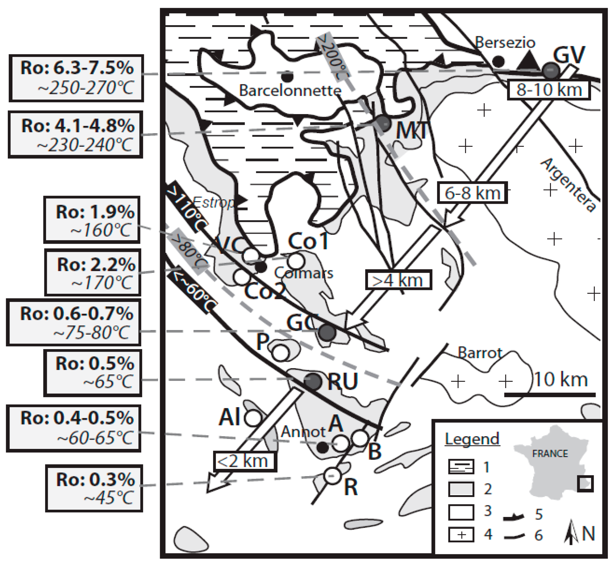
2.1. Geological Setting
2.2. Burial History

3. Methods
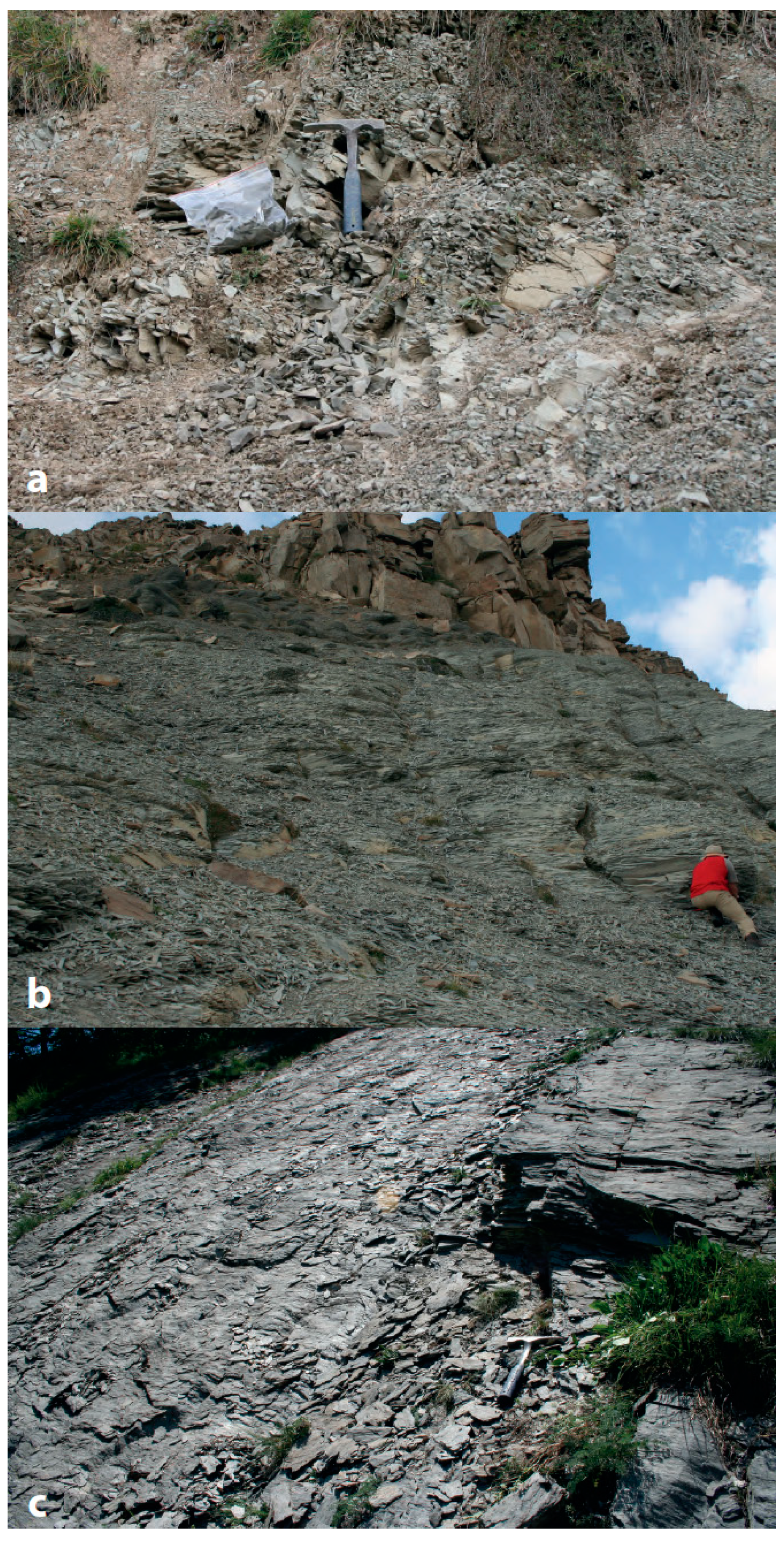
3.1. Sampling and Mineralogy

| Sampling Site | Sample | Latitude | Longitude | Lithology | Mean Ro (%) | SD Ro | TOC (%) |
|---|---|---|---|---|---|---|---|
| Allons (Al) | 1A | N 43°59′05.9″ | E 6°34′56.3″ | Marl (MBl) | |||
| 2A | N 44°00′09.4″ | E 6°34′11.1″ | Marl (MBl) | ||||
| Rouaine (R) | 4A | N 43°56′01.1″ | E 6°40′12.9″ | Marl (MBl) | |||
| 5A | N 43°56′06.6″ | E 6°40′29.4″ | Fine-grained sandstone (GA) | ||||
| Braux (B) | 6A | N 43°58′16″ | E 6° 42′17.2″ | Marl (MBl) | |||
| 7A | N 43°58′16″ | E 6°42′17.2″ | Fine-grained sandstone (GA) | ||||
| Annot (A) | A0 | N 43°57′43.1″ | E 6°40′34.6″ | Marl (MBl) | |||
| 11A | ND | ND | Marl (MBl) | ||||
| 12A | ND | ND | Fine-grained sandstone (GA) | ||||
| Le Ruch (RU) | RUmg | N 44°02′40.5″ | E 6°40′28.1″ | Marl (MBl) | |||
| RUmb | N 44°02′40.5″ | E 6°40′28.1″ | Turbiditic pelite (MBr) | 0.54 | 0.1 | 0.23 | |
| Grand Coyer (GC) | CY1p | N 44°05′09.1″ | E 6°41′0″ | Turbiditic pelite (GA) | 0.61 | 0.07 | 0.35 |
| CY3p | N 44°05′09.1″ | E 6°41′0″ | Turbiditic pelite (GA) | 0.65 | 0.06 | 0.68 | |
| CY5 | N 44°05′09.1″ | E 6°41′0″ | Turbiditic pelite (GA) | ||||
| CY6 | N 44°05′09.1″ | E 6°41′0″ | Turbiditic pelite (GA) | ||||
| CY7 | N 44°05′09.1″ | E 6°41′0″ | Turbiditic pelite (GA) | ||||
| CY8 | N 44°05′09.1″ | E 6°41′0″ | Turbiditic pelite (GA) | ||||
| CY10 | N 44°05′09.1″ | E 6°41′0″ | Turbiditic pelite (GA) | ||||
| CY11 | N 44°05′09.1″ | E 6°41′0″ | Turbiditic pelite (GA) | ||||
| Peyresq (P) | 20A | N 44°02′12.0″ | E 6°36′25.6″ | Marl (MBl) | |||
| Colmars (Co1) | 21A | N 44°09′11.1″ | E 6°32′41.2″ | Marl (MBl) | |||
| 22A | N 44°09′26.2″ | E 6°32′29.1″ | Marl (MBl) | ||||
| 23A | N 44°09′49.3″ | E 6°31′50.7″ | Marl (MBl) | ||||
| Colmars (Co2) | 25A | N 44°09′07.7″ | E 6°40′28.7″ | Marl (MBl) | |||
| 26A | N 44°09′26.9″ | E 6°39′19.0″ | Marl (MBl) | ||||
| Villars-Colmars (VC) | 13A | ND | ND | Turbiditic pelite (GA) | |||
| 14A | ND | ND | Turbiditic pelite (GA) | ||||
| 15A | ND | ND | Fine-grained sandstone (GA) | ||||
| La Moutière (MT) | MT12 | N 44°18′58″ | E 6°47′46″ | Turbiditic pelite (GA) | 4.06 | 0.16 | 0.34 |
| MT17 | N 44°18′58″ | E 6°47′46″ | Turbiditic pelite (GA) | ||||
| MT29 | N 44°18′58″ | E 6°47′46″ | Turbiditic pelite (GA) | 4.13 | 0.17 | 0.28 | |
| MT120 | N 44°18′58″ | E 6°47′46″ | Turbiditic pelite (GA) | ||||
| MTmg | N 44°18′58″ | E 6°47′46″ | Marl (MBl) | ||||
| Gias Vallonetto (GV) | GV1 | N 44°21′41.7″ | E 7°03′32.4″ | Turbiditic pelite (GA) | 6.29 | 0.41 | 0.61 |
| GV11 | N 44°21′41.7″ | E 7°03′32.4″ | Turbiditic pelite (GA) | 7.47 | 0.4 | 0.43 | |
| GVmg | N 44°21′41.7″ | E 7°03′32.4″ | Marl (MBl) |
3.2. Analytical Methods
4. Results
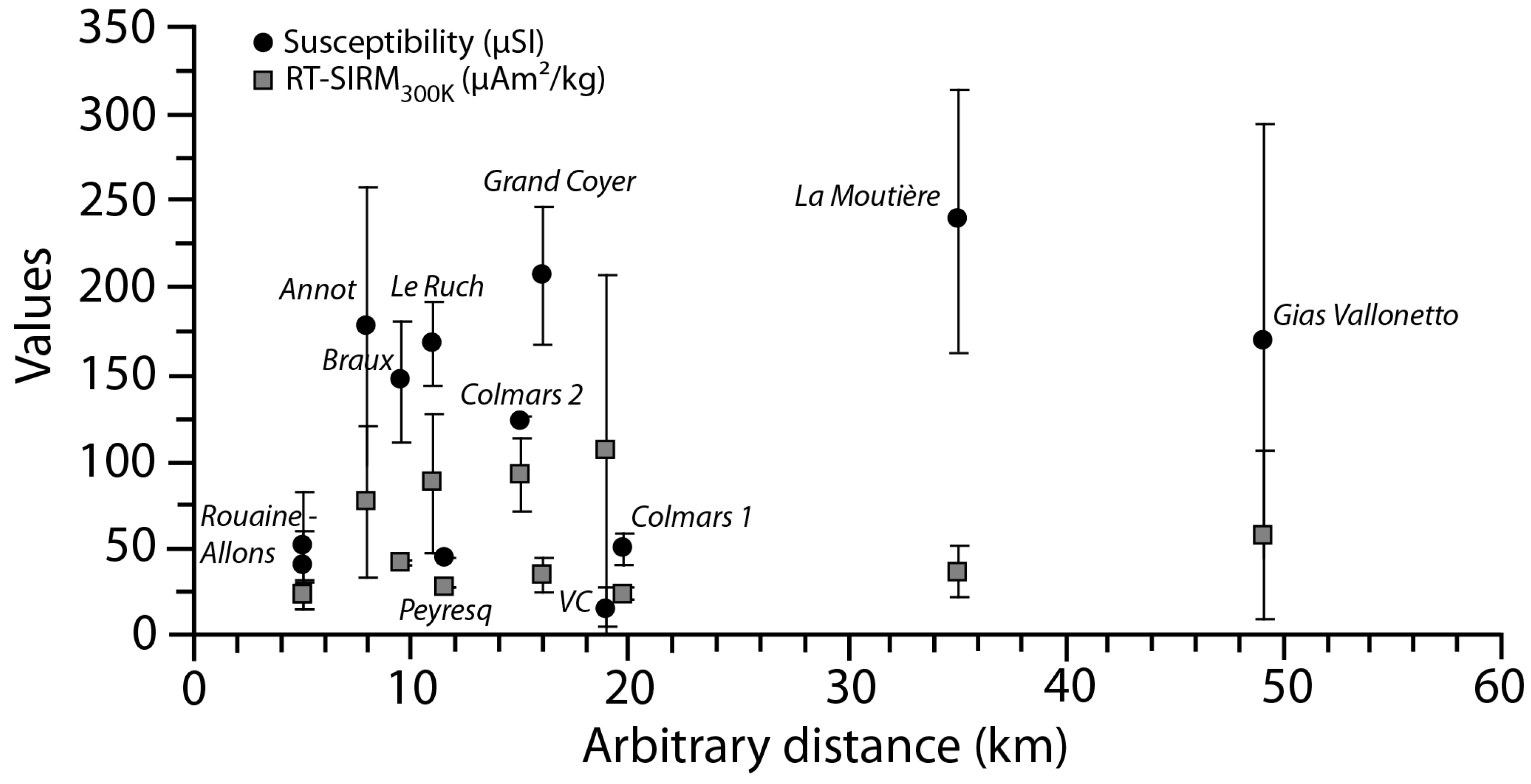
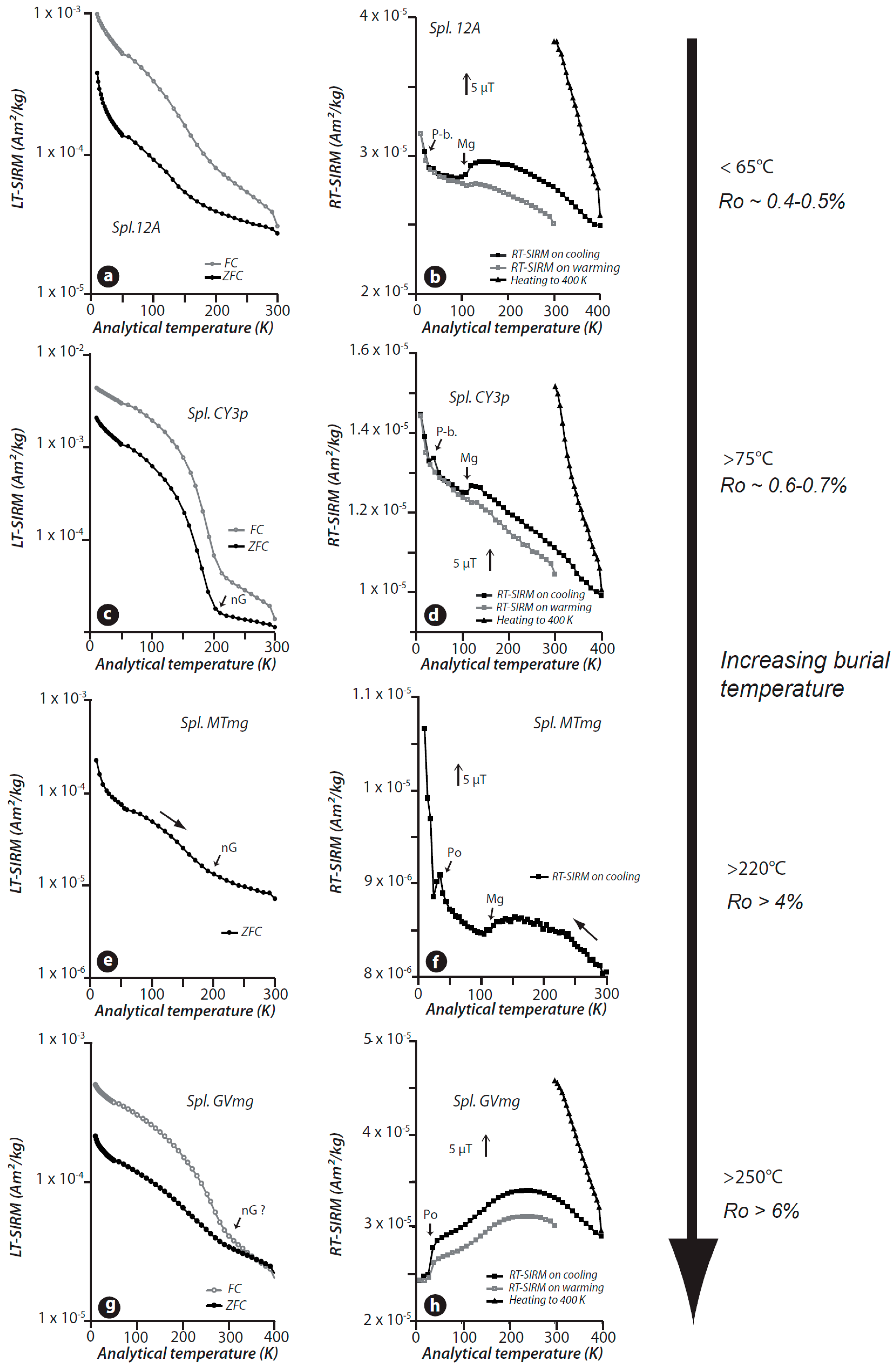
4.1. General Trends
| Sampling Site | Sample | χ (μSI) | RT-SIRM300 K (μAm2/kg) | LT-SIRM10 K (μAm2/kg) |
|---|---|---|---|---|
| Allons (Al) | 1A | 74 | 18 | 387 |
| 2A | 29 | 29 | 317 | |
| Rouaine (R) | 4A | 54 | 18 | 314 |
| 5A | 28 | 29 | 148 | |
| Braux (B) | 6A | 171 | 42 | 572 |
| 7A | 123 | 41 | 399 | |
| Annot (A) | A0 | 101 | 28 | 378 |
| 11A | 261 | 111 | 702 | |
| 12A | 170 | 92 | 905 | |
| Le Ruch (RU) | RUmg | 152 | 116 | 642 |
| RUmb | 185 | 59 | 2,894 | |
| Grand Coyer (GC) | CY1p | 201 | 32 | 1,372 |
| CY3p | 220 | 37 | 5,077 | |
| CY5 | 229 | 37 | 1,334 | |
| CY6 | 251 | 22 | 1,534 | |
| CY7 | 231 | 35 | 1,976 | |
| CY8 | 229 | 51 | 4,613 | |
| CY10 | 168 | 24 | 329 | |
| CY11 | 133 | 45 | 353 | |
| Peyresq (P) | 20A | 45 | 29 | 422 |
| Colmars (Co1) | 21A | 50 | 20 | 238 |
| 22A | 60 | 25 | 205 | |
| 23A | 41 | 28 | 311 | |
| Colmars (Co2) | 25A | 121 | 78 | 1,212 |
| 26A | 125 | 108 | 901 | |
| Villars-Colmars (VC) | 13A | 194 | 27 | 783 |
| 14A | 128 | 72 | 981 | |
| 15A | 205 | 220 | 816 | |
| La Moutière (MT) | MT12 | 307 | 35 | 2,249 |
| MT17 | 259 | 59 | 7,148 | |
| MT29 | 301 | 34 | 3,208 | |
| MT120 | 198 | 39 | 3,359 | |
| MTmg | 130 | 17 | 467 | |
| Gias Vallonetto (GV) | GV1 | 239 | 23 | 10,265 |
| GV11 | 244 | 39 | 709 | |
| GVmg | 26 | 113 | 538 | |
| La Moutière (MT) | MT12 | 307 | 35 | 2,249 |
| MT17 | 259 | 59 | 7,148 | |
| MT29 | 301 | 34 | 3,208 | |
| MT120 | 198 | 39 | 3,359 | |
| MTmg | 130 | 17 | 467 | |
| Gias Vallonetto (GV) | GV1 | 239 | 23 | 10,265 |
| GV11 | 244 | 39 | 709 | |
| GVmg | 26 | 113 | 538 |


4.2. Allons, Rouaine and Annot Areas

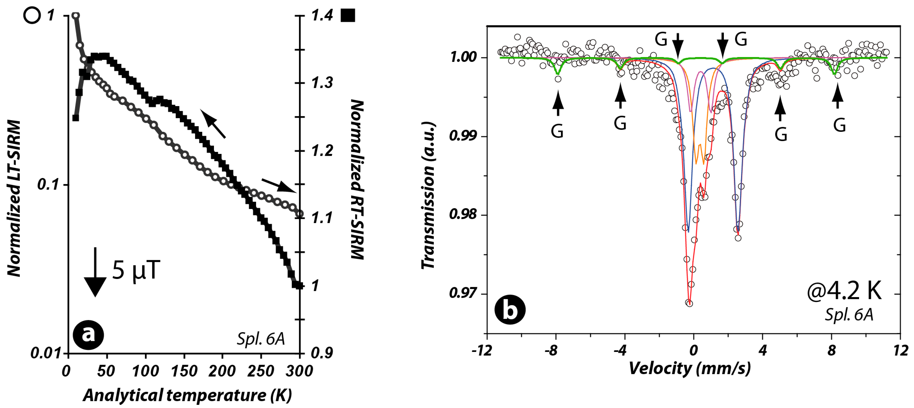
4.3. Braux Area

4.4. Le Ruch and Grand Coyer Areas
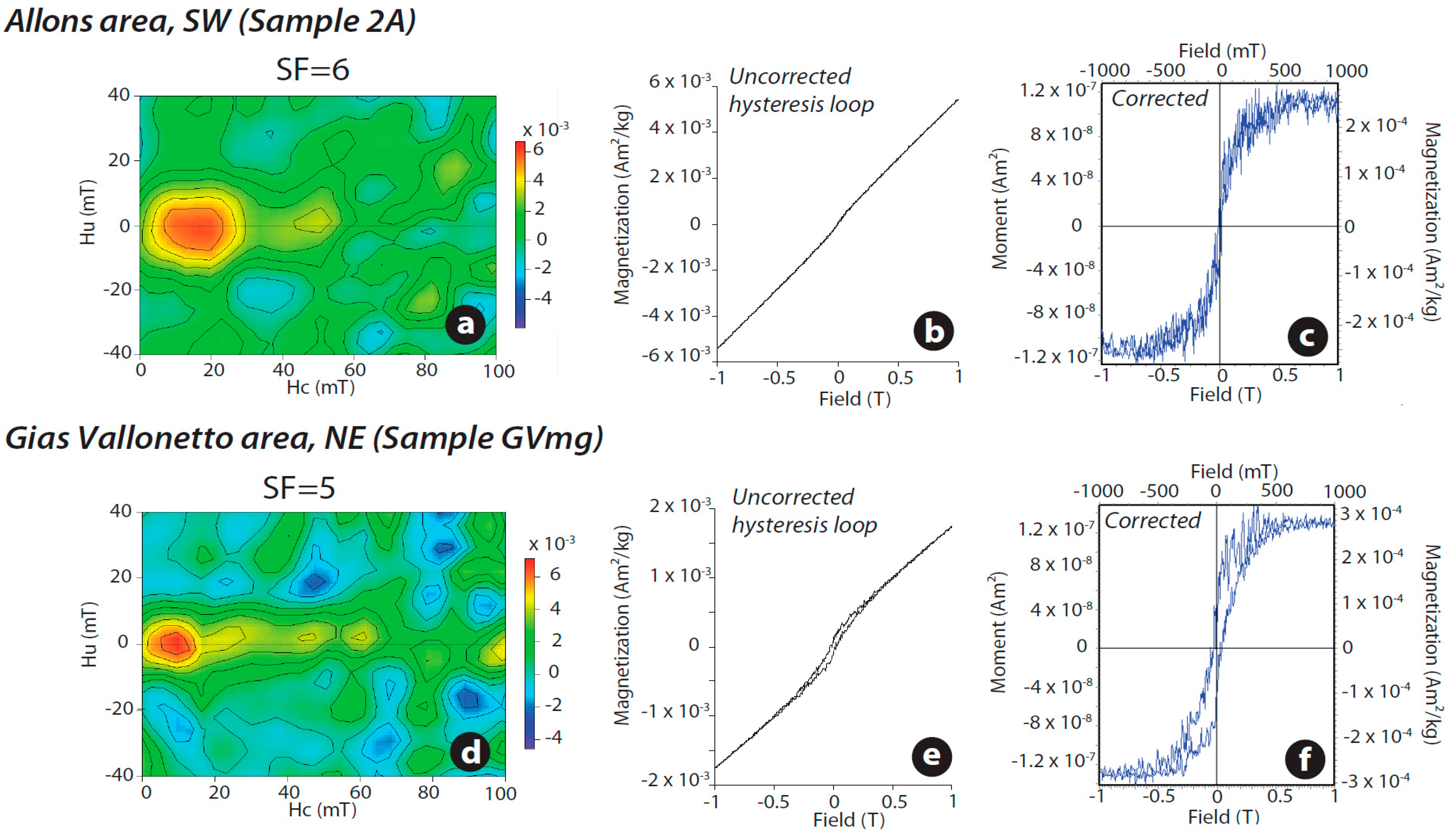
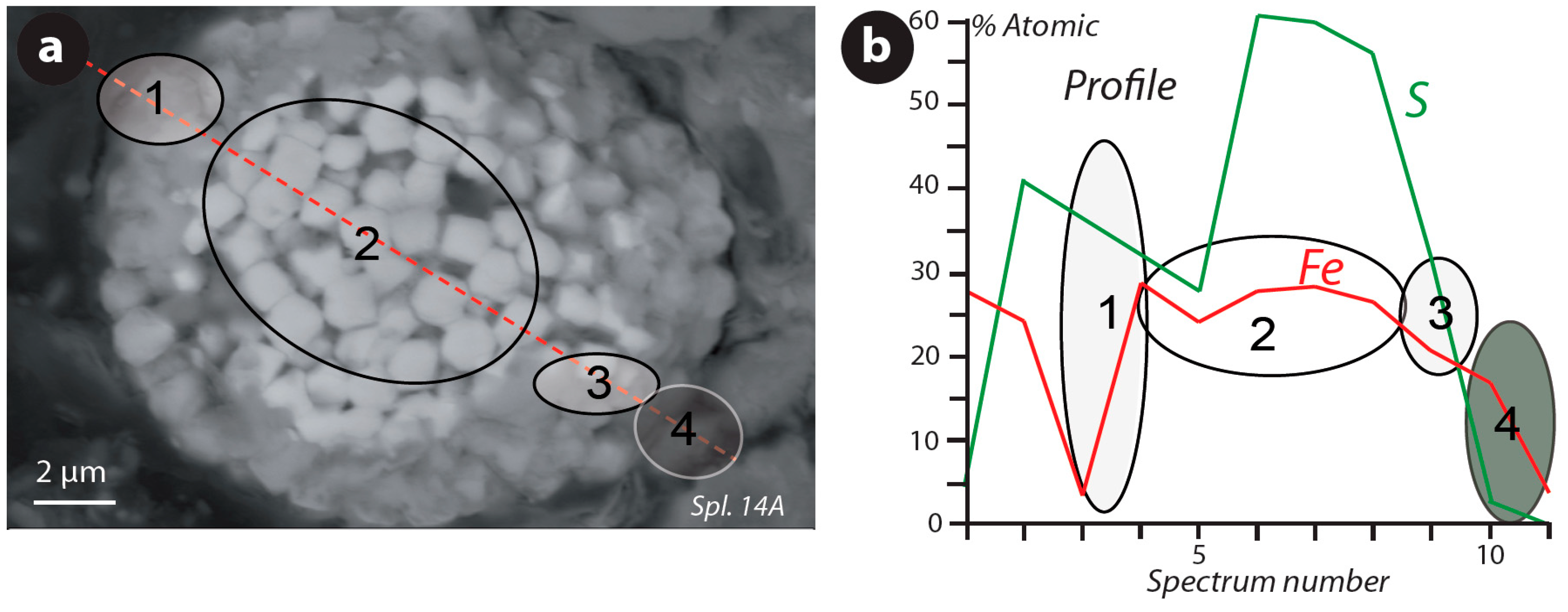
4.5. Villars-Colmars, La Moutière and Gias Vallonetto Areas
5. Discussion
5.1. Origin of the Magnetic Assemblage

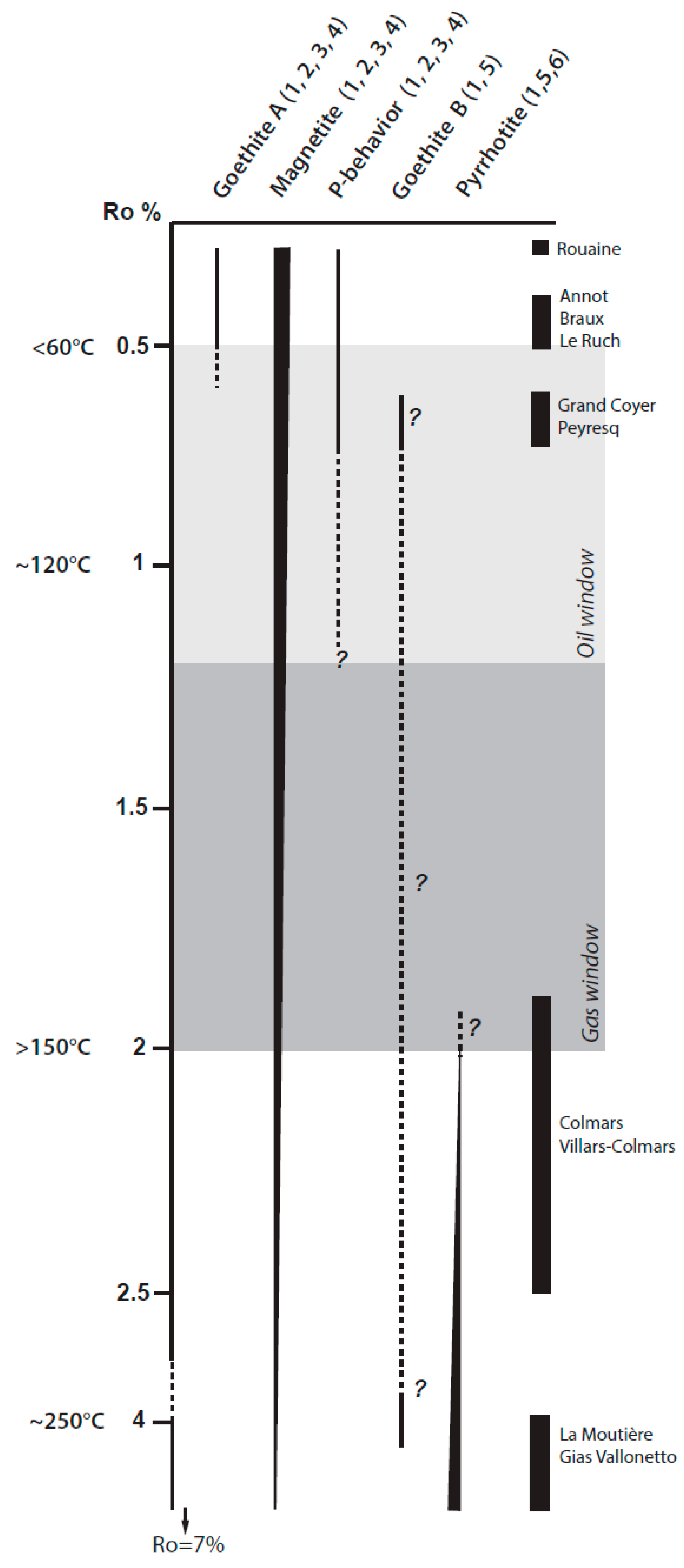
5.2. Toward a Burial Model
5.3. Analog for a Gas Shale System?
6. Conclusions
Acknowledgments
Author Contributions
Conflicts of Interest
References
- Rochette, P. Metamorphic control of the magnetic mineralogy of black shales in the Swiss Alps: Toward the use of “magnetic isogrades”. Earth Planet. Sci. Lett. 1987, 84, 446–456. [Google Scholar] [CrossRef]
- Dunlop, D.; Özdemir, O.; Clark, D.; Schmidt, P. Time-temperature relations for the remagnetization of pyrrhotite (Fe7S8) and their use in estimating paleotemperatures. Earth Planet. Sci. Lett. 2000, 176, 107–116. [Google Scholar] [CrossRef]
- Crouzet, C.; Ménard, G.; Rochette, P. Cooling history of the Dauphinoise Zone (Western Alps, France) deduced from the thermopaleomagnetic record: Geodynamic implications. Tectonophysics 2001, 340, 79–93. [Google Scholar] [CrossRef]
- Schill, E.; Appel, E.; Gautam, P. Towards pyrrhotite/magnetite geothermometry in low-grade metamorphic carbonates of the Thethyan Himalayas (Shiar Khola, Central Nepal). J. Asian Earth Sci. 2002, 20, 195–201. [Google Scholar] [CrossRef]
- Roberts, A.P.; Weaver, R. Multiple mechanisms of remagnetization involving sedimentary greigite (Fe3S4). Earth Planet. Sci. Lett. 2005, 231, 263–277. [Google Scholar]
- Rowan, C.J.; Roberts, A.P.; Broadbent, T. Reductive diagenesis, magnetite dissolution, greigite growth and paleomagnetic smoothing in marine sediments: A new view. Earth Planet. Sci. Lett. 2009, 277, 223–235. [Google Scholar] [CrossRef]
- Roberts, A.P.; Chang, L.; Rowan, C.J.; Horng, C.S.; Florindo, F. Magnetic properties of sedimentary greigite (Fe3S4): An update. Rev. Geophys. 2011, 49. [Google Scholar] [CrossRef]
- Aubourg, C.; Pozzi, J.-P.; Kars, M. Burial, claystones remagnetization and some consequence for magnetostratigraphy. In Remagnetization and Chemical Alteration of Sedimentary Rocks; Elmore, R.D., Muxworthy, A.R., Aldana, M.M., Mena, M., Eds.; Special Publications Volume 371; Geological Society London: London, UK, 2012; pp. 181–188. [Google Scholar]
- Aubourg, C.; Pozzi, J.-P. Toward a new <250 °C pyrrhotite-magnetite geothermometer for claystones. Earth Planet. Sci. Lett. 2010, 294, 47–57. [Google Scholar] [CrossRef]
- Abdelmalak, M.; Aubourg, C.; Geoffroy, L.; Laggoun-Defarge, F. A new oil window indicator? The magnetic assemblage of claystones from the Baffin Bay volcanic margin (Greenland). AAPG Bull. 2012, 96, 205–215. [Google Scholar] [CrossRef]
- Blaise, T.; Barbarand, J.; Kars, M.; Ploquin, F.; Aubourg, C.; Brigaud, B.; Cathelineau, M.; El Albani, A.; Gautheron, C.; Izart, A.; et al. Reconstruction of low temperature (<100 °C) burial in sedimentary basins: A comparison of geothermometer sensibility in the intracontinental Paris Basin. Mar. Pet. Geol. 2014, 53, 71–87. [Google Scholar]
- Kars, M.; Aubourg, C.; Suárez-Ruiz, I. Neoformed magnetic minerals as an indicator of moderate burial: The key example of Middle Paleozoic sedimentary rocks, West Virginia, WV, USA. AAPG Bull. 2014. accepted for publication. [Google Scholar]
- Rochette, P.; Lamarche, G. Evolution des propriétés magnétiques lors des transformations minérales dans les roches: Exemple du Jurassique Dauphinois (Alpes françaises). Bull. Mineral. 1986, 109, 687–696. (In French) [Google Scholar]
- Appel, E.; Crouzet, C.; Schill, E. Pyrrhotite Remagnetizations in the Himalaya: A Review. In Remagnetization and Chemical Alteration of Sedimentary Rocks; Elmore, R.D., Muxworthy, A.R., Aldana, M.M., Mena, M., Eds.; Special Publications Volume 371; Geological Society London: London, UK, 2012; pp. 163–180. [Google Scholar]
- Gillett, S.L. Paleomagnetism of the Notch Peak contact metamorphic aureole revisited: Pyrrhotite form magnetite+pyrite under sbmetamorphic conditions. J. Geophys. Res. 2003, 108, 2446. [Google Scholar] [CrossRef]
- Horng, C.S.; Torii, M.; Shea, K.S.; Kao, S.J. Inconsistent magnetic polarities between greigite and pyrrhotite/magnetite-bearing marine sediments from the Tsailiao-chi section. Earth Planet. Sci. Lett. 1998, 164, 467–481. [Google Scholar] [CrossRef]
- Horng, C.S.; Huh, C.A.; Chen, K.H.; Lin, C.H.; Shea, K.S.; Hsiung, K.H. Pyrrhotite as a tracer for denudation of the Taiwan orogen. Geochem. Geophys. Geosyst. 2012, 13. [Google Scholar] [CrossRef]
- Roberts, A.P.; Chang, L.; Heslop, D.; Florindo, F.; Larrasoaña, J.C. Searching for single domain magnetite in the “pseudo-single-domain” sedimentary haystack: Implications of biogenic magnetite preservation for sediment magnetism and relative paleointensity determinations. J. Geophys. Res. 2012, 117. [Google Scholar] [CrossRef]
- Canfield, D.E.; Berner, R.A. Dissolution and pyritization of magnetite in anoxic marine sediments. Geochim. Cosmochim. Acta 1987, 51, 645–659. [Google Scholar]
- Bloemendal, J.; King, J.; Hunt, A.; DeMenocal, P.; Hayashida, A. Origin of the sedimentary magnetic record at Ocean Drilling Program sites on the Owen Ridge, western Arabian Sea. J. Geophys. Res. 1993, 98, 4199–4219. [Google Scholar] [CrossRef]
- Dunlop, D.; Özdemir, O. Rock Magnetism: Fundamentals and Frontiers; Cambridge University Press: Cambridge, UK, 1997. [Google Scholar]
- Ford, M.; Lickorish, W.H.; Kusznir, N.J. Tertiary foreland sedimentation in the southern subalpine chains, SE France: A geodynamic appraisal. Basin Res. 1999, 11, 315–336. [Google Scholar] [CrossRef]
- Ford, M.; Lickorish, W.H. Foreland basin evolution around the western Alpine arc. In Deep-water Sedimentation in the Alpine Basin of SE France: New Perspectives on the Grès d’Annot and Related Systems; Joseph, P., Lomas, S.A., Eds.; Special Publications Volume 221; Geological Society London: London, UK, 2004; pp. 39–63. [Google Scholar]
- Joseph, P.; Lomas, S. Deep-water sedimentation in the Alpine Foreland Basin of SE France: A new perspective on the Grès d’Annot and related systems: An introduction. In Deep-water Sedimentation in the Alpine Basin of SE France: New Perspectives on the Grès d’Annot and Related Systems; Joseph, P., Lomas, S.A., Eds.; Geological Society London, Special Publications: London, UK, 2004; Volume 221, pp. 1–16. [Google Scholar]
- Ravenne, C.; Vially, R.; Riche, P.; Trémolières, P. Sédimentation et tectonique dans le bassin éocène sup-oligocène des Alpes du Sud. Rev. Inst. Fr. Pet. 1987, 42, 529–553. (In French) [Google Scholar]
- Apps, G.; Peel, F.; Elliott, T. The structural setting and palaeogeographical evolution of the Grès d’Annot basin. In Deep-water Sedimentation in the Alpine Basin of SE France: New Perspectives on the Grès d’Annot and Related Systems; Joseph, P., Lomas, S.A., Eds.; Geological Society London, Special Publications: London, UK, 2004; Volume 221, pp. 65–96. [Google Scholar]
- Du Fornel, E.; Joseph, P.; Desaubliaux, G.; Eschard, R.; Guillocheau, F.; Lerat, O.; Muller, C.; Ravenne, C.; Sztrakos, K. The southern Grès d’Annot out crops (French Alps): An attempt at regional correlation. In Deep-water Sedimentation in the Alpine Basin of SE France: New Perspectives on the Grès d’Annot and Related Systems; Joseph, P., Lomas, S.A., Eds.; Geological Society London, Special Publications: London, UK, 2004; Volume 221, pp. 137–160. [Google Scholar]
- Jean, S.; Kerckhove, C.; Perriaux, J.; Ravenne, C. Un modèle Paléogène de bassin à turbidites: Les Grès d’Annot du NW du massif de l’Argentera-Mercantour. Geol. Alp. 1985, 61, 115–143. (In French) [Google Scholar]
- Garcia, D.; Joseph, P.; Maréchal, B.; Moutte, J. Patterns of geochemical variability in relation to turbidite facies in the Grès d’Annot Formation. In Deep-water Sedimentation in the Alpine Basin of SE France: New Perspectives on the Grès d’Annot and Related Systems; Joseph, P., Lomas, S.A., Eds.; Geological Society London, Special Publications: London, UK, 2004; Volume 221, pp. 349–365. [Google Scholar]
- Kerckhove, C. La “zone du flysch” dans les nappes de l’Embrunais-Ubaye (Alpes occidentales). Geol. Alp. 1969, 45, 1–202. [Google Scholar]
- Sztrakos, K.; Du Fornel, E. Stratigraphie, paléoécologie et foraminifères du Paléogène des Alpes Maritimes et des Alpes de Haute-Provence (Sud-Est de la France). Rev. Micropaleontol. 2003, 46, 229–267. (In French) [Google Scholar] [CrossRef]
- Labaume, P.; Ritz, J.-F.; Philip, H. Failles normales récentes dans les Alpes sud-occidentales: Leurs relations avec la tectonique compressive. Comptes Rendus Acad. Sci. 1989, 308, 1553–1560. (In French) [Google Scholar]
- Labaume, P.; Jolivet, M.; Souquière, F.; Chauvet, A. Tectonic control on diagenesis in a foreland basin: Combined petrologic and thermochronologic approaches in the Grès d’Annot basin (late Eocene—Early Oligocene, French-Italian external Alps). Terra Nova 2008, 20, 95–101. [Google Scholar] [CrossRef]
- Labaume, P.; Arnaud, N.; Buatier, M.; Charpentier, D.; Chauvet, A.; Chirouze, F.; Jolivet, M.; Monié, P.; Sizun, J.-P.; Travé, A. Contrôle Tectonique de la Diagenèse d’une Formation Turbiditique d’Avant-Chaine, Exemple des Grés d’Annot, Alpes Externes Franco-Italiennes; Unpublished Internal Report; TOTAL: Pau, France, 2008. (In French) [Google Scholar]
- Labaume, P.; Sizun, J.-P.; Charpentier, D.; Travé, A.; Chirouze, F.; Buatier, M.; Chauvet, A.; Walgenwitz, F.; Jolivet, M.; Monié, P.; et al. Diagenesis controlled by tectonic burial in a foreland basin turbidite formation. The case example of the Grès d’Annot, French-Italian external Alps. In Proceedings of the European Geosciences Union (EGU) General Assembly, Vienna, Austria, 19–24 April 2009.
- Cavailhes, T. Architecture et Propriétés Pétrophysiques des Zones de Failles dans une Série Gréso Pélitique Turbiditique Profondément Enfouie: Rôle de la Déformation et des Interactions Fluide-Roche. Ph.D. Thesis, Université de Montpellier 2, Montpellier, France, 2012. [Google Scholar]
- Souquière, F. Relations Tectonique/Diagenèse dans un Bassin d’Avant-Chaîne, Exemple des Grès d’Annot: Approche Pétrologique et Thermochronologiques. Master’s Thesis, Université de Montpellier 2, Montpellier, France, 2005. [Google Scholar]
- Barlier, J.; Ragot, J.-P.; Thouray, J.-C. L’évolution des Terres Noires subalpines méridionales d’après l’analyse minéralogique des argiles et la réflectance des particules carbonées. Bull. BRGM 1974, 6, 533–548. [Google Scholar]
- Pickering, K.; Hilton, V. Turbidite Systems of Southern France: Application to Hydrocarbon Prospectivity; Vallis Press: London, UK, 1998. [Google Scholar]
- Vassoyevitch, N.B.; Korchagina, N.V.; Lopatin, N.V.; Chernyshev, V.V. Principal phase of oil formation. Int. Geol. Rev. 1970, 12, 1276–1297. [Google Scholar] [CrossRef]
- Leclère, H.; Buatier, M.; Charpentier, D.; Sizun, J.-P.; Labaume, P.; Cavailhes, T. Formation of phyllosilicates in fault zone affecting deeply buried arkosic sandstones. Their influence on fault zone petrophysic properties (Annot sandstones, late Eocene-early Oligocene, external Alps). Swiss J. Geosci. 2012, 105, 299–312. [Google Scholar] [CrossRef]
- Cavailhes, T.; Sizun, J.-P.; Labaume, P.; Chauvet, A.; Buatier, M.; Soliva, R.; Gout, C. Influence of fault rock foliation on fault zone permeability: The case of deeply buried arkosic sandstones (Grès d’Annot, SE France). AAPG Bull. 2013, 97, 1521–1543. [Google Scholar] [CrossRef]
- Özdemir, O.; Dunlop, D. Thermoremanence and Néel temperature of goethite. Geophys. Res. Lett. 1996, 23, 921–924. [Google Scholar]
- Kars, M.; Aubourg, C.; Pozzi, J.-P. Low temperature magnetic behaviour near 35 K in unmetamorphosed claystones. Geophys. J. Int. 2011, 186, 1029–1035. [Google Scholar] [CrossRef]
- Harrison, R.; Feinberg, J. FORCinel: An improved algorithm for calculating first-order reversal curve distributions using locally weighted regression smoothing. Geochem. Geophys. Geosyst. 2008, 9. [Google Scholar] [CrossRef]
- Tauxe, L.; Mullender, T.; Pick, T. Potbellies, wasp-waists, and superparamagnetism in magnetic hysteresis. J. Geophys. Res. 1996, 101, 571–583. [Google Scholar] [CrossRef]
- Muxworthy, A.R.; McClelland, E. Review of the low-temperature magnetic properties of magnetite from a rock magnetic perspective. Geophys. J. Int. 2000, 140, 101–114. [Google Scholar]
- Özdemir, O.; Dunlop, D.; Moskowitz, B. Changes in remanence, coercivity and domain state at low-temperature in magnetite. Earth Planet. Sci. Lett. 2002, 194, 343–358. [Google Scholar]
- Dekkers, M.J. Magnetic properties of natural goethite—II. TRM behaviour during thermal and alternating field demagnetization and low-temperature treatment. Geophys. J. Int. 1989, 97, 341–355. [Google Scholar] [CrossRef]
- Özdemir, O.; Dunlop, D. Hallmarks of maghemitization in low-temperature remanence cycling of partially oxidized magnetite nanoparticles. J. Geophys. Res. 2010, 115. [Google Scholar] [CrossRef]
- Dekkers, M.J.; Mattéi, J.-L.; Fillion, G.; Rochette, P. Grain-size dependence of the magnetic behavior of pyrrhotite during its low-temperature transition at 34 K. Geophys. Res. Lett. 1989, 16, 855–858. [Google Scholar] [CrossRef]
- Rochette, P.; Fillion, G.; Mattéi, J.-L.; Dekkers, M.J. Magnetic transition at 30–34 Kelvin in pyrrhotite: Insight into a widespread occurrence of this mineral in rocks. Earth Planet. Sci. Lett. 1990, 98, 319–328. [Google Scholar] [CrossRef]
- Wolfers, P.; Fillion, G.; Ouladdiaf, B.; Ballou, R.; Rochette, P. The pyrrhotite 32 K magnetic transition. Solid State Phenom. 2011, 170, 174–179. [Google Scholar] [CrossRef]
- Kars, M.; Aubourg, C.; Pozzi, J.-P.; Janots, D. Continuous production of nanosized magnetite through low grade burial. Geochem. Geophys. Geosyst. 2012, 13. [Google Scholar] [CrossRef]
- Murad, E.; Cashion, J. Mössbauer Spectroscopy of Environmental Materials and Their Industrial Utilization; Kluwer Academic Publishers: Boston, MA, USA, 2013. [Google Scholar]
- Guyodo, Y.; Mostrom, A.; Penn, R.L.; Banerjee, S.K. From nanodots to nanorods: Oriented aggregation and magnetic evolution of nanocrystalline goethite. Geophys. Res. Lett. 2003, 30. [Google Scholar] [CrossRef]
- Maher, B.A.; Thompson, R. Quaternary Climates, Environments and Magnetism; Cambridge University Press: Cambridge, UK, 1999. [Google Scholar]
- Guyodo, Y.; LaPara, T.M.; Anschutz, A.J.; Penn, R.L.; Banerjee, S.K. Rock magnetic, chemical and bacterial community analysis of a modern soil from Nebraska. Earth Planet. Sci. Lett. 2006, 251, 168–178. [Google Scholar] [CrossRef]
- Evans, M.; Elmore, R.D. Fluid control of localized mineral domains in limestone pressure solution structures. J. Struct. Geol. 2006, 28, 284–301. [Google Scholar] [CrossRef]
- Wilkin, R.; Barnes, H. Formation processes of framboidal pyrite. Geochim. Cosmochim. Acta 1997, 61, 323–339. [Google Scholar] [CrossRef]
- Suk, D.; Peacor, D.; Van der Voo, R. Replacement of pyrite framboids by magnetite in limestones and implications for paleomagnetism. Nature 1990, 345, 611–613. [Google Scholar] [CrossRef]
- Suk, D.; Van der Voo, R.; Peacor, D. Origin of magnetite responsible for remagnetization of Early Paleozoic limestones of New York State. J. Geophys. Res. 1993, 98, 419–434. [Google Scholar] [CrossRef]
- Rowan, C.J.; Roberts, A.P. Magnetite dissolution, diachronous greigite formation, and secondary magnetizations from pyrite oxidation: Unraveling complex magnetizations in Neogene marine sediments from New Zealand. Earth Planet. Sci. Lett. 2006, 241, 119–137. [Google Scholar] [CrossRef]
- Manning, E.B.; Elmore, R.D. Rock Magnetism and Identification of Remanence Components in the Marcellus Shale, Pennsylvania. In Remagnetization and Chemical Alteration of Sedimentary Rocks; Elmore, R.D., Muxworthy, A.R., Aldana, M.M., Mena, M., Eds.; Special Publications Volume 371; Geological Society London: London, UK, 2012; pp. 271–282. [Google Scholar]
- Cairanne, G.; Aubourg, C.; Pozzi, J.-P.; Moreau, M.-G.; Decamps, T.; Marolleau, G. Laboratory chemical remanent magnetization in natural claystones: A record of two polarities. Geophys. J. Int. 2004, 159, 909–916. [Google Scholar] [CrossRef]
- Moreau, M.-G.; Ader, M.; Enkin, R. The remagnetization of clay-rich rocks in sedimentary basins: Low-temperature experimental formation of magnetic carriers in natural sample. Earth Planet. Sci. Lett. 2005, 230, 193–210. [Google Scholar] [CrossRef]
- Aubourg, C.; Pozzi, J.-P.; Janots, D.; Sahraoui, L. Imprinting chemical remanent magnetization in claystones at 95 °C. Earth Planet. Sci. Lett. 2008, 272, 172–180. [Google Scholar] [CrossRef]
- Crouzet, C.; Ménard, G.; Rochette, P. High-precision three-dimensional paleothermometry derived from paleomagnetic data in Alpine metamorphic unit. Geology 1999, 27, 503–506. [Google Scholar] [CrossRef]
- Beyssac, O.; Simoes, M.; Avouac, J.-P.; Farley, K.A.; Chen, Y.G.; Chan, Y.C.; Goffé, B. Late Cenozoic metamorphic evolution and exhumation of Taiwan. Tectonics 2007, 26. [Google Scholar] [CrossRef] [Green Version]
- Sweeney, J.J.; Burnham, A.K. Evaluation of a simple model of vitrinite reflectance based on chemical kinetics. AAPG Bull. 1990, 74, 1559–1570. [Google Scholar]
- Barker, C.E.; Pawlewicz, M.J. Calculation of vitrinite reflectance from thermal histories and peak temperatures. In Reevaluation of Vitrinite Reflectance; Mukhopadhyay, P.K., Dow, W.G., Eds.; Series 570; American Chemical Society Symposium: Washington, DC, USA, 1994; pp. 216–229. [Google Scholar]
- Bruner, K.R.; Smosna, R. A Comparative Study of the Mississippian Barnett Shale, Fort Worth Basin, and Devonian Marcellus Shale, Appalachian Basin. Available online: http://www.netl.doe.gov/File%20Library/Research/Oil-Gas/publications/brochures/DOE-NETL-2011-1478-Marcellus-Barnett.pdf (accessed on 1 July 2014).
© 2014 by the authors; licensee MDPI, Basel, Switzerland. This article is an open access article distributed under the terms and conditions of the Creative Commons Attribution license (http://creativecommons.org/licenses/by/3.0/).
Share and Cite
Kars, M.; Aubourg, C.; Labaume, P.; Berquó, T.S.; Cavailhes, T. Burial Diagenesis of Magnetic Minerals: New Insights from the Grès d’Annot Transect (SE France). Minerals 2014, 4, 667-689. https://doi.org/10.3390/min4030667
Kars M, Aubourg C, Labaume P, Berquó TS, Cavailhes T. Burial Diagenesis of Magnetic Minerals: New Insights from the Grès d’Annot Transect (SE France). Minerals. 2014; 4(3):667-689. https://doi.org/10.3390/min4030667
Chicago/Turabian StyleKars, Myriam, Charles Aubourg, Pierre Labaume, Thelma S. Berquó, and Thibault Cavailhes. 2014. "Burial Diagenesis of Magnetic Minerals: New Insights from the Grès d’Annot Transect (SE France)" Minerals 4, no. 3: 667-689. https://doi.org/10.3390/min4030667