Article

Use of Iron Powder to Obtain High Yields of Leptothrix Sheaths in Culture

1
Core Research for Evolutionary Science and Technology (CREST), Japan Science and Technology Agency (JST), Okayama 700-8530, Japan
2
Graduate School of Natural Science and Technology, Okayama University, Okayama 700-8530, Japan
*
Author to whom correspondence should be addressed.
Minerals 2015, 5(2), 335-345; https://doi.org/10.3390/min5020335
Submission received: 27 February 2015 / Accepted: 1 June 2015 / Published: 4 June 2015

Abstract

:
The Leptothrix species, Fe-oxidizing bacteria, produce an extracellular, microtubular sheath with a complicated organic–inorganic hybrid nature. We have discovered diverse industrial functions for this material, e.g., electrode material for Li-ion batteries, catalyst enhancers, pigments, plant growth promoters, and plant protectants. To consistently obtain material with the qualitative and quantitative stability needed for industrial applications, we focused on developing an optimum culture system for sheath synthesis by the Leptothrix sp. strain OUMS1. Although we have used Fe plates as an Fe source in the liquid silicon-glucose-peptone medium (SGP), the plates do not yield a consistent quality or precise mass, and formation of Fe-encrusted sheath is restricted to a surface of the plates, which limits harvest yield. In this study, to obtain a high yield of sheaths, we cultured OUMS1 in SGP supplemented with Fe powders. The addition of Fe powders to the medium (up to 14.0 g/L) did not adversely influence growth of OUMS1. The final yield of sheaths was about 10-fold greater than in the Fe plate culture. The sheaths also maintained a microtubular form and crystalline texture similar to those produced on Fe plates in SGP. The results proved the usefulness of Fe powder for consistently high yields of Fe-encrusted sheaths of stable quality.

1. Introduction

The Leptothrix species, one of the Fe/Mn-oxidizing bacteria, are ubiquitous in aqueous environments, especially at sites characterized by a circumneutral pH, an oxygen gradient and a source of reduced iron and manganese minerals [1,2]. The filamentous growth of Leptothrix species, the capability to form microtubular sheaths, and the precipitation of copious amounts of oxidized Fe or Mn distinguish the genus from other phylogenetically related genera [2,3]. To date, many researchers have reported that the major inorganic component of these sheaths is in the form of iron oxides that bind other inorganics such as Si, P, and often Ca and S [4,5,6,7,8]. Our previous microscopic and spectroscopic studies proved that the sheath was an ingenious hybrid of organic and inorganic materials produced through the interaction of bacterial exopolymers with aqueous-phase inorganics [5,8,9]. Intriguingly, natural Leptothrix sheaths were discovered to have a variety of the industrial functions: lithium-ion battery anode material [10], catalyst enhancer [11,12,13], and porcelain pigment [14]. The quality and quantity of the natural sheaths, however, are inevitably influenced by environmental changes such as temperature, quantity of supplied groundwater or spring water, concentrations of inorganics in water, as was noted by Vollrath et al. [15]. For industrial applications, a constant supply system of qualitatively and quantitatively determinate material is critical.
Our current project was aimed at establishing a basic system for culturing an isolated strain of Leptothrix sp. to produce reliable quantities of Leptothrix sheaths with stable quality sufficient for industrial use. Routinely, we have used small pieces of commercial Fe plates in liquid medium as the Fe source [16]. However, these heterogeneous plates do not provide a precise amount of Fe to the medium, and sheath yield is at most 10–12 mg/100 mL of medium (dry mass), far too low for industrial applications. In this paper, we present a culture method to increase yield of the harvested sheaths using Fe powder, which has a greater surface area than the Fe plates.

2. Experimental Section

2.1. Strain, Medium and Preculturing

Leptothrix sp. strain OUMS1 (NITE BP-860) (hereafter, referred to as OUMS1), isolated from flocculent, ocherous deposits in a biological freshwater purification plant in Joyo City, Kyoto Prefecture, Japan [16], was used in this study after recovery from frozen stock. For preculturing, OUMS1 was streaked on silicon–glucose–peptone (hereafter, referred to as SGP; liquid medium, unless otherwise stated) agar medium (pH 7.0) (for components, see supplemental Table S1) [16] and incubated at 20 °C for seven days. Single colonies were independently picked up with autoclaved toothpicks, transferred to 25 mL of SGP in 50 ml conical tubes (BD Bioscience, Bedford, MA, USA), and incubated on a rotary shaker at 20 °C and 70 rpm. Two days later, the cell suspension was adjusted to 103 cfu/mL using fresh SGP by densitometry (Nanodrop 2000C, Thermo Fisher Scientific Inc., Waltham, MA, USA) for use as inoculum.

2.2. Calculation of the Amount of Powdery Iron to Add to SGP and Cultures

Two types of Fe powder (99.5% purity; 45 and 150 μm diameter) (hereafter, referred to as 45 and 150 μm Fe powders, respectively) were purchased from Wako Pure Chemical Industries, Ltd. (Osaka, Japan). To calculate the amount of powder to provide a total surface area equivalent to that of three small Fe plates (size: 1 cm × 1 cm × 0.1 cm; total surface area: 2.4 cm2 × 3 = 7.2 cm2), we treated both particles as spherical based on their almost spherical shape under a light microscope with an estimated surface area of 6.4 × 10⁻⁹ for the 45 μm Fe powder and 7.1 × 108 cm2 for the 150 μm. Based on these surface areas, the mass of the 45 μm powder to use was calculated as 4.3 and 14 mg/L for the 150 μm. For better accuracy in weighing, a 101-, 102-, or 103-fold amount of the powder (0.043, 0.43, 4.3 g/L of 45 μm Fe powder; 0.14, 1.4, and 14.0 g/L of 150 μm Fe powder) was added to 100 mL of SGP (hereafter, referred to as Fe powder medium) (Figure 1D). One milliliter of the inoculum suspension was then transferred to 99 mL of the respective medium in a 200 mL glass flask (final inoculum density = 10 cfu/mL), followed by incubation with one of several shaking modes as described later. Both powders remained stable on the flask bottom during culturing (Figure 1D). For reference, we used SGP plus three Fe plates (hereafter referred to as Fe plate medium) (Figure 1A) inoculated and incubated in the same way.
Figure 1. SGP containing Fe plates or powder before and after two week incubation and sheaths encompassing cells of Leptothrix sp. strain OUMS1 harvested from media after incubation. (A) Flask of Fe plate medium containing three Fe plates before incubation. (B) Brownish masses of Fe-encrusted sheaths cover the Fe plate surface, and whitish masses of primitive sheaths in the upper periphery of the medium after a 24 h incubation. (C) Light micrograph of primitive sheaths with thin, nearly transparent walls encompassing chained bacterial cells seen in the whitish mass at periphery of medium. (D) Flask of Fe powder medium containing 1.4 g/flask of 150 μm Fe powders before incubation. (E) Brownish masses of Fe-encrusted sheaths on inner surface of flask in contact with shaken medium. (F) Light micrograph of Fe-encrusted sheaths with thick walls in brownish masses (E). Scale bar = 10 μm.
Minerals 05 00335 g001

2.3. Effects of Fe Powder Medium and Shaking Mode on Bacterial Growth

Initially, we examined whether the Fe powders and shaking modes affected growth of OUMS1. The Fe powder media inoculated with the bacterial suspension were incubated on rotary shakers at 70, 140, and 180 rpm or reciprocal shakers at 115 and 140 rpm at 20 °C for two weeks. On days 0, 1–4, 7, and 14 after the onset of incubation, growing cells in the media were harvested. When the cells were harvested, the Fe powders in the media were pulled to the flask bottom using a magnet to avoid powder contamination in the sampled suspension. According to the method of Emerson and Ghiorse [17], 1 mL samples of the culture were collected using 23-gauge needles on a 10 mL syringe, then expressed into 1.5 mL Eppendorf tubes to disrupt aggregates. The cell suspensions were serially diluted 10-fold with fresh SGP and immediately spot-inoculated onto SGP agar plates. After a 4–5 day incubation at 20 °C, colonies were counted for three replicates and the average cfu was recorded (hereafter, referred to as cfu-test). For reference, the inoculum suspension was added to the Fe plate medium and incubated on a rotary shaker at 70 rpm and 20 °C for 2 weeks as routinely performed [6,8,16], and cfu was counted as described.

2.4. Removal of Fe Powders from Culture Flasks, Collection and Weighing of Sheaths

After the two week incubation, Fe powders in culture flasks were collected and removed using a magnet held to the outer surface of the flasks. To harvest sheaths, the brownish sheath mass on the inner surface of flask was gently rubbed up and down with a cushiony silicon spatula to make a sheath suspension. The Fe powder was then removed and repeatedly washed with ultrapure water (UPW), to make an additional sheath suspension. Both suspensions were combined, washed with UPW 10 times by centrifugation at 2600 ×g, freeze-dried, and weighed.
In the Fe plate medium, however, the brownish mass of sheaths adhered only to the surface of the plates, although a whitish sheath mass adhered to the inner wall of the flask (Figure 1B). To avoid contamination with this whitish mass, the Fe plates were carefully transferred from the flask to a glass beaker of UPW, and their surfaces were rubbed gently with a silicon spatula. This sheath suspension was washed repeatedly with UPW by centrifugation at 2600 ×g and freeze-dried as above. After the dried specimens were weighed, they were subjected to the following microscopy and X-ray diffraction analyses.

2.5. Scanning Electron Microscopy (SEM) and X-Ray Diffractometry (XRD)

Dried specimens were resuspended in 100% ethanol, mounted onto a specimen stub, and air-dried in a clean bench. The specimens were observed with an SEM equipped with energy dispersive X-ray spectrometer (EDX) (S-4300, Hitachi, Tokyo, Japan). The SEM images were obtained after Pt-coating and the EDX mapping without Pt-coating. Atomic percentages of major inorganics (Fe, Si, and P) detectable in sheaths were calculated by EDX, and their variations among 10 spot values were expressed.
Following the previous protocol [7], X-ray diffraction patterns of sheaths harvested from Fe powder and -plate media were obtained using an RINT-2000 (Rigaku, Tokyo, Japan) with Cu-Kα radiation (voltage, 40 kV; current, 200 mA). The dried specimens were pressed into an XRD holder with a glass plate to flatten the surface of the compacted powders. Scans were performed from 10° to 80° (2θ value) with a step size of 0.02° at a fixed time of 1 s. The XRD pattern of the XRD holder was measured, and the pattern was subtracted from the samples’ XRD patterns.

3. Results and Discussion

3.1. Influence of Fe Powders and Culture-Shaking Modes on Exponential Cell Growth and Medium pH

The cfu-test revealed that cell growth increased exponentially by day 3 and reached the stationary phase thereafter in both Fe powder and -plate media regardless of the shaking mode (rotary at ≤140 rpm or reciprocal ≤115 rpm) (Figure 2). However, cell growth was not detected with more rapid agitations (180 rpm rotary and 140 rpm reciprocal) probably due to excessive oxygenation of the medium. This result was expected because low oxygen has been reported to favor growth and Fe(II) oxidation of aerobic Leptothrix [2,15,18,19,20], consistent with their good growth at the natural air–water interface [17]. The present study confirmed that use of a proper amount of Fe powders in the medium did not adversely affect the cell growth under favorable shaking modes.
Changes in pH in the media was examined during incubation. In both Fe plate and -powder media, the pH remained at 7.0 ± 0.2 during the two week incubation.
Figure 2. Time course of growth of cells of Leptothrix sp. strain OUMS1 cultured in SGP supplemented with 14 g/L of 150 μm Fe powder under different shaking modes. (ro = rotary and recip = reciprocal).
Minerals 05 00335 g002

3.2. Harvest of Sheaths from Culture

As reported previously [16], in Fe plate medium, brownish masses of sheaths formed on the smooth surface of the plates (Figure 1B). Consistent with Rogers and Anderson’s observation [3] that cultured Sphaerotilus discophorus strain SS1 (hereafter, referred to as SS1) formed brown colonies as a result of precipitation of Mn and/or Fe oxides, sheaths with thick brownish walls were observed in these brown masses of Fe encrustation on the plates. In contrast, sheaths with thin, nearly transparent walls (hereafter, referred to as primitive sheaths) were observed in the whitish masses attached to the inner surface of the flask (Figure 1C). Although the whitish masses were exposed to the agitated medium during culture, appreciable Fe encrustation did not occur, probably due to insufficient concentration of ionic Fe in the circulating medium, as described below. The mean dry mass of the harvested brownish sheaths was 10–12 mg per flask in this case.
In the Fe powder medium during shaking, however, brownish masses of Fe-encrusted sheaths adhered to the inner surface of the flask (Figure 1E). These brownish sheaths were confirmed by light microscopy to have a seemingly sturdy, brownish frame (Figure 1F). The Fe powders at the flask bottom were also covered with a brownish mass of sheaths (Figure 1E). The total mass of the brownish sheaths harvested from both inner surface of flask and surface of Fe powders averaged 100–120 mg per flask, 10-fold more than the case of Fe plate medium. Light microscopy revealed numerous fine Fe particles attached to the sheath surfaces in the 45 μm Fe powder medium. It was difficult to separate both sheaths and particles physically by the present harvesting protocol, which made it impossible to accurately estimate the yield of sheaths from the 45 μm Fe powder medium. Therefore, we decided to use only the 150 μm Fe powder hereafter.

3.3. Morphology and Fe Distribution of Sheaths

As readily expected from light micrographs (Figure 1C,F), the sheaths had a microtubular form (Figure 3) with a mean diameter of 2–5 μm in the Fe plate medium and 1.5–2.5 μm in the Fe powder medium. The entire sheath wall was 200–250 nm thick in the former and 100–150 nm in the latter. The outer coat of both sheaths was composed of thin, loosely woven fibrils and thick, aggregated fibrils. The inner surface of the sheaths looked globular (Figure 3B,D). Numerous fine particles (Figure 3B,D arrows) were often seen attached to the outer coat in both media. These particles probably correspond to aggregated ionic Fe (probably Fe(II) and/or Fe(III) hydroxides/oxyhydroxides) [21]. As illustrated in Figure 3E,F, EDX analysis revealed a homogeneous distribution of Fe in a sheath that was harvested from the Fe powder medium. This distribution pattern was very similar to that in sheaths harvested from a two week culture of OUMS1 in the Fe plate medium as reported earlier [7,16]. The average atomic percentages of Fe in 10 spots of sheaths harvested after two weeks were 66.9 ± 9.9%, 82.4 ± 3.8%, and 88.9 ± 6.1% in 0.14, 1.4, and 14 g/L Fe powder medium, respectively, showing that Fe encrustation increased with increasing amount of Fe source in the medium.

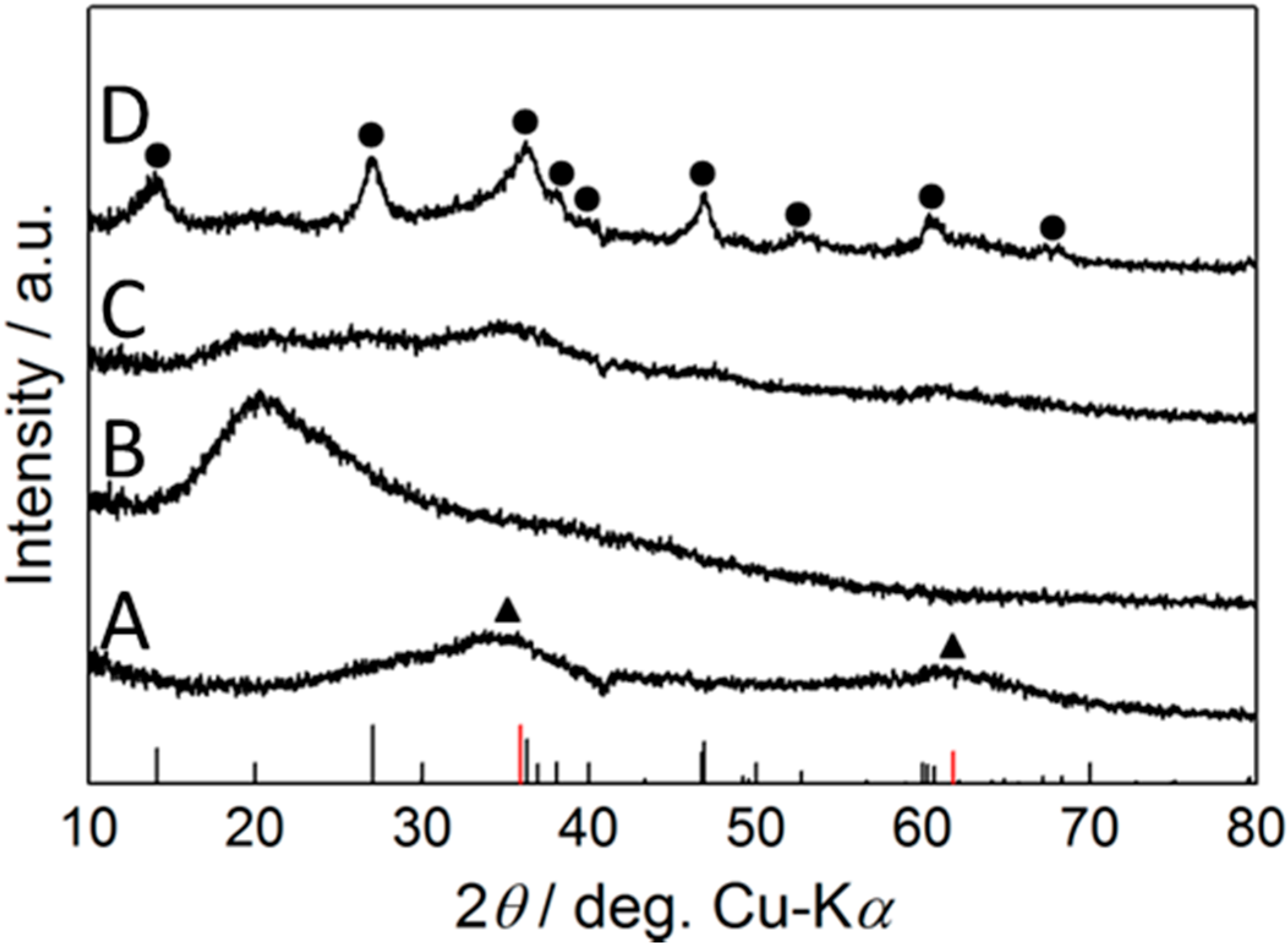
3.4. Crystallinity of Sheaths Detected by XRD

Similar to the finding by Eggleton et al. [22], a very broad peak at 30–40° and around 61° (black triangle in line A of Figure 4), plausibly was derived from two-line ferrihydrite (2Fh) [7], was observed for sheaths formed in the Fe plate medium. The sheaths that formed in the 0.14 g/L Fe powder medium showed a broad peak between 2θ = 15° and 31° (highest at 2θ = 20°), which was probably derived from C-containing constituents of bacterial and/or medium origin [23], and no remarkable peaks for the presence of Fe oxides were detected (line B of Figure 4). The sheaths in the 1.4 g/L Fe powder medium showed slight peaks at 2θ = 35° and 61° suggesting the presence of XRD-amorphous 2Fh (line C of Figure 4), similar to those from the Fe plate medium. In contrast, in the 14 g/L Fe powder medium, major diffraction peaks at 2θ were detected at several points (line D of Figure 4), which correspond to those of γ-FeOOH (lepidocrocite) (ICDD database, PDF 00-044-1415). Results show that the crystallinity of sheaths is affected by the amount of Fe powder added to the medium. Ishihara et al. [7] found that by culturing OUMS1 in Fe plate medium containing varied amounts of Si source, the constitutional Fe oxide phase changed from poorly crystalline lepidocrocite at 0 ppm Si to XRD-amorphous 2Fh at 100–300 ppm Si via their mixture phase with intermediate Si content. In a supplemental experiment, we confirmed that sheaths were composed of the mixture phase of lepidocrocite and 2Fh that formed in the Fe powder medium supplemented with 100 μg/mL Si (Supplemental material Figure S1). These results show that the chemical character and crystallinity of the sheath can be regulated by the medium component, in particular, the balance of Fe and Si, as Châtellier et al. [24] and Ishihara et al. [7] emphasized.
Figure 3. SEM and EDX mapping images of sheaths formed in Fe plate and -powder media during two week incubation. A, B: Sheath with fibrous outer surface (A) and enlarged image of sheath with a thick wall (B) in Fe plate medium. C, D: Sheath with fibrous outer surface (C) and enlarged image of sheath with relatively thin wall (D) in Fe powder medium. Note fine Fe particles on the sheath surface (arrows in B and D). E, F: SEM image (E) and Fe distribution map (F) of sheath formed in Fe powder medium. Note that particles attached to the sheath surface (arrows) contain a detectable level of Fe. Scale: A, C, E, F, 1 μm; B, D, 500 nm.
Minerals 05 00335 g003
Because the crystallinity of synthetic 2Fh is lowered when Si is doped in the structure [25,26,27], the Si-dependent changes in the texture suggested in the present study may not directly be associated with the presence of the bacterial cell but may result from direct autocatalytic oxidation and/or chemical/physical interactions among elements. Châtellier et al. [24] reported that the presence of bacterial cells did not modify the mineralogy of the Fe-oxides but that the size of the Fe-oxide particles tended to be reduced and that the presence of the cells also affected the spatial organization and the morphology of the particles. Although the present XRD analysis showed that the crystallinity of the texture of the sheath obtained in the Fe powder medium was similar to that obtained in the Fe plate medium, further analyses are obviously needed to examine physical and chemical characters of the harvested sheaths.
Toner et al. [28] reported that organics were responsible for the poor crystallinity of microbe-associated Fe minerals. Thus, the crystallinity change observed in this study could be dependent not only on the balance of Fe and Si but also on the bacterial and/or medium organics. To verify this hypothesis, we need to determine how Fe and Si interact with each other in the medium and thus affect sheath texture and how the bacterial organics interact with aqueous-phase elements, in particular Fe.
Figure 4. XRD patterns of cultured sheaths in SGP containing different Fe sources: (A) three Fe plates or 150 μm Fe powder at (B) 0.14 g/L, (C) 1.4 g/L, (D) 14 g/L. The main diffraction peaks for lepidocrocite (black circles) were assigned using ICDD database of lepidocrocite (PDF 00-044-1415). The position of the diffraction peaks for 2Fh (black triangle) was assigned using the data of 2Fh [29] and is given as black and red bars at the bottom of the figure, respectively. The break near 40° corresponds to the peaks derived from the sample holder.
Minerals 05 00335 g004

4. Conclusions

The present study demonstrated that use of 150 μm Fe powder was conducive to the production of 100–120 mg/flask (dry mass) of Fe-encrusted sheaths, enough to supply test trials for industrial application. The microtubular sheaths in the Fe powder medium were similar in morphology to sheaths in the Fe plate medium. Crystallinity of the texture of the Fe powder sheaths was similar but not the same as the Fe plate sheaths. However, the crystallinity of sheaths can be regulated by adjusting the quantitative ratio of Fe to Si in the medium. Because the powder is precisely weighed, the desired amount of Fe source was accurately supplied to the medium. In addition, the powder was readily collected by using a magnet, minimizing Fe contamination in the harvested sheaths. The present results demonstrate the advantages of using Fe powder to obtain sufficient quantities of Fe-encrusted sheaths of stable quality needed for experimental trials on industrial applications of the sheaths.

Acknowledgments

We thank Mikio Takano for instructive advice about Fe behavior in aquatic systems and Keiko Toyoda for technical support. We also acknowledge Beth E. Hazen for reviewing and editing the manuscript. This study was financially supported by JST-CREST (2012–2016) (J.T.).

Author Contributions

Tomoko Suzuki, Tatsuki Kunoh, and Katsunori Tamura conceived the overall experimental strategy and performed all physiological and microscopic experiments. Hideki Hashimoto and Daisuke Nakatsuka did the XRD and microscopic analyses. Hitoshi Kunoh and Jun Takada developed the original concept of the project and provided technical advice. All authors participated in writing the manuscript.

Conflicts of Interest

The authors declare no conflict of interest.

References

  1. Ghiorse, W.C.; Hirsch, P. An ultrastructural study of iron and manganese deposition associated with extracellular polymers of pedomicrobium-like budding bacteria. Arch. Microbiol. 1979, 123, 213–226. [Google Scholar] [CrossRef]
  2. Spring, S. The general Leptothrix and Sphaerotilus. Prokaryotes 2006, 5, 758–777. [Google Scholar]
  3. Rogers, S.R.; Anderson, J.J. Measurement of growth and iron deposition in Sphaerotilus discophorus. J. Bacteriol. 1976, 126, 257–263. [Google Scholar] [PubMed]
  4. Emerson, D.; Ghiorse, W.C. Ultrastructure and chemical composition of the sheath of Leptothrix discophora SP-6. J. Bacteriol. 1993, 175, 7808–7818. [Google Scholar] [PubMed]
  5. Suzuki, T.; Hashimoto, H.; Ishihara, H.; Kasai, T.; Kunoh, H.; Takada, J. Structural and spatial associations between Fe, O, and C in the network structure of the Leptothrix ochracea sheath surface. Appl. Environ. Microbiol. 2011, 77, 7873–7875. [Google Scholar] [CrossRef] [PubMed]
  6. Suzuki, T.; Ishihara, H.; Furutani, M.; Shiraishi, T.; Kunoh, H.; Takada, J. A novel method for culturing of Leptothrix sp. strain OUMS1 in natural conditions. Minerals 2012, 2, 118–128. [Google Scholar] [CrossRef]
  7. Ishihara, H.; Hashimoto, H.; Taketa, E.; Suzuki, T.; Mandai, K.; Kunoh, H.; Takada, J. Silicon-rich, iron oxide microtubular sheath produced by an iron-oxidizing bacterium, Leptothrix sp. strain OUMS1. Minerals 2014, 4, 565–577. [Google Scholar] [CrossRef]
  8. Furutani, M.; Suzuki, T.; Ishihara, H.; Hashimoto, H.; Kunoh, H.; Takada, J. Initial assemblage of bacterial saccharic fibrils and element deposition to form an immature sheath in cultured Leptothrix sp. strain OUMS1. Minerals 2011, 1, 157–166. [Google Scholar] [CrossRef]
  9. Furutani, M.; Suzuki, T.; Ishihara, H.; Hashimoto, H.; Kunoh, H.; Takada, J. Assemblage of bacterial saccharic microfibrils in sheath skeleton formed by cultured Leptothrix sp. strain OUMS1. J. Mar. Sci. Res. Dev. 2011, 5. [Google Scholar] [CrossRef]
  10. Hashimoto, H.; Kobayashi, G.; Sakuma, R.; Fujii, T.; Hayashi, N.; Kanno, R.; Takano, M.; Takada, J. Bacterial nanometric amorphous Fe-based oxide: A potential lithium-ion battery anode material. ACS Appl. Mater. Interfaces 2014, 6, 5374–5378. [Google Scholar] [CrossRef] [PubMed]
  11. Ema, T.; Miyazaki, Y.; Kozuki, I.; Sakai, T.; Hashimoto, H.; Takada, J. Highly active lipase immobilized on biogenous iron oxide via an organic bridging group: The dramatic effect of the immobilization support on enzymatic function. Green Chem. 2011, 13, 3187–3195. [Google Scholar] [CrossRef]
  12. Ema, T.; Miyazaki, Y.; Taniguchi, T.; Takada, J. Robust porphyrin catalysts immobilized on biogenous iron oxide for the repetitive conversions of epoxides and CO2 into cyclic carbonates. Green Chem. 2013, 15, 2485–2492. [Google Scholar] [CrossRef]
  13. Mandai, K.; Korenaga, T.; Ema, T.; Sakai, T.; Furutani, M.; Hashimoto, H.; Takada, J. Biogenous iron oxide-immobilized palladium catalyst for the solvent-free Suzuki–Miyaura coupling reaction. Tetrahedron Lett. 2012, 53, 329–332. [Google Scholar] [CrossRef]
  14. Hashimoto, H.; Asaoka, H.; Nakano, T.; Kusano, Y.; Ishihara, H.; Ikeda, Y.; Nakanishi, M.; Fujii, T.; Yokoyama, T.; Horiishi, N.; Nanba, T.; Takada, J. Preparation, microstructure, and color tone of microtubule material composed of hematite/amorphous-silicate nanocomposite from iron oxide of bacterial origin. Dye. Pigment. 2012, 95, 639–643. [Google Scholar] [CrossRef]
  15. Vollrath, S.; Behrends, T.; Koch, C.B.; van Cappellen, P. Effects of temperature on rates and mineral products of microbial Fe(II) oxidation by Leptothrix cholodnii at microaerobic conditions. Geochim. Cosmochim. Acta 2013, 108, 107–124. [Google Scholar] [CrossRef]
  16. Sawayama, M.; Suzuki, T.; Hashimoto, H.; Kasai, T.; Furutani, M.; Miyata, N.; Kunoh, H.; Takada, J. Isolation of a Leptothrix strain, OUMS1, from ocherous deposits in groundwater. Curr. Microbiol. 2011, 63, 173–180. [Google Scholar] [CrossRef] [PubMed]
  17. Emerson, D.; Ghiorse, W.C. Isolation, cultural maintenance, and taxonomy of Leptothrix discophora and characterization of manganese-oxidizing activity associated with the sheath. Appl. Environ. Microbiol. 1992, 58, 4001–4010. [Google Scholar] [PubMed]
  18. Vollrath, S.; Behrends, T.; van Cappellen, P. Oxygen dependency of neutrophilic Fe(II) oxidation by Leptothrix differs from abiotic reaction. Geomicrobiol. J. 2012, 29, 550–560. [Google Scholar] [CrossRef]
  19. Emerson, D.; Moyer, C. Isolation and characterization of novel iron-oxidizing bacteria that grow at a circumneutral pH. Appl. Environ. Microbiol. 1997, 63, 4784–4792. [Google Scholar] [PubMed]
  20. Druschel, G.K.; Emerson, D.; Sutka, R.; Suvchecki, P.; Luther, G.W., III. Low-oxygen and chemical kinetic constraints on the geochemical niche of neutrophilic iron(II) oxidizing microorganism. Geochim. Cosmochim. Acta 2008, 72, 3358–3370. [Google Scholar] [CrossRef]
  21. Pourbaix, M. Atlas of Electrochemical Equilibria in Aqueous Solutions; Pergamon Press: Oxford, UK, 1966. [Google Scholar]
  22. Eggleton, R.A.; Fitzpatrick, R.W. New data and a revised structural model for ferrihydrite. Clays Clay Miner. 1988, 36, 111–124. [Google Scholar] [CrossRef]
  23. Grangeon, S.; Lanson, B.; Miyata, N.; Tani, Y.; Manceau, A. Structure of nanocrystalline phyllomanganates produced by freshwater fungi. Am. Mineral. 2010, 95, 1608–1616. [Google Scholar] [CrossRef] [Green Version]
  24. Châtellier, X.; West, M.M.; Rose, J.; Fortin, D.; Leppard, G.G.; Ferris, F.G. Characterizations of iron-oxides formed by oxidation of ferrous ions in the presence of various bacterial species and inorganic ligands. Geomicrobiol. J. 2004, 21, 99–112. [Google Scholar] [CrossRef]
  25. Dyer, L.; Fawell, P.D.; Newman, O.M.G.; Richmond, W.R. Synthesis and characterization of ferrihydrite/silica co-precipitates. J. Colloid Interface Sci. 2010, 348, 65–70. [Google Scholar] [CrossRef] [PubMed]
  26. Dyer, L.M.; Chapman, K.W.; English, P.; Saunders, M.; Richmond, W. Insights into the crystal and aggregate structure of Fe(III) oxide/silica co-precipitates. Am. Mineral. 2012, 97, 63–69. [Google Scholar] [CrossRef]
  27. Seehra, M.S.; Roy, P.; Raman, A.; Manivannan, A. Structural investigations of synthetic ferrihydrite nanoparticles doped with Si. Solid State Commun. 2004, 130, 597–601. [Google Scholar] [CrossRef]
  28. Toner, B.M.; Santelli, C.M.; Marcus, M.A.; Wirth, R.; Chan, C.S.; McCollon, T.; Bach, W.; Edwards, K.J. Biogenic iron oxyhydroxide formation at mid-ocean ridge hydrothermal vents: Juan de Fuca Ridge. Geochim. Cosmochim. Acta 2009, 73, 388–403. [Google Scholar] [CrossRef]
  29. Kennedy, C.B.; Scott, S.D.; Ferris, F.G. Characterization of bacteriogenic iron oxide deposits from axial volcano, Juan de Fuca Ridge, Northeast Pacific Ocean. Geomicrobiol. J. 2003, 20, 199–214. [Google Scholar] [CrossRef]

