Article

Effects of Dietary l-Glutamine Supplementation on the Intestinal Function and Muscle Growth of Piglets

1
Key Laboratory of Agro-Ecological Processes in Subtropical Region, Institute of Subtropical Agriculture, Chinese Academy of Sciences, Changsha 410125, China
2
University of Chinese Academy of Sciences, Beijing 100039, China
3
Hubei Collaborative Innovation Center for Animal Nutrition and Feed Safety, Hubei Key Laboratory of Animal Nutrition and Feed Science, Wuhan Polytechnic University, Wuhan 430023, China
*
Authors to whom correspondence should be addressed.
These authors contributed equally to this work.
Life 2024, 14(3), 405; https://doi.org/10.3390/life14030405
Submission received: 28 January 2024 / Revised: 6 March 2024 / Accepted: 11 March 2024 / Published: 19 March 2024
(This article belongs to the Section Animal Science)

Abstract

:
The aim of this study was to investigate the effects of dietary l-glutamine (Gln) supplementation on the morphology and function of the intestine and the growth of muscle in piglets. In this study, sixteen 21-day-old piglets were randomly divided into two groups: the Control group (fed a basal diet) and the Gln group (fed a basal diet supplemented with 0.81% Gln). Blood, gut, and muscle samples were collected from all piglets on Day 20 of the trial. Compared with the Control group, the supplementation of Gln increased (p < 0.05) the villus height, villus width, villus surface area, and villus height/crypt depth ratio of the small intestine. Furthermore, the supplementation of Gln increased (p < 0.05) total protein, total protein/DNA, and RNA/DNA in both the jejunum and ileum. It also increased (p < 0.05) the concentrations of carnosine and citrulline in the jejunal mucosa, as well as citrulline and cysteine concentrations in the ileum. Conversely, Gln supplementation decreased (p < 0.05) Gln concentrations in both the jejunum and ileum, along with β-aminoisobutyric acid and 1-Methylhistidine concentrations, specifically in the ileum. Subsequent research revealed that Gln supplementation increased (p < 0.05) the mRNA levels for glutathione-S-transferase omega 2 and interferon-β in the duodenum. In addition, Gln supplementation led to an increase (p < 0.05) in the number of Lactobacillus genus in the colon, but a decrease (p < 0.05) in the level of HSP70 in the jejunum and the activity of diamine oxidase in plasma. Also, Gln supplementation reduced (p < 0.05) the mRNA levels of glutathione-S-transferase omega 2 and interferon stimulated genes, such as MX1, OAS1, IFIT1, IFIT2, IFIT3, and IFIT5 in both the jejunum and ileum, and the numbers of Clostridium coccoides, Enterococcus genus, and Enterobacterium family in the colon. Moreover, Gln supplementation enhanced (p < 0.05) the concentrations of total protein, RNA/DNA, and total protein/DNA ratio in the longissimus dorsi muscle, the concentrations of citrulline, ornithine, arginine, and hydroxyproline, and the mRNA level of peptide transporter 1, while reducing the contents of hydrogen peroxide and malondialdehyde and the mRNA level of glutathione-S-transferase omega 2 in the longissimus dorsi muscle. In conclusion, dietary Gln supplementation can improve the intestinal function of piglets and promote the growth of the longissimus dorsi muscle.

1. Introduction

Piglets are subjected to continuous nutritional, psychological, and environmental stress during weaning, adversely affecting future growth performance, which can lead to significant economic losses in the pig industry. It is important to develop strategies which reduce the negative effects of stress after weaning. Gln is regarded as the most abundant free amino acid in the body. It is mainly synthesised in skeletal muscle which is the most important site for Gln storage [1]. Gln provides nitrogen and carbon skeletons for endogenous arginine synthesis [2], stimulates the mammalian target of rapamycin (mTOR) pathway [3], takes part in the synthesis of glutathione [4], and acts as a precursor for the synthesis of purine and pyrimidine nucleotides. In addition, Gln is involved in maintaining the integrity and function of the intestinal barrier and promoting cell proliferation [5], and is essential for the optimal growth of newborn piglets.
Numerous studies have shown that Gln can regulate the intestinal function of piglets [6,7,8]. As we know, the small intestine is the central place to absorb Gln [9,10,11], Gln not only promotes intestinal growth and development [12,13], but also increases the expression of anti-oxidation-related genes in the intestine [14]. In addition, Gln can regulate the gut microbiota [6,15,16], ultimately promoting the performance of weaned pigs. In this study, the expression levels of genes related to neutralizing glutamine metabolism in the duodenum, jejunum, ileum, and longissimus were analysed using Quantitative Real-Time PCR, verifying the direct regulatory effect of glutamine on intestinal and musculature-related genes. Amino acid concentrations in the jejunum, ileum, and longissimus tuneus were detected using an amino acid analyser. The obtained data can directly reflect the changes of various amino acid concentrations after adding glutamine. Finally, the combination of these two aspects can verify the beneficial effect of glutamine on intestinal health and muscle growth and provide a reference for its application.

2. Materials and Methods

2.1. Animal and Experimental Design

The experiment was approved by the Institutional Animal Care and Use Committee of Hubei Province (Approval Code WPU201809001). In this experiment, all pigs were born naturally at term (114 days of gestation). Sixteen 21 day-old healthy piglets (Duroc × Landrace × Yorkshire, 5.49 ± 0.35 kg) were purchased from commercial pig farms. All piglets were kept in individual cages and the ambient temperature was maintained at 22–25 °C [17]. Each cage was equipped with a plastic trough floor, a nipple water bottle, and a feeder to allow the pigs to feed and drink freely. According to the requirement of the National Research Council in 2012, the diet formula consists of corn and soybean meal, with specific additions, as shown in Table 1. After a 3-day adaptation period, piglets were randomly divided into two groups: the Control group and the Gln group. Each group contained 8 replicates with 1 pig per replicate. The experiment lasted for 20 days. Piglets in the Control group were fed a basal diet containing 0.99% l-alanine for isonitrogenous control, and the experimental group were fed a basal diet supplemented with 0.81% Gln.

2.2. Sample Collection

At the end of the experiment, blood samples were collected from the anterior vena cava of the piglets using 10 mL heparinised vacuum tubes after 12 h fasting, and it was placed at 37 °C for 30 min. The plasma was then separated by centrifuging at 3500× g for 10 min, and then stored at −80 °C until analysis. Piglets were then euthanised with an intravenous injection of pentobarbital sodium [18] (50 mg/kg BW). As described by Yi et al. [17], 1 cm intestinal segments of the descending duodenum, middle jejunum, and ileum were taken and placed in 4% paraformaldehyde for the preparation of gastrointestinal tract sections. Then, an approximately 10 cm long segment of the intestine was opened lengthwise and carefully rinsed with cold phosphate-buffered saline, followed by the scraping of the mucosa. Intestinal mucosal samples were wrapped in tinfoil, frozen with liquid nitrogen at −80 °C, and stored until use. At the same time, the longissimus dorsi muscle was collected from the left caudal side of the carcass to the tenth rib with a 3 cm long section and then stored at −80 °C for further experiments. Moreover, colonic chyme was collected into a 2 mL EP tube with a small hole, totalling 5 tubes of colonic chyme per piglet. These EP tubes were frozen in liquid nitrogen and stored for later use.

2.3. Intestinal Morphology Measurement

The intestine was dehydrated and paraffin-embedded [12]. The intestinal morphology was determined after 5 μm sections were stained with haematoxylin and eosin (H.E.) [19]. The intestinal villus morphology was measured using light microscopy (American Optical Co., Scientific Instrument Div., Buffalo, NY, USA) with a computer-assisted morphometry system (BioScan Optimetric, BioScan Inc., Edmonds, WA, USA). Villus height (VH) is the vertical distance from the tip of the villi to the crypt; crypt depth (CD) is the vertical distance from the crypt to the base; villus width (VW) is measured using an auxiliary linear measurement tool to take average values of uniform parts in the whole villi; and villus surface area (VS) is measured using the trace point of the regular polygon measuring tool in the auxiliary measuring system. The results of these indicators were determined by the average of ten villi from eight samples in each group.

2.4. Determination of DAO Activity in Plasma

According to Hampson et al. [20], the activity of diamine oxidase (DAO) in plasma was determined using spectrophotometry. The assay mixture (3.8 mL) contained 3 mL of phosphate buffer (0.2 M, pH 7.2), 0.1 mL (0.004%) of horseradish peroxidase solution (Sigma Chemicals, St. Louis, MI, USA), 0.1 mL of o-dianisidine–methanol solution [0.5% of o-dianisidine (Sigma Chemicals) in methanol], 0.5 mL of plasma, and 0.1 mL of substrate solution (0.175% of cadaverine dihydrochloride, Sigma Chemicals). The mixture was incubated at 37 °C for 30 min, and absorbance at 436 nm was measured to calculate the activity of DAO [21].

2.5. Assay of DNA, RNA, and Protein Contents in Intestinal Mucosa and Longissimus Dorsi Muscle

According to Hou et al. [22], the DNA, RNA, and protein contents were extracted from the mucosa and longissimus dorsi muscle using the TRI REAGENT-RNA/DNA/Protein isolation reagent (Nanjing Jiancheng Bioengineering Institute, Nanjing, China). The modified Schmidt–Tannhauser method, described by Johnson and Chandller [23], was used to determine DNA and RNA using spectrophotometry. Mucosa and muscle samples (~0.1 g) were homogenised in 1 mL of ice-cold PBS-EDTA buffer (0.05 mol/L Na3PO4, 2.0 mol/L NaCl, 2.0 mmol/L EDTA, pH 7.4). The homogenate was centrifuged at 12,000× g at 4 °C for 10 min to obtain the supernatant for assays. The contents of mucosal and muscle proteins were analysed according to the method of Lowry et al. [24].

2.6. Determination of Amino Acid Contents in Intestinal Mucosa and Longissimus Dorsi Muscle

Amino acids in the jejunum, ileum, and longissimus dorsi muscle were determined using an automated amino acid analyser (S433D, Sykam GmbH, Eresing, Germany), as described by Xie et al. [25], with minor modifications. Briefly, 1 mL of homogenate was thoroughly mixed with 1 mL of salicylsulphonic acid (2%); after a 15 min ice bath, the supernatant was collected by means of centrifugation at 10,000× g at 4 °C for 15 min, and then the lithium hydroxide solution was added to the supernatant to adjust to pH 7.0. The mixture was filtered through a 0.22 µm filter membrane and used for the determination of amino acid content. The chromatographic system consisted of a Waters Breeze HPLC system (Waters Corporation, Milford, MA, USA), including 1525 binary HPLC pumps, a 2487 Dual-λ Absorbance Detector, 717 plus autosampler and Breeze system software (version 3.30 SPA), Waters XTerra MS C18 column (5 μm × 4.6 mm × 150 mm), mobile phase A (0.1 mol/L sodium acetate, pH 7.2), and mobile phase B (100% methanol).

2.7. Quantitative Real-Time PCR Analyses for Gene Expression

Total RNA was extracted from the intestinal mucosa and longissimus dorsi muscle using TRIzol Reagent (Invitrogen, Carlsbad, CA, USA), according to the method provided by the manufacturer. The integrity of RNA was verified using agarose gel electrophoresis. The purity and concentration of RNA were assessed through the spectrophotometric determination of the OD260/OD280 ratio. The OD260/OD280 ratio of all samples was above 1.8, which corresponds to 90–100% pure nucleic acid [26,27]. Moreover, total RNA was reverse-transcribed by using the PrimeScript®RT kit combined with gDNA Eraser (Takara, Dalian, China). The cDNA was stored at −20 °C until use. To amplify the cDNA fragments of the mucosa, longissimus dorsi muscle, and microbia, qPCR was performed by using primer pairs (Table 2 as previously described by Ott et al. [28]. The analysis for gene expression was performed on the Applied Biosystems 7500 Real-Time PCR System (Foster City, CA, USA) by using SYBR® Premix Ex Taq™ (Takara, Dalian, China), according to the manufacturer’s instructions. The specificity of the qRT-PCR reactions was assessed by analysing the melting curves of the products and verifying the size of the amplicons [29]. To ensure the sensitivity and accuracy of the qPCR results, both mucosal and longissimus dorsi muscle samples were internally normalised using the average cycle threshold (Ct) of ribosomal protein L4 and glyceraldehyde-3-phosphate dehydrogenase [30]; to avoid any artifacts caused by variants in the target genes, the average Ct of 16S rDNA [31] was used as the reference for each sample for normalisation of microbial samples. The analysis of the results was performed using the 2−ΔΔCt method [32].

2.8. Quantitative PCR Analyses for Colon Bacteria

As described by Castillo et al. [33], the microbial DNA was extracted and purified from colon chyme using the QIAamp DNA Stool Mini Kit (Qiagen, West Sussex, UK). In short, each sample of frozen digestive fluid (0.3–0.5 g) was thawed and homogenised in the InhibitEX buffer and then centrifuged to obtain the supernatant. Proteinase K was added to the supernatant, mixed sufficiently, and centrifuged to collect the supernatant. Then, 200 μL of ethanol (96–100%) was added to the supernatant (200 μL), and the DNA was purified using the QIAamp spin column. The total DNA was quantified using a NanoDrop® ND-1000A UV-vis spectrophotometer (Thermo Scientific, Wilmington, DE, USA) at an OD value of 260 nm, and its purity was evaluated by measuring the OD260/OD280 ratio. The OD260/OD280 ratios of all samples were between 1.7 and 1.9. In addition, the genomic DNA length of each sample was determined using 1% denatured agarose gel electrophoresis. Samples were stored at −20 °C until qPCR analysis.

2.9. Protein Immunoblot Analysis

Western blotting was used to analyse the HSP70 protein, as described by Hou et al. [21,34]. In short, the frozen intestinal mucosa samples were ground into powder using a mortar and pestle under liquid nitrogen conditions. A jejunum mucosal sample of approximately 100 mg was taken and mixed with 1 mL lysis buffer, after which the homogeniser was used to configure the supernatant at 12,000× g at 4 °C for 15 min. The supernatant was added to microcentrifuge tubes, and 2 × sodium dodecyl sulphate buffer was added in a 1:1 ratio. Samples were boiled for 5 min and cooled on ice prior to Western blot analysis. Proteins (50 µg/sample for HSP70) were separated using 10% polyacrylamide gel electrophoresis. The proteins were transferred to polyvinylidene fluoride (PVDF) membranes using electrophoresis. Also, the PVDF membrane was blocked to membranes at room temperature by using skim-milk powder in TBST buffer for at least 1 h [35]. The HSP70 antibody (Enzo Life Sciences, Inc., New York, NY, USA) and β-actin antibody (Sigma Chemicals, Saint Louis, MO, USA) were added and incubated at 4 °C overnight. The membranes were washed three times with TBST (0.1% Tween and 2% 1 × Tris-buffered Hydrochloric acid) and incubated with anti-rabbit (mouse) immunoglobulin G horseradish peroxidase-conjugated secondary antibody for 1 h at room temperature (purchased from Beijing Zhong Shan Golden Bridge Biological Technology Co., Ltd., Beijing, China, dissolved in 5% non-fat dry milk and TBST at 1:5000 dilution). Then, the membranes were rinsed with TBST buffer 3 times for 10 min after incubation with a primary antibody and 5 times for 8 min after incubation with a secondary antibody. The enhanced chemiluminescence Western blotting kit (ECL-plus, Amersham Biosciences, Uppsala, Sweden) was used for blotting, the Gene Genome bioimaging system was used for imaging, and the GeneTools software (version 4.02 SPA) (Syngene, Frederick, MD, USA) was used for analysis.

2.10. Determination of the Antioxidant Capacity of Longissimus Dorsi Muscle

The concentrations of malondialdehyde (MDA) and hydrogen peroxide (H2O2), as well as the activities of superoxide dismutase (SOD) and catalase (CAT) were determined using commercially available kits (Nanjing Jiancheng Bioengineering Institute, Nanjing, China). The experiment was performed in triplicate.

2.11. Statistical Analysis

Data were reported as means with SD and were analysed using the SPSS 13.0 statistical software package (SPSS Inc., Chicago, IL, USA). The normality and constant variance for experimental data were tested using Levene’s test. Differences between means were determined using an unpaired t-test. Probability values < 0.05 indicated statistical significance.

3. Results

3.1. Determination the Activity of DAO in Plasma and Intestinal Morphology

The intestinal section morphology of the weaned piglets in the Control group and the Gln group is shown in Figure 1. Compared with the Control group, dietary Gln supplementation decreased (p < 0.05) DAO activity in plasma and increased VH, VH/CD, and VS in the small intestine of piglets (p < 0.05) (Figure 1 and Table 3).

3.2. The Contents of TP, RNA, and DNA in the Intestinal Mucosa of Piglets

Compared with the Control group, the dietary supplementation of Gln increased (p < 0.05) the content of TP, the RNA/DNA ratio, and the TP/DNA ratio in the mucosa of the jejunum and ileum, while decreasing the DNA content in the ileum mucosa (p < 0.05). There were no significant differences in the levels of these indexes in the duodenum (Table 4).

3.3. Concentrations of Amino Acids in the Jejunum and Ileum

Data on free amino acid concentrations in the jejunum and ileum are summarised in Table 5. Dietary Gln supplementation increased (p < 0.05) citrulline concentrations and decreased (p < 0.05) Gln and carnosine concentrations in the jejunal mucosa. In addition, the concentrations of citrulline, cystine, and β-alanine in the ileum mucosa were increased (p < 0.05), and the concentrations of Gln, β-aminoisobutyric acid, and 1-Methylhistidine in the ileum mucosa were decreased in the Gln group (p < 0.05).

3.4. Intestinal Gene Expression and Colon Bacterial Abundance

Data of the expression levels of genes in the duodenum, jejunum, and ileum are summarised in Table 6. Compared with the Control group, the dietary supplementation of Gln reduced (p < 0.05) the mRNA levels of MX1, OAS1, IFIT1, IFIT2, IFIT3, and IFIT5 in the jejunum, and IFIT1 and MX1 in the ileum; and increased (p < 0.05) the mRNA levels of GSTO2 and IFN-β in the duodenum, GSTO2, IFN-α, and SLC7A6 in the jejunum, and GSTO2, IFN-β, SLC6A19, and SLC7A7 in the ileum. In terms of colonic bacterial abundance, Gln supplementation piglets had a higher number of Lactobacillus genus than the Control group (p < 0.05) and lower numbers of Enterobacterium family, Enterococcus genus, and Clostridium coccoides than the Control group (p < 0.05) (Table 4).

3.5. Concentrations of Amino Acids in the Longissimus Dorsi Muscle

Data on the concentrations of free amino acid in the longissimus dorsi muscle are summarised in Table 7. Gln supplementation in diets increased (p < 0.05) the concentrations of citrulline, ornithine, arginine, and hydroxyproline, and decreased (p < 0.05) the concentration of Gln in the longissimus dorsi muscle.

3.6. TP, RNA, and DNA in the Longissimus Dorsi Muscle and Muscle Gene Expression

As shown in Table 8, Gln supplementation in diets not only increased (p < 0.05) the concentration of TP, DNA, and TP/DNA in the longissimus dorsi muscle, but also increased (p < 0.05) the mRNA level of PepT1.

3.7. Redox Status of Longissimus Dorsi Muscle

Data on the redox state of the longissimus dorsi muscle of piglets is summarized in Table 9. Gln supplementation in diets increased (p < 0.05) the activity of SOD. In addition, the concentrations of oxidative products, such as MDA and H2O2, were decreased (p < 0.05).

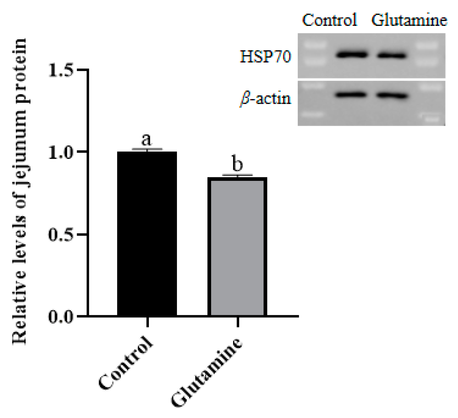
3.8. Effect of Gln Supplementation on the Abundance of the HSP70 Protein in the Jejunum Mucosa of Piglets

The expression level of HSP70 in the jejunal mucosa of piglets is shown in Figure 2. The expression level of HSP70 in the jejunal mucosa of the Gln group was significantly lower than that of the Control group.

4. Discussion

With increasing research into amino acids, there is growing recognition of the important impacts of amino acids on the health, growth, development, reproduction, and homeostasis of living organisms. In recent decades, biomedical and nutritional science have increasingly focused their attention on the beneficial effects of amino acids on animal intestinal health. Gln is widely recognised as a major metabolic fuel for the small intestine and serves as a substrate for intestinal cells to produce purines [9]. In addition, research has shown that Gln could stimulate enterocyte protein synthesis through the activation of the mTOR signalling pathway [8]. The aim of this study was to investigate the effects of Gln on improving intestinal function and promoting muscle growth. Our experimental results demonstrated that Gln increased VS, VW, and VH in the small intestine of piglets. Gln supplementation also enhanced mucosal TP levels as well as the RNA/DNA ratio and TP/DNA ratio in the jejunum and ileum [36]. Furthermore, we observed lower plasma DAO activity in piglets fed with Gln; also, DAO can be used to monitor mucosal injury severity [37]. Intriguingly, supplementation with Gln also led to increased levels of citrulline in both the jejunum and ileum, which possess protective effects on intestinal epithelial cells [38]. This increase in citrulline could potentially be attributed to referential enhancement by glutamine supplements within the intestine’s production capacity for citrulline itself [39]. It was also noted that the concentration of Gln decreased in both the jejunum and ileum; this result may be attributed to the fact that exogenous Gln promotes the proliferation of mucosal cells, which utilises Gln as an energy source, leading to the increased consumption and decreased content of Gln. These results suggest that Gln can enhance intestinal development and maintain its integrity.
Gln plays a role in regulating the redox status of the small intestine in piglets. As a precursor of glutathione synthesis [40], Gln protects the intestinal mucosa from peroxidation damage [41]. In this study, dietary supplementation with Gln was found to elevate the mRNA level of GSTO2 in the small intestine of piglets. GSTO2 can be directly reduced by glutathione or enzymatically by various thiol transferases and NADPH-dependent reductases [42]. Therefore, Gln supplementation improves the antioxidant capacity of the intestine in piglets. Furthermore, HSP70 is considered a marker for oxidative stress [43]; it serves as an important protective factor for intestinal epithelial cells against harmful substances and ulcer formation, and its expression promotes cell proliferation while inhibiting apoptosis and protein synthesis [44]. Notably, HSP70 is well known for its ability to protect organisms from heat-induced toxicity [45]. It also enhances cellular tolerance under conditions such as heat stress, oxidative stress, metal ion stress infection, and tumour growth through anti-apoptotic mechanisms and antioxidative properties. The decrease in HSP70 protein abundance observed in the jejunal mucosa supports the notion that dietary supplementation with Gln benefits intestinal function by reducing intestinal stress effects compared to Control group conditions.
The dietary supplementation of glutamine increased the mRNA levels of IFN-α in the jejunum and ileum and the mRNA levels of IFN-β in the duodenum and ileum, whereas it decreased interferon-stimulated genes (ISGs) [46], including MX1, OAS1, IFIT1, IFIT2, IFIT3, and IFIT5. Previous evidence has shown that virus infection can promote the expression of different ISGs in host cells [47,48,49]. In this study, the decrease in mRNA levels of some ISGs may be due to the fact that Gln can create a healthier environment within the intestinal tract.
In terms of intestinal nutrient transporters, dietary supplementation with Gln resulted in the increased expression of genes encoding amino acid transporters (SLC6A19, SLC7A7, and SLC7A6). SLC6A19 is responsible for the transport of cationic amino acids across the apical membrane. Simultaneously, the main role of SLC7A7 and SLC7A6 is to not only transport neutral and cationic amino acids across the basolateral membrane enterocytes [50], but to also facilitate the absorption of peptides and amino acids [51]. Gln supplementation also elevated the mRNA levels of PepT1 in the longissimus dorsi muscle which is elevated for piglet muscle growth [52]. PepT1 belongs to the proton-dependent oligopeptide transporter family (POT). Many studies have confirmed its critical role in dipeptide, tripeptide, and pseudopeptide absorption within the intestinal tract [53]. Furthermore, the function of PepT1 is associated with cellular networks regulating amino acid homeostasis, including the proteolysis of dietary proteins by proteases and peptidases, mTOR signalling, de novo protein synthesis, and the unfolded protein response [54]. As mentioned earlier, the dietary supplementation of Gln may enhance the intestinal absorption of water and amino acids by regulating the expression of epithelial aquaporin and amino acid transporters, thereby affecting the intestinal health of piglets.
Intestinal bacteria and amino acids are closely interconnected within the intestine. Their interaction significantly impacts host amino acid homeostasis and health as well as the efficiency of dietary amino acid supplementation [55]. The dietary supplementation of glutamine altered the piglet intestinal flora composition by reducing the abundance of Clostridium coccoides, the Enterobacterium family, and the Enterococcus genus, while increasing Lactobacillus abundance in the colon. The ratio between lactobacilli and intestinal bacteria can serve as an indicator of piglet intestinal health [33]. It has been suggested that effects of Gln on intestinal bacteria may be achieved by regulating specific enzyme activity [56], improving protein and energy balance [55], as well as enhancing the utilisation and metabolism of amino acids in the small intestine [6]. In summary, a 0.81% Gln dietary supplementation can maintain the intestinal health of piglets by improving gut microbiota composition.
One notable finding from this study is that Gln promotes muscle growth. The group supplemented with Gln exhibited increased concentrations of TP, RNA/DNA, and TP/DNA ratios in the longissimus dorsi muscle, indicating enhanced muscle synthesis capability [57]. The amino acid content in the longissimus dorsi muscle exhibited a significant decrease. Previous studies have demonstrated that the content of Gln in the longissimus dorsi muscle may significantly decrease during the growing phase of pigs due to its requirement for growth and development [52], which also indicates an increase in tissue protein synthesis [58]. Dietary supplementation with Gln resulted in an increase in the concentration of citrulline, ornithine, arginine, and hydroxyproline. Some studies of these amino acids have shown that citrulline can stimulate muscle protein synthesis by redistributing ATP consumption towards the process of muscle protein synthesis [59]; ornithine can inhibit the breakdown of proteins in skeletal muscle; and hydroxyproline constitutes a substantial proportion of total amino acids in collagen, which is an important component of muscle [60].
SOD and CAT are the key enzymes of the antioxidant system, playing crucial roles in scavenging free radicals and reducing oxidative damage. Under stress conditions, the activity of SOD and CAT in the serum decreased and the content of MDA increased [61]. He et al. [62] found that the supplementation of 1% Gln increased the activity of SOD but decreased the content of MDA. In this study, Gln supplementation improved the activity of SOD, but reduced the content of MDA, indicating that Gln may benefit from the growth of weaning pigs by enhancing antioxidant capacity and preventing peroxide damage to improve antioxidant capacity in the longissimus dorsi muscle.

5. Conclusions

These results suggest that Gln supplementation promotes the intestinal function of piglets by improving intestinal morphology, enhancing intestinal antioxidant capacity, increasing amino acid transporters and water channel transporters, and regulating intestinal flora. In addition, it also accelerates the growth of the longissimus dorsi muscle.

Author Contributions

Conceptualization, S.G., K.Y. and Y.H.; Methodology, Z.Z., D.Y., B.D., D.Z., K.Y. and Y.H.; Formal analysis, J.L., D.Z., K.Y. and Y.H.; Investigation, B.L., D.Y. and B.D.; Resources, S.G., B.D., T.W., D.Z. and Y.H.; Data curation, L.W.; Writing—original draft, L.W., S.G. and Y.H.; Writing—review and editing, L.W., M.S., J.L., Y.Z., Z.Z., S.G., D.Y., K.Y. and Y.H.; Visualization, T.W. and D.Z.; Funding acquisition, S.G. and Y.H. All authors have read and agreed to the published version of the manuscript.

Funding

This research was funded by the National Natural Science Foundation of China (32172763, U22A20514), the National Key R&D Program of China (2022YFD130040302), the Hubei Provincial Key R&D Program (2021BBA083), and the Open Project of Hubei Key Laboratory of Animal Nutrition and Feed Science (202302).

Institutional Review Board Statement

All experimental procedures were approved by the Institutional Animal Care and Use Committee at Wuhan Polytechnic University (20 March 2018, approval code WPU2018001).

Informed Consent Statement

Not applicable.

Data Availability Statement

Data will be made available upon request.

Acknowledgments

We are grateful to all the funds that have helped with this research and to all the students and technicians who have helped with this research.

Conflicts of Interest

All authors consent to participate and publish this article.

Abbreviations

ASCT2Sodium-dependent neutral amino acid transporter 2
CATCatalase
DAODiamine oxidase
Glnl-glutamine
GSTO2Glutathione-S-transferase omega-2
H2O2Hydrogen peroxide
HSP70Heat shock protein-70
IFITInterferon induced protein with tetratricopeptide repeats
ISGsInterferon stimulated genes
MDAMalondialdehyde
MSTNMyostatin
mTORMammalian target of rapamycin
MX1MX dynamin Like GTPase 1
MX2MX dynamin like GTPase 2
Myf5Myogenic Factor 5
OAS12′-5′-Oligoadenylate Synthetase 1
OASL2′-5′-Oligoadenylate Synthetase Like
PepT1Peptide transporter 1
SLC7A6l-type amino acid transporter 2
SLC7A7l-type amino acid transporter 1
SLC6A19Sodium-independent amino acid transporter
SODSuperoxide dismutase
TPTotal protein
VHVillus height
VH/CDVillus height/crypt depth ratio
VSVillus surface area
VWVillus width

References

  1. Ji, F.J.; Wang, L.X.; Yang, H.S.; Hu, A.; Yin, Y.L. Review: The roles and functions of glutamine on intestinal health and performance of weaning pigs. Animal 2019, 13, 2727–2735. [Google Scholar] [CrossRef]
  2. Wu, G.; Morris, S.M., Jr. Arginine metabolism: Nitric oxide and beyond. Biochem. J. 1998, 336 Pt 1, 1–17. [Google Scholar] [CrossRef]
  3. Marc Rhoads, J.; Wu, G. Glutamine, arginine, and leucine signaling in the intestine. Amino Acids 2009, 37, 111–122. [Google Scholar] [CrossRef]
  4. Wu, G.; Fang, Y.Z.; Yang, S.; Lupton, J.R.; Turner, N.D. Glutathione metabolism and its implications for health. J. Nutr. 2004, 1134, 489–492. [Google Scholar] [CrossRef]
  5. Wu, G.; Bazer, F.W.; Dai, Z.; Li, D.; Wang, J.; Wu, Z. Amino acid nutrition in animals: Protein synthesis and beyond. Annu. Rev. Anim. Biosci. 2014, 2, 387–417. [Google Scholar] [CrossRef] [PubMed]
  6. Ren, W.K.; Duan, J.L.; Yin, J.; Liu, G.; Cao, Z.; Xiong, X.; Chen, S.; Li, T.; Yin, Y.; Hou, Y.Q.; et al. Dietary L-glutamine supplementation modulates microbial community and activates innate immunity in the mouse intestine. Amino Acids 2014, 46, 2403–2413. [Google Scholar] [CrossRef]
  7. Ren, W.K.; Yin, J.; Wu, M.; Liu, G.; Yang, G.; Xion, Y.; Su, D.; Wu, L.; Li, T.; Chen, S.; et al. Serum amino acids profile and the beneficial effects of L-arginine or L-glutamine supplementation in dextran sulfate sodium colitis. PLoS ONE 2014, 9, e88335. [Google Scholar] [CrossRef]
  8. Yi, D.; Hou, Y.; Wang, L.; Ouyang, W.; Long, M.; Zhao, D.; Ding, B.; Liu, Y.; Wu, G. L-Glutamine enhances enterocyte growth via activation of the mTOR signaling pathway independently of AMPK. Amino Acids 2015, 47, 65–78. [Google Scholar] [CrossRef]
  9. Wu, G. Amino acids: Metabolism, functions, and nutrition. Amino Acids 2009, 37, 1–17. [Google Scholar] [CrossRef] [PubMed]
  10. Dai, Z.L.; Zhang, J.; Wu, G.; Zhu, W.Y. Utilization of amino acids by bacteria from the pig small intestine. Amino Acids 2010, 39, 1201–1215. [Google Scholar] [CrossRef] [PubMed]
  11. Dai, Z.L.; Li, X.L.; Xi, P.B.; Zhang, J.; Wu, G.; Zhu, W.Y. Metabolism of select amino acids in bacteria from the pig small intestine. Amino Acids 2012, 42, 1597–1608. [Google Scholar] [CrossRef]
  12. Wu, G.; Meier, S.A.; Knabe, D.A. Dietary glutamine supplementation prevents jejunal atrophy in weaned pigs. J. Nutr. 1996, 126, 2578–2584. [Google Scholar] [CrossRef]
  13. Wang, J.; Chen, L.; Li, P.; Li, X.; Zhou, H.; Wang, F.; Li, D.; Yin, Y.; Wu, G. Gene expression is altered in piglet small intestine by weaning and dietary glutamine supplementation. J. Nutr. 2008, 138, 1025–1032. [Google Scholar] [CrossRef]
  14. Chamorro, S.; de Blas, C.; Grant, G.; Badiola, I.; Menoyo, D.; Carabaño, R. Effect of dietary supplementation with glutamine and a combination of glutamine-arginine on intestinal health in twenty-five-day-old weaned rabbits. J. Anim. Sci. 2010, 88, 170–180. [Google Scholar] [CrossRef] [PubMed]
  15. Xue, H.; Sawyer, M.B.; Field, C.J.; Dieleman, L.A.; Murray, D.; Baracos, V.E. Bolus oral glutamine protects rats against CPT-11-induced diarrhea and differentially activates cytoprotective mechanisms in host intestine but not tumor. J. Nutr. 2008, 138, 740–746. [Google Scholar] [CrossRef] [PubMed]
  16. Zhang, Y.; Lu, T.; Han, L.; Zhao, L.; Niu, Y.; Chen, H. L-Glutamine supplementation alleviates constipation during late gestation of mini sows by modifying the microbiota composition in feces. Biomed Res. Int. 2017, 2017, 4862861. [Google Scholar] [CrossRef] [PubMed]
  17. Yi, D.; Li, B.; Hou, Y.; Wang, L.; Zhao, D.; Chen, H.; Wu, T.; Zhou, Y.; Ding, B.; Wu, G. Dietary supplementation with an amino acid blend enhances intestinal function in piglets. Amino Acids 2018, 50, 1089–1100. [Google Scholar] [CrossRef]
  18. Whiting, T.L.; Steele, G.G.; Wamnes, S.; Green, C. Evaluation of methods of rapid mass killing of segregated early weaned piglets. Can. Vet. J. 2011, 52, 753–758. [Google Scholar] [PubMed]
  19. Liu, P.; Piao, X.S.; Kim, S.W.; Wang, L.; Shen, Y.B.; Lee, H.S.; Li, S.Y. Effects of chito-oligosaccharide supplementation on the growth performance, nutrient digestibility, intestinal morphology, and fecal shedding of Escherichia coli and Lactobacillus in weaning pigs. J. Anim. Sci. 2008, 86, 2609–2618. [Google Scholar] [CrossRef]
  20. Hampson, D.J. Alterations in piglet small intestinal structure at weaning. Res. Vet. Sci. 1986, 40, 32–40. [Google Scholar] [CrossRef]
  21. Hou, Y.; Yao, K.; Wang, L.; Ding, B.; Fu, D.; Liu, Y.; Zhu, H.; Liu, J.; Li, Y.; Kang, P.; et al. Effects of α-ketoglutarate on energy status in the intestinal mucosa of weaned piglets chronically challenged with lipopolysaccharide. Br. J. Nutr. 2011, 106, 357–363. [Google Scholar] [CrossRef]
  22. Hou, Y.; Wang, L.; Zhang, W.; Yang, Z.; Ding, B.; Zhu, H.; Liu, Y.; Qiu, Y.; Yin, Y.; Wu, G. Protective effects of N-acetylcysteine on intestinal functions of piglets challenged with lipopolysaccharide. Amino Acids 2012, 43, 1233–1242. [Google Scholar] [CrossRef]
  23. Johnson, L.R.; Chandler, A.M. RNA and DNA of gastric and duodenal mucosa in antrectomized and gastrin-treated rats. Am. J. Physiol. 1973, 224, 937–940. [Google Scholar] [CrossRef]
  24. Lowry, O.H.; Rosebrough, N.J.; Farr, A.L.; Randall, R.J. Protein measurement with the Folin phenol reagent. J. Biol. Chem. 1951, 193, 265–275. [Google Scholar] [CrossRef]
  25. Xie, C.; Zhang, S.; Zhang, G.; Zhang, F.; Chu, L.; Qiao, S. Estimation of the optimal ratio of standardized ileal digestible threonine to lysine for finishing barrows fed low crude protein diets. Asian-Australas. J. Anim. Sci. 2013, 59, 26–34. [Google Scholar] [CrossRef] [PubMed]
  26. Hou, Y.; Wang, L.; Yi, D.; Ding, B.; Yang, Z.; Li, J.; Chen, X.; Qiu, Y.; Wu, G. N-acetylcysteine reduces inflammation in the small intestine by regulating redox, EGF and TLR4 signaling. Amino Acids 2013, 45, 513–522. [Google Scholar] [CrossRef] [PubMed]
  27. Yi, D.; Hou, Y.; Wang, L.; Long, M.; Hu, S.; Mei, H.; Yan, L.; Hu, C.A.; Wu, G. N-acetylcysteine stimulates protein synthesis in enterocytes independently of glutathione synthesis. Amino Acids 2016, 48, 523–533. [Google Scholar] [CrossRef] [PubMed]
  28. Ott, S.J.; Musfeldt, M.; Ullmann, U.; Hampe, J.; Schreiber, S. Quantification of intestinal bacterial populations by real-time PCR with a universal primer set and minor groove binder probes: A global approach to the enteric flora. J. Clin. Microbiol. 2004, 42, 2566–2572. [Google Scholar] [CrossRef] [PubMed]
  29. Meurens, F.; Berri, M.; Auray, G.; Melo, S.; Levast, B.; Virlogeux-Payant, I.; Chevaleyre, C.; Gerdts, V.; Salmon, H. Early immune response following Salmonella enterica subspecies enterica serovar Typhimurium infection in porcine jejunal gut loops. Vet. Res. 2009, 40, 5. [Google Scholar] [CrossRef]
  30. Nygard, A.B.; Jørgensen, C.B.; Cirera, S.; Fredholm, M. Selection of reference genes for gene expression studies in pig tissues using SYBR green qPCR. BMC. Mol. Bio. 2007, 8, 67. [Google Scholar] [CrossRef]
  31. Tako, E.; Glahn, R.P.; Welch, R.M.; Lei, X.; Yasuda, K.; Miller, D.D. Dietary inulin affects the expression of intestinal enterocyte iron transporters, receptors and storage protein and alters the microbiota in the pig intestine. Br. J. Nutr. 2008, 99, 472–480. [Google Scholar] [CrossRef]
  32. Livak, K.J.; Schmittgen, T.D. Analysis of relative gene expression data using real-time quantitative PCR and the 2(-Delta Delta C(T)) Methods. Methods 2001, 25, 402–408. [Google Scholar] [CrossRef]
  33. Castillo, M.; Martín-Orúe, S.M.; Manzanilla, E.G.; Badiola, I.; Martín, M.; Gasa, J. Quantification of total bacteria, enterobacteria and lactobacilli populations in pig digesta by real-time PCR. Vet. Microbiol. 2006, 114, 165–170. [Google Scholar] [CrossRef] [PubMed]
  34. Hou, Y.; Wang, L.; Ding, B.; Liu, Y.; Zhu, H.; Liu, J.; Li, Y.; Wu, X.; Yin, Y.; Wu, G. Dietary alpha-ketoglutarate supplementation ameliorates intestinal injury in lipopolysaccharide-challenged piglets. Amino Acids 2010, 39, 555–564. [Google Scholar] [CrossRef]
  35. Yao, K.; Yin, Y.; Li, X.; Xi, P.; Wang, J.; Lei, J.; Hou, Y.; Wu, G. Alpha-ketoglutarate inhibits glutamine degradation and enhances protein synthesis in intestinal porcine epithelial cells. Amino Acids 2012, 42, 2491–2500. [Google Scholar] [CrossRef] [PubMed]
  36. Yamauchi, K. Review of a histological intestinal approach to assessing the intestinal function in chickens and pigs. Anim. Sci. J. 2007, 78, 356–370. [Google Scholar] [CrossRef]
  37. Luk, G.D.; Bayless, T.M.; Baylin, S.B. Plasma postheparin diamine oxidase. Sensitive provocative test for quantitating length of acute intestinal mucosal injury in the rat. J. Clin. Investig. 1983, 71, 1308–1315. [Google Scholar] [CrossRef] [PubMed]
  38. Chapman, J.C.; Liu, Y.; Zhu, L.; Rhoads, J.M. Arginine and citrulline protect intestinal cell monolayer tight junctions from hypoxia-induced injury in piglets. Pediatr. Res. 2012, 72, 576–582. [Google Scholar] [CrossRef]
  39. Beaufrère, A.M.; Neveux, N.; Patureau Mirand, P.; Buffière, C.; Marceau, G.; Sapin, V.; Cynober, L.; Meydinal-Denis, D. Long-term intermittent glutamine supplementation repairs intestinal damage (structure and functional mass) with advanced age: Assessment with plasma citrulline in a rodent model. J. Nutr. Health Aging 2014, 18, 814–819. [Google Scholar] [CrossRef]
  40. Wu, G. Intestinal mucosal amino acid catabolism. J. Nutr. 1998, 128, 1249–1252. [Google Scholar] [CrossRef]
  41. Gleeson, M. Dosing and efficacy of glutamine supplementation in human exercise and sport training. J. Nutr. 2008, 138, 2045S–2049S. [Google Scholar] [CrossRef]
  42. Hamilton, L.E.; Zigo, M.; Mao, J.; Xu, W.; Sutovsky, P.; O’Flaherty, C.; Oko, R. GSTO2 Isoforms Participate in the Oxidative Regulation of the Plasmalemma in Eutherian Spermatozoa during Capacitation. Antioxidants 2019, 8, 601. [Google Scholar] [CrossRef] [PubMed]
  43. Sepponen, K.; Pösö, A.R. The inducible form of heat shock protein 70 in the serum, colon and small intestine of the pig: Comparison to conventional stress markers. Vet. J. 2006, 171, 519–524. [Google Scholar] [CrossRef] [PubMed]
  44. Zhong, X.; Zhang, X.H.; Li, X.M.; Zhou, Y.M.; Li, W.; Huang, X.X.; Zhang, L.L.; Wang, T. Intestinal growth and morphology is associated with the increase in heat shock protein 70 expression in weaning piglets through supplementation with glutamine. J. Anim. Sci. 2011, 89, 3634–3642. [Google Scholar] [CrossRef] [PubMed]
  45. Al-Aqil, A.; Zulkifli, I. Changes in heat shock protein 70 expression and blood characteristics in transported broiler chickens as affected by housing and early age feed restriction. Poult. Sci. 2009, 88, 1358–1364. [Google Scholar] [CrossRef] [PubMed]
  46. Yang, E.; Li, M.M.H. All about the RNA: Interferon-Stimulated Genes that interfere with viral RNA processes. Front. Immunol. 2020, 9, 605024. [Google Scholar] [CrossRef] [PubMed]
  47. Pidugu, V.K.; Pidugu, H.B.; Wu, M.M.; Liu, C.J.; Lee, T.C. Emerging functions of human IFIT proteins in cancer. Front. Mol. Biosci. 2019, 19, 148. [Google Scholar] [CrossRef]
  48. Ashley, C.L.; Abendroth, A.; McSharry, B.P.; Slobedman, B. Interferon-Independent Upregulation of Interferon-Stimulated Genes during Human Cytomegalovirus Infection is Dependent on IRF3 Expression. Viruses 2019, 11, 246. [Google Scholar] [CrossRef]
  49. Pingale, K.D.; Kanade, G.D.; Karpe, Y.A. Hepatitis E virus polymerase binds to IFIT1 to protect the viral RNA from IFIT1-mediated translation inhibition. J. Gen. Virol. 2019, 100, 471–483. [Google Scholar] [CrossRef]
  50. Osmanyan, A.K.; Ghazi Harsini, S.; Mahdavi, R.; Fisinin, V.I.; Arkhipova, A.L.; Glazko, T.T.; Kovalchuk, S.N.; Kosovsky, G.Y. Intestinal amino acid and peptide transporters in broiler are modulated by dietary amino acids and protein. Amino Acids 2018, 50, 353–357. [Google Scholar] [CrossRef]
  51. Zhang, C.; Wang, C.; Chen, K.; Zhao, X.; Geng, Z. Effect of L-theanine on growth performance, intestinal development and health, and peptide and amino acid transporters expression of broilers. J. Sci. Food Agric. 2020, 100, 1718–1725. [Google Scholar] [CrossRef]
  52. Liu, Y.; Kong, X.; Li, F.; Tan, B.; Li, Y.; Duan, Y.; Yin, Y.; He, J.; Hu, C.; Blachier, F.; et al. Co-dependence of genotype and dietary protein intake to affect expression on amino acid/peptide transporters in porcine skeletal muscle. Amino Acids 2016, 48, 75–90. [Google Scholar] [CrossRef]
  53. Wang, C.Y.; Liu, S.; Xie, X.N.; Tan, Z.R. Regulation profile of the intestinal peptide transporter 1 (PepT1). Drug Des. Dev. Ther. 2017, 8, 3511–3517. [Google Scholar] [CrossRef]
  54. Spanier, B.; Rohm, F. Proton coupled oligopeptide transporter 1 (PepT1) function, regulation, and influence on the intestinal homeostasis. Compr. Physiol. 2018, 8, 843–869. [Google Scholar] [PubMed]
  55. Dai, Z.L.; Wu, G.; Zhu, W.Y. Amino acid metabolism in intestinal bacteria: Links between gut ecology and host health. Front. Biosci. 2011, 16, 1768–1786. [Google Scholar] [CrossRef]
  56. Perna, S.; Alalwan, T.A.; Alaali, Z.; Alnashaba, T.; Gasparri, C.; Infantino, V.; Hammad, L.; Riva, A.; Petrangolini, G.; Allegrini, P.; et al. The role of Glutamine in the complex interaction between gut microbiota and health: A narrative review. Int. J. Mol. Sci. 2019, 20, 5232. [Google Scholar] [CrossRef] [PubMed]
  57. Preedy, V.R.; Peters, T.J. Changes in protein, RNA and DNA and rates of protein synthesis in muscle-containing tissues of the mature rat in response to ethanol feeding: A comparative study of heart, small intestine and gastrocnemius muscle. Alcohol Alcohol. 1990, 25, 489–498. [Google Scholar]
  58. Guay, F.; Trottier, N.L. Muscle growth and plasma concentrations of amino acids, insulin-like growth factor-I, and insulin in growing pigs fed reduced-protein diets. J. Anim. Sci. 2006, 84, 3010–3019. [Google Scholar] [CrossRef] [PubMed]
  59. Goron, A.; Lamarche, F.; Blanchet, S.; Delangle, P.; Schlattner, U.; Fontaine, E.; Moinard, C. Citrulline stimulates muscle protein synthesis, by reallocating ATP consumption to muscle protein synthesis. J. Cachexia Sarcopenia Muscle 2019, 10, 919–928. [Google Scholar] [CrossRef] [PubMed]
  60. Li, P.; Wu, G. Roles of dietary glycine, proline, and hydroxyproline in collagen synthesis and animal growth. Amino Acids 2018, 50, 29–38. [Google Scholar] [CrossRef] [PubMed]
  61. Yang, C.M.; Xu, W.D.; Chen, A.G. Effect of Gly-L-Gln on growth performance and intestinal absorption ability in weaned piglets. China J. Anim. Sci. 2005, 41, 6–8. [Google Scholar]
  62. He, J.; Feng, G.D.; Ao, X.; Li, Y.F.; Qian, H.X.; Liu, J.B.; Bai, G.Y.; He, Z.Z. Effects of L-glutamine on growth performance, antioxidant ability, immunity and expression of genes related to intestinal health in weanling pigs. Livest. Sci. 2016, 189, 102–109. [Google Scholar] [CrossRef]
Figure 1. Intestinal villus morphology of the duodenum, jejunum, and ileum of piglets. The intestinal morphology of the duodenum, jejunum, and ileum in the Control group is shown in (AC), respectively. The intestinal morphology of the duodenum, jejunum, and ileum of the Gln group is shown in (DF), respectively. The morphology and structure of villi were complete and clear in outline.
Life 14 00405 g001
Figure 2. Effect of Gln supplementation on the abundance of the HSP70 protein in the jejunum mucosa of piglets. Data are mean ± SD, n = 3. a,b Mean values with different letters for same protein differ significantly (p < 0.05).
Life 14 00405 g002
Table 1. Ingredients and nutrient composition of the basal diet (air-dry basis).
ItemsContentItemsContent
Ingredient Nutrient level
Corn38.4DE (MJ/kg)14.27
Soybean meal16CP18.54
Flour12Lys1.5
Whey powder (low protein)8Met0.42
Soybean protein concentrate5Cys0.29
Wheat middling5Thr0.93
Fish meal4.5Trp0.23
Glucose3Ca0.75
CaHPO41.33AP0.49
Limestone0.37TP0.68
NaCl0.25Na0.31
Plant oil3.95CF6.26
Premix *1NaCl0.61
Lys0.64
Met0.13
Thr0.21
Choline0.12
Mildew preventive0.1
* The premix provided the following amounts of vitamins and trace minerals per kg of complete diet: Fe, 100 mg (FeSO4·H2O); Cu, 150 mg (CuSO4·5H2O); Mn, 40 mg (MnSO4·5H2O); Zn, 100 mg (ZnSO4·7H2O); I, 0.5 mg (KI); Se, 0.3 mg (Na2SeO3·5H2O); VA, 3.72 mg; VD3, 0.10 mg; DL-α-tocopheryl acetate, 26.7 mg; VK3, 4 mg; VB1, 6 mg; VB2, 12 mg; VB6, 6 mg; VB12, 0.05 mg; biotin, 0.2 mg; folic acid, 2 mg; niacin, 50 mg; and d-calcium pantothenate, 5 mg.
Table 2. The sequences of primers used in the present study.
ItemsPrimer Sequence (5′-3′)
Total bacteria count(F) CGGTCCAGACTCCTACGGG
(R) TTACCGCGGCTGCTGGCAC
Enterococcus genus(F) CCCTTATTGTTAGTTGCCATCATT
(R) ACTCGTTGTACTTCCCATTGT
Enterobacterium family(F) CATTGACGTTACCCGCAGAAGAAGC
(R) CTCTACGAGACTCAAGCTTGC
Clostridium coccoides(F) AATGACGGTACCTGACTAA
(R) CTTTGAGTTTCATTCTTGCGAA
Lactobacillus genus(F) TCGCGTC(C/T)GGTGTGAAAG
(R) CCACATCCAGC(A/G)TCCAC
Internal reference(F) CAGAAATGGGAATGGAAAGTTG
(R) CCATTGGTCAGGTCATTCAATACA
ASCT2(F) GCCAGCAAGATTGTGGAGAT
(R) GAGCTGGATGAGGTTCCAAA
GSTO2(F) GCCTTGAGATGTGGGAGAGAA
(R) AAGATGGTGTTCTGATAGCCAAGA
IFIT1(F) GCTAAACCAAACACCGCAGAA
(R) GGAACTCAATCTCCTCCAAGACC
IFIT2(F) CAGAAGGCGGCAGAGAATG
(R) ACACAGAGGCAGGCGAGATAG
IFIT3(F) GCATTTTCCAGCCAGCATC
(R) TCTGTTCCTTTCCTTTCCTTCCT
IFIT5(F) CAGAAAATACAGCCATCCACCA
(R) AGGGCACTTAAACTCTGCACATC
IFN-α(F) ACTCCATCCTGGCTGTGAGGAAAT
(R) ATCTCATGACTTCTGCCCTGACGA
IFN-β(F) AGCAGATCTTCGGCATTCTC
(R) GTCATCCATCTGCCCATCAA
MSTN(F) GAAGTCAAGGTAACAGACACACCAA
(R) GCAATAATCCAGTCCCATCCA
MX1(F) AGTGCGGCTGTTTACCAAG
(R) TTCACAAACCCTGGCAACTC
MX2(F) CGCATTCTTTCACTCGCATC
(R) CCTCAACCCACCAACTCACA
Myf5(F) CCACGACTAACCCCAACCA
(R) TTTTCCACCTGCTCCCTCA
OAS1(F) TGGTGGTGGAGACACACACA
(R) CCAACCAGAGACCCATCCA
OASL(F) GGCACCCCTGTTTTCCTCT
(R) AGCACCGCTTTTGGATGG
PepT1(F) ATTCTCAGGCTCCTTCCAACA
(R) GCAACCCCGCAAACAGA
SLC7A6(F) CTGCCGCCTGCATGTGT
(R) TGTGCCCCACTTGACATAGG
SLC7A7(F) TTTGGTTCCCAAGGTTGCA
(R) GCAGCTTCCTGGCATTGC
SLC6A19(F) CGAGTACCCGTACCTGATGGA
(R) TGCGTAGAAGGGCGAAGAA
Table 3. Effects of dietary supplementation with Gln on plasma DAO and intestinal morphology in piglets.
ItemsControl GroupGln Groupp-Value
Plasma
          DAO (mmol/L)15.36 ± 2.49 a9.38 ± 3.54 b0.002
Duodenum
          Villus height (μm)282 ± 24 b349 ± 13 a<0.01
          Crypt depth (μm)119 ± 7121 ± 70.574
          Villus height/crypt depth2.45 ± 0.16 b2.89 ± 0.23 a<0.01
          Villus width (μm)140 ± 8 b161 ± 5 a<0.01
          Villus surface area (μm2)32,431 ± 3438 b44,744 ± 3273 a<0.01
Jejunum
          Villus height (μm)260 ± 21 b311 ± 31 a0.002
          Crypt depth (μm)99 ± 7103 ± 160.564
          Villus height/crypt depth2.60 ± 0.14 b3.06 ± 0.36 a0.006
          Villus width (μm)104 ± 5 b124 ± 8 a<0.01
          Villus surface area (μm2)20,481 ± 3519 b26,809 ± 3987 a0.005
Ileum
          Villus height (μm)231 ± 16 b290 ± 22 a<0.01
          Crypt depth (μm)110 ± 12114 ± 160.631
          Villus height/crypt depth2.18 ± 0.10 b2.59 ± 0.30 a0.002
          Villus width (μm)106 ± 6 b120 ± 10 a0.005
          Villus surface area (μm2)18,462 ± 1742 b25,847 ± 3745 a<0.01
Data are mean ± SD, n = 8. a,b Means within rows with different superscripts differ signficantly (p < 0.05).
Table 4. Effects of dietary supplementation with Gln on TP, RNA, and DNA in the intestinal mucosa in piglets.
ItemsControl GroupGln Groupp-Value
Duodenum
          TP (mg/g)67.20 ± 7.1066.00 ± 3.900.694
          RNA (mg/g)2.51 ± 0.322.58 ± 0.340.522
          DNA (mg/g)0.36 ± 0.070.40 ± 0.070.234
          RNA/DNA7.27 ± 1.666.91 ± 1.840.256
          TP/DNA196.64 ± 45.10175.37 ± 35.970.716
Jejunum
          TP (mg/g)51.50 ± 7.60 b62.70 ± 6.10 a0.006
          RNA (mg/g)1.70 ± 0.331.75 ± 0.240.744
          DNA (mg/g)0.21 ± 0.020.19 ± 0.020.162
          RNA/DNA7.79 ± 1.04 b8.80 ± 0.46 a<0.01
          TP/DNA255.12 ± 61.68 b319.59 ± 52.72 a0.001
Ileum
          TP (mg/g)31.90 ± 6.70 b58.90 ± 7.30 a<0.01
          RNA (mg/g)2.24 ± 0.362.22 ± 0.300.709
          DNA (mg/g)0.18 ± 0.026 a0.14 ± 0.01 b0.001
          RNA/DNA11.49 ± 2.67 b15.25 ± 2.85 a0.020
          TP/DNA166.67 ± 30.90 b402.96 ± 61.66 a<0.01
Data are mean ± SD, n = 8. a,b Means within rows with different superscripts differ significantly (p < 0.05).
Table 5. Effects of dietary supplementation with Gln on the concentrations of amino acids in the jejunum and ileum of piglets.
Amino Acids (μg/g)Control GroupGln Groupp-Value
Jejunum
          1-Methylhistidine4.081 ± 1.8964.103 ± 2.8930.986
          Arginine216.335 ± 61.647210.695 ± 66.2190.863
          Carnosine6.582 ± 1.502 a4.55 ± 1.153 b0.009
          Citrulline94.349 ± 26.045 b160.174 ± 47.633 a0.004
          Cystine2.489 ± 0.6494.731 ± 3.4060.089
          Glutamine833.294 ± 133.851 a638.748 ± 108.339 b0.006
          Hydroxyproline112.917 ± 5.053110.441 ± 15.0410.666
          Ornithine45.68 ± 7.743.54 ± 5.490.533
          β-Alanine60.883 ± 874.941 ± 21.4130.104
          β-Aminoisobutyric acid17.703 ± 6.11720.05 ± 23.7950.791
Ileum
          1-Methylhistidine2.956 ± 0.463 a1.869 ± 0.566 b0.001
          Arginine187.077 ± 19.474184.844 ± 39.1830.887
          Carnosine7.565 ± 2.587.607 ± 2.2560.973
          Citrulline62.951 ± 10.771 b87.347 ± 22.556 a0.015
          Cystine2.939 ± 0.6133.598 ± 1.0690.152
          Glutamine983.157 ± 186.976 a679.397 ± 140.461 b0.003
          Hydroxyproline167.301 ± 17.16 a142.792 ± 7.786 b0.002
          Ornithine110.647 ± 18.887120.151 ± 52.1050.635
          β-Alanine67.41 ± 16.363 b86.473 ± 17.62 a0.042
          β-Aminoisobutyric acid2.676 ± 0.613 a1.425 ± 0.133 b<0.01
Data are mean ± SD, n = 8. a,b Means within rows with different superscripts differ significantly (p < 0.05).
Table 6. Effects of dietary supplementation with Gln on the expression levels of genes in the duodenum, jejunum, and ileum and bacterial genes in the colon in piglets.
ItemsControl GroupGln Groupp-Value
Duodenum
          GSTO21.000 ± 0.213 b3.749 ± 0.913 a<0.01
          IFN-β1.000 ± 0.119 b2.684 ± 0.649 a<0.01
Jejunum
          GSTO21.000 ± 0.2571.254 ± 0.2740.077
          IFIT11.000 ± 0.281 a0.532 ± 0.138 b0.001
          IFIT21.000 ± 0.248 a0.613 ± 0.163 b0.002
          IFIT31.000 ± 0.163 a0.661 ± 0.139 b0.001
          IFIT51.000 ± 0.119 a0.659 ± 0.158 b<0.01
          IFN-α1.000 ± 0.245 b1.286 ± 0.272 a0.044
          MX11.000 ± 0.143 a0.464 ± 0.106 b<0.01
          MX21.000 ± 0.265 a0.600 ± 0.155 b0.002
          OAS11.000 ± 0.272 a0.670 ± 0.175 b0.012
          OASL1.000 ± 0.281 a0.446 ± 0.111 b<0.01
          SLC7A61.000 ± 0.154 b1.227 ± 0.236 a0.039
Ileum
          GSTO21.000 ± 0.118 b2.005 ± 0.399 a<0.01
          IFIT11.000 ± 0.224 a0.607 ± 0.135 b0.001
          IFN-α1.000 ± 0.169 b1.321 ± 0.275 a0.014
          IFN-β1.000 ± 0.101 b1.392 ± 0.293 a0.003
          MX11.000 ± 0.161 a0.712 ± 0.148 b0.002
          SLC7A71.000 ± 0.235 b1.291 ± 0.251 a0.031
          Colon bacteria
          Clostridium coccoides1.000 ± 0.154 a0.335 ± 0.059 b<0.01
          Enterobacterium family1.000 ± 0.136 a0.354 ± 0.044 b<0.01
          Enterococcus genus1.000 ± 0.246 a0.728 ± 0.181 b0.024
          Lactobacillus genus1.000 ± 0.242 b2.450 ± 0.449 a<0.01
Data are mean ± SD, n = 8. The expression level of the Control group was regarded as 1. a,b Means within rows with different superscripts differ significantly (p < 0.05).
Table 7. Effects of dietary supplementation with Gln on the concentrations of amino acids in the longissimus dorsi muscle of piglets.
Amino AcidsControl GroupGln Groupp-Value
1-Methylhistidine (μg/g)3.955 ± 1.2093.743 ± 1.4370.755
Arginine (μg/g)56.966 ± 8.866 b107.416 ± 52.704 a0.018
Carnosine (ng/g)13.65 ± 0.91614.044 ± 1.4110.519
Citrulline (μg/g)57.191 ± 11.348 b82.69 ± 14.84 a0.002
Glutamine (ng/g)2.075 ± 0.328 a1.629 ± 0.233 b0.007
Hydroxyproline (μg/g)94.618 ± 12.03 b124.028 ± 23.401 a0.007
Ornithine (μg/g)155.918 ± 33.51 b222.05 ± 59.788 a0.016
β-Alanine (ng/g)0.962 ± 0.1070.857 ± 0.2250.254
β-Aminoisobutyric acid (μg/g)14.926 ± 5.31216.039 ± 3.3850.625
Data are mean ± SD, n = 8. a,b Means within rows with different superscripts differ significantly (p < 0.05).
Table 8. Effects of dietary supplementation with Gln on TP, RNA, and DNA and the expression levels of genes in the longissimus dorsi muscle in piglets.
ItemsControl GroupGln Groupp-Value
          TP (mg/g)29.1 ± 2.2 b37.4 ± 5.1 a0.001
          RNA (mg/g)0.060 ± 0.017 a0.035 ± 0.007 b<0.01
          DNA (mg/g)0.336 ± 0.021 b0.386 ± 0.049 a0.003
          RNA/DNA0.170 ± 0.042 a0.091 ± 0.020 b<0.01
          TP/DNA87.771 ± 5.219 b100.078 ± 11.917 a<0.01
Genes
          ASCT21.000 ± 0.2120.847 ± 0.1790.140
          SLC6A191.000 ± 0.2301.102 ± 0.1850.343
          MSTN1.000 ± 0.2020.964 ± 0.2170.738
          Myf51.000 ± 0.1641.087 ± 0.1870.339
          PepT11.00 ± 0.176 b1.569 ± 0.408 a0.003
          SLC7A71.00 ± 0.1890.878 ± 0.1970.228
          SLC7A61.000 ± 0.2501.065 ± 0.2490.611
Data are mean ± SD, n = 8. The expression level of the Control group was regarded as 1. a,b Means within rows with different superscripts differ significantly (p < 0.05).
Table 9. Effects of dietary supplementation with Gln on antioxidant capacity in the longissimus dorsi muscle of piglets.
ItemsControl GroupGln Groupp-Value
CAT (U/mg protein)1.11 ± 0.331.09 ± 0.300.910
MDA (nmol/mg protein)0.65 ± 0.15 a0.41 ± 0.20 b0.020
H2O2 (μmol/mg protein)1.59 ± 0.53 a1.11 ± 0.25 b0.009
SOD (U/mg protein)73.1 ± 12.03 b83.8 ± 7.11 a0.048
Data are mean ± SD, n = 8. a,b Means within rows with different superscripts differ significantly (p < 0.05).
Disclaimer/Publisher’s Note: The statements, opinions and data contained in all publications are solely those of the individual author(s) and contributor(s) and not of MDPI and/or the editor(s). MDPI and/or the editor(s) disclaim responsibility for any injury to people or property resulting from any ideas, methods, instructions or products referred to in the content.

