Article

Complete Mitochondrial Genome and Its Phylogenetic Position in Red Algae Fushitsunagia catenata from South Korea

1
Industry-University Cooperation Foundation, Pukyong National University, 45 Yongso-ro, Nam-gu, Busan 48513, Republic of Korea
2
Study Program of Environmental Engineering, Faculty of Science and Technology, Universitas Airlangga, Surabaya 60115, Indonesia
3
Marine Eco-Technology Institute, Busan 48520, Republic of Korea
4
Dokdo Fisheries Research Center, National Institute of Fisheries Science, Pohang 37709, Republic of Korea
5
Department of Ocean Engineering, Pukyong National University, 45 Yongso-ro, Nam-gu, Busan 48513, Republic of Korea
6
Department of Microbiology, Pukyong National University, 45 Yongso-ro, Nam-gu, Busan 48513, Republic of Korea
7
School of Marine and Fisheries Life Science, Pukyong National University, 45 Yongso-ro, Nam-gu, Busan 48513, Republic of Korea
*
Authors to whom correspondence should be addressed.
Life 2024, 14(4), 534; https://doi.org/10.3390/life14040534
Submission received: 20 February 2024 / Revised: 5 April 2024 / Accepted: 10 April 2024 / Published: 22 April 2024
(This article belongs to the Section Genetics and Genomics)

Abstract

:
The mitogenome is an important tool in taxonomic and evolutionary studies. Only a few complete mitogenomes have been reported for red algae. Herein, we reported the complete mitochondrial genome sequence of Fushitsunagia catenata (Harvey) Filloramo, G.V. and Saunders, G.W. 2016, a monospecific genus. The genome was 25,889 bp in circumference and had a strongly biased AT of 70.4%. It consisted of 2 rRNAs, 23 tRNAs, and 24 protein-coding genes (PCGs). nad5 (1986 bp) was the largest and atp9 (231 bp) was the smallest PCG. All PCGs used ATG as an initiation codon and TAA as a termination codon, except TAG, which was the termination codon used in the sdh3, rps3, and rps11 genes. The general structure and gene content of the present findings were almost identical to those of other red algae genomes, particularly those of the Rhodymeniales order. The maximum likelihood analysis showed that F. catenata was closely related to Rhodymenia pseudopalmata. The mitochondrial genome data presented in this study will enhance our understanding of evolution in Rhodophyta species.

1. Introduction

The phylum Rhodophyta, also known as red algae, is a monophyletic group largely comprising multicellular photosynthetic eukaryotes. The seven groups, Rhodophytes-Bangiophyceae, Compsopogonophyceae, Cyanidiophyceae, Florideophyceae, Porpyridiophyceae, Rhodellophyceae, and Stylonematophyceae, comprising over 7538 species, comprise a diverse group of algae [1]. The class Florideophyceae encompasses many species (7141), mostly multicellular sea algae. As eukaryotic members of the Archaeplastida supergroup, red algae are not real plants; they share similar ancestors with the green lineage (Chloroplastida) [2]. Red algae are uncommon in freshwater environments but ubiquitous in marine ecosystems (98%) [3].
Recent studies that combined morphological and genomic data resulted in numerous taxonomic revisions. The genus Fushitsunagia was recently isolated from the genus Lomentaria [4]. Fushitsunagia catenata is larger, measuring 10–15 cm in height, with straight apices, a turgid texture, and irregular branches [5]. The taxonomy of the genus Fushitsunagia is still unclear because morphological traits have limited taxonomic relevance. To date, only a few mitochondrial genes (cob, cox1, and cox3) of F. catenata have been reported. Phylogenetic analysis using complete mitochondrial genomes is more informative for determining evolutionary relatedness than single-gene sequencing [6]. Therefore, we analyzed the whole mitochondrial genome of F. catenata and discussed the evolutionary connections between rhodophytes.
Fushitsunagia catenata (Harvey) Filloramo, G.V. & Saunders, G.W. 2016 is a red macroalga and belongs to the phylum Rhodophyta (Florideophyceae; Rhodymeniophycidae; Rhodymeniales; Lomentariaceae) [7]. Fushitsunagia is a monospecific genus that is naturally found in China, Japan, and South Korea [8] and may be found in the Gulf of California [9], New South Wales, Australia [10], and Spain [11]. The cytochrome oxidase subunits and phylogenetic resolutions based on these genes have been reported [4].
Mitochondrial genes are valuable for phylogenetic research. However, a more precise understanding of phylogenetic relationships may be obtained by analyzing the full mitochondrial genome. There have been no reports of the full mitochondrial genome or phylogenetic analyses of F. catenata. This study included the construction of the first comprehensive mitochondrial genome of F. catenata using de novo assembly on an Illumina platform. The findings of this study will be important for future phylogenetic analysis, in-depth comprehension of gene content and structure, and comparative mitochondrial genome analyses.

2. Materials and Methods

2.1. Sample Collection and Genomic DNA Extraction

The red macroalga F. catenata sample (Figure S1) used in this study was collected from the coastal region of Gijang, Busan, South Korea (35.284634 N, 129.259071 E) in August 2022. The samples were subsequently deposited in the Ecological Restoration Group, Marine Eco-Technology Institute, Busan, South Korea (specimen number PU-T01-S-MA-05). Genomic DNA was isolated using a DNeasy Blood and Tissue kit (Qiagen, Germany) according to the manufacturer’s instructions. The concentration and purity of the extracted DNA were evaluated using a NanoDrop spectrophotometer (Thermo Fisher Scientific D1000, Waltham, MA, USA). The extracted genomic DNA was stored at a temperature of −4 °C and transported to Macrogen (Daejeon, South Korea; https://www.macrogen.com/ko/) for library creation and sequencing.

2.2. Mitochondrial Genome Sequencing

DNA libraries were created using the TrueSeq Nano DNA Kit and then subjected to sequencing on the Illumina platform (Illumina, HiSeq 2500, San Diego, CA, USA) using paired-end reads with a length of 150 bp. To reduce analytical bias, the acquired reads were trimmed using the Trimmomatic v0.36 (http://www.usadellab.org/cms/?page=trimmomatic, accessed on 15 October 2023) [12]. This included the removal of adapter sequences and low-quality reads with quality scores below 20 (Q < 20). The trimmed reads were randomly sampled to assemble the mitochondrial genome. In this case, only the sampled reads were used for de novo assembly. The overall quality of sequencing reads was assessed using FastQC v0.11.5 (http://www.bioinformatics.babraham.ac.uk/projects/fastqc, accessed on 15 October 2023) [13]. High-quality reads were assembled using k-mers and SPAdes v3.15.0 (http://cab.spbu.ru/software/spades/, accessed on 15 October 2023) [14,15]. After the complete genome was assembled, BLAST analysis was performed to identify the contigs containing the mitogenome sequences in the NCBI database (https://blast.ncbi.nlm.nih.gov/Blast.cgi, accessed on 15 October 2023).

2.3. Mitochondrial Genome Assembly and Annotation

The contig was annotated using the online platform MFannot (https://megasun.bch.umontreal.ca/apps/mfannot/, accessed on 15 October 2023) [16]. Protein-coding genes (PCGs) were identified and validated using the open reading frame finder (https://www.ncbi.nlm.nih.gov/orffinder/, accessed on 15 October 2023) and verified manually using BLAST homology searches against the NCBI protein database [17]. The RNAweasel tool (https://megasun.bch.umontreal.ca/apps/rnaweasel/, accessed on 15 October 2023) was used to validate the annotated RNAs and detect introns [18]. The Tandem Repeats Finder tool (https://tandem.bu.edu/trf/, accessed on 15 October 2023) was used to detect and analyze repetitive sequences [19]. The assembled contig was subjected to identification analysis by querying BlastN and comparing its size with that of the previously reported mitochondrial genomes of Rhodophyta.

2.4. Physical Mapping and Codon Usage Analysis

Map visualization of the genetic information identified in the mitochondria of F. catenata (GenBank accession number OR045827) was generated using OGDRAW (https://chlorobox.mpimp-golm.mpg.de/OGDraw.html, accessed on 15 October 2023) [20]. The nucleotide content of the mitochondrial genome was determined using MEGA11 v11.0.8 software [21]. The codon usage of PCGs was analyzed using the Sequence Manipulation Suite program (https://www.bioinformatics.org/sms2/codon_usage.html, accessed on 15 October 2023) [22]. The skew analysis was determined using the following formulas: AT-skew = (A − T)/(A + T) and GC-skew = (G − C)/(G + C) [23]. Intergenic spacers between genes and overlapping areas were manually calculated.

2.5. Phylogenetic Analysis

The phylogenetic tree was constructed using the complete mitochondrial genome and the cox1, cox3, and cob gene sequences of 12 selected red algae from the subclass Rhodymeniophycidae, together with one outgroup member from the family Glaucocystaceae (Table 1). The mitochondrial genomes and gene sequences used in this study were retrieved from the NCBI GenBank database (https://www.ncbi.nlm.nih.gov/, accessed on 10 November 2023). Multiple sequence alignments were performed using ClustalW [24], and a maximum likelihood (ML) phylogenetic tree was created using MEGA11 [25]. ML analysis was conducted using the Tamura–Nei model with default settings and 1000 bootstrap replications [21].

3. Results

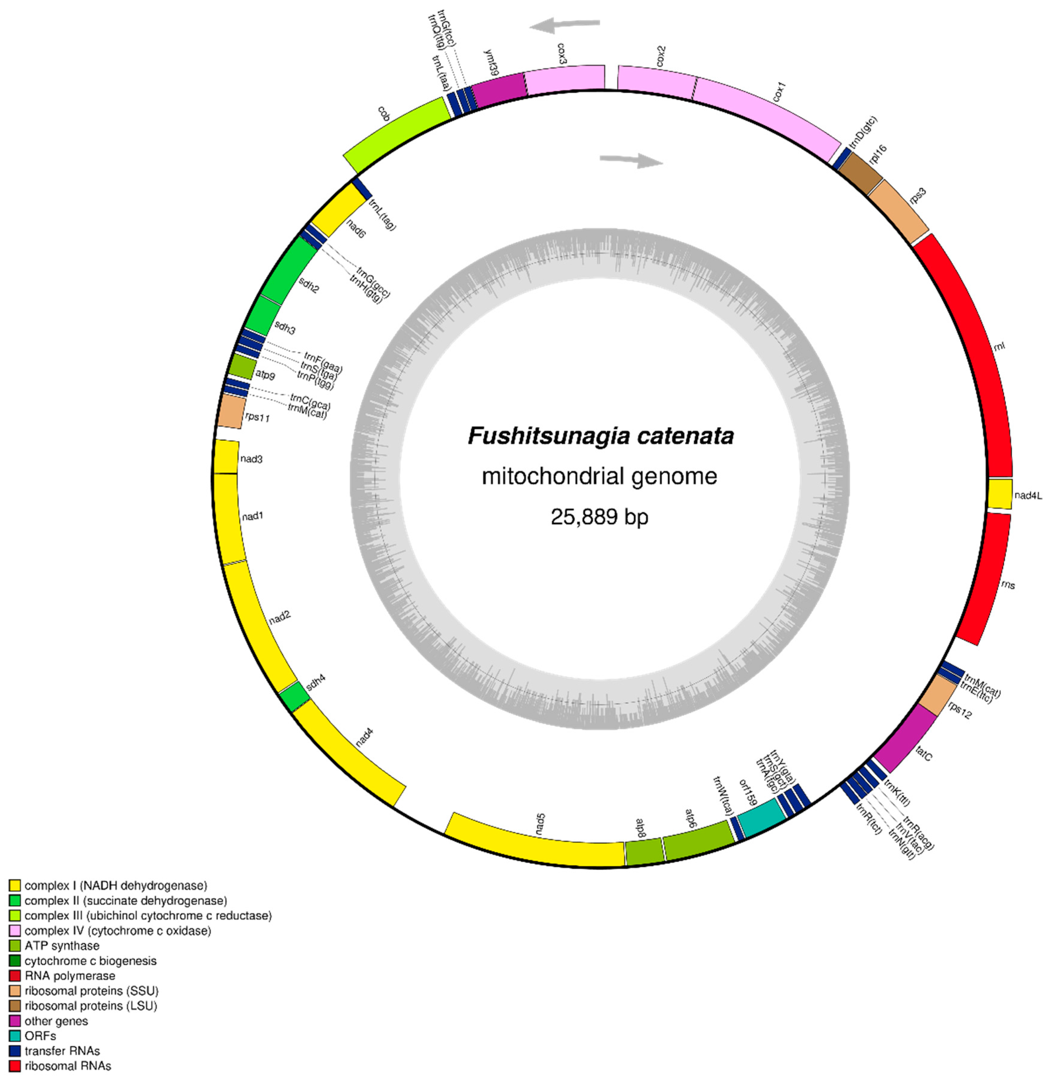
3.1. Mitochondrial Genome Characterization

The F. catenata library was subjected to next-generation sequencing using an Illumina HiSeq 2500 sequencer, resulting in 22,066,120 raw reads. The GC content of the reads was 43.39%, with a Q20 score of 93.81% and a Q30 score of 87.08%. After removing low-quality sequences, 14,944,788 filtered reads were obtained with a GC content of 42.65%, Q20 accuracy of 98.86%, and Q30 accuracy of 95.78%. The reads were subjected to de novo assembly (100% coverage with a depth of 479.84), resulting in a contig consisting of 25,889 bases, with a GC content of 29.59%. The mitochondrial genome of F. catenata (GenBank: OR045827) was circular with a length of 25,889 bp (Figure 1). It contained 49 genes, consisting of 24 PCGs, 23 tRNAs, and 2 rRNAs. The H-strands contained 10 PCGs, 11 tRNAs, and 2 rRNAs. In contrast, the L strand consisted of 14 PCGs and 12 tRNAs. The nucleotide content of the whole genome was determined to be 37.1% A, 33.3% T, 15.1% G, and 14.5% C, as shown in Table 1. The analysis of nucleotide composition revealed a biased composition of A + T, which accounted for 70.4% of the total genome. The whole genome exhibited positive AT and GC skewness, suggesting a preference for using As over Ts and Gs over Cs.

3.2. Protein-Coding Genes

Twenty-four PCGs comprised 69.35% of the mitochondrial genome of F. catenata. These genes comprised a total length of 17,955 bp. There were clusters of genes such as NADH dehydrogenase subunits, succinate dehydrogenase, apocytochrome b, cytochrome c oxidase, ATP synthase, small- and large-subunit ribosomal proteins, independent protein translocase, and a gene encoding a hypothetical protein (Table 2). The nad5 and atp9 genes were the largest and smallest, respectively, in terms of length within the whole mitochondrial genome. Nad5 accounted for 7.67% (1986 bp) of the genome, whereas atp9 accounted for 0.89% (231 bp). ATG and TAA served as the initiation and termination codons for all PCGs, except TAG, which served as the termination codon specifically for the sdh3, rps3, and rps11 genes. The mitochondrial genome of F. catenata contained only rpl16 among the ribosomal protein genes (Table 3). The genes rpl5 and rpl20 do not exist, and no intronic coding sequences were identified.

3.3. Codon Usage Analysis

A codon usage analysis of the mitochondrial genome of F. catenata showed that 5961 amino acids were expressed in the PCGs (Table S1). The amino acid composition indicated that leucine (N = 900, 15.10%), phenylalanine (N = 587, 9.85%), and isoleucine (N = 582, 9.76%) were the amino acids most often found. Conversely, cysteine (N = 78, 1.31%), histidine (N = 121, 2.03%), and tryptophan (N = 129, 2.16%) showed the lowest levels of abundance among the identified amino acids. The codons TTA (leucine, N = 568, 9.53%), TTT (phenylamine, N = 515, 8.64%), and ATT (alanine, N = 388, 6.51%) were the most frequently used codons in PCGs.

3.4. RNAs

In the mitochondrial genome of F. catenata, the rRNA genes were located on the H-strand and identified as rnl (large subunit, 2604 bp) and rns (small subunit, 1361 bp) (Table 2). These genes had a combined length of 3965 bp, accounting for 15.32% of the whole mitochondrial genome. The rRNA genes were separated using the nad4L gene. A total of 23 tRNAs, ranging from 71 to 93 bp in length, were found in the mitochondrial genome; trnI was not identified in the F. catenata mitochondrial genome. Among these, arginine (trnR-TCT and trnR-ACG), glycine (trnG-TCC and trnG-GCC), leucine (trnL-TAA and trnL-TAG), methionine (trnM-CAT), and serine (trnS-TGA and trnS-GCT) had two copies with distinct anticodons, with the exception of methionine, which had the same anticodon. The tRNA cysteine (trnC-GCA, 71 bp) was the shortest and serine (trnS-GCT, 93 bp) was the longest. The total tRNA length was 1741 bp, accounting for 6.73% of the whole genome length, and no intronic RNA sequences were detected.

3.5. Overlapping and Intergenic Spacer Regions

An examination of the intergenic nucleotides of the F. catenata mitochondrial genome sequence revealed that only two gene junctions exhibited an overlap of 21 bp: trnLnad6 (1 bp overlap) and TatCrps12 (20 bp overlap). In addition, we observed intergenic gaps ranging from 1 to 607 bp. The largest intergenic gap, measuring 607 bp, was observed between nad4 and nad5 genes (Table 2).

3.6. Phylogenetic Analysis

ML phylogenetic trees were constructed with complete mitochondrial genome sequences based on single-gene sequences of the species within the Rhodymeniales order. The ML phylogenetic analysis indicated that F. catenata was most closely related to Rhodymenia pseudopalmata with strong bootstrap support (Figure 2). Rhodymeniales species (F. catenata and R. pseudopalmata) formed a monophyletic clade with Halymeniales species (Grateloupia elliptica, G. turuturu) with high bootstrap support but not with other species.
A phylogenetic analysis, using gene sequences of cox1 (Figure S2), cox3 (Figure S3), and cob (Figure S4), revealed differences in the relationships among the species in the group. However, the bootstrap values supporting each node were often modest, except for a substantial bootstrap value that supported the relationship between sister taxa of Grateloupia and Gelidium species. Most algal orders formed a monophyletic group in the phylogenies of cox1, cox3, and cob, except for Gigartinales and Rhodymeniales in the cox1 phylogeny.

4. Discussion

The mitochondrial genome of F. catenata conformed to characteristics often observed in red algae, and the quality of the sequenced genome was comparable to that of other species belonging to the Rhodymeniophycidae subfamily (Table 1). The size and base composition of monospecific F. catenata were consistent with those of a previously reported Rhodymeniales species, R. pseudopalmata (KC875852; 26,166 bp, 29.5% GC) [29].
The long intergenic nucleotide region between nad4 and nad5 in F. catenata (Table 2) was similar to the intergenic regions of the previously reported species, G. elliptica, Gelidium coulteri, G. sinicola, Gracilariopsis andersonii, G. turuturu, and Sarcopeltis skottsbergii. In contrast, the other species investigated in this study possessed intronic tRNA genes between nad4 and nad5 (specifically, trnI in Agarophyton chilense, Hydropuntia rangiferina, Rhodomelopsis africana, R. pseudopalmata, and trnH in Gloiopeltis furcate) [6,26,27,28,29,30]. The lack of trnI in some species may be attributed to the inaccurate annotation of tRNA. The most closely related species, R. pseudopalmata (Figure S5, circular mitogenome), had gene content similar to that of F. catenata [12]. Three tRNAs (trnH, trnW, and trnY) were present in R. pseudopalmata but absent in F. catenata, whereas two tRNAs (trnI and trnU) were absent in F. catenata but present in R. pseudopalmata. Additionally, we found that both species had two copies of each trnL, trnG, trnS, and trnM gene and that F. catenata alone had two copies of the trnR gene.
In general, the arrangement of mitochondrial genomes in Rhodymeniophycidae is highly conserved in terms of genome size and gene content (Table 3), as often observed in other red algal groups [6,26,29]. No intronic PCGs or tRNA were detected in the mitochondrial genome of F. catenata. However, group II intronic cox1 genes have been identified in red algae, such as G. elliptica and G. turuturu, as reported by Patil et al. [27,28]. Additionally, intronic tRNA genes have been reported, including the intronic trnI gene in A. chilense (MZ336082), H. rangiferina (MZ336092), R. africana (OP748274), and R. pseudopalmata (KC875852), and the intronic trnH gene in G. furcate (OP612669). Most red algal species that have been sequenced have two rRNAs, rnl and rns. However, two species of the Halymeniales order have an additional rRNA called rns5 [27,28]. rRNA has also been detected in other Florideophyceae red algal species. However, they only exist in certain orders and species of the same genus or family [6,31]. Among the ribosomal protein genes, the rpl20 gene seems to be the least conserved in red algae [6]. rpl5 and rpl20 were not identified in the mitochondrial genome sequence of F. catenata, which is consistent with the mitogenome characteristics of G. coultery (MG922857), G. sinicola (KX427233), R. africana (OP748274), and R. pseudopalmata (KC875852). These differences may play important roles in the mitogenomic evolution of Florideophyceae red algae.
The ML phylogenetic analysis, based on the complete genome sequence (Figure 2) of selective members of Rhodymeniophycidae, revealed that F. catenata is closely related to R. pseudopalmata, which belongs to the Rhodymeniales order. Furthermore, F. catenata formed a monophyletic group with species from the Halymeniales order, namely G. elliptica (OP479979) and G. turuturu (OQ972988). A previous study documented the emergence of a monophyletic cluster, including the Rhodymeniales and Halymeniales orders [31]. However, the phylogenetic tree based on individual genes (Figures S2–S4) shows that the cob and cox3 genes have a phylogenetic structure similar to that of the complete genome-based phylogeny. However, the phylogeny based on the cox1 gene sequence separated the sister species of the monophyletic clade of Gigartinales (G. furcate, S. skottsbergii) and Rhodymeniales (F. catenata, R. pseudopalmata). The resulting topology was aligned with a phylogenetic tree constructed from multiple genes and the complete mitochondrial genome sequence [27,28,31]. Filloramo and Sanders [4] found that the Rhodymeniales order is monophyletic and divided into two major lineages: Fryeellaceae, which is a sister of Faucheaceae and Lomentariaceae, and Rhodymeniaceae, which is allied to Champiaceae and Hymenocladiaceae. However, complete mitochondrial genome sequences of several Rhodymeniales families are unavailable. Therefore, to understand the differences and relationships between species, an extensive phylogenetic study of the whole mitochondrial genome of Rhodymeniales is necessary.

5. Conclusions

The present study investigated the complete mitochondrial genome of the red alga F. catenata (NCBI GenBank accession no. OR045827.1) and analyzed their genomic and phylogenetic relationships with other species. In this study, we observed that the F. catenata mitochondrial genome has lost ribosomal protein genes (rpl5 and rpl20), in contrast with other red algae. Furthermore, the trnI gene was not identified, which may have been due to annotation errors. This study demonstrated that the F. catenata mitochondrial genome exhibits characteristics common to red algae and that the quality of its sequenced mitochondrial genome is similar to that of other species within the Rhodymeniophycidae subfamily. The phylogenies presented in this study, which were based on the mitochondrial genome, indicate that it is not possible to accurately determine species relationships within algal orders by analyzing individual gene sequences, such as cox1, cox3, cob, and entire genome sequences. Therefore, it is necessary to explore multi-gene approaches. It is expected that the mitochondrial genome data reported in this study will be valuable for enhancing our understanding of evolution in Rhodophyta species.

Supplementary Materials

The following supporting information can be downloaded at: https://www.mdpi.com/article/10.3390/life14040534/s1, Figure S1. A specimen image of Fushitsunagia catenata, a macroalga that was collected from the East Sea in South Korea. It is about 11 to 14 cm tall, has straight and hard apices, and irregular branching. Figure S2. The phylogenetic tree generated from maximum likelihood (ML) analysis for the cox1 gene sequences of several algae species. The sequence generated in this study is in bold. Numbers at nodes represent the bootstrap values based on 1000 replicates. Figure S3. The phylogenetic tree generated from maximum likelihood (ML) analysis for the cox3 gene sequences of several algae species. The sequence generated in this study is in bold. Numbers at nodes represent the bootstrap values based on 1000 replicates. Figure S4. The phylogenetic tree generated from maximum likelihood (ML) analysis for the cob gene sequences of several algae species. The sequence generated in this study is in bold. Numbers at nodes represent the bootstrap values based on 1000 replicates. Figure S5. The circular mitochondrial genome of Rhodymenia pseudopalmata (GenBank accession no. KC875852). Map visualization was produced using OGDRAW. The colors reflect the grouping of functional genes together with their acronyms. Table S1. Codon usage of Fushitsunagia catenata (OR045827) mitochondrial protein-coding genes.

Author Contributions

Conceptualization, M.P.P. and J.-O.K.; methodology, M.P.P. and J.-O.K.; software, M.P.P. and N.I.O.; validation, J.-O.K., Y.-R.K. and S.Y.; formal analysis, M.P.P. and J.-O.K.; investigation, J.-O.K., S.Y. and K.K.; resources, Y.-R.K., S.Y., I.-C.L. and K.K.; data curation, M.P.P., Y.-R.K. and S.Y.; writing—original draft preparation, M.P.P.; writing—review and editing, N.I.O., J.-O.K., Y.-R.K., S.Y., I.-C.L. and K.K.; visualization, M.P.P. and N.I.O.; supervision, J.-O.K., S.Y. and K.K.; project administration, S.Y., I.-C.L. and K.K.; funding acquisition, K.K. All authors have read and agreed to the published version of the manuscript.

Funding

This research was supported by the Pukyong National University, Industry-University Cooperation Research Fund in 2023 (grant number 202311650001). This research was partially funded by the National Institute of Fisheries Science, Korea (grant number R2024060).

Institutional Review Board Statement

Not applicable.

Informed Consent Statement

Not applicable.

Data Availability Statement

The mitogenome sequence data supporting the findings of this study are openly available in GenBank at https://www.ncbi.nlm.nih.gov/ under the accession number OR045827. The associated BioProject, BioSample, and SRA numbers are PRJNA1046667, SAMN38505473, and SRR26992496, respectively.

Conflicts of Interest

The authors declare no conflicts of interest.

References

  1. Guiry, M.D.; Guiry, G.M. AlgaeBase. World-Wide Electronic Publication, National University of Ireland, Galway. Available online: https://www.algaebase.org/about/ (accessed on 6 December 2023).
  2. Adl, S.M.; Simpson, A.G.; Farmer, M.A.; Andersen, R.A.; Anderson, O.R.; Barta, J.R.; Bowser, S.S.; Brugerolle, G.U.Y.; Fensome, R.A.; Fredericq, S.; et al. The new higher level classification of eukaryotes with emphasis on the taxonomy of protists. J. Eukaryot. Microbiol. 2005, 52, 399–451. [Google Scholar] [CrossRef]
  3. Gurgel, C.F.D.; Lopez-Bautista, J. Red algae. In Encyclopedia of Life Science; John Willey and Sons, Ltd.: Hoboken, NJ, USA, 2007; pp. 1–5. [Google Scholar] [CrossRef]
  4. Filloramo, G.V.; Saunders, G.W. Application of multigene phylogenetics and site-stripping to resolve intraordinal relationships in the Rhodymeniales (Rhodophyta). J. Phycol. 2016, 52, 339–355. [Google Scholar] [CrossRef]
  5. D’Archino, R.; Zuccarello, G.C. Two red macroalgae newly introduced into New Zealand: Pachymeniopsis lanceolata (K. Okamura) Y. Yamada ex S. Kawabata and Fushitsunagia catenata Filloramo et GW Saunders. Bot. Mar. 2021, 64, 129–138. [Google Scholar] [CrossRef]
  6. Salomaki, E.D.; Lane, C.E. Red algal mitochondrial genomes are more complete than previously reported. Genome Biol. Evol. 2017, 9, 48–63. [Google Scholar] [CrossRef]
  7. Schoch, C.L.; Ciufo, S.; Domrachev, M.; Hotton, C.L.; Kannan, S.; Khovanskaya, R.; Leipe, D.; Mcveigh, R.; O’Neill, K.; Robbertse, B.; et al. NCBI Taxonomy: A comprehensive update on curation, resources and tools. Database 2020, 2020, baaa062. [Google Scholar] [CrossRef]
  8. Masuda, M.; Kudo, T.; Kawaguchi, S.; Guiry, M.D. Lectotypification of some marine red algae described by WH Harvey from Japan. Phycol. Res. 1995, 43, 191–202. [Google Scholar] [CrossRef]
  9. Norris, J.N.; Rosas, L.E.A.; Pedroche, F.F. Conspectus of the benthic marine algae of the Gulf of California: Rhodophyta, Phaeophyceae, and Chlorophyta. Smithson. Contrib. Bot. 2017, 106, 1–125. [Google Scholar] [CrossRef]
  10. Millar, A.J.K.; Kraft, G.T. Catalogue of marine and freshwater red algae (Rhodophyta) of New South Wales, including Lord Howe Island, south-western Pacific. Aust. Syst. Bot. 1993, 6, 1–90. [Google Scholar] [CrossRef]
  11. Gallardo, T.; Bárbara, I.; Alfonso-Carrillo, J.; Bermejo, R.; Altamirano, M.; Gómez-Garreta, A.; Barceló, C.; Rull, J.; Ballesteros, E.; De la Rosa, J. Nueva lista crítica de las algas bentónicas marinas de España. A new checklist of benthic marine algae of Spain. Algas. Boletín Inf. De La Soc. Española De Ficología 2016, 51, 7–52. [Google Scholar]
  12. Bolger, A.M.; Lohse, M.; Usadel, B. Trimmomatic: A flexible trimmer for Illumina sequence data. Bioinformatics 2014, 30, 2114–2120. [Google Scholar] [CrossRef]
  13. Andrews, S. FastQC: A Quality Control Tool for High Throughput Sequence Data; Babraham Institute: Cambridge, UK, 2010. [Google Scholar]
  14. Chikhi, R.; Medvedev, P. Informed and automated k-mer size selection for genome assembly. Bioinformatics 2014, 30, 31–37. [Google Scholar] [CrossRef]
  15. Bankevich, A.; Nurk, S.; Antipov, D.; Gurevich, A.A.; Dvorkin, M.; Kulikov, A.S.; Lesin, V.M.; Nikolenko, S.I.; Pham, S.; Prjibelski, A.D.; et al. SPAdes: A new genome assembly algorithm and its applications to single-cell sequencing. J. Comput. Biol. 2012, 19, 455–477. [Google Scholar] [CrossRef]
  16. Lang, B.F.; Beck, N.; Prince, S.; Sarrasin, M.; Rioux, P.; Burger, G. Mitochondrial genome annotation with MFannot: A critical analysis of gene identification and gene model prediction. Front. Plant Sci. 2023, 14, 1222186. [Google Scholar] [CrossRef]
  17. Gish, W.; States, D.J. Identification of protein coding regions by database similarity search. Nat. Genet. 1993, 3, 266–272. [Google Scholar] [CrossRef]
  18. Lang, B.F.; Laforest, M.J.; Burger, G. Mitochondrial introns: A critical view. Trends Genet. 2007, 23, 119–125. [Google Scholar] [CrossRef]
  19. Benson, G. Tandem repeats finder: A program to analyze DNA sequences. Nucleic Acids Res. 1999, 27, 573–580. [Google Scholar] [CrossRef]
  20. Greiner, S.; Lehwark, P.; Bock, R. OrganellarGenomeDRAW (OGDRAW) version 1.3. 1: Expanded toolkit for the graphical visualization of organellar genomes. Nucleic Acids Res. 2019, 47, W59–W64. [Google Scholar] [CrossRef]
  21. Tamura, K.; Stecher, G.; Kumar, S. MEGA11: Molecular evolutionary genetics analysis version 11. Mol. Biol. Evol. 2021, 38, 3022–3027. [Google Scholar] [CrossRef]
  22. Stothard, P. The sequence manipulation suite: JavaScript programs for analyzing and formatting protein and DNA sequences. Biotechniques 2000, 28, 1102–1104. [Google Scholar] [CrossRef]
  23. Perna, N.T.; Kocher, T.D. Patterns of nucleotide composition at fourfold degenerate sites of animal mitochondrial genomes. J. Mol. Evol. 1995, 41, 353–358. [Google Scholar] [CrossRef]
  24. Thompson, J.D.; Gibson, T.J.; Higgins, D.G. Multiple sequence alignment using ClustalW and ClustalX. Curr. Protoc. Bioinf. 2003, 1, 2–3. [Google Scholar] [CrossRef] [PubMed]
  25. Felsenstein, J. Evolutionary trees from DNA sequences: A maximum likelihood approach. J. Mol. Evol. 1981, 17, 368–376. [Google Scholar] [CrossRef] [PubMed]
  26. Boo, G.H.; Hughey, J.R.; Miller, K.A.; Boo, S.M. Mitogenomes from type specimens, a genotyping tool for morphologically simple species: Ten genomes of agar-producing red algae. Sci. Rep. 2016, 6, 35337. [Google Scholar] [CrossRef] [PubMed]
  27. Patil, M.P.; Kim, J.O.; Kim, K.; Kim, Y.R. The complete sequence of the mitochondrial DNA and phylogenetic analysis of the marine red alga Grateloupia elliptica (Rhodophyta: Halymeniales). Mitochondrial DNA Part B 2023, 8, 222–223. [Google Scholar] [CrossRef] [PubMed]
  28. Patil, M.P.; Kim, J.O.; Kim, Y.R.; Yoon, S.; Kim, K. Complete Mitogenome Sequencing, Annotation, and Phylogeny of Grateloupia turuturu, a Red Alga with Intronic cox1 Gene. Life 2023, 13, 1642. [Google Scholar] [CrossRef]
  29. Kim, K.M.; Yang, E.C.; Yi, G.; Yoon, H.S. Complete mitochondrial genome of sublittoral macroalga Rhodymenia pseudopalmata (Rhodymeniales, Rhodophyta). Mitochondrial DNA 2014, 25, 273–274. [Google Scholar] [CrossRef]
  30. Hughey, J.R.; Leister, G.L.; Gabrielson, P.W.; Hommersand, M.H. Sarcopeltis gen. nov. (Gigartinaceae, Rhodophyta), with S. skottsbergii comb. nov. from southern South America and S. antarctica sp. nov. from the Antarctic Peninsula. Phytotaxa 2020, 468, 75–88. [Google Scholar] [CrossRef]
  31. Walford, S.R.M. Characterisation of Red Algal Parasite Mitochondria from Aotearoa. Ph.D. Thesis, Open Access Te Herenga Waka-Victoria University of Wellington, Wellington, New Zealand, 2023. [Google Scholar]
Figure 1. The circular mitochondrial genome of Fushitsunagia catenata. The arrow direction shows gene orientation, and the different colors reflect the groupings of functional genes together with their acronyms.
Life 14 00534 g001
Figure 2. Maximum likelihood phylogenetic tree based on complete mitochondrial genome sequence of Rhodymeniophycidae. The sequence generated in this study is in bold. The support value on each node represents the bootstrap value.
Life 14 00534 g002
Table 1. List of algal mitochondrial genomes with nucleotide compositions.
Algae NameAccession NumberLength (bp)Nucleotide Composition (%)AT-SkewGC-SkewRef.
ATGCATGC
Agarophyton chilenseMZ33608225,94237.934.614.013.472.527.40.04550.0219-
Fushitsunagia catenataOR04582725,88937.133.315.114.570.429.60.05400.0203This study
Gelidium coulteriMG92285724,96336.833.015.314.969.830.20.05440.0132-
Gelidium sinicolaKX42723324,96936.833.015.314.969.830.20.05440.0132[26]
Gloiopeltis furcateOP61266926,60032.829.219.318.762.038.00.05810.0158-
Gracilariopsis andersoniiKX68787827,01137.534.514.413.672.028.00.04170.0286[6]
Grateloupia ellipticaOP47997928,50336.232.615.915.368.831.20.05230.0192[27]
Grateloupia turuturuOQ97298828,26536.132.716.115.168.831.20.04940.0321[28]
Hydropuntia rangiferinaMZ33609225,90839.135.512.912.574.625.40.04830.0157-
Rhodomelopsis africanaOP74827426,39439.735.812.512.075.524.50.05170.0204-
Rhodymenia pseudopalmataKC87585226,16636.933.615.014.570.529.50.04680.0169[29]
Sarcopeltis skottsbergiiMT03218125,90837.534.014.613.971.528.50.04900.0246[30]
Glaucocystis nostochinearumHQ90842534,08738.435.913.012.774.325.70.03360.0117-
Table 2. Fushitsunagia catenata (OR045827) mitochondrial gene annotation.
Gene Group GeneThree Letter CodeStrandLocationSize (bp)No. of Amino AcidsStart CodonStop CodonAnticodonIntergenic Nucleotides *
StartEnd
rRNALarge subunit of a ribosomernl-H2026232604----36
Small subunit of a ribosomerns-H24,17625,5361361----47
tRNATransfer RNA genestrnDAspH3765383672---GTC55
trnGGlyH7789786072---TCC7
trnQGlnH7868793972---TTG19
trnLLeuH7959804385---TAA42
trnLLeuL9266934883---TAG−1
trnGGlyL998110,05272---GCC15
trnHHisL10,06810,14275---GTG3
trnFPheL11,30211,37574---GAA8
trnSSerL11,38411,46885---TAG20
trnPProL11,48211,55473---TGG26
trnCCysL11,85411,92471---GCA12
trnMMetL11,93712,01074---CAT15
trnWTrpL20,92820,99972---TCA13
trnAAlaL21,51721,59074---TGC21
trnSSerL21,61221,70493---GCT19
trnYTyrL21,72421,80481---GTA351
trnRArgH22,15622,23075---TCT17
trnNAsnH22,24822,32174---GTT5
trnVValH22,32722,39872---TAC14
trnRArgH22,41322,48674---ACG33
trnKLysH22,52022,59374---TTT35
trnEGluH23,74423,81572---TTC8
trnMMetH23,82423,89673---CAT279
PCGsNADH dehydrogenase subunits (complex 1)nad6-L93489956609202ATGTAA-24
nad3-L12,51912,884366121ATGTAA-1
nad1-L12,88613,866981326ATGTAA-8
nad2-L13,87515,3591485494ATGTAA-12
nad4-L15,61517,0961482493ATGTAA-607
nad5-L17,70419,6891986661ATGTAA-14
nad4L-H25,58425,889306101ATGTAA-19
Succinate dehydrogenase (complex 2)sdh2-L10,14610,895750249ATGTAA-9
sdh3-L10,90511,285381126ATGTAG-16
sdh4-L15,37215,61124079ATGTAA-3
Apocytochrome b (complex 3)cob-H808692461161386ATGTAA-19
Cytochrome c oxidase (complex 4)cox1-H389254871596531ATGTAA-4
cox2-H54926289798265ATGTAA-131
cox3-H64217239819272ATGTAA-3
ATP synthase (complex 5)ymf39-H72437785543180ATGTAA-3
atp9-L11,58111,81123176ATGTAA-42
atp8-L19,70420,114411136ATGTAA-18
atp6-L20,13320,891759252ATGTAA-36
SSU ribosomal proteinrps3-H26603340681226ATGTAG-12
rps11-L12,02612,379354117ATGTAG-139
rps12-H23,36323,737375124ATGTAA-6
LSU ribosomal proteinrpl16-H33533757405134ATGTAA-7
Independent protein translocase TatC-H22,62923,384756251ATGTAA-−20
Hypothetical proteinsorf159-L21,01321,492480159ATGTAA-24
Note: H and L indicate that the genes were transcribed on the heavy and light strands, respectively; * denotes the number of nucleotides between a given gene and the next, with a negative value indicating an overlap.
Table 3. Fushitsunagia catenata and reference algal mitochondrial genome features.
Algae NameRNAProtein-Coding Genes (PCGs)Intronic PCG/tRNA
Total tRNArRNA Total PCGatp4, atp6, atp8, atp9cob, cox1, cox2, cox3nad1, nad2, nad3, nad4, nad4L, nad5, nad6Ribosomal proteinssdh2, sdh3, sdh4tatCNo. of ORF*
rnl,
rns
rrn5rpl5rpl16rpl20rps3, rps11, rps12
A. chilense23+-25+++-+++++1trnI
F. catenata23+-24+++-+-+++1-
G. coulteri20+-23+++-+-+++--
G. sinicola18+-23+++-+-+++--
G. furcata23+-24+++-+++++-trnH
G. andersonii18+-25+++-+++++1-
G. elliptica20++26+++++++++1cox1
G. turuturu20++26+++-+++++2cox1
H. rangiferina24+-25+++-+++++1trnI
R. africana22+-23+++-+-+++-trnI
R. pseudopalmata a21+-24+++-+-+++1trnI
S. skottsbergii22+-24+++-+++++--
G. nostochinearum a,b25++34+++++-+sdh2--4-
Note: ‘+’ indicates present and ‘-’ indicates absent. ORF* represents the hypothetical proteins. ‘a’ indicates PCGs atp4/ymf39 is annotated as a hypothetical protein. ‘b’ indicates additional NADH dehydrogenase subunits (nad7, nad9, and nad11) and additional PCG including rpl (rpl2, rpl6) and rps (rps7, rps10, rps13, and rps19).
Disclaimer/Publisher’s Note: The statements, opinions and data contained in all publications are solely those of the individual author(s) and contributor(s) and not of MDPI and/or the editor(s). MDPI and/or the editor(s) disclaim responsibility for any injury to people or property resulting from any ideas, methods, instructions or products referred to in the content.

