Article

The Anabaena sp. PCC 7120 Exoproteome: Taking a Peek outside the Box

by
Paulo Oliveira
1,2,*,†,
Nuno M. Martins
1,2,3,†,
Marina Santos
1,2,
Narciso A. S. Couto
4,
Phillip C. Wright
4 and
Paula Tamagnini
1,2,5
1
Instituto de Investigação e Inovação em Saúde, Universidade do Porto, Porto 4150-180, Portugal
2
IBMC-Instituto de Biologia Molecular e Celular, Universidade do Porto, Porto 4150-180, Portugal
3
Laboratoire du Biologie du Noyau, Institut de Biologie & de Médecine Moléculaire, Université Libre de Bruxelles, Charleroi B-6041, Belgium
4
ChELSI Institute, Chemical and Biological Engineering, University of Sheffield, Sheffield S1 3JD, UK
5
Faculdade de Ciências, Departamento de Biologia, Universidade do Porto, Porto 4169-007, Portugal
*
Author to whom correspondence should be addressed.
These authors contributed equally to this work.
Life 2015, 5(1), 130-163; https://doi.org/10.3390/life5010130
Submission received: 29 October 2014 / Accepted: 31 December 2014 / Published: 8 January 2015
(This article belongs to the Special Issue Cyanobacteria: Ecology, Physiology and Genetics)

Abstract

:
The interest in examining the subset of proteins present in the extracellular milieu, the exoproteome, has been growing due to novel insights highlighting their role on extracellular matrix organization and biofilm formation, but also on homeostasis and development. The cyanobacterial exoproteome is poorly studied, and the role of cyanobacterial exoproteins on cell wall biogenesis, morphology and even physiology is largely unknown. Here, we present a comprehensive examination of the Anabaena sp. PCC 7120 exoproteome under various growth conditions. Altogether, 139 proteins belonging to 16 different functional categories have been identified. A large fraction (48%) of the identified proteins is classified as “hypothetical”, falls into the “other categories” set or presents no similarity to other proteins. The evidence presented here shows that Anabaena sp. PCC 7120 is capable of outer membrane vesicle formation and that these vesicles are likely to contribute to the exoproteome profile. Furthermore, the activity of selected exoproteins associated with oxidative stress has been assessed, suggesting their involvement in redox homeostasis mechanisms in the extracellular space. Finally, we discuss our results in light of other cyanobacterial exoproteome studies and focus on the potential of exploring cyanobacteria as cell factories to produce and secrete selected proteins.

1. Introduction

Cyanobacteria are prokaryotes that distinctively perform oxygenic photosynthesis. However, the phylum is highly heterogeneous, including strains that are morphologically diverse and metabolically plastic, and some even present the capacity of cellular differentiation. Given their minimal nutritional requirements, cyanobacteria can be found in a wide range of habitats and ecological niches, mainly free-living in aquatic environments, but they can also be found in extreme environments, as well as in symbiotic associations. Their cosmopolitan ecological distribution and their long evolutionary history are strong arguments for placing cyanobacteria among the most successful group of microorganisms.
The total collection of proteins in a cell, tissue or organism, under specific conditions of time, space and environment represents the proteome [1]. Several aspects of the cyanobacterial proteome have been carefully examined over the years. The initial studies aiming at identifying the maximum amount of proteins present in cyanobacteria [2,3] quickly led to more sophisticated analyses of the proteome dynamics in response to changes in environmental cues (e.g., [4,5,6,7]), as well as to the result of deleting or over-expressing a particular gene of interest (e.g., [8,9,10]). Several cyanobacterial strains have been the focus of proteomic studies, including Synechocystis, Anabaena, Nostoc, Cyanothece, Gloeothece and Euhalothece [5,6,11,12,13,14,15,16,17]. In addition, the protein composition of various subcellular compartments plus their dynamics in response to environmental and/or genetic changes has also been described, including in the outer-membrane [18,19], periplasm [20], cytoplasmic membrane [21,22], cytosol [23] and thylakoid membrane [9,24,25]. Additionally, various reports corroborate that cyanobacteria have the capacity of translocating proteins from the cytoplasm to the extracellular space (e.g., [26,27,28,29,30,31]). Once translocated across the outer membrane, a protein can remain anchored to the membrane, associate (non)-covalently with outer membrane components, assemble into macromolecular structures on the cell surface or be released into the surrounding environment [32].
The subset of proteins present in the extracellular milieu, the exoproteome, has been poorly studied in cyanobacteria: not only is the number of cyanobacterial strains examined restricted (Synechocystis, Anabaena and Nostoc), but also the growth conditions tested are limited [26,27,30]. Some difficulties arise when studying the exoproteome: many of the exoproteins of interest are transported to the extracellular milieu as the result of active protein secretion via a specific secretion system (secretome). Nevertheless, it may also be possible that cytosolic proteins or even membrane proteins end up accumulating in the extracellular space as the result of cell lysis, being relatively stable to proteolytic cleavage and, thus, contributing to the exoproteome. Thus, as pointed out by Desvaux et al. [32], an exoprotein is not necessarily translocated. Therefore, the major challenge is comprised of distinguishing between exoproteins that are secreted from those that accumulate in the extracellular milieu, but that are not actively transported. One strategy to address this challenge is to eliminate the cell’s protein secretion systems and evaluate which exoproteins are not transported. Such an analysis has already been adopted for Anabaena sp. PCC 7120 [26]. In that work, Hahn et al. [26] report on the secretome analysis and the involvement of the TolC-like protein, HgdD, a type I secretion system component, on protein secretion.
With the present work, we aim to profile the cyanobacterial exoproteome in the direction of unveiling novel cyanobacterial extracellular processes implicated in cell wall biogenesis, morphology and physiology. Therefore, here, we report on the isolation and identification of the exoproteome of Anabaena sp. PCC 7120 grown in medium with different nitrogen sources. Remarkably, a significant number of proteins were identified, belonging to various functional categories. In the present work, in addition to validating the mass spectrometry protein identifications by assessing some of the respective enzymes’ activities, we also analyse the potential of using cyanobacterial protein secretion for biotechnological applications.

2. Experimental Section

Organism and growth conditions: The filamentous, heterocyst-forming cyanobacterium Anabaena sp. PCC 7120 (also known as Nostoc sp. PCC 7120) was grown in liquid BG11 [33], BG110 (BG11 medium without nitrate) or BG110 supplemented with 5 mM NH4Cl and 10 mM HEPES-NaOH, pH 7.5 (BG110 + NH4+), under a continuous light regime of 30–40 µmol photons m−2 s−1, at 28 °C. For exoproteome sample collection and concentration, cultures of Anabaena sp. PCC 7120 were inoculated with pre-cultures that had been growing in 100-mL Erlenmeyer flasks containing approximately 50 mL of culture volume. To minimize filament shearing and mechanical cell lysis, these pre-cultures were maintained either in static conditions or in an orbital shaker under mild shaking conditions (60–80 rpm). Pre-cultures were used as the inoculum when a final chlorophyll a concentration of 5–10 µg mL−1 was reached (approximately the same concentration as cultures should reach for exoproteome recovery and concentration; see below). Then, an aliquot of 4 mL of pre-culture was diluted with fresh growth medium to a final culture volume of 400 mL (1:100 dilution) in 500-mL glass gas washing bottles. Cultures were grown with aeration with air (flux of approximately 1 L min−1). In the case of strain BSMPo1, the Anabaena sp. PCC 7120 strain over-expressing hesF under the control of the nirA promoter [34], cultivation was done in BG11 medium supplemented with 30 µg mL−1 of neomycin.
Exoproteome isolation and analysis: The exoproteome of Anabaena sp. PCC 7120 was isolated as described previously [34]. Briefly, between 100 and 200 mL of Anabaena sp. PCC 7120 cultures grown in BG11, BG110 or BG110 + NH4+ to a final chlorophyll a concentration of approximately 8–10 µg mL−1 were collected by centrifugation (4000 ×g). The supernatants were filtered through 0.2-µm pore size filters and further concentrated by centrifugation with Amicon Ultra-15 Centrifugal Filter units (Merck Millipore) with a nominal molecular weight limit of 3 kDa. Concentrated exoproteome samples were either used immediately for zymographic and enzyme activity analyses (see below) or saved at −20 °C until further analysis. Exoproteome samples were separated by electrophoresis on SDS (sodium dodecyl sulphate)-polyacrylamide gels: in brief, electrophoresis was performed on a vertical mini Protean system (Bio-Rad, Hercules, CA, USA) according to the method of Laemmli [35]. A 10% gel was prepared, and samples that were mixed with Laemmli sample buffer and heated at 95 °C for 5 min were loaded on the gel. Protein separation was carried out at 20 mA. After protein separation, proteins were visualized using colloidal Coomassie Brilliant Blue G (Sigma-Aldrich, Poole Dorset, UK).
In-gel digestion: In-gel digestion was performed according to Shevchenko et al. [36] with some minor modifications. Briefly, Coomassie Blue-stained bands, observed consistently across at least 3 biological replicates, were excised and cut into cubes (ca. 1 × 1 mm), which were de-stained by successive washes and incubations (20 min) with a 50:50 (v/v) solution of 100 mM ammonium bicarbonate and acetonitrile. After the reduction and alkylation steps, digestion was performed in the presence of 0.2 μg of trypsin (Promega, Fitchburg, WI, USA) for each gel piece. After digestion, peptides were extracted with 50 μL of 5% (v/v) acetonitrile, 0.1% (v/v) formic acid and then with 50 μL of 50% (v/v) acetonitrile 0.1% (v/v) formic acid solution. The combined extracts were concentrated in a vacuum centrifuge at room temperature and stored at −20 °C until mass spectrometric analysis.
High-performance liquid chromatography (HPLC) mass spectrometry (MS) analysis: Extracted peptides were reverse-phase separated on an Ultimate 3000 capillary HPLC system (Dionex, Surrey, UK) and MS analysed on an Amazon ion trap mass spectrometer (Bruker, Bremen, Germany) interfaced with and electrospray ion source. On the LC system, a 75 μm × 15 cm C18 analytical column (LC Packings) preceded by a C18 trap column (LC Packings) was used to separate peptides at 30 °C using a flow rate of 300 nL min−1. Aqueous buffer (Buffer A, 3% (v/v) HPLC acetonitrile and 97% (v/v) HPLC water incorporating 0.1% (v/v) formic acid) and organic buffer (Buffer B, 97% (v/v) HPLC acetonitrile and 3% (v/v) HPLC water incorporating 0.1% (v/v) formic acid) were used to perform reverse-phase separation using a 90-min binary gradient started with 0% Buffer B for 5 min, followed by a linear ramp from 5% to 40% (v/v) Buffer B for 70 min, then an isocratic wash with 90% (v/v) Buffer B for 7 min and column re-equilibration at 5% (v/v) Buffer B for 8 min. Between every two sample injections, the HPLC ran a 40-min isocratic wash with 100% Buffer A to act as a blank and purge the system. LC eluate was sprayed on the MS using a capillary voltage of 2700 V, an end plate offset of 500 V, a dry gas temperature of 180 °C and a dry gas of 6 L min−1. The MS was set to detect positive ions using the standard enhanced mode with an m/z range of 200–2000. Ions were accumulated in the trap until the ion charge count (ICC) reached 20,000 with a maximum accumulation time of 200 ms. Peptide sequence information was inferred by collision-induced dissociation (CID) tandem mass spectrometry (MS/MS) experiments in a data-dependent acquisition fashion by selecting auto MS(n), where the top four most intense peaks were chosen for dissociation with a total ion count (TIC) absolute threshold of 25,000 and a relative threshold of 5% of the base peak.
MS/MS database search: After acquisition, raw data were converted into a mascot generic file (mgf) using DataAnalysis™ software, Version 4.2 (Bruker, Bremen, Germany). These peak lists containing the smoothed and centroided m/z and signal intensities were used for peptide identification using the EasyProt search algorithm [37] (developed between the Biomedical Proteomics Research Group of the Human Protein Science department at the University of Geneva, the Swiss Center for Applied Human Toxicology and the Swiss Institute of Bioinformatics) and the Anabaena sp. PCC 7120 protein database available at and downloaded from UniProt (http://www.uniprot.org/; accessed in July 2013). On EasyProt, trypsin was set as the enzyme, allowing for two missed cleavages; carbamidomethyl cysteine was considered as a fixed modification and oxidation of methionine as variable modification. The peptide tolerance and MS/MS tolerance were set to 0.6 Da. Data files containing CID were searched using the ESI-trap. Data quality was filtered, allowing only peptide identification with a Z-score >5 and a minimum length of 6 amino acids. To determine which proteins were present in the sample, a false discovery rate of no more than 1% was chosen, calculated using a reverse target-decoy [38] automatically generated by EasyProt. A further stringency of a minimum of two peptides identified per protein was applied. Subcellular localization prediction of proteins identified in the different exoproteomes was carried out using the online tool, PSORTb version 3.0 [39].
Superoxide dismutase (SOD) zymography and catalase activity analyses: Total protein extractions from Anabaena sp. PCC 7120 were performed using an extraction buffer containing either 50 mM Tris-HCl pH 7.5, 1 mM EDTA, 2 mM DTT, 10% glycerol and supplemented with a protease inhibitor cocktail (cOmplete Mini, EDTA-free, Roche, Basel, Switzerland) [40] for SOD measurements or 50 mM potassium phosphate pH 7.0 for catalase activity determination. In both cases, cells were lysed by sonication (Branson sonifier, Genève, Switzerland). Superoxide dismutase activity was assessed by in-gel zymography in the following way: the protein content present in approximately 15–20 mL of each cell-free growth medium (of cultures grown to a chlorophyll a concentration of 7–10 µg mL−1) and 100–550 µg of total protein were separated by electrophoresis on 10% (w/v) native-acrylamide gels. After electrophoresis, the gel was incubated 20 min in a 2.5 mM nitroblue-tetrazolium solution, followed by a 15-min incubation in SOD developing solution (36 mM potassium phosphate buffer, pH 7.8; 28 mM tetramethylethylenediamine and 86 µM riboflavin; for inhibition of the Fe containing SOD, 5 mM H2O2 was added to the SOD developing solution according to [41]). The gel was then exposed to a 60-W light source until full development. For catalase activity assessment, global catalase activity was determined following the H2O2 dissociation by measuring absorbance at 240 nm. In brief, between 300 and 450 µg of total protein or the protein content present in approximately 10–20 mL of culture were mixed in a quartz cuvette with 50 mM potassium phosphate buffer (pH 7.0) and H2O2 to a final concentration of 20 mM. The reaction was then followed spectrophotometrically at 240 nm for 3 min. Catalase activity is expressed as units per mg of total protein (when cell-free extracts were used) or units per µg of chlorophyll a (in the case of exoproteome samples), defining a unit as the amount of enzyme that catalyses the dissociation of 1 µmol of H2O2 per minute at pH 7.0 at room temperature. For both SOD and catalase activity determinations, protein samples obtained from at least three biological replicates were used.
Determination of reactive oxygen species (ROS): To determine the total amount of ROS found in BG11, BG110 and BG110 + NH4+, the general oxidative stress indicator 2’,7’-dichlorodihydrofluorescein diacetate (H2DCF-DA) (Life Technologies, Carlsbad, CA, USA) was used. When used in intact cells, intracellular esterases cleave the ester groups and remove the acetate, readily converting the non-fluorescent molecule H2DCF-DA to carboxy-dichlorofluorescein (DCF), a green-fluorescent form of the molecule upon oxidation by the activity of ROS. To obtain DCF in vitro, H2DCF-DA was hydrolysed with 0.01 M NaOH for 30 min at 37 °C in the dark [42]. Autoclaved BG11, BG110 and BG110 + NH4 media were kept sterile in the same conditions as cultures of Anabaena sp. PCC 7120, i.e., in glass gas washing bottles with aeration, at 28 °C, under a continuous light regime of 30–40 µmol photons m−2 s−1. Samples of each sterile medium were collected and loaded on 96-well microtiter plates and mixed with DCF to a final concentration of 5 µM. ROS levels of medium samples supplemented with 0.5 mM H2O2 were also determined by DCF fluorescence in the same microtiter plate. After the addition of DCF, plates were incubated 1 h at 30 °C in the dark, followed by determination of DCF fluorescence at 528/20 nm after excitation at 485/20 nm on a Synergy 2 multi-mode microplate reader (BioTek, Winooski, VT, USA).
Negative-staining transmission electron microscopy: For negative staining transmission electron microscopy, 10 µL of concentrated exoproteome samples were mounted on Formvar/carbon film-coated mesh nickel grids (Electron Microscopy Sciences, Hatfield, PA, USA) and left standing for 2 min. The liquid in excess was removed with filter paper, and 10 µL of 1% uranyl acetate were added on to the grids and left standing for 10 s, after which, liquid in excess was removed with filter paper. Visualization was carried out on a Jeol JEM-1400 at 80 kV.

3. Results and Discussion

3.1. The Exoproteome of Anabaena sp. PCC 7120

Currently, in our group, we are identifying the exoproteome of various cyanobacterial strains, aiming at understanding the role of exoproteins on the cell structure and physiology. Briefly, the cells were grown under different conditions; samples were taken periodically during the culture growth and the growth medium was separated from the cells by mild centrifugation and filtration. The resulting cell-free medium was then concentrated by ultrafiltration (for details, see the Experimental Section). The exoproteome of Anabaena sp. PCC 7120 grown in medium with different combined nitrogen sources (nitrate or ammonia) or in nitrogen-fixing conditions separated by SDS-polyacrylamide gel electrophoresis is presented in Figure 1. The total amount of proteins accumulating in the growth medium, regardless of the cultivating conditions, is substantial, supporting the notion that Anabaena sp. PCC 7120 can indeed export and release proteins to the extracellular space with a specific function in that particular environment. This hypothesis gets further support from the observation that the exoproteome profiles in the three conditions tested present differential compositions, indicating that some proteins are specifically expressed in a given condition and could be exported to the extracellular milieu to fulfil a particular task.
When studying the exoproteome of a microorganism, the possible contribution of cell lysis or leakage to the whole exoproteome is an issue that deserves careful consideration. In the present work, this question was addressed experimentally, trying to minimize its contribution. For that purpose, growth medium from cultures of Anabaena sp. PCC 7120 cultivated in the three different growth conditions described was periodically sampled and the respective exoproteomes analysed (data not shown). This was done in order to evaluate whether some exoproteins could accumulate differentially in the extracellular space with respect to particular growth phases, but also to assess the contribution of cell lysis during the cultivation period (we hypothesized that as cultures get older, more cytoplasmic content ends up accumulating in the medium as a result of cell death and/or cumulative cell lysis). Hence, cultures that had reached a chlorophyll a concentration of approximately 8–10 µg mL−1 were then used for exoproteome analyses. Moreover, cells of Anabaena sp. PCC 7120 were also cultivated in different types of systems. Initially, cultivation was carried out in glass gas washing bottles with continuous aeration (see Experimental Section). However, since culture aeration may result in filament shearing and ultimately lead to mechanical cell lysis, we also grew Anabaena sp. PCC 7120 cultures in milder conditions, namely in 1-L Erlenmeyer flasks (with 300 mL of culture volume) in an orbital shaker with gentle shaking (100 rpm). However, no significant differences could be observed in the overall exoproteome composition between the various conditions as assessed by observation of colloidal Coomassie Brilliant Blue G-stained SDS-polyacrylamide gels (data not shown). Thus, cultivation in glass gas washing bottles was preferred due to the higher growth rates observed for Anabaena sp. PCC 7120.
In order to identify the proteins in the Anabaena sp. PCC 7120 exoproteomes, we started by performing mass spectrometric analyses directly on concentrated culture supernatants. However, given the high biomolecular heterogeneity of the cyanobacterial growth medium after cultivation, which is composed not only of proteins, but also of, e.g., extracellular polysaccharides [43,44], this approach resulted in poor protein identification. In fact, most of the results obtained could not be matched to any peptide. Thus, we decided to separate the various components of the concentrated supernatants by SDS-polyacrylamide gel electrophoresis and to proceed for protein identification by selecting Coomassie Blue-stained bands and/or gel portions that had been observed consistently across at least three biological replicates (see Figure 1).
Figure 1. Exoproteome profiles of Anabaena sp. PCC 7120 cultivated under various growth conditions. Wild-type cells of Anabaena sp. PCC 7120 were grown in nitrogen-fixing conditions (N2) or in medium supplemented with nitrate (NO3) or ammonia (NH4+). The protein content present in 5 mL of each cell-free growth medium was separated by SDS-polyacrylamide gel electrophoresis and the exoproteomes visualized by Coomassie Blue staining. Bands and gel areas selected for in-gel trypsin digestion and further protein identification by mass spectrometry are highlighted on the right of each panel by arrowheads and lines, respectively. Proteins identified in each band and the gel portion are listed in the Supplementary Information. The molecular masses of the Precision Plus Protein All Blue standard (Bio-Rad, Hercules, CA, USA) are indicated on the left.
Life 05 00130 g001
Proteins identified in the extracellular milieu of Anabaena sp. PCC 7120 cells grown in nitrogen-fixing conditions or in medium supplemented with nitrate or ammonia are listed in Table 1 (for details, see the Supporting Information). Altogether, peptides from 139 different proteins, belonging to 16 functional categories, were identified.
Table 1. List of proteins identified in the exoproteome of Anabaena sp. PCC 7120 grown in nitrogen-fixing conditions or in media supplemented with nitrate or ammonia.
Cyanobase aAnnotation bGrowth Condition cFunctional Category dPreviously Identified in e
All0004ATP synthase gamma chainNH4+Photosynthesis and respiration-
All0005ATP synthase subunit alphaN2, NH4+Photosynthesis and respirationAnabaena
All0167Maltooligosyltrehalose synthaseNH4+Other categories-
All0168Alpha-amylaseNO3Other categories-
All0207All0207 proteinNO3Conserved hypothetical protein-
All0259Cytochrome c-550NH4+Photosynthesis and respiration-
All0268All0268 proteinNH4+Conserved hypothetical protein-
All0275Glycerophosphoryl diester phosphodiesteraseNO3, NH4+Other categoriesAnabaena
All0458Uncharacterized low temperature-induced protein all0458NH4+Conserved hypothetical proteinAnabaena
All0875All0875 proteinN2, NH4+Other categoriesAnabaena
All1220All1220 proteinNH4+Conserved hypothetical protein-
All1342All1342 proteinNO3No similarity-
All1380All1380 proteinNO3, NH4+No similarity-
All1683Phosphoserine aminotransferaseN2Amino acid biosynthesis-
All1750All1750 proteinNO3No similarity-
All1951Substrate-binding protein of ABC transporterN2, NO3, NH4+Transport and binding proteins-
All2105FMN-dependent NADH-azoreductaseNH4+Fatty acid, phospholipid and sterol metabolismAnabaena
All2108All2108 proteinNO3Conserved hypothetical protein-
All2315Ketol-acid reductoisomeraseNO3, NH4+Amino acid biosynthesis-
All2316Aldo/keto reductaseNH4+Other categories-
All2375All2375 proteinNH4+Transport and binding proteins-
All2425All2425 proteinNH4+No similarity-
All2453Cytochrome b6-f complex iron-sulphur subunit 1NH4+Photosynthesis and respiration-
All2498N-acetyl-gamma-glutamyl-phosphate reductase 2NH4+Transport and binding proteins-
All2533Prolyl endopeptidaseN2, NH4+Translation-
All2563TransaldolaseN2, NO3, NH4+Energy metabolismAnabaena
All2567Probable phosphoketolase 2NH4+Conserved hypothetical protein-
All2655All2655 proteinNH4+No similarity-
All2843Alkaline phosphataseNO3, NH4+Other categoriesN. commune
All3149All3149 proteinNO3, NH4+Conserved hypothetical protein-
All3325All3325 proteinNH4+Conserved hypothetical protein-
All3538EnolaseN2, NH4+Energy metabolismN. punctiforme, Anabaena
All3556Succinate-semialdehyde dehydrogenaseN2, NO3Energy metabolism-
All3643All3643 proteinNH4+No similarity-
All3653Allophycocyanin subunit alpha-BNH4+Photosynthesis and respiration-
All3791Ribonuclease DNH4+Transcription-
All3797Beta-Ig-H3/fasciclin (Fragment)N2, NH4+Conserved hypothetical proteinAnabaena
All3909Uroporphyrinogen decarboxylaseNO3, NH4+Biosynthesis of cofactors, prosthetic groups, and carriers-
All3964Phosphoglucomutase/phosphomannomutaseN2, NO3, NH4+Central intermediary metabolismAnabaena
All3984All3984 proteinN2Conserved hypothetical proteinN. punctiforme
All4038All4038 proteinNH4+No similarity-
All4050All4050 proteinNH4+Conserved hypothetical proteinAnabaena
All4121Ferredoxin--NADP reductaseN2, NO3, NH4+Photosynthesis and respirationAnabaena
All4131Phosphoglycerate kinaseN2, NO3, NH4+Regulatory functionsN. commune, N. punctiforme, Anabaena
All4145All4145 proteinNH4+Other categories-
All4191DNA-directed RNA polymerase subunit alphaNH4+Transcription-
All421450S ribosomal protein L4NH4+Translation-
All4287Peptidyl-prolyl cis-trans isomerase BNH4+TranslationN. punctiforme
All4388All4388 proteinN2Conserved hypothetical protein-
All4464Phosphoadenosine phosphosulfate reductaseNO3, NH4+Amino acid biosynthesis-
All4499All4499 proteinN2, NO3Conserved hypothetical protein-
All4539l-sorbosone dehydrogenaseN2, NO3, NH4+Other categoriesN. punctiforme
All4563Fructose-bisphosphate aldolaseNH4+Other categories-
All4575Phosphate-binding periplasmic protein of phosphate ABC transporterN2, NO3, NH4+Transport and binding proteins-
All4749All4749 proteinNO3, NH4+Conserved hypothetical protein-
All4906Phosphoglycerate mutaseN2Energy metabolism-
All4968Glutathione reductaseN2, NO3Biosynthesis of cofactors, prosthetic groups, and carriersAnabaena
All4985Sucrose synthaseNH4+Energy metabolism-
All5039ATP synthase subunit betaN2, NO3, NH4+Photosynthesis and respirationAnabaena
All5062Glyceraldehyde-3-phosphate dehydrogenase 2N2, NH4+Photosynthesis and respiration-
All7614All7614 proteinN2, NO3Conserved hypothetical protein-
All7633All7633 proteinNO3, NH4+Conserved hypothetical protein-
Alr0020Phycobiliprotein ApcENH4+Photosynthesis and respiration-
Alr0021Allophycocyanin subunit alpha 1N2, NO3, NH4+Photosynthesis and respirationN. punctiforme, Anabaena
Alr0022Allophycocyanin subunit betaN2, NO3, NH4+Photosynthesis and respirationAnabaena
Alr0051IMP dehydrogenaseNO3Purines, pyrimidines, nucleosides, and nucleotides-
Alr0069Ribonuclease PHNH4+Transcription-
Alr0132Alr0132 proteinNO3, NH4+Conserved hypothetical proteinAnabaena
Alr0140Periplasmic oligopeptide-binding protein of oligopeptide ABC transporterN2, NO3, NH4+Transport and binding proteins-
Alr0169Cyclomaltodextrin glucanotransferaseNH4+Other categories-
Alr0237Probable cytosol aminopeptidaseN2, NH4+Translation-
Alr0267Alr0267 proteinN2, NO3No similarityN. punctiforme, Anabaena
Alr0474Alr0474 proteinNO3, NH4+No similarity-
Alr0523Phycoerythrocyanin subunit betaN2, NO3, NH4+Photosynthesis and respirationN. commune
Alr0525Phycobilisome 34.5 kDa linker polypeptide, phycoerythrocyanin-associated, rodNO3Photosynthesis and respirationN. commune
Alr0528C-phycocyanin subunit betaN2, NO3, NH4+Photosynthesis and respirationN. punctiforme, Anabaena
Alr0529C-phycocyanin alpha chainN2, NO3, NH4+Photosynthesis and respirationN. punctiforme, Anabaena
Alr0530Phycobilisome 32.1 kDa linker polypeptide, phycocyanin-associated, rodN2, NO3, NH4+Photosynthesis and respirationN. commune
Alr0534Phycobilisome rod-core linker polypeptide CpcG1NO3, NH4+Photosynthesis and respirationN. commune
Alr0608Nitrate transport protein NrtAN2, NO3, NH4+Amino acid biosynthesisAnabaena
Alr0782Ribulose-phosphate 3-epimeraseNH4+Central intermediary metabolismN. commune, Anabaena
Alr0834Porin major outer membrane proteinN2, NO3Cell envelope-
Alr0880Oligopeptidase ANH4+Translation-
Alr0996ProteaseN2Translation-
Alr1004Alanine--glyoxylate aminotransferaseN2Amino acid biosynthesis-
Alr1050Glucose-6-phosphate isomeraseN2, NO3, NH4+Energy metabolismAnabaena
Alr1080Acetylornithine aminotransferaseN2Amino acid biosynthesis-
Alr1299Phosphoribosylglycinamide formyltransferase 2N2Purines, pyrimidines, nucleosides, and nucleotides-
Alr1310Alr1310 proteinNH4+Conserved hypothetical protein-
Alr13133-isopropylmalate dehydrogenaseNH4+Amino acid biosynthesis-
Alr1329Alr1329 proteinN2No similarity-
Alr1348Ferredoxin-sulphite reductaseNO3Other categories-
Alr1362Alr1362 proteinNO3Other categories-
Alr1364Alr1364 proteinNH4+Conserved hypothetical protein-
Alr1381Calcium-dependent proteaseNO3Translation-
Alr1520Alr1520 proteinNO3Conserved hypothetical protein-
Alr1524Ribulose bisphosphate carboxylase large chainN2, NO3Photosynthesis and respirationN. punctiforme
Alr1548Alr1548 proteinNO3, NH4+Conserved hypothetical protein-
Alr1742Chaperone protein DnaK2N2, NH4+Cellular processes-
Alr1834Alr1834 proteinN2, NO3, NH4+Transport and binding proteins-
Alr1965ATP phosphoribosyltransferaseNO3, NH4+Other categories-
Alr2190Alpha-amylaseN2, NO3Other categories-
Alr2313Alr2313 proteinNO3, NH4+No similarity-
Alr2328Glutamine synthetaseN2, NO3, NH4+Amino acid biosynthesisN. punctiforme, Anabaena
Alr2535Branched-chain amino-acid ABC transport system periplasmic binding proteinN2Transport and binding proteins-
Alr2709Alr2709 proteinN2Conserved hypothetical protein-
Alr2771Dihydroxy-acid dehydrataseNO3Amino acid biosynthesisAnabaena
Alr2877Bicarbonate transport bicarbonate-binding proteinN2, NO3, NH4+Transport and binding proteins-
Alr2887Alr2887 proteinN2, NO3Conserved hypothetical protein-
Alr2938Superoxide dismutaseN2, NO3, NH4+Cellular processesN. commune, N. punctiforme, Anabaena
Alr2948Alr2948 proteinNO3, NH4+Other categories-
Alr2973GlucokinaseNH4+Energy metabolism-
Alr3090Alr3090 proteinNH4+Conserved hypothetical proteinAnabaena
Alr3344TransketolaseN2, NH4+Other categories-
Alr3402Nucleoside diphosphate kinaseNH4+Purines, pyrimidines, nucleosides, and nucleotides-
Alr3539Alr3539 proteinN2, NO3, NH4+No similarityAnabaena
Alr3588Alr3588 proteinNO3, NH4+No similarityAnabaena
Alr3607Alr3607 proteinNO3, NH4+No similarity-
Alr3608Alr3608 proteinN2Other categories-
Alr3659Alr3659 proteinN2, NH4+Energy metabolismN. punctiforme
Alr3808Nutrient stress-induced DNA-binding proteinN2, NH4+Other categoriesN. commune, Anabaena
Alr4072Alr4072 proteinN2, NO3, NH4+Other categories-
Alr4123PhosphoribulokinaseN2, NH4+Photosynthesis and respiration-
Alr4238Alr4238 proteinNH4+Other categoriesAnabaena
Alr4385Triosephosphate isomeraseNH4+Energy metabolism-
Alr4448Endo-1,4-beta-xylanaseN2Other categories-
Alr4550Uncharacterized protein alr4550N2, NO3, NH4+Conserved hypothetical proteinN. punctiforme, Anabaena
Alr4641PeroxiredoxinNO3, NH4+Cellular processesAnabaena
Alr4745Dihydrolipoyl dehydrogenaseN2, NO3, NH4+Energy metabolism-
Alr4794Alr4794 proteinNO3, NH4+Conserved hypothetical proteinAnabaena
Alr4853Aspartate aminotransferaseN2Amino acid biosynthesis-
Alr4907Ornithine carbamoyltransferaseN2, NO3, NH4+Amino acid biosynthesis-
Alr4976Phosphodiesterase/alkaline phosphatase DNO3Other categoriesN. commune
Alr4979Alr4979 proteinNH4+Conserved hypothetical proteinAnabaena
Alr5103ll-diaminopimelate aminotransferase 1N2, NO3Other categoriesAnabaena
Alr5182OxidoreductaseNH4+Other categories-
Alr7261Alr7261 proteinN2, NO3, NH4+Other categories-
Alr7346Alr7346 proteinN2, NH4+No similarity-
Alr7524Alr7524 proteinN2, NO3, NH4+Conserved hypothetical protein-
a Protein IDs according to Cyanobase (http://genome.microbedb.jp/cyanobase); b protein annotation according to the UniProt database (http://www.uniprot.org/); c indicates in which growth condition a particular protein has been identified, but should not be considered as a reference to whether that protein is found in that condition only; d functional category of each exoprotein, as found in Cyanobase (http://genome.microbedb.jp/cyanobase); e proteins (or orthologues) that have already been identified in previously-studied cyanobacterial exoproteomes or secretomes are highlighted; N. commune, Nostoc commune DRH1 [27]; N. punctiforme, Nostoc punctiforme PCC 73102 [30]; Anabaena, Anabaena sp. PCC 7120 [26].
In Section 3.1.1 and Section 3.1.2, a detailed analysis and discussion of the different proteins identified is presented, followed by some considerations regarding non-classical protein secretion, namely via outer membrane vesicle formation.

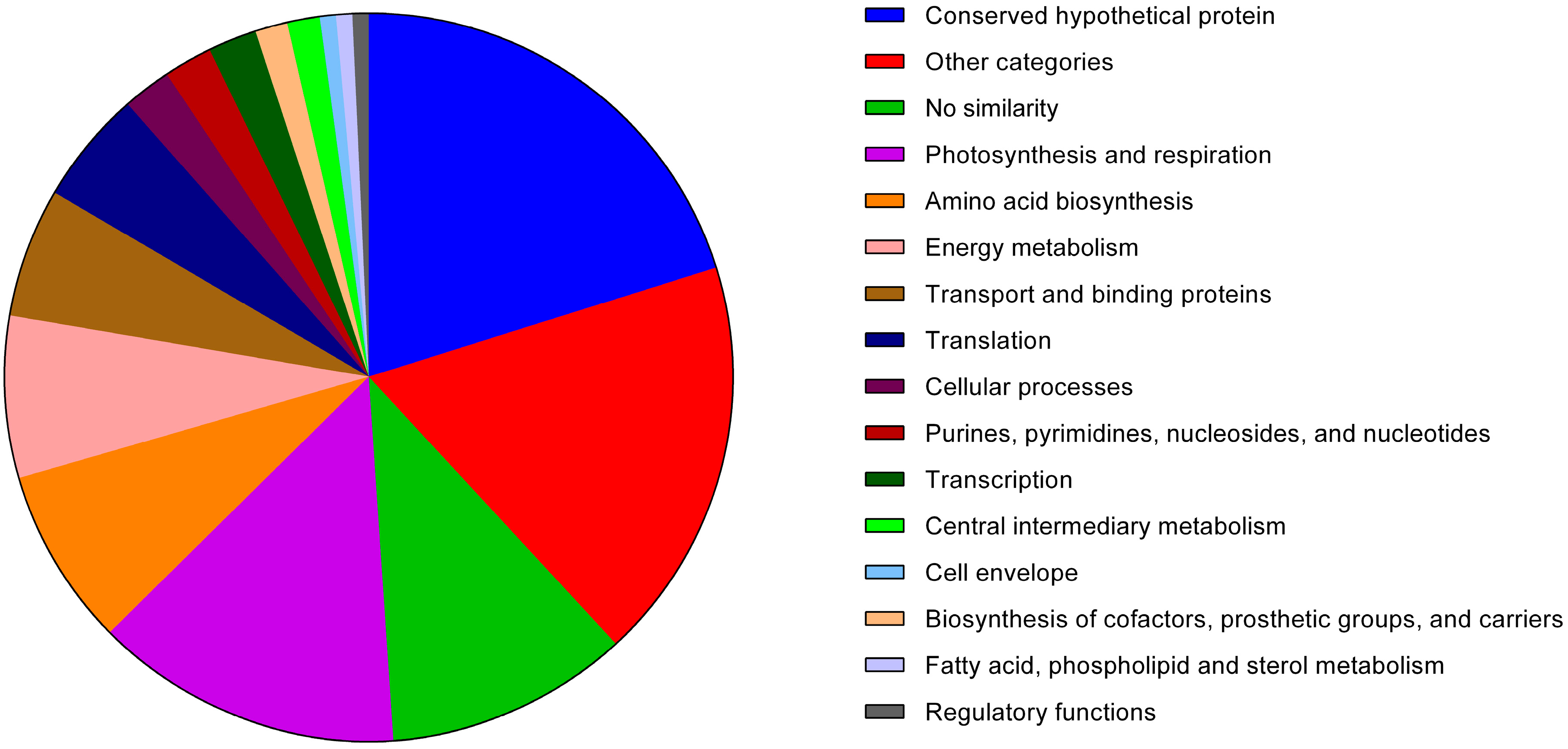
3.1.1. Analysis of the Anabaena sp. PCC 7120 Identified Exoproteins

One question to bear in mind when analysing in detail the exoproteome composition of Anabaena sp. PCC 7120 is to know how a particular protein reached the extracellular milieu. Different scenarios may be considered: within the whole group of exoproteins, some may reach the extracellular space via active translocation across the cell wall and accumulate in this environment, because they play a role there. Others may also be translocated across the outer membrane, but instead are structural parts of the cell wall itself (or are involved in cell wall biogenesis) and are released to the extracellular milieu as a result of cell wall turnover. In addition, outer membrane and periplasmic proteins may also contribute to the exoproteome composition as a result of, e.g., passive protein leakage during cell division and cytokinesis, or by rupture of filament integrity, or even by active disposal of non-functional proteins/enzymes. Ultimately, cytoplasmic proteins may also contribute significantly to the composition of the exoproteome: cell death and mechanical shearing may lead to cell lysis. In that case, proteins that do not have a specific function in this environment may simply accumulate outside of the cells during cultivation, because they are relatively stable to proteolysis. However, in this work, knowing exactly how the identified exoproteins ended up in the extracellular milieu remains to be determined. Despite all of our efforts to experimentally minimize the contribution of proteins accumulating in the extracellular space as a result of cell lysis, we cannot exclude that some of the proteins identified are indeed cytoplasmic proteins. Nevertheless, the proteins listed in Table 1 are those identified from gel bands that could be detected consistently across several biological replicates. Thus, the analysis that will follow below regards them as exoproteins, even though the origin and extracellular function (if any) of most require additional experimental work.
A distribution analysis of the proteins listed in Table 1 based on their respective functional categories (Figure 2) reveals that approximately 48% of the identified exoproteome is classified as “conserved hypothetical” (20%), presents no similarity to other proteins (10%) or falls into the “other categories” set (18%). The latter group of proteins comprises members associated with several unrelated processes, such as “transposon-related functions” and “adaptation and atypical conditions”, or are simply categorized as “other”. Thus, this group includes proteins with completely different functions and that share no obvious metabolic connection. Overall, the high frequency (48%) of identified proteins belonging to categories of unknown function further exposes our lack of knowledge regarding the impact of exoproteins and respective extracellular processes on the cell structure and physiology.
Among the identified extracellular proteins with assigned function, it is possible to find various examples of proteins involved in the degradation and processing of different types of biomolecules, including nucleic acids, along with interconversion and salvage of nucleosides (All3791, Alr0069, Alr7261, Alr3402, Alr0051, Alr1299, Alr1520), degradation of proteins and peptides, as well as processes regarding amino acid biosynthesis and processing (All2533, Alr0237, Alr0880, Alr0996, Alr1381, All4287, Alr1742, Alr4853, All2315, Alr1004, Alr1313, Alr2771, Alr0608, Alr1080, Alr2328 Alr4907, All1683, All4464), sugar breakdown and processing (Alr3608, Alr4448, All3964, All0167, All0168, All0875, Alr0169, Alr2190, All4539) and phosphor scavenge and transport (All0207, All2843, All4575, Alr4238, Alr4976). Detection of these proteins suggests that the processes carried out by Anabaena sp. PCC 7120 extracellularly are intimately associated with recycling of valuable nutrients and compounds present in the environment. These substrates may accumulate in the medium as the result of either active secretion across the cyanobacterial cell wall (e.g., extracellular polysaccharides) or by passive release as a consequence of cell lysis. In addition, proteins typically involved in ROS detoxification and redox homeostasis have also been identified, and a detailed analysis and discussion can be found in Section 3.2.
Figure 2. Absolute frequency of the various functional categories (as ascribed by Cyanobase, http://genome.microbedb.jp/cyanobase) of the proteins identified in the exoproteome of Anabaena sp. PCC 7120.
Life 05 00130 g002
The three growth conditions used to cultivate Anabaena sp. PCC 7120 clearly induced the differential compositions of the exoproteomes (Figure 1). This observation indicates that some proteins are specifically expressed in a given condition and exported to the extracellular milieu to fulfil a particular task. However, the approach adopted and presented here does not allow performing direct comparisons of exoproteome differential compositions between culture conditions. In fact, relative abundance variances analyses should be preceded by better refined and resolved exoproteome separations, e.g., by two-dimensional gel electrophoresis, shotgun proteins and tagging [14,15,24] or label-free workflows [45]. Nevertheless, some differences can be easily identified: for example, Band 7 in the exoproteome of cells grown in BG110 (Lane 1, N2), Bands 5 and 7 in the exoproteome of cells cultivated in BG11 (Lane 2, NO3) and Bands 7, 9 and 13 in the exoproteome of cells grown in BG110 + NH4+ (Lane 3, NH4+) contain proteins that accumulate in higher amounts relatively to the other growth conditions (Figure 1). Based on the number of unique peptides identified for each protein detected in each gel band/gel portion and on its respective protein sequence coverage (see the Supplementary Information), the most abundant protein in Band 7 in BG110 is Alr0267. This exoprotein was recently shown to be involved in filament adhesion and aggregation in Anabaena sp. PCC 7120 cells grown in nitrogen-fixing conditions [34]. The protein was named HesF (for heterocyst specific attachment factor), and its secretion was shown to be dependent on the TolC-like HgdD protein, part of the type I secretion system [34]. Moreover, the respective gene transcription was shown to be highly upregulated upon a transition from non-nitrogen fixing to nitrogen fixing conditions [34], which could explain the protein’s higher abundance in BG110 compared to BG11 or BG110 + NH4+. The proteins identified in higher amounts in Band 5 in BG11 and with approximately the same abundance (according to the criteria outlined above; see the Supplementary Information) are Alr0608 (NrtA), the solute-binding component of the high-affinity nitrate ABC transporter [46], and Alr2877 (CmpA), the bicarbonate transport bicarbonate-binding protein [47]. In a medium containing nitrate, such as BG11, it is not surprising that NrtA is found in higher amounts than in any of the other two growth conditions. Interestingly, Band 7 in BG11 also contains high levels of NrtA, even though the molecular mass of this band is much lower than expected for the full-length NrtA (see the discussion below). Band 7 in BG110 + NH4+, presents an intensity level comparable to Band 5 in BG11; however, the most abundant protein in the former is Alr2877. The gene encoding for protein Alr2877 has been shown to be regulated mainly by the inorganic carbon supply [47]. Nevertheless, it was also shown in the same work that expression of alr2877 is significantly lower in Anabaena sp. PCC 7120 cells experiencing both inorganic carbon and combined nitrogen limiting conditions, as compared to cells growing in a medium limited in inorganic carbon, but replete with combined nitrogen [47]. Thus, these observations could account for the significant differences observed in the amounts of Alr2877 (CmpA) between the exoproteomes of cells cultivated in BG11 and BG110 + NH4+ and the exoproteome of Anabaena sp. PCC 7120 cells grown in BG110 (see Figure 1 and the Supplementary Information). Finally, and still in BG110 + NH4+, Band 9 is majorly Alr0530 (phycobilisome linker polypeptide, phycocyanin associated) and Band 13 is composed mainly of Alr0022 (allophycocyanin subunit beta). The function of these two latter proteins on the extracellular space is unknown; however, it is interesting to notice that the concentrated supernatant of cells grown in BG110 + NH4+ presented a bluish colour (data not shown), in agreement with the higher amounts of phycocyanin-related proteins. In general, when analysing the list of proteins identified in a given exoproteome, one question frequently raised concerns whether the proteins identified correspond to full-length, active enzymes or if they correspond to proteolytic fragments. To address that point, we have combined the analysis of the SDS-polyacrylamide gel electrophoresis separation of the exoproteome content (Figure 1) with the examination of the proteins identified in each band/area of the gel (see the Supplementary Information) and respective peptides detected by mass spectrometry (see the Supplementary Information). As mentioned above, Band 7 in BG11 is mostly represented by NrtA (Alr0608). NrtA is a lipoprotein that is tethered to the cytoplasmic membrane, facing the periplasm [46]. The NrtA predicted molecular mass is 48 kDa, while after cleavage of its signal peptide, it is estimated to be approximately 45 kDa [48], which matches well with the molecular mass of Band 5. However, Band 7 has an estimated molecular mass of 28 kDa. After analysing the NrtA, unique peptides detected in Band 5 the amino acid sequence between positions 87 and 422 were covered, while for Band 7, only peptides covering position 184 to 422 were obtained. Therefore, we believe that NrtA in Band 5 may correspond to the full-length protein, while in Band 7, NrtA is most likely an N-terminus truncated proteolytic fragment. Nevertheless, it remains elusive whether proteolysis occurred outside or inside the cell. Another interesting case is the unknown protein Alr0474, detected in the exoproteome of Anabaena sp. PCC 7120 cells grown in medium with nitrate (Bands 6 and 8). While Alr0474 (596 amino acids) peptides found in Band 6 cover amino acid positions 325 to 590, the ones identified in Band 8 correspond to positions 54 to 189. These results indicate that Alr0474 is likely to be released to the extracellular milieu and then be subjected to proteolysis. Future exoproteome studies will certainly help to clarify these matters and will further unveil the novel molecular mechanisms of protein turnover and recycling.
Something that cannot go unnoticed is the fact that some exoproteins are encoded by adjacent genes (located in the same or opposite strands) or genes that are located relatively close in the genome. These include cases in which the respective genes are located contiguously and in the same strand, such as All0004 and All0005, which are subunits of ATP synthase; All0167, All0168 and Alr0169 (the first two are encoded by genes located in one strand, while the latter is in the other) are all involved in carbohydrate metabolic processes; All2315 and All2316 are the ketol-acid reductoisomerase and the aldo/keto reductase, respectively; Alr0020 (phycobilisome subunit), Alr0021 and Alr0022 (both allophycocyanin subunits); and Alr0528, Alr0529 and Alr0530 are phycocyanin-related proteins. Alternatively, we have also registered cases in which the genes are contiguous, but divergently oriented in the genome: All1380 (no similarity) and Alr1381 (calcium-dependent protease); All2533 (prolyl endopeptidase) and Alr2535 (branched-chain amino-acid ABC transport system periplasmic binding protein), even though in this case, the short gene asl2534 is located in between the respective genes; All3538 (enolase) and Alr3539 (unknown protein); All4121 (ferredoxin-NADP(+) reductase) and Alr4123 (phosphoribulokinase), even though the short gene asl4122 is located in between the respective genes; All4906 (phosphoglycerate mutase) and Alr4907 (ornithine carbamoyltransferase); and Alr0267 and All0268, both unknown proteins. In addition, it is also possible to find proteins encoded by genes not located contiguously, but still in very close genomic proximity, such as All0875 and Alr0878, All2105 and All2108, All2563 and All2567, Alr1362 and Alr1364, Alr1520 and Alr1524 and Alr4976, Alr4979 and All4985. It is not uncommon to find contiguous or adjacent genes encoding extracellular proteins in other bacteria (e.g., [49]), but whether these observations in Anabaena sp. PCC 7120 represent fortuitous occurrences or reflect defined molecular mechanisms that may render gene transcription, protein translation and subsequent secretion more efficient remain to be determined.
Another interesting aspect worth mentioning regards the identification of proteins encoded by plasmid-borne genes. Proteins Alr7261 and Alr7346 (the respective genes are in the Alpha plasmid) and Alr7524, All7614 and All7633 (the respective genes are in the Beta plasmid) all belong to one of the categories “conserved hypothetical proteins”, “no similarity” or “other functions”. Even though the information available about these proteins is scarce, it is remarkable that based on the PSORTb subcellular localization prediction tool [39], Alr7261 is predicted to be an extracellular protein, while the others are predictably periplasmic (All7633), outer membrane (All7614), cytoplasmic (Alr7524) or unknown (Alr7346) proteins (see the Supplementary Information). In addition, Alr7261 and Alr7524 were found in the exoproteome of Anabaena sp. PCC 7120 cells grown under all tested conditions (Table 1). Despite the general lack of knowledge concerning proteins encoded by plasmid-borne genes, alr7261 putatively encodes an extracellular sugar-non-specific nuclease NucA homologue (Cyanobase), whose most characterized member in Anabaena sp. PCC 7120 is All7362 [50,51]. Alternatively, All7614 has homology to carbohydrate-selective porins (OprB); members of this protein family have been shown in Nostoc punctiforme ATCC 29133 to function as sugar porins, being needed for the optimal uptake of both fructose and glucose [52].
The exoproteome of the cyanobacterium N. punctiforme PCC 73102 was recently reported [30]. In that work, the proteins accumulating in the extracellular milieu of cells grown with nitrate or in nitrogen-fixing conditions are presented, in a total of 33 proteins [30]. Furthermore, the secretome analysis of Anabaena sp. PCC 7120 was also recently presented by Hahn and co-workers [26], but they report the detection of secreted proteins (total of 50) from cells grown in a medium containing nitrate. In addition, the extracellular protein content (total of 13 proteins) of Nostoc commune DRH1, another filamentous, heterocyst-forming cyanobacterium, has also been studied, but here, the work was focused essentially on the protein content associated with its extracellular polysaccharide matrix [27]. Approximately 33% of the proteins identified in the present work have already been detected (or their orthologues) at least in one of the works mentioned above (see Table 1). The extended exoproteome profile presented here, as compared to the ones reported by others, may be accounted for in the three different growth conditions studied. Interestingly, two proteins have been identified in all of the studies focusing on the extracellular proteome of filamentous, nitrogen-fixing cyanobacteria: superoxide dismutase (Alr2938; see the discussion below) and phosphoglycerate kinase (All4131). The latter belongs to the “regulatory functions” category, even though it is a crucial enzyme in the energy metabolism. The presence of this enzyme in the extracellular space of three different cyanobacteria suggests a possible important function in that environment; however, it remains undetermined whether the enzyme is actually active and, if it is, what its function may be in such an environment.
Examining with further detail the work carried out on the secretome of Anabaena sp. PCC 7120, the closest to this work that we could find, a total of 72% of the proteins identified in that study could also be detected in this work. From those, it is worth referencing that proteins belonging to the categories “photosynthesis and respiration” (including ATP synthase and various phycobilisome subunits), “amino acid biosynthesis”, “central intermediary metabolism” (including phosphoglucomutase/phosphomannomutase and ribulose-phosphate 3-epimerase) and “energy metabolism” (transaldolase, glucose-6-phosphate isomerase and enolase) are found in both studies, supporting the notion that these proteins are indeed part of the exoproteome, despite being part of the primary metabolism of the organism. From the secreted proteins reported [26] that were not identified here, we can highlight All0070, a Mn-containing superoxide dismutase whose activity we were able to detect instead in the exoproteome (see below), a ferritin-like Dps protein (All1173), while here, we found instead the ferritin-like Dps proteins, All0458, Alr3808 and All4145; and the putative alkaline phosphatases, Alr2234 and Alr5291; while in our work, we identified All0207 and Alr4976.

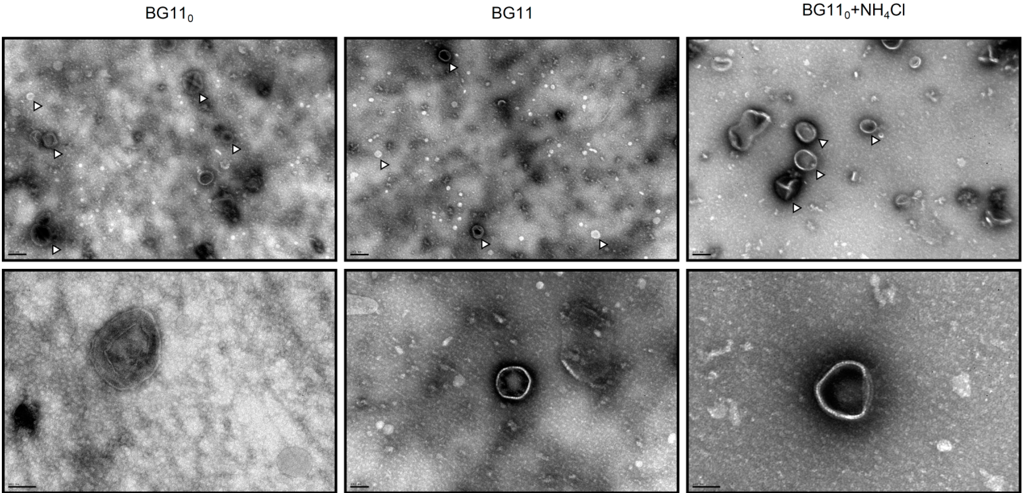
3.1.2. Membrane Vesicle Formation

When analysing the proteins identified in the various exoproteome samples of Anabaena sp. PCC 7120 (Table 1) and also reported by others [26,27,30], we could find several proteins predicted to be localized in the cytoplasm (Supplementary Information). Furthermore, few outer membrane proteins could also be observed in the exoproteome (Supplementary Information). At first glance, detection of such proteins in the extracellular space seems to be an indication of cell lysis and the passive release of proteins to the extracellular space, as already highlighted above. Nevertheless, identification of predicted cytosolic proteins in the extracellular milieu has been reported several times in a wide spectrum of bacteria, e.g., [53,54,55,56,57,58]. In the case of Anabaena sp. PCC 7120, the extracellular processes are so poorly studied and understood that it is possible that some of these proteins are indeed actively secreted [26], fulfilling a specific function yet to be described. Alternatively, it may also represent a mechanism for the removal of such proteins (or respective proteolytic degradation products) from the cytoplasm [26] or other subcellular locations (e.g., see NrtA, discussed above).
A good example to demonstrate how much is unknown in terms of extracellular processes is outer membrane vesicles (OMV) formation. A large number of heterotrophic Gram-negative bacteria naturally produce OMV, spherical bilayered vesicles released from the outer membrane, ranging in size from 50 to 250 nm in diameter [59]. Their suggested functions include toxin trafficking, DNA transfer and uptake, protein delivery and communication (reviewed by [59,60]). OMV formation by photoautotrophs was not described until earlier in 2014, when Biller and co-workers [61] were able to show it in cyanobacteria of the genera Prochlorococcus and Synechococcus. In that work, not only cultures of Prochlorococcus were shown to continuously release lipid vesicles containing proteins, DNA and RNA, but also that these vesicles could be found abundantly in coastal and open-ocean sea water samples. Most interestingly, Prochlorococcus vesicles were demonstrated to support the growth of heterotrophic bacteria, as well as to have the capacity of being recognized and infected by cyanophages [61]. Thus, as recognized by the authors, the ability to form vesicles by marine photoautotrophs adds another layer of complexity to the flow of information, energy and biomolecules in marine microbial ecosystems [61]. However, this may not be restricted to marine ecosystems, if OMV formation proves to be a common mechanism widespread to other cyanobacteria occupying different ecological niches. Proteomic analyses of Prochlorococcus MED4 vesicles identified a diverse set of proteins, including membrane nutrient transporters and porins, but also, predictably, soluble proteases and hydrolases and several proteins of unknown function [61]. Interestingly, from the 40 Prochlorococcus MED4 reported vesicle proteins, eight homologues could be found in the exoproteome of Anabaena sp. PCC 7120, including the porin TolC-like HgdD (Alr2887), the membrane transporters All4575 (phosphate), All1951 (ABC-type) and All4388 (putative polysaccharide exporter), the phosphoribosylglycinamide formyltransferase 2 (Alr1299), the ATP synthase α-subunit (All0005), the RuBisCO large subunit RbcL (Alr1524) and the hypothetical protein, All0268. Furthermore, it is also noteworthy that even though it was not possible to find the respective homologues for some of the Prochlorococcus MED4 vesicles’ proteins, related counterparts could be found for proteases (Alr1381, Alr0996), aminotransferases (All1683, Alr1004, Alr1080, Alr4853, Alr5103) and even ribosomal proteins (All4214) (see the Supplementary Information). Until now, there have been no reports describing the ability of Anabaena sp. PCC 7120 to form and release vesicles. Therefore, the different Anabaena sp. PCC 7120 concentrated exoproteome samples were subjected to negative staining transmission electron microscopy, in an attempt to find OMVs. Remarkably, the three samples presented numerous small spherical structures (Figure 3) that strongly resemble the membrane vesicles reported for Prochlorococcus [61].
This observation, together with the detection of membrane vesicles in the Synechocystis sp. PCC 6803 concentrated exoproteome (data not shown; [62]), suggests that outer membrane vesicle formation may represent an ability widely distributed in cyanobacteria. Hence, we propose that the set of membrane vesicles’ proteins identified in Prochlorococcus MED4 that could also be found in the exoproteome of Anabaena sp. PCC 7120 may be considered as part of the cyanobacterial membrane vesicle proteome core. However, Prochlorococcus and Anabaena are cyanobacterial genera that occupy very different ecological niches (marine vs. freshwater), present obvious morphological differences (unicellular vs. filamentous) and have different capabilities concerning cellular differentiation (no differentiated cells vs. heterocysts). In addition, the genome size of Prochlorococcus MED4 is approximately 2.4 Mbp, while the one of Anabaena sp. PCC 7120 is about three-times larger (7.2 Mbp). These differences may contribute to membrane vesicle proteomic variances and even account for an extended membrane vesicle proteome repertoire in organisms with larger genomes and more complex lifestyles. Therefore, it is possible that other proteins identified in the present work may also be originating from membrane vesicles. However, this will only be elucidated after the Anabaena sp. PCC 7120 outer membrane vesicles’ proteome becomes fully characterized. Therefore, in some prokaryotes, membrane vesicle formation represents a useful, highly efficient and effective, but yet largely overlooked, instrument of protein secretion: the fact that soluble proteins included in the OMV lumen, as well as bound to the OMV surface are co-released with insoluble material (lipids) may represent a protective mechanism, by which OMV-mediated transport can allow less stable molecules, such as protease-susceptible proteins, to reach their destination or behave like time-release capsules, which provide a beneficial activity at a later time [60]. Even more interesting is the capacity of OMV to be specifically targeted to a particular distal site through the binding specificity between surface-exposed bacterial proteins and environmental ligands or receptors [60].
Figure 3. Negative staining electron micrographs of Anabaena sp. PCC 7120 concentrated exoproteome samples collected from cultures grown in nitrogen-fixing conditions (BG110), or in medium supplemented with nitrate (BG11) or ammonia (BG110 + NH4Cl). (Top) The presence and abundance of outer membrane vesicles (some highlighted by white arrowheads) in each sample (size bar, 200 nm); (bottom) the details of selected vesicles (size bar, 100 nm).
Life 05 00130 g003

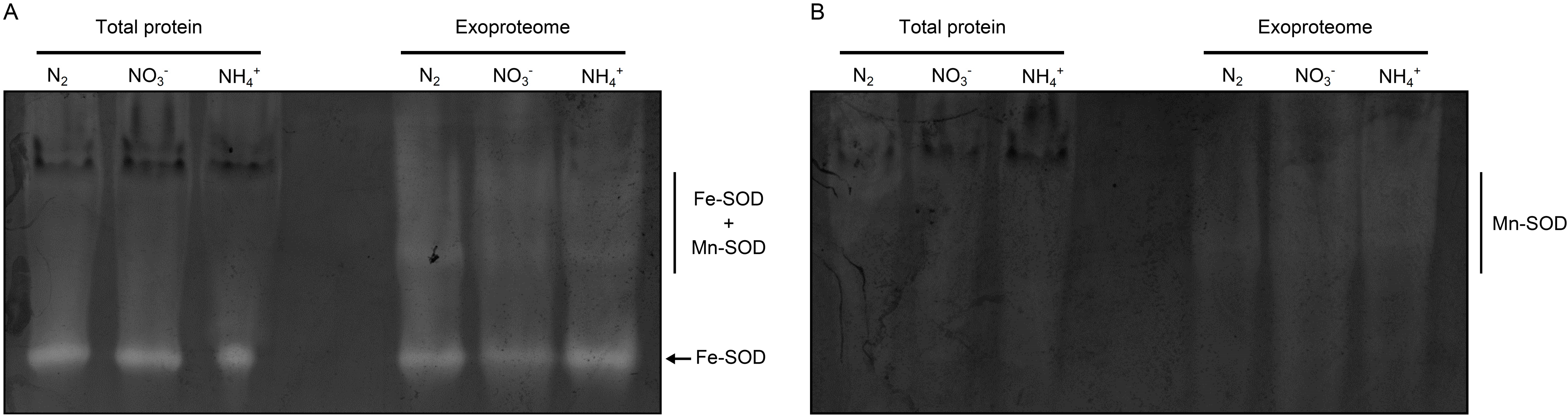
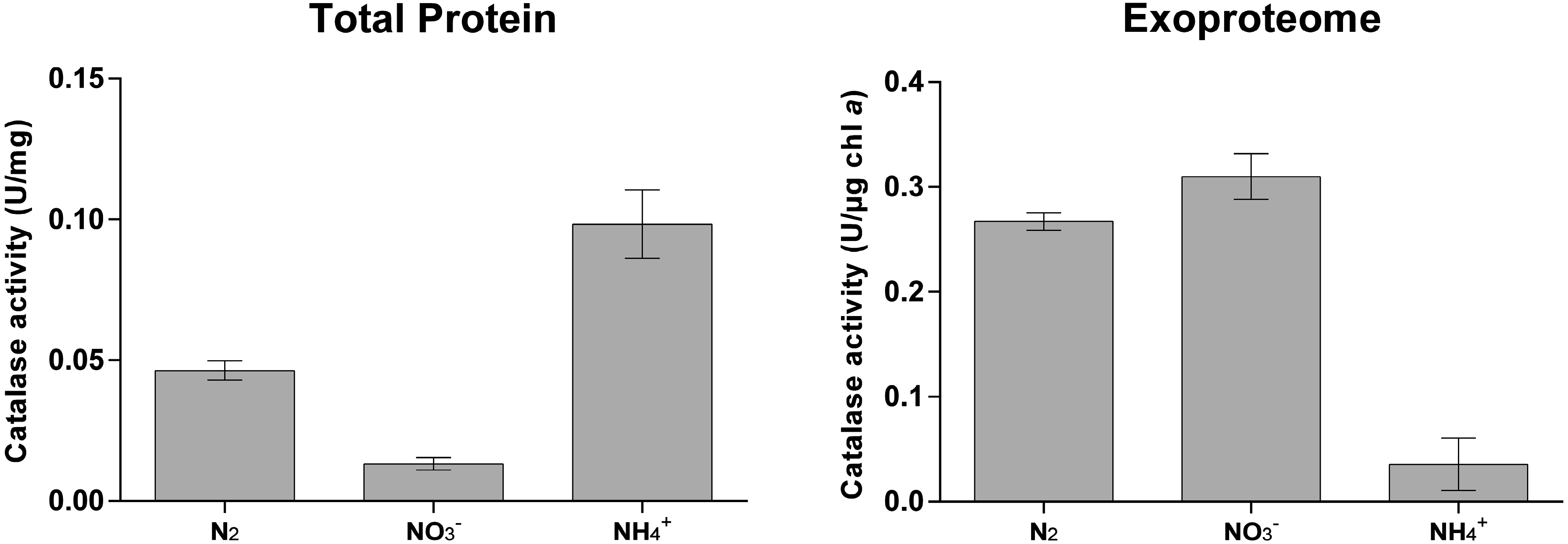
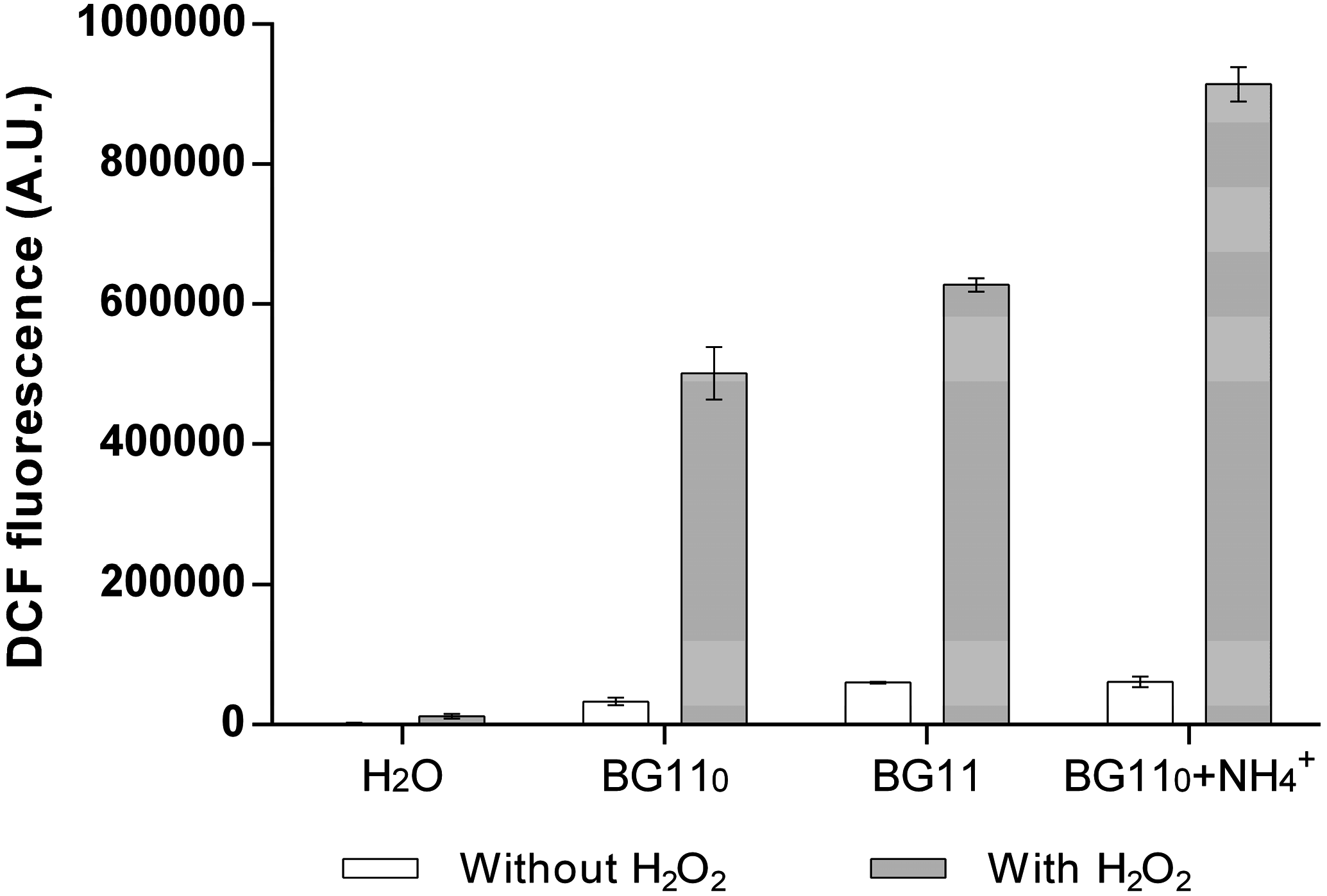
3.2. Cyanobacterial Exoproteins Involved in Redox Homeostasis

One aspect on which we focused our attention was the identification in the extracellular milieu of proteins typically involved in ROS detoxification and redox homeostasis. The identified exoproteins include the Fe superoxide dismutase (Fe-SOD–Alr2938), a hypothetical Mn-catalase (Alr3090), a peroxiredoxin (Alr4641), the ferritin-like, nutrient stress-induced DNA binding proteins All0458, Alr3808 and All4145 and glutathione reductase (All4968).
The ability of prokaryotes to export ROS protein scavengers to the extracellular space is well documented in the literature, particularly for pathogenic bacteria, such as mycobacteria [63,64], streptococci [65], Campylobacter [66] and Corynebacterium [58]. Since phagocytic cells produce reactive oxygen intermediates to kill invading bacteria, it is not surprising that these enzymes are important for virulence [63,64]. In contrast, reports describing the presence and activity of ROS detoxifying proteins in the extracellular space of cyanobacteria are limited to the work of Shirkey et al. [67]. In this work, the exudates (supernatant fractions) of desiccated colonies of N. commune ENG/1996 and cultures of N. commune DRH1 were shown to have high amounts of SOD, as well as high levels of enzyme activity [67]. It was also demonstrated that the extensive extracellular polysaccharide matrix of N. commune DRH1 generated superoxide radicals upon exposure to ultra-violet irradiation [67]. Hence, it was proposed that the SOD released by N. commune is crucial for the cyanobacterium to deal with the oxidative stress imposed by multiple cycles of desiccation and rehydration of the extracellular matrix during ultra-violet irradiation in situ [67]. Anabaena sp. PCC 7120 does not form a complex extracellular polysaccharide matrix similar to that described for N. commune strains, and still, SOD could be identified accumulating in the extracellular space. Fe-SOD was detected in the exoproteomes of cells grown under all tested conditions (Table 1) and not just in the exoproteome of cells grown under conditions eliciting the synthesis and secretion of large amounts of polysaccharides, as in nitrogen-fixing conditions. Therefore, we have decided to evaluate whether SOD activity could be detected in the three isolated exoproteomes. Our in-gel activity results clearly show the presence of SOD activity in all three isolated exoproteomes (Figure 4); in addition to the clear SOD activity band, other fainter bands/smear could also be observed (Figure 4A). We hypothesized that the additional fainter bands could be resulting from the activity of Fe-SOD (Alr2938) and Mn-containing SOD (All0070; not identified in this work) complexes. To test that hypothesis, the Fe-SOD activity was inhibited with the presence of H2O2; in such conditions, the intensity of the clearest SOD activity band completely disappeared, as well as part of the signal from the other fainter bands/smear, remaining however as part of the signal (Figure 4B). This result supports our initial suggestion that Fe-SOD and the Mn-containing SOD may indeed form complexes in the extracellular milieu.
Figure 4. In-gel analysis of the SOD activities of Anabaena sp. PCC 7120. Cells were grown in nitrogen-fixing conditions (N2) or in medium with nitrate (NO3) or ammonia (NH4+). Cell-free extracts were obtained from the collected cells, while the exoproteome was concentrated from the respective cell-free medium. Three hundred micrograms of total protein (total protein) and the protein content present in approximately 20 mL of culture (exoproteome) were separated by native-polyacrylamide gel electrophoresis. (A) Zymogram depicting total SOD activity. The Fe-SOD and Fe-SOD/Mn-SOD complexes’ activity bands are highlighted; (B) Zymogram showing the SOD activity bands, as a result of the specific inhibition of Fe-SOD with 5 mM H2O2 [41]. The Mn-SOD activity bands/smear are highlighted.
Life 05 00130 g004
In addition to the detection of Fe-SOD protein in the extracellular space of Anabaena sp. PCC 7120 (both in this work and in [26]) and in N. commune ENG/1996 and N. commune DRH1 [27,67], an SOD homologue was also identified recently in the exoproteome of N. punctiforme PCC 73102 [30]. Based on these results, it is tempting to assume that secretion of SOD may be a generalized mechanism adopted by cyanobacteria. The exoproteome of the unicellular, freshwater, non-nitrogen-fixing cyanobacterium, Synechocystis sp. PCC 6803, was isolated as described for Anabaena sp. PCC 7120, and the SOD activity was equally assessed by zymography. However, no SOD activity bands could be observed in Synechocystis sp. PCC 6803 (data not shown), suggesting that only a limited number of cyanobacterial species (thus far limited to filamentous and heterocyst-forming strains) are capable of exporting and accumulating active SOD in the extracellular milieu, at least under the conditions tested.
We have also extended our analyses to evaluate catalase activity (Figure 5). Our results indicate that some factor in the Anabaena sp. PCC 7120 growth medium does possess the capacity of decomposing H2O2 (Figure 5), likely catalase. These results validate the identification of Alr3090 in the exoproteome of Anabaena sp. PCC 7120 and further support the suggestion that a complex oxidative stress defence mechanism exists extracellularly in this cyanobacterium. Nevertheless, total catalase activity levels determined here are low as compared to what is described for other cyanobacteria [68,69], even when using total protein extracts (Figure 5). However, catalase has been shown not to be the main mechanism to cope with H2O2 by the filamentous, heterocyst-forming cyanobacterium, Anabaena sp. PCC 7120; instead, peroxiredoxins are reported to be the main H2O2 detoxifying pathway [70], one of which (Alr4641) has been identified in the exoproteome of Anabaena sp. PCC 7120 (see the discussion below).
Figure 5. Catalase activity measured in cell-free extracts (total protein) or in concentrated exoproteomes of Anabaena sp. PCC 7120. Activities are expressed as units (defined as the amount of enzyme that catalyses the dissociation of 1 µmol of H2O2 per minute) per mg of total protein (left) or units per µg chlorophyll a (right).
Life 05 00130 g005
These results combined indicate that Anabaena sp. PCC 7120 is dealing with oxidative stress even outside of the cells. This notion may not be surprising for cyanobacteria in defined ecological circumstances, such as in mats, biofilms, symbiotic associations or even when free-living in freshwater bodies or marine ecosystems, since competition with other microorganisms for nutrients and even other cyanobacteria for light may involve ROS release and attack. However, in controlled laboratory conditions, in which the organism is cultivated axenically, it becomes difficult to interpret the observation that Anabaena sp. PCC 7120 exports and accumulates such high amounts of functional ROS detoxifying agents. Consequently, we became interested in analysing the ROS usually formed in normal growth medium under ordinary cultivating conditions, but without the presence of cells. Thus, the three media used to grow Anabaena sp. PCC 7120 (BG110, BG11, BG110 + NH4+) were kept sterile in the same conditions as cyanobacterial cultures. ROS levels were determined using the fluorescent redox indicator, DCF (dichlorodihydrofluorescein) (Figure 6).
Figure 6. Reactive oxygen species (ROS) level determination in sterile cyanobacterial growth medium. Fluorescence of the molecular probe, DCF, which is directly related to its oxidation state and to the amount of ROS in the medium, was used to measure total ROS levels (A.U., arbitrary units). Cyanobacterial media BG110, BG11 and BG110 + NH4+ were kept sterile under the same conditions of temperature and aeration as cyanobacterial cultures. Water was used as the control. White bars, water or medium without H2O2; grey bars, water or medium with 0.5 mM H2O2. Given the heterogeneity in DCF fluorescence between different experiments, the data shown are from a single experiment, but the observed pattern is typical (at least three separate observations). The bar values are means, and the error bars correspond to the standard deviation from three technical replicates.
Life 05 00130 g006
Interestingly, as compared to distilled and deionized water, all three media presented higher levels of total ROS as indicated by a higher DCF fluorescence (Figure 6). In addition, all media presented approximately the same DCF fluorescence levels (white bars in Figure 6), suggesting that all present about the same amount of ROS. In order to monitor the probe’s responsiveness to the presence of ROS, 0.5 mM H2O2 were added exogenously to each medium and to water. The addition of H2O2 induced a much stronger fluorescence signal in any of the tested media as compared to water. Despite its evident utility and simplicity of use, DCF presents some disadvantages, one of which lies in its differential fluorescence response to various ROS (Life Technologies). This could account for the differences observed between water supplemented with H2O2 and all tested media supplemented with H2O2: since the reference BG11 medium is composed of minerals and several metals in solution [33], it is possible that H2O2 reacts with, e.g., iron (Fe2+) present in the medium in the so-called Fenton reaction, resulting in the formation of hydroxyl radicals. The DCF redox indicator has been shown to respond approximately 40-times more efficiently to the presence of hydroxyl radicals than to H2O2 (Life Technologies). Overall, these results show that ROS are generated naturally in ordinary cyanobacterial growth medium (something also observed for cultivating media of other organisms; [71] and the references therein) and that the interaction of H2O2 with medium components generates additional ROS. Moreover, these results also show that some cyanobacteria may release enzymes into the extracellular milieu to specifically detoxify exogenous ROS.
Regarding the other proteins involved in redox homeostasis identified in the exoproteome, the peroxiredoxin Alr4641 (2-Cys Prx) has been suggested to be the main mechanism by which Anabaena sp. PCC 7120 is capable of coping with H2O2, which presents abundant and sensitive intracellular levels of 2-Cys Prx, but low catalase activity [70]. Therefore, it may be possible that Anabaena sp. PCC 7120 secretes both catalase and 2-Cys Prx in order to cope efficiently with H2O2 exposure.
Furthermore, proteins All0458, Alr3808 and All4145 are ferritin-like, nutrient stress-induced DNA binding proteins (Dps). One of the main components providing structural integrity to biofilms, in addition to exopolysaccharides and proteins, is extracellular DNA [72]. Interestingly, Dps proteins have been shown to have an important role in Campylobacter jejuni in vitro biofilm formation [73], and Dps proteins have been identified in the exoproteomes of Staphylococcus aureus [74] and Bacillus anthracis [75] and even in the OMV of Brucella melitensis [76]. Thus, it is possible that Anabaena sp. PCC 7120 releases DNA to the extracellular space to promote biofilm formation, and Dps proteins are simultaneously translocated to protect extracellular DNA from structural damage elicited by oxidative stress.
Finally, the glutathione reductase (All4968) is the enzyme that catalyses the reduction of glutathione disulfide to glutathione with NADPH as the reducing cofactor. Glutathione is an extremely important antioxidant agent, preventing the damage of core cellular components caused by ROS. Interestingly, glutathione reductase has also been found in the exoproteome of the marine bacterium, Roseobacter sp. MED193 [77]. However, the presence of the protein in the extracellular milieu is rather puzzling: if glutathione may indeed play a role in maintaining redox homeostasis in the extracellular environment as a protection barrier in the cell vicinity against ROS (and, in fact, a bacterial glutathione transporter has been reported to export reductant to the periplasm of Escherichia coli [78]), it becomes difficult to interpret how glutathione reductase may be active when glutathione disulfide and reducing power in the form of NADPH are required. Additional work is needed to shed light on whether glutathione reductase is actively secreted by Anabaena sp. PCC 7120 and, if so, what may be the physiological impact/advantage of All4968 presence in the extracellular milieu of this cyanobacterium.
In general, it remains to be elucidated how these identified proteins involved in ROS detoxification and redox homeostasis reach the extracellular space, since no leader peptides can be detected in their primary amino acid sequences. One possibility could be that these proteins represent non-classical secreted proteins, and so, their secretory mechanisms remain elusive; alternatively, their transport to the extracellular milieu may be mediated by OMV, as described for SOD in B. melitensis [76] and Acinetobacter baumannii [79].

3.3. Modulating Cyanobacterial Protein Secretion: Future Perspectives

The competence for secreting proteins to the extracellular milieu is not exclusive to cyanobacteria, and it has already been used in certain prokaryotes to produce and secrete selected protein targets. In the case of applications where purified recombinant proteins are used directly, secretion of these proteins extracellularly could significantly reduce the complexity of a production process by eliminating the need for cell lysis and reducing the burden of removing host proteins [80]. Furthermore, secretion of highly expressed proteins minimizes the formation of inclusion bodies, aids in folding, allows for disulfide bond formation, reduces the effects of intracellular protein degradation and lessens the detrimental effects of cytotoxic proteins (reviewed by [80]).
Given the extraordinary physiological, metabolic and genetic qualities presented by cyanobacteria, we believe that if properly engineered, protein secretion by these photoautotrophs represents an attractive approach to further explore biotechnological solutions. In fact, the ability of cyanobacteria to use sunlight and carbon dioxide as energy and carbon sources, respectively, together with faster growth rates (compared to plants) and the relative ease with which they can be genetically engineered (compared to algae), make cyanobacteria stand out from all other organisms so far used in biotechnological applications [44,81]. In particular, cyanobacterial protein secretion may be a valuable alternative for solving various challenges in bioremediation, biomass recovery and biofuel production.
In addition to the strategies already in course for using cyanobacteria as efficient agents of bioremediation (e.g., making use of their exopolysaccharides [82]), cyanobacterial protein secretion could contribute significantly towards that goal; for example, nitrogen-fixing cyanobacteria could be genetically modified to express and secrete specific heavy metal chelators, contributing to detoxifying heavy-metal contaminated soils, as well as enriching their combined nitrogen content.
In order to reach commercially attractive figures in cyanobacteria-based biotechnological applications, the cultivation of cyanobacteria needs to be done at a large scale, which brings the issue of biomass recovery in large culture volumes. One of the strategies to separate growth medium from the valuable biomass has been to trigger cyanobacterial cells to adhere, aggregate and flocculate, easing its recovery. Some studies have shown that cyanobacterial cells adhesion and aggregation are facilitated by extracellular proteins [28,31,34]; thus, activation of those exoproteins expression and secretion with a precisely defined timing may aid in biomass recovery and reduce total biomass production costs.
In the case that whole microbial cells expressing recombinant proteins are used to interact with a polymeric material, extracellular secretion of proteins is necessary due to the inability of microbial cells to uptake polymer substrates [80]. Therefore, deconstruction of, e.g., polymeric lignocellulosic biomass to fermentable sugars is an important area with increasing interest in biofuel production from renewable resources. In this case, “polysaccharase” or “cellulase” secreting cyanobacteria would contribute to degrading these complex polymeric substances to simpler sugars; since cyanobacteria are capable of fixing their own carbon, products of lignocellulosic deconstruction would not be taken up by cyanobacteria, being instead fully available for other, more suitable fermenting bacteria that could then convert the recently released sugars into valuable biofuels.
In free-living nitrogen-fixing cyanobacteria, once atmospheric nitrogen is fixed to ammonia and assimilated to glutamine, it is believed to be partly converted to arginine, which is then polymerized into cyanophycin [83]. Cyanophycin synthesis after nitrogen fixation has been suggested to serve an important function by removing from solution the products of nitrogen fixation, which could have a negative feedback effect on nitrogenase [83,84]. Analogously, generating nitrogen-fixing cyanobacteria capable of secreting large amounts of a target protein would cause the organism to be drained off of fixed nitrogen and probably relieve repression and feedback on nitrogen fixation. Consequently, from the continuous protein secretion, one could expect higher nitrogen fixation rates and/or extended periods of nitrogenase activity to sustain growth, which would be an advantage for nitrogenase-based technological processes, such as the production of molecular hydrogen, a by-product of nitrogenase activity [85].
Investigations recently carried out in our laboratory strongly indicate that there is room for engineering and modulating cyanobacterial protein secretion. As presented in Figure 7, the simple fusion of a nitrate inducible promoter with the exoprotein encoding gene, hesF, resulted in the overexpression of the protein, leading to an over-accumulation of HesF in the extracellular milieu. Furthermore, this result also highlights that even though a signal peptide could not be identified in the sequence of HesF, whatever addresses the protein for secretion seems to be highly efficient [34].
Figure 7. Exoproteome profiles of Anabaena sp. PCC 7120 wild-type (WT) and BSMPo1 [34] cultivated in BG11. The Coomassie-stained SDS-polyacrylamide gel shows the exoproteome of both strains grown for three days in a medium containing nitrate. The protein content present in approximately 3 mL of the growth medium was loaded on each lane. The molecular masses of the Precision Plus Protein All Blue standard (Bio-Rad), Lane M, are indicated on the left, while the arrowhead points to the overexpressed and secreted protein HesF.
Life 05 00130 g007
In order to explore protein secretion by cyanobacteria, a number of aspects need to be addressed and finely tuned. As for other bacterial systems, signal peptides or protein determinants that can address the translocation of a newly-synthesized protein from the cyanobacterial cytoplasm to the extracellular space represents one of the most crucial steps towards optimization of the secretion process. Signal peptides recognized by specific secretion systems are described for some bacteria (for a review, see [80]), and these could be tested and validated in cyanobacteria. Alternatively, in the specific case of HesF presented above, no signal peptide could be identified, and still, it must bear an efficient secretion determinant judging by the amounts of protein that accumulated in the growth medium. This represents a good example of how screening for novel signal peptides may open the door to more efficient secretion systems. Another aspect that deserves close attention when designing a protein secretion module is the selection of a promoter that will drive the transcription of the protein of interested coding gene. Cell-type specificity (in the case of cyanobacteria with the capacity of cellular differentiation), timing of expression and promoter strength are just a few of the points that need to be considered and coordinated with the particular protein to be secreted. Efforts to design suitable promoters for the most varied purposes and applications in cyanobacteria are being made [86,87] and will surely aid in engineering competent protein secretion microorganisms.
However, a few challenges loom ahead before successful implementation of engineered cyanobacterial cells to secrete selected target proteins can be attained. Vilhauer and co-workers showed that proteases are present and active in the extracellular space of Nostoc punctiforme, demonstrating as well that the extent of such proteolytic activity was dependent on the growth condition [30]. Thus, extracellular protease activity has to be considered and duly evaluated, so as to avoid poor protein recovery yields, low quality protein purifications or extracellular modules of low functionality, depending on the final application of the secreted protein. Still, this may be overcome by a balanced regulation between the rate of gene transcription, protein expression and protein secretion, as well as the rate of protein degradation, similarly to what happens in the cytoplasm of protein-(over)expressing bacteria. Moreover, the natural production and release of polysaccharides by cyanobacterial cells may also represent an obstacle towards the use of secreted proteins to fulfil particular goals.
Finally, the membrane vesicles that Anabaena sp. PCC 7120 is shown here to be able to produce represent an additional point of interest regarding protein secretion. The molecular mechanisms behind membrane vesicle formation, including the cellular structures and components responsible for packing those vesicles or even the determinants that govern what goes in and what remains out of the vesicles, are completely unknown in cyanobacteria. However, it is tempting to imagine the possibility of cyanobacteria forming membrane vesicles enriched in a target cargo that has been recombinantly produced: not only would this ease purification and increase the stability of the cargo, but also the membrane vesicles could act as delivery vectors. Certainly, many fundamental aspects need to be clarified before it becomes possible to be fully explored, but such a scenario is not completely unrealistic, since the delivery of foreign antigens by engineered outer membrane vesicle vaccines has already been demonstrated [88].

4. Conclusions

The exoproteome of the filamentous, heterocyst-forming cyanobacterium, Anabaena sp. PCC 7120, grown in nitrogen-fixing conditions or in medium supplemented either with nitrate or ammonia is presented in this work. Strikingly, as many as 139 proteins have been identified, belonging to 16 different functional categories. Proteins involved in oxidative stress detoxification and redox homeostasis are among the identified exoproteins, including SOD and catalase, whose activities we were able to detect in the concentrated supernatant. This suggests that Anabaena sp. PCC 7120 invests valuable cellular resources to eliminate reactive oxygen species, even outside of the cell. Furthermore, Anabaena sp. PCC 7120 is shown here, for the first time, to be able to release outer membrane vesicles that likely contribute to the whole exoproteome content. Finally, cyanobacteria seem to have the potential of becoming robust protein secretion factories, and future studies focusing on the cyanobacterial exoproteome will certainly contribute towards that goal.

Acknowledgments

We wish to express our gratitude to Rui Fernandes and Franscisco Figueiredo (IBMC) for practical help with electron microscopy analyses and the Parasite Disease group (IBMC) for the use of the Synergy 2 microplate reader. This work was funded by FEDER funds through the Operational Competitiveness Programme, COMPETE, and by National Funds through FCT, Fundação para a Ciência e a Tecnologia, under the scholarships, SFRH/BPD/74894/2010 (Paulo Oliveira) and PTDC/BIA-MIC/2889/2012-PR451402-BIL (Marina Santos). Nuno M. Martins acknowledges financial support by the European Union under the Erasmus program (2013). Phillip C. Wright and Narciso Couto acknowledge the Engineering and Physical Sciences Research Council (EPSRC) for funding under the Chemical Engineering at the Life Science Interface (ChELSI) and S&I award (EP/E036252/1).

Author Contributions

Paulo Oliveira, Nuno M. Martins, Marina Santos, Phillip C. Wright and Paula Tamagnini conceived and designed the experiments. Nuno M. Martins, Paulo Oliveira, Marina Santos and Narciso A. S. Couto performed the experiments. Nuno M. Martins, Paulo Oliveira, Marina Santos and Narciso A. S. Couto, Phillip C. Wright and Paula Tamagnini analysed the data. Paulo Oliveira, Narciso A. S. Couto, Phillip C. Wright and Paula Tamagnini contributed to the writing of the manuscript. All authors have read and approved the final manuscript.

Supplementary Materials

Supplementary materials can be accessed at: https://www.mdpi.com/2075-1729/5/1/130/s1.

Conflicts of Interest

The authors declare no conflict of interest.

References

  1. Varki, A. Essentials of Glycobiology, 2nd ed.; Cold Spring Harbor Laboratory Press: New York, NY, USA, 2009. [Google Scholar]
  2. Sazuka, T.; Ohara, O. Towards a proteome project of cyanobacterium Synechocystis sp. strain PCC6803: Linking 130 protein spots with their respective genes. Electrophoresis 1997, 18, 1252–1258. [Google Scholar] [CrossRef] [PubMed]
  3. Sazuka, T.; Yamaguchi, M.; Ohara, O. Cyano2Dbase updated: Linkage of 234 protein spots to corresponding genes through N-terminal microsequencing. Electrophoresis 1999, 20, 2160–2171. [Google Scholar] [CrossRef] [PubMed]
  4. Choi, J.S.; Kim, D.S.; Lee, J.; Kim, S.J.; Kim, S.I.; Kim, Y.H.; Hong, J.; Yoo, J.S.; Suh, K.H.; Park, Y.M. Proteome analysis of light-induced proteins in Synechocystis sp. PCC 6803: Identification of proteins separated by 2D-PAGE using N-terminal sequencing and MALDI-TOF MS. Mol. Cells 2000, 10, 705–711. [Google Scholar] [CrossRef] [PubMed]
  5. Wegener, K.M.; Singh, A.K.; Jacobs, J.M.; Elvitigala, T.; Welsh, E.A.; Keren, N.; Gritsenko, M.A.; Ghosh, B.K.; Camp, D.G., II; Smith, R.D.; et al. Global proteomics reveal an atypical strategy for carbon/nitrogen assimilation by a cyanobacterium under diverse environmental perturbations. Mol. Cell. Proteomics 2010, 9, 2678–2689. [Google Scholar] [CrossRef] [PubMed]
  6. Pandhal, J.; Ow, S.Y.; Wright, P.C.; Biggs, C.A. Comparative proteomics study of salt tolerance between a nonsequenced extremely halotolerant cyanobacterium and its mildly halotolerant relative using in vivo metabolic labeling and in vitro isobaric labeling. J. Proteome Res. 2009, 8, 818–828. [Google Scholar] [CrossRef] [PubMed]
  7. Pandhal, J.; Wright, P.C.; Biggs, C.A. Proteomics with a pinch of salt: A cyanobacterial perspective. Saline Syst. 2008, 4. [Google Scholar] [CrossRef]
  8. Rowland, J.G.; Simon, W.J.; Prakash, J.S.; Slabas, A.R. Proteomics reveals a role for the RNA helicase CrhR in the modulation of multiple metabolic pathways during cold acclimation of Synechocystis sp. PCC6803. J. Proteome Res. 2011, 10, 3674–3689. [Google Scholar] [CrossRef] [PubMed]
  9. Zhang, L.; Selão, T.T.; Pisareva, T.; Qian, J.; Sze, S.K.; Carlberg, I.; Norling, B. Deletion of Synechocystis sp. PCC 6803 leader peptidase LepB1 affects photosynthetic complexes and respiration. Mol. Cell. Proteomics 2013, 12, 1192–1203. [Google Scholar] [CrossRef] [PubMed]
  10. Li, T.; Yang, H.M.; Cui, S.X.; Suzuki, I.; Zhang, L.F.; Li, L.; Bo, T.T.; Wang, J.; Murata, N.; Huang, F. Proteomic study of the impact of hik33 mutation in Synechocystis sp. PCC 6803 under normal and salt stress conditions. J. Proteome Res. 2012, 11, 502–514. [Google Scholar] [CrossRef] [PubMed]
  11. Barrios-Llerena, M.E.; Reardon, K.F.; Wright, P.C. 2-DE proteomic analysis of the model cyanobacterium Anabaena variabilis. Electrophoresis 2007, 28, 1624–1632. [Google Scholar] [CrossRef] [PubMed]
  12. Gan, C.S.; Reardon, K.F.; Wright, P.C. Comparison of protein and peptide prefractionation methods for the shotgun proteomic analysis of Synechocystis sp. PCC 6803. Proteomics 2005, 5, 2468–2478. [Google Scholar] [CrossRef] [PubMed]
  13. Pereira, S.B.; Ow, S.Y.; Barrios-Llerena, M.E.; Wright, P.C.; Moradas-Ferreira, P.; Tamagnini, P. iTRAQ-based quantitative proteomic analysis of Gloeothece sp. PCC 6909: Comparison with its sheathless mutant and adaptations to nitrate deficiency and sulfur limitation. J. Proteomics 2011, 75, 270–283. [Google Scholar] [CrossRef] [PubMed]
  14. Ow, S.Y.; Noirel, J.; Cardona, T.; Taton, A.; Lindblad, P.; Stensjo, K.; Wright, P.C. Quantitative overview of N2 fixation in Nostoc punctiforme ATCC 29133 through cellular enrichments and iTRAQ shotgun proteomics. J. Proteome Res. 2009, 8, 187–198. [Google Scholar] [CrossRef] [PubMed]
  15. Ow, S.Y.; Cardona, T.; Taton, A.; Magnuson, A.; Lindblad, P.; Stensjo, K.; Wright, P.C. Quantitative shotgun proteomics of enriched heterocysts from Nostoc sp. PCC 7120 using 8-plex isobaric peptide tags. J. Proteome Res. 2008, 7, 1615–1628. [Google Scholar] [CrossRef] [PubMed]
  16. Anderson, D.C.; Campbell, E.L.; Meeks, J.C. A soluble 3D LC/MS/MS proteome of the filamentous cyanobacterium Nostoc punctiforme. J. Proteome Res. 2006, 5, 3096–3104. [Google Scholar] [CrossRef] [PubMed]
  17. Aryal, U.K.; Callister, S.J.; McMahon, B.H.; McCue, L.A.; Brown, J.; Stockel, J.; Liberton, M.; Mishra, S.; Zhang, X.; Nicora, C.D.; et al. Proteomic profiles of five strains of oxygenic photosynthetic cyanobacteria of the genus Cyanothece. J. Proteome Res. 2014, 13, 3262–3276. [Google Scholar] [CrossRef] [PubMed]
  18. Huang, F.; Hedman, E.; Funk, C.; Kieselbach, T.; Schroder, W.P.; Norling, B. Isolation of outer membrane of Synechocystis sp. PCC 6803 and its proteomic characterization. Mol. Cell. Proteomics 2004, 3, 586–595. [Google Scholar] [CrossRef] [PubMed]
  19. Moslavac, S.; Bredemeier, R.; Mirus, O.; Granvogl, B.; Eichacker, L.A.; Schleiff, E. Proteomic analysis of the outer membrane of Anabaena sp. strain PCC 7120. J. Proteome Res. 2005, 4, 1330–1338. [Google Scholar] [CrossRef] [PubMed]
  20. Fulda, S.; Huang, F.; Nilsson, F.; Hagemann, M.; Norling, B. Proteomics of Synechocystis sp. strain PCC 6803. Identification of periplasmic proteins in cells grown at low and high salt concentrations. Eur. J. Biochem. 2000, 267, 5900–5907. [Google Scholar] [CrossRef] [PubMed]
  21. Zhang, L.F.; Yang, H.M.; Cui, S.X.; Hu, J.; Wang, J.; Kuang, T.Y.; Norling, B.; Huang, F. Proteomic analysis of plasma membranes of cyanobacterium Synechocystis sp. strain PCC 6803 in response to high pH stress. J. Proteome Res. 2009, 8, 2892–2902. [Google Scholar] [CrossRef] [PubMed]
  22. Huang, F.; Parmryd, I.; Nilsson, F.; Persson, A.L.; Pakrasi, H.B.; Andersson, B.; Norling, B. Proteomics of Synechocystis sp. strain PCC 6803: Identification of plasma membrane proteins. Mol. Cell. Proteomics 2002, 1, 956–966. [Google Scholar] [CrossRef] [PubMed]
  23. Simon, W.J.; Hall, J.J.; Suzuki, I.; Murata, N.; Slabas, A.R. Proteomic study of the soluble proteins from the unicellular cyanobacterium Synechocystis sp. PCC6803 using automated matrix-assisted laser desorption/ionization-time of flight peptide mass fingerprinting. Proteomics 2002, 2, 1735–1742. [Google Scholar] [CrossRef] [PubMed]
  24. Agarwal, R.; Matros, A.; Melzer, M.; Mock, H.P.; Sainis, J.K. Heterogeneity in thylakoid membrane proteome of Synechocystis 6803. J. Proteomics 2010, 73, 976–991. [Google Scholar] [CrossRef] [PubMed]
  25. Srivastava, R.; Pisareva, T.; Norling, B. Proteomic studies of the thylakoid membrane of Synechocystis sp. PCC 6803. Proteomics 2005, 5, 4905–4916. [Google Scholar] [CrossRef] [PubMed]
  26. Hahn, A.; Stevanovic, M.; Brouwer, E.; Bublak, D.; Tripp, J.; Schorge, T.; Karas, M.; Schleiff, E. Secretome analysis of Anabaena sp. PCC 7120 and the involvement of the TolC-homologue HgdD in protein secretion. Environ. Microbiol. 2014. [Google Scholar] [CrossRef]
  27. Helm, R.D.; Potts, M. Extracellular matrix (ECM). In Ecology of Cyanobacteria II: Their Diversity in Space and Time; Whitton, B.A., Ed.; Springer: Berlin/Heidelberg, Germany, 2012; pp. 461–480. [Google Scholar]
  28. Kehr, J.C.; Zilliges, Y.; Springer, A.; Disney, M.D.; Ratner, D.D.; Bouchier, C.; Seeberger, P.H.; de Marsac, N.T.; Dittmann, E. A mannan binding lectin is involved in cell-cell attachment in a toxic strain of Microcystis aeruginosa. Mol. Microbiol. 2006, 59, 893–906. [Google Scholar] [CrossRef] [PubMed]
  29. Sergeyenko, T.V.; Los, D.A. Identification of secreted proteins of the cyanobacterium Synechocystis sp. strain PCC 6803. FEMS Microbiol. Lett. 2000, 193, 213–216. [Google Scholar] [CrossRef] [PubMed]
  30. Vilhauer, L.; Jervis, J.; Ray, W.K.; Helm, R.F. The exo-proteome and exo-metabolome of Nostoc punctiforme (cyanobacteria) in the presence and absence of nitrate. Arch. Microbiol. 2014, 196, 357–367. [Google Scholar] [CrossRef] [PubMed]
  31. Zilliges, Y.; Kehr, J.C.; Mikkat, S.; Bouchier, C.; de Marsac, N.T.; Borner, T.; Dittmann, E. An extracellular glycoprotein is implicated in cell-cell contacts in the toxic cyanobacterium Microcystis aeruginosa PCC 7806. J. Bacteriol. 2008, 190, 2871–2879. [Google Scholar] [CrossRef] [PubMed]
  32. Desvaux, M.; Hebraud, M.; Talon, R.; Henderson, I.R. Secretion and subcellular localizations of bacterial proteins: A semantic awareness issue. Trends Microbiol. 2009, 17, 139–145. [Google Scholar] [CrossRef] [PubMed]
  33. Rippka, R.; Deruelles, J.; Waterbury, J.B.; Herdman, M.; Stanier, R.Y. Generic assignments, strain histories and properties of pure cultures of cyanobacteria. J. Gen. Microbiol. 1979, 111, 1–61. [Google Scholar] [CrossRef]
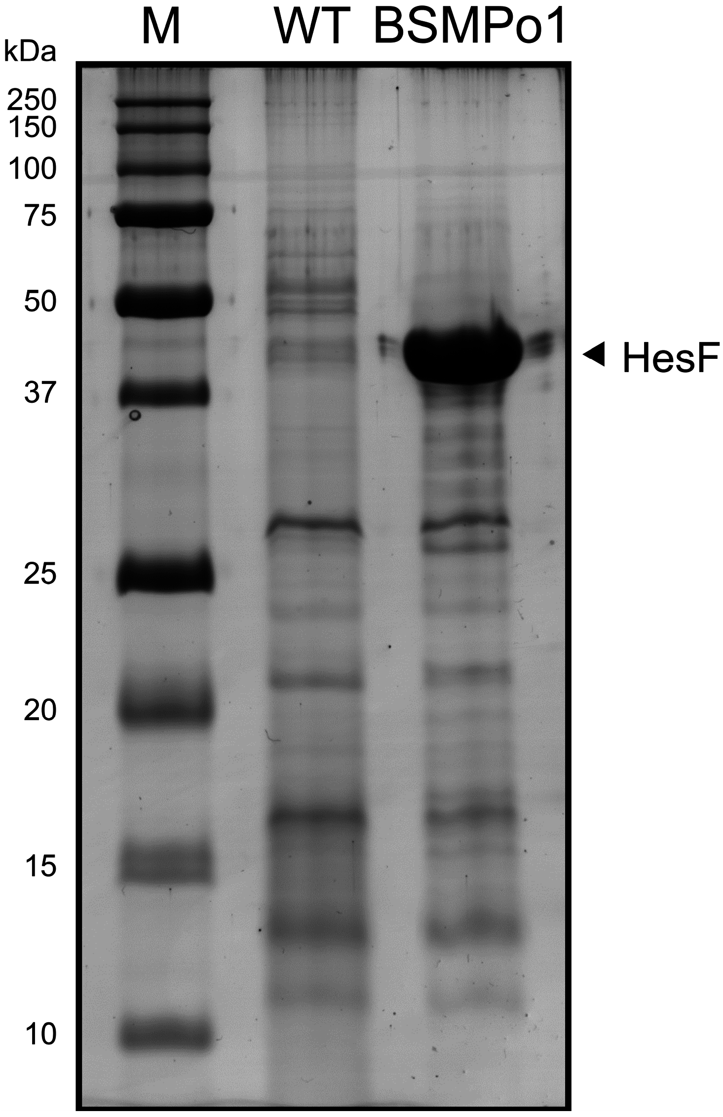
  34. Oliveira, P.; Pinto, F.; Pacheco, C.C.; Mota, R.; Tamagnini, P. HesF, an exoprotein required for filament adhesion and aggregation in Anabaena sp. PCC 7120. Environ. Microbiol. 2014. [Google Scholar] [CrossRef]
  35. Laemmli, U.K. Cleavage of structural proteins during the assembly of the head of bacteriophage T4. Nature 1970, 227, 680–685. [Google Scholar] [CrossRef] [PubMed]
  36. Shevchenko, A.; Wilm, M.; Vorm, O.; Mann, M. Mass spectrometric sequencing of proteins silver-stained polyacrylamide gels. Anal. Chem. 1996, 68, 850–858. [Google Scholar] [CrossRef] [PubMed]
  37. Gluck, F.; Hoogland, C.; Antinori, P.; Robin, X.; Nikitin, F.; Zufferey, A.; Pasquarello, C.; Fetaud, V.; Dayon, L.; Muller, M.; et al. EasyProt—An easy-to-use graphical platform for proteomics data analysis. J. Proteomics 2013, 79, 146–160. [Google Scholar] [CrossRef] [PubMed]
  38. Elias, J.E.; Gygi, S.P. Target-decoy search strategy for increased confidence in large-scale protein identifications by mass spectrometry. Nat. Methods 2007, 4, 207–214. [Google Scholar] [CrossRef] [PubMed]
  39. Yu, N.Y.; Wagner, J.R.; Laird, M.R.; Melli, G.; Rey, S.; Lo, R.; Dao, P.; Sahinalp, S.C.; Ester, M.; Foster, L.J.; et al. PSORTb 3.0: Improved protein subcellular localization prediction with refined localization subcategories and predictive capabilities for all prokaryotes. Bioinformatics 2010, 26, 1608–1615. [Google Scholar] [CrossRef] [PubMed]
  40. Lopes Pinto, F.; Erasmie, S.; Blikstad, C.; Lindblad, P.; Oliveira, P. FtsZ degradation in the cyanobacterium Anabaena sp. strain PCC 7120. J. Plant Physiol. 2011, 168, 1934–1942. [Google Scholar] [CrossRef] [PubMed]
  41. Pitcher, L.H.; Brennan, E.; Zilinskas, B.A. The antiozonant ethylenediurea does not act via superoxide dismutase induction in bean. Plant Physiol. 1992, 99, 1388–1392. [Google Scholar] [CrossRef] [PubMed]
  42. Hempel, S.L.; Buettner, G.R.; O’Malley, Y.Q.; Wessels, D.A.; Flaherty, D.M. Dihydrofluorescein diacetate is superior for detecting intracellular oxidants: Comparison with 2’,7’-dichlorodihydrofluorescein diacetate, 5(and 6)-carboxy-2’,7’-dichlorodihydrofluorescein diacetate, and dihydrorhodamine 123. Free Radic. Biol. Med. 1999, 27, 146–159. [Google Scholar] [CrossRef] [PubMed]
  43. Pereira, S.; Zille, A.; Micheletti, E.; Moradas-Ferreira, P.; De Philippis, R.; Tamagnini, P. Complexity of cyanobacterial exopolysaccharides: Composition, structures, inducing factors and putative genes involved in their biosynthesis and assembly. FEMS Microbiol. Rev. 2009, 33, 917–941. [Google Scholar] [CrossRef] [PubMed]
  44. Heidorn, T.; Camsund, D.; Huang, H.H.; Lindberg, P.; Oliveira, P.; Stensjo, K.; Lindblad, P. Synthetic biology in cyanobacteria engineering and analyzing novel functions. Methods Enzymol. 2011, 497, 539–579. [Google Scholar] [PubMed]
  45. Nanni, P.; Levander, F.; Roda, G.; Caponi, A.; James, P.; Roda, A. A label-free nano-liquid chromatography-mass spectrometry approach for quantitative serum peptidomics in Crohn’s disease patients. J. Chromatogr. B Anal. Technol. Biomed. Life Sci. 2009, 877, 3127–3136. [Google Scholar] [CrossRef]
  46. Koropatkin, N.M.; Pakrasi, H.B.; Smith, T.J. Atomic structure of a nitrate-binding protein crucial for photosynthetic productivity. Proc. Natl. Acad. Sci. USA 2006, 103, 9820–9825. [Google Scholar] [CrossRef] [PubMed]
  47. Lopez-Igual, R.; Picossi, S.; Lopez-Garrido, J.; Flores, E.; Herrero, A. N and C control of ABC-type bicarbonate transporter Cmp and its LysR-type transcriptional regulator CmpR in a heterocyst-forming cyanobacterium, Anabaena sp. Environ. Microbiol. 2012, 14, 1035–1048. [Google Scholar] [CrossRef] [PubMed]
  48. Maeda, S.; Omata, T. Substrate-binding lipoprotein of the cyanobacterium Synechococcus sp. strain PCC 7942 involved in the transport of nitrate and nitrite. J. Biol. Chem. 1997, 272, 3036–3041. [Google Scholar] [CrossRef] [PubMed]
  49. Kellett, L.E.; Poole, D.M.; Ferreira, L.M.; Durrant, A.J.; Hazlewood, G.P.; Gilbert, H.J. Xylanase B and an arabinofuranosidase from Pseudomonas fluorescens subsp. cellulosa contain identical cellulose-binding domains and are encoded by adjacent genes. Biochem. J. 1990, 272, 369–376. [Google Scholar]
  50. Meiss, G.; Gimadutdinow, O.; Haberland, B.; Pingoud, A. Mechanism of DNA cleavage by the DNA/RNA-non-specific Anabaena sp. PCC 7120 endonuclease NucA and its inhibition by NuiA. J. Mol. Biol. 2000, 297, 521–534. [Google Scholar] [CrossRef] [PubMed]
  51. Muro-Pastor, A.M.; Flores, E.; Herrero, A.; Wolk, C.P. Identification, genetic analysis and characterization of a sugar-non-specific nuclease from the cyanobacterium Anabaena sp. PCC 7120. Mol. Microbiol. 1992, 6, 3021–3030. [Google Scholar] [CrossRef] [PubMed]
  52. Ekman, M.; Picossi, S.; Campbell, E.L.; Meeks, J.C.; Flores, E. A Nostoc punctiforme sugar transporter necessary to establish a cyanobacterium-plant symbiosis. Plant Physiol. 2013, 161, 1984–1992. [Google Scholar] [CrossRef] [PubMed]
  53. Mastronunzio, J.E.; Huang, Y.; Benson, D.R. Diminished exoproteome of Frankia spp. in culture and symbiosis. Appl. Environ. Microbiol. 2009, 75, 6721–6728. [Google Scholar] [CrossRef] [PubMed]
  54. Osbourne, D.; Aruni, A.W.; Dou, Y.; Perry, C.; Boskovic, D.S.; Roy, F.; Fletcher, H.M. VimA-dependent modulation of the secretome in Porphyromonas gingivalis. Mol. Oral Microbiol. 2012, 27, 420–435. [Google Scholar] [CrossRef] [PubMed]
  55. Tjalsma, H. Feature-based reappraisal of the Bacillus subtilis exoproteome. Proteomics 2007, 7, 73–81. [Google Scholar] [CrossRef] [PubMed]
  56. Tjalsma, H.; Antelmann, H.; Jongbloed, J.D.; Braun, P.G.; Darmon, E.; Dorenbos, R.; Dubois, J.Y.; Westers, H.; Zanen, G.; Quax, W.J.; et al. Proteomics of protein secretion by Bacillus subtilis: Separating the “secrets” of the secretome. Microbiol. Mol. Biol. Rev. 2004, 68, 207–233. [Google Scholar] [CrossRef] [PubMed]
  57. Ziebandt, A.K.; Kusch, H.; Degner, M.; Jaglitz, S.; Sibbald, M.J.; Arends, J.P.; Chlebowicz, M.A.; Albrecht, D.; Pantucek, R.; Doskar, J.; et al. Proteomics uncovers extreme heterogeneity in the Staphylococcus aureus exoproteome due to genomic plasticity and variant gene regulation. Proteomics 2010, 10, 1634–1644. [Google Scholar] [CrossRef] [PubMed]
  58. Silva, W.M.; Seyffert, N.; Santos, A.V.; Castro, T.L.; Pacheco, L.G.; Santos, A.R.; Ciprandi, A.; Dorella, F.A.; Andrade, H.M.; Barh, D.; et al. Identification of 11 new exoproteins in Corynebacterium pseudotuberculosis by comparative analysis of the exoproteome. Microb. Pathog. 2013, 61–62, 37–42. [Google Scholar] [CrossRef] [PubMed]
  59. Mashburn-Warren, L.M.; Whiteley, M. Special delivery: Vesicle trafficking in prokaryotes. Mol. Microbiol. 2006, 61, 839–846. [Google Scholar] [CrossRef] [PubMed]
  60. Kulp, A.; Kuehn, M.J. Biological functions and biogenesis of secreted bacterial outer membrane vesicles. Annu. Rev. Microbiol. 2010, 64, 163–184. [Google Scholar] [CrossRef] [PubMed]
  61. Biller, S.J.; Schubotz, F.; Roggensack, S.E.; Thompson, A.W.; Summons, R.E.; Chisholm, S.W. Bacterial vesicles in marine ecosystems. Science 2014, 343, 183–186. [Google Scholar] [CrossRef] [PubMed]
  62. Walda, C.; Stucken, K.; Dagan, T. Characterization of outer membrane vesicles in cyanobacteria. In Proceedings of the 9th European Workshop on the Molecular Biology of Cyanobacteria, Texel, The Netherlands, 7–11 September 2014; p. 91.
  63. Raynaud, C.; Etienne, G.; Peyron, P.; Laneelle, M.A.; Daffe, M. Extracellular enzyme activities potentially involved in the pathogenicity of Mycobacterium tuberculosis. Microbiology 1998, 144, 577–587. [Google Scholar] [CrossRef] [PubMed]
  64. Smith, I. Mycobacterium tuberculosis pathogenesis and molecular determinants of virulence. Clin. Microbiol. Rev. 2003, 16, 463–496. [Google Scholar] [CrossRef] [PubMed]
  65. Gerlach, D.; Reichardt, W.; Vettermann, S. Extracellular superoxide dismutase from Streptococcus pyogenes type 12 strain is manganese-dependent. FEMS Microbiol. Lett. 1998, 160, 217–224. [Google Scholar] [CrossRef] [PubMed]
  66. Kaakoush, N.O.; Man, S.M.; Lamb, S.; Raftery, M.J.; Wilkins, M.R.; Kovach, Z.; Mitchell, H. The secretome of Campylobacter concisus. FEBS J. 2010, 277, 1606–1617. [Google Scholar] [CrossRef] [PubMed]
  67. Shirkey, B.; Kovarcik, D.P.; Wright, D.J.; Wilmoth, G.; Prickett, T.F.; Helm, R.F.; Gregory, E.M.; Potts, M. Active Fe-containing superoxide dismutase and abundant sodF mRNA in Nostoc commune (cyanobacteria) after years of desiccation. J. Bacteriol. 2000, 182, 189–197. [Google Scholar] [CrossRef] [PubMed]
  68. Regelsberger, G.; Obinger, C.; Zoder, R.; Altmann, F.; Peschek, G.A. Purification and characterization of a hydroperoxidase from the cyanobacterium Synechocystis PCC 6803: Identification of its gene by peptide mass mapping using matrix assisted laser desorption ionization time-of-flight mass spectrometry. FEMS Microbiol. Lett. 1999, 170, 1–12. [Google Scholar] [CrossRef] [PubMed]
  69. Tichy, M.; Vermaas, W. In vivo role of catalase-peroxidase in Synechocystis sp. strain PCC 6803. J Bacteriol. 1999, 181, 1875–1882. [Google Scholar] [PubMed]
  70. Pascual, M.B.; Mata-Cabana, A.; Florencio, F.J.; Lindahl, M.; Cejudo, F.J. Overoxidation of 2-Cys peroxiredoxin in prokaryotes: Cyanobacterial 2-Cys peroxiredoxins sensitive to oxidative stress. J. Biol. Chem. 2010, 285, 34485–34492. [Google Scholar] [CrossRef] [PubMed]
  71. Halliwell, B. Are polyphenols antioxidants or pro-oxidants? What do we learn from cell culture and in vivo studies? Arch. Biochem. Biophys. 2008, 476, 107–112. [Google Scholar] [CrossRef] [PubMed]
  72. López, D.; Vlamakis, H.; Kolter, R. Biofilms. Cold Spring Harb. Perspect. Biol. 2010, 2. [Google Scholar] [CrossRef]
  73. Theoret, J.R.; Cooper, K.K.; Zekarias, B.; Roland, K.L.; Law, B.F.; Curtiss, R., III; Joens, L.A. The Campylobacter jejuni Dps homologue is important for in vitro biofilm formation and cecal colonization of poultry and may serve as a protective antigen for vaccination. Clin. Vaccine Immunol. 2012, 19, 1426–1431. [Google Scholar] [CrossRef] [PubMed]
  74. Enany, S.; Yoshida, Y.; Magdeldin, S.; Bo, X.; Zhang, Y.; Enany, M.; Yamamoto, T. Two dimensional electrophoresis of the exo-proteome produced from community acquired methicillin resistant Staphylococcus aureus belonging to clonal complex 80. Microbiol. Res. 2013, 168, 504–511. [Google Scholar] [CrossRef] [PubMed]
  75. Kim, S.K.; Park, M.K.; Kim, S.H.; Oh, K.G.; Jung, K.H.; Hong, C.H.; Yoon, J.W.; Chai, Y.G. Identification of stringent response-related and potential serological proteins released from Bacillus anthracis overexpressing the RelA/SpoT homolog, RshBant. Curr. Microbiol. 2014, 69, 436–444. [Google Scholar] [CrossRef] [PubMed]
  76. Avila-Calderon, E.D.; Lopez-Merino, A.; Jain, N.; Peralta, H.; Lopez-Villegas, E.O.; Sriranganathan, N.; Boyle, S.M.; Witonsky, S.; Contreras-Rodriguez, A. Characterization of outer membrane vesicles from Brucella melitensis and protection induced in mice. Clin. Dev. Immunol. 2012, 2012. [Google Scholar] [CrossRef]
  77. Christie-Oleza, J.A.; Pina-Villalonga, J.M.; Bosch, R.; Nogales, B.; Armengaud, J. Comparative proteogenomics of twelve Roseobacter exoproteomes reveals different adaptive strategies among these marine bacteria. Mol. Cell. Proteomics 2012, 11. [Google Scholar] [CrossRef] [PubMed]
  78. Pittman, M.S.; Robinson, H.C.; Poole, R.K. A bacterial glutathione transporter (Escherichia coli CydDC) exports reductant to the periplasm. J. Biol. Chem. 2005, 280, 32254–32261. [Google Scholar] [CrossRef] [PubMed]
  79. Kwon, S.O.; Gho, Y.S.; Lee, J.C.; Kim, S.I. Proteome analysis of outer membrane vesicles from a clinical Acinetobacter baumannii isolate. FEMS Microbiol. Lett. 2009, 297, 150–156. [Google Scholar] [CrossRef] [PubMed]
  80. Reed, B.; Chen, R. Biotechnological applications of bacterial protein secretion: From therapeutics to biofuel production. Res. Microbiol. 2013, 164, 675–682. [Google Scholar] [CrossRef] [PubMed]
  81. Lindblad, P.; Lindberg, P.; Oliveira, P.; Stensjo, K.; Heidorn, T. Design, engineering, and construction of photosynthetic microbial cell factories for renewable solar fuel production. Ambio 2012, 41, 163–168. [Google Scholar] [CrossRef] [PubMed]
  82. Ozturk, S.; Aslim, B.; Suludere, Z.; Tan, S. Metal removal of cyanobacterial exopolysaccharides by uronic acid content and monosaccharide composition. Carbohydr. Polym. 2014, 101, 265–271. [Google Scholar] [CrossRef] [PubMed]
  83. Haselkorn, R. Heterocyst differentiation and nitrogen fixation in cyanobacteria. In Associative and Endophytic Nitrogen-Fixing Bacteria and Cyanobacterial Associations; Elmrich, C., Newton, W.E., Eds.; Springer: Dordrecht, The Netherlands, 2007; Volume 5, pp. 233–255. [Google Scholar]
  84. Burnat, M.; Herrero, A.; Flores, E. Compartmentalized cyanophycin metabolism in the diazotrophic filaments of a heterocyst-forming cyanobacterium. Proc. Natl. Acad. Sci. USA 2014, 111, 3823–3828. [Google Scholar] [CrossRef] [PubMed]
  85. Tamagnini, P.; Leitão, E.; Oliveira, P.; Ferreira, D.; Pinto, F.; Harris, D.J.; Heidorn, T.; Lindblad, P. Cyanobacterial hydrogenases: Diversity, regulation and applications. FEMS Microbiol. Rev. 2007, 31, 692–720. [Google Scholar] [CrossRef] [PubMed]
  86. Camsund, D.; Heidorn, T.; Lindblad, P. Design and analysis of LacI-repressed promoters and DNA-looping in a cyanobacterium. J. Biol. Eng. 2014, 8. [Google Scholar] [CrossRef]
  87. Huang, H.H.; Lindblad, P. Wide-dynamic-range promoters engineered for cyanobacteria. J. Biol. Eng. 2013, 7. [Google Scholar] [CrossRef]
  88. Chen, D.J.; Osterrieder, N.; Metzger, S.M.; Buckles, E.; Doody, A.M.; DeLisa, M.P.; Putnam, D. Delivery of foreign antigens by engineered outer membrane vesicle vaccines. Proc. Natl. Acad. Sci. USA 2010, 107, 3099–3104. [Google Scholar] [CrossRef] [PubMed]

