Article

Biodiversity and Abundance of Cultured Microfungi from the Permanently Ice-Covered Lake Fryxell, Antarctica

1
School of Marine Sciences and Molecular and Biomedical Sciences, University of Maine, Orono, ME 04469, USA
2
Adaptive Symbiotic Technologies, University of Washington, Seattle, WA 98195, USA
3
Scripps Institute of Oceanography, La Jolla, CA 92037, USA
*
Author to whom correspondence should be addressed.
Submission received: 4 June 2018 / Revised: 31 August 2018 / Accepted: 1 September 2018 / Published: 6 September 2018
(This article belongs to the Special Issue Fungi from Extreme Environments)

Abstract

:
In this work, we explore the biodiversity of culturable microfungi from the water column of a permanently ice-covered lake in Taylor Valley, Antarctica from austral field seasons in 2003, 2008 and 2010, as well as from glacial stream input (2010). The results revealed that there was a sharp decline in total culturable fungal abundance between 9 and 11 m lake depth with a concurrent shift in diversity. A total of 29 species were identified from all three water sources with near even distribution between Ascomycota and Basidomycota (15 and 14 respectively). The most abundant taxa isolated from Lake Fryxell in 2008 were Glaciozyma watsonii (59%) followed by Penicillium spp. (10%), both of which were restricted to 9 m and above. Although seven species were found below the chemocline of 11 m in 2008, their abundance comprised only 10% of the total culturable fungi. The taxa of isolates collected from glacial source input streams had little overlap with those found in Lake Fryxell. The results highlight the spatial discontinuities of fungal populations that can occur within connected oligotrophic aquatic habitats.

1. Introduction

Lake Fryxell in Taylor Valley, geographically part of the McMurdo Dry Valleys region of Antarctica is a permanently ice-covered lake with an average of 6 m of ice. This area is the focus of decades of research and more recently tourism, therefore may be vulnerable to human impacts. This lake is primarily fed by melt water from the Canada and Commonwealth Glaciers (Figure 1—photo of area with glaciers feeding lake). A marked chemocline exists at 11 m with increases in both salts and SO42− concentrations [1]. An active microbial loop exists in Lake Fryxell as described by Bowman and coworkers [2] is based on decades of microbial research on prokaryotes [3,4,5,6,7,8,9,10] and eukaryotes [7,11,12,13,14,15,16,17,18]. However, as with most lakes worldwide, the study of fungal biodiversity and ecological significance lags far behind [19]. One of the earliest studies of fungi associated with Lake Fryxell [20] described culturable yeasts in the algal mats that form on the lake shore but, sampling did not include either lake water or sediment.
The uniqueness of the McMurdo Dry Valleys ice covered lakes and the recent human activity, both research and tourist, highlights the importance of assessing the status of and potential human impact on these microbial communities. Here we describe cultivable fungal species isolated from Lake Fryxell water at several depths below a permanent ice cover as well as from streams at the terminus of the two major glaciers feeding Lake Fryxell during austral field seasons 2003, 2008 and 2010.

2. Materials and Methods
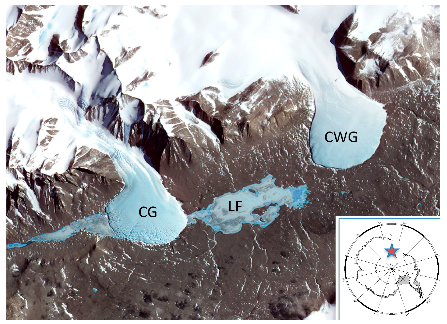
2.1. Field Location

Lake Fryxell is located in Taylor Valley, McMurdo Dry Valleys, South Victoria Land, Antarctica (−77.62; 163.15). The glacial streams were sampled within 5 m of the glacial front of Canada Glacier (−77.72; 163.11), and Commonwealth Glacier (−77.69; 163.41) (Figure 1).

2.2. Sample Collection and Processing

Water samples were collected from Lake Fryxell through a hole drilled in the lake ice during 2003, 2008 and 2010 using a 5-L Niskin bottle. All sampling was done during the Austral summer, specifically during the months of December and January. The water depth was determined from the level that the lake water raised in the sample hole. Samples were collected at 7, 8 and 9.5 m during 2003; at 7, 8, 9, 11 and 12 m during 2008; and at 7, 8, 9, 10, 11, 12, 13 and 14 m as well as glacier terminus streams during 2010. Stream samples were obtained by collecting 10 L of running stream water in sterile 1 L bottles. The sample size for all samples, regardless of source, was considered to be 5 L. Each 5-L sample was filtered onto Metricel black (Gelman) 47 mm 0.45 sterile membrane filters that were then incubated on media plates of YPD (BD, Franklin Lakes, NJ, USA) or 50% YPD each containing chloramphenicol (100 mg−1·mL) at 10 °C. The 2008 samples were filtered in 100 mL aliquots with all filters plated separately and all emerging isolates were enumerated. For the other field seasons some water samples clogged the filters with 100-mL volumes and those samples were filtered with smaller volumes per filter and each complete water sample was utilized. Each filter was plated separately and at least five of each morphotype, if there were five on a plate, were selected from each plate for further processing.
Culture plates were shipped refrigerated from Antarctica to University of Maine and incubated at 10 °C for up to one year. Each culture plate was checked for fungal growth and photographed at weekly interval for up to 18 months from time of collection.
Multiple isolates of each observed morphotype were subcultured from 2003 and 2010 samples. The number of isolates investigated varied based on the number of morphotypes observed. If available at least five of each morphotype were identified. A subculture from every isolate obtained in 2008 was generated and identified using either F-ARISA (fungal-automated ribosomal intergenic spacer analysis) as previously described [21] or by DNA sequencing of the ribosomal internal transcribed spacer region (ITS) for those isolates that could not be positively identified using F-ARISA. The F-ARISA is based on the variability of ITS fragment sizes. In this method, polymerase chain reaction (PCR) is carried out and the resultant fragments are compared using high resolution capillary DNA fingerprinting. Multiple isolates from cultures acquired in 2003, 2010 and from stream samples were subcultured and identified by sequencing the ITS region. Sets of primer combinations including ITS4, ITS5, ITS1F, EF3 [22,23,24,25] were used for PCR amplification. PCR for DNA sequenced fragments were carried out as previously described [21]. Specific ITS region amplicons were produced by PCR completed with 100 ng genomic DNA in 25 μL reactions using Illustra PuReTaq Ready-To-Go™ PCR Beads (GE Lifesciences, Pittsburgh, USA). PCR primer set ITS5-ITS4 (White et al. 1990) were used to target the ITS region for sequencing (Table 1). Initial denaturation completed for 2 min at 95 °C and 35 cycles with a PTC-200 thermal cycler (MJ Research, Watertown, MA, USA) under the following conditions: 30 s at 95 °C, 30 s at 52.3 °C, 1 min at 72 °C with a final 72 °C 10 min extension. DNA extraction and F-ARISA from all cultured 2008 isolates, regardless of origin, was carried out as previously described [26] and at least one isolate from each taxa identified was sequenced, even for those identified using the F-ARISA method. DNA sequencing was done by the University of Maine Sequencing facility (https://umaine.edu/dnaseq/). The internal transcribed spacer (ITS) sequences, including ITS1 through ITS2, were aligned using MUSCLE [27]. The resulting alignment was used to create a phylogenetic tree through the Seaview version 4.3.1 [28]. A rooted neighbor-joining distance tree was generated using the Seaview option of Jukes–Cantor distance measure [28]. Bootstrap values were based on 100 replicates. Venn’s diagram was made using Venny 2.1 [29]. ITS sequences were submitted to GenBank [21] and are shown in Table 1 for all identified taxa.

3. Results

3.1. Fungal Diversity

Thirty species of fungi were cultured from Lake Fryxell waters and glacial streams feeding Lake Fryxell (Table 1, Figure 2). There was about equal representation between Ascomycota and Basidiomycota species found from all the sources (53% to 47% respectively).
During 2008 we were able to determine the culture-based abundance of fungal taxa identified from Lake Fryxell. The 2008 Lake Fryxell samples showed Glaciozyma species representing 69% of the total abundance and Toxicocladosporidium, Acremonium, and Geomyces each representing 1% (Figure 3).

3.2. Fungal Distribution

The distribution of fungi was explored both by depth in Lake Fryxell as well as potential seeding by input by glacial streams. Isolates from all three field seasons were used for distribution determination. The total fungal abundance above the sharp chemocline of 11 m was higher than below with little over all abundance difference between 7 m and 9 m (Figure 4 and Figure 5, Table 1). Although Glaciozyma spp. and Penicillium spp. comprised the largest number of the fungal abundance above 11 m, they were not found in samples below 11 m. There were several species found both above and below the 11-m chemocline boundary, however only three species were found exclusively below 11 m (Clavispora lusitaniae, Holtermanniella nyarrowii, and Toxicocladosporium strelitziae).
Potential fungal seeding of Lake Fryxell by stream input was explored by sampling stream water near the fronts of the two primary glaciers that feed the lake. A total of 13 species were identified from Canada Glacier stream and Commonwealth Glacier stream (Table 1). There was no overlap of species found between either of these sources (Figure 5). In addition, only five of these 13 species were found in Lake Fryxell samples, while 17 species found in lake water were not found in either of the glacial stream samples.

4. Discussion

Although few studies have examined fungal diversity in Antarctic lake water columns, there have been studies that have isolated fungi from soil surrounding lakes, lake sediment, or nearby by microbial mats [30]. For example, lake sediment samples from the Skarvsnes Oasis region of East Antarctica revealed a diversity of five Ascomycetes and five Basidiomycetes genera similar to that found in Lake Fryxell [30]. Even over such a large geographic distance some of the same genera (Mrakia & Thelebolus) were isolated from the Skarvsnes Oasi lake sediments and Lake Fryxell (the Skarvsnes Oasi study characterized isolates only to genus). This suggests that these genera are cold adapted and common across Antarctica. More intriguing are the isolates from Skarvsnes Oasi soils that overlap with Lake Fryxell isolates (e.g., Geomyces, Cryptococcus, Mrakia, Theolobus, Diozegia). The fact that glaciers and stream do not appear to be a major source of fungi to the lake, the overlap between the studies suggest that soil may be a significant source of fungi in Lake Fryxell. Fungi may move from the soil to the lake by wind dispersion, melt pond run-off, or direct movement via snowmelt or growth.
Lake Fryxell has increasing H2S and decreasing dissolved O2 below the 11-m chemocline [31]. The abundance of only a few viable fungal species that are found primarily below the chemocline suggests that these organisms are not only able to tolerate the local environment but may be adapted to it. This is supported by the fact that, although most fungi are inhibited by H2S, a few species have been reported to metabolize and grow at high levels of H2S [32,33]. Additional research is needed to determine if these fungi are actively metabolizing and/or growing below the chemocline. There have been several studies of the microbial composition in Lake Fryxell, focusing on bacteria and Archaea [4,5,12,14,34,35]. Methanogenic Archaeal diversity [4] in Lake Fryxell were found to be clustered in either the water column or the sediment, demonstrating functional separation. In addition, a diversity of phototropic purple bacteria was identified from the water column [5,36] and nutrient cycling both above and below the chemocline.
The fact that there are at least four sources of microbial introduction into Lake Fryxell (e.g., CG, CWG, soil, and air) suggests that either: (1) The spatial distribution of source fungi is extreme and there is little or no homogenization of microbes over this area. (2) The level of detection may be so low that the error rates are high creating a detection bias from the small sample sizes that are used in these kinds of studies. (3) Soil may be a significant source of fungi as indicated above.
The list of fungi found in Antarctica continues to grow with the suggestion that “fungi might be the most diverse biota in Antarctica” [37]. There remain many challenges to determine both fungal abundance and activity from Antarctic environmental samples yet is clear that they must play a role in nutrient recycling and decomposition. Based on the extreme habitat below the Lake Fryxell chemocline, it is clear that a limited number of fungal species are able to adapt yet it remains to be determined how closely the Lake Fryxell isolates are to those found in the northern hemisphere.

Author Contributions

Conceptualization, L.C., R.J.R. and R.R.; Methodology, L.C., R.J.R. and R.R.; Formal Analysis, L.C., R.J.R. and B.S.; Investigation, L.C., R.J.R., R.R., B.S. and H.S.; Resources, L.C.; Data Curation, L.C. and B.S.; Writing—Original Draft Preparation, L.C.; Writing—Review & Editing, L.C., R.J.R., R.R. and H.S.; Supervision, L.C., and R.J.R.; Project Administration, L.C.; Funding Acquisition, L.C., R.J.R., R.R. and H.S.

Funding

This work was partially supported by Grant# NSF OPP-0125611 and NSF ANT-0739696 and its REU supplement.

Acknowledgments

Thanks to: Scott Craig, Megan Altenritter, Leslie Astbury, Amy Chichilo, Tristy Vick-Majors, and Andrew Barber for field and laboratory help.

Conflicts of Interest

The authors declare no conflicts of interest.

References

  1. Angino, E.E.; Armitage, K.B.; Tash, J.C. Chemical Stratification in Lake Fryxell, Victoria Land, Antarctica. Science 1962, 138, 34–36. [Google Scholar] [CrossRef] [PubMed]
  2. Bowman, J.S.; Vick-Majors, T.J.; Morgan-Kiss, R.; Takacs-Vesbach, C.; Ducklow, H.W.; Priscu, J.C. Microbial community dynamics in two polar extremes: The lakes of the McMurdo Dry Valleys and the West Antarctic Peninsula marine ecosystem. BioScience 2016, 66, 829–847. [Google Scholar] [CrossRef]
  3. Brambilla, E.; Hippe, H.; Hagelstein, A.; Tindall, B.J.; Stackebrandt, E. 16S rDNA diversity of cultured and uncultured prokaryotes of a mat sample from Lake Fryxell, McMurdo Dry Valleys, Antarctica. Extremophiles 2001, 5, 23–33. [Google Scholar] [CrossRef] [PubMed]
  4. Karr, E.A.; Ng, J.M.; Belchik, S.M.; Sattley, W.M.; Madigan, M.T.; Achenbach, L.A. Biodiversity of methanogenic and other archaea in the permanently frozen Lake Fryxell, Antarctica. Appl. Environ. Microbiol. 2006, 72, 1663–1666. [Google Scholar] [CrossRef] [PubMed]
  5. Karr, E.A.; Sattley, W.M.; Jung, D.O.; Madigan, M.T.; Achenbach, L.A. Remarkable diversity of phototrophic purple bacteria in a permanently frozen Antarctic lake. Appl. Environ. Microbiol. 2003, 69, 4910–4914. [Google Scholar] [CrossRef] [PubMed]
  6. Karr, E.A.; Sattley, W.M.; Rice, M.R.; Jung, D.O.; Madigan, M.T.; Achenbach, L.A. Diversity and Distribution of Sulfate-Reducing Bacteria in Permanently Frozen Lake Fryxell, McMurdo Dry Valleys, Antarctica. Appl. Environ. Microbiol. 2005, 71, 6353–6359. [Google Scholar] [CrossRef] [PubMed] [Green Version]
  7. Laybourn-Parry, J.; James, M.R.; McKnight, D.M.; Priscu, J.; Spaulding, S.A.; Shiel, R. The microbial plankton of Lake Fryxell, southern Victoria Land, Antarctica during the summers of 1992 and 1994. Polar Biol. 1996, 17, 54–61. [Google Scholar] [CrossRef]
  8. Stackebrandt, E.; Brambilla, E.; Cousin, S.; Dirks, W.; Pukall, R. Culture-independent analysis of bacterial species from an anaerobic mat from Lake Fryxell, Antarctica: Prokaryotic diversity revisited. Cell Mol. Biol. 2004, 50, 517–524. [Google Scholar] [PubMed]
  9. Taton, A.; Grubisic, S.; Brambilla, E.; De Wit, R.; Wilmotte, A. Cyanobacterial diversity in natural and artificial microbial mats of Lake Fryxell (McMurdo Dry Valleys, Antarctica): A morphological and molecular approach. Appl. Environ. Microbiol. 2003, 69, 5157–5169. [Google Scholar] [CrossRef] [PubMed]
  10. Wharton, R.A.; Parker, B.C.; Simmons, G.M.; Seaburg, K.G.; Love, F.G. Biogenic calcite structures forming in Lake Fryxell, Antarctica. Nature 1982, 295, 403–405. [Google Scholar] [CrossRef]
  11. Ayres, E.; Wall, D.H.; Adams, B.J.; Barrett, J.E.; Virginia, R.A. Unique similarity of faunal communities across aquatic–terrestrial interfaces in a polar desert ecosystem. Ecosystems 2007, 10, 523–535. [Google Scholar] [CrossRef]
  12. McKnight, D.M.; Howes, B.L.; Taylor, C.D.; Goehringer, D.D. Phytoplankton Dynamics in a Stably Stratified Antarctic Lake during Winter Darkness. J. Phycol. 2000, 36, 852–861. [Google Scholar] [CrossRef]
  13. Priscu, J.C.; Priscu, L.R.; Howard-Williams, C.H.; Vincent, W.F. Diel patterns of photosynthate biosynthesis by phytoplankton in permanently ice-covered Antarctic lakes under continuous sunlight. J. Plankton Res. 1988, 10, 333–340. [Google Scholar] [CrossRef]
  14. Priscu, J.C.; Priscu, L.R.; Vincent, W.F.; Howard-Williams, C.H. Photosynthate distribution by microplankton in permanently ice-covered Antarctic desert lakes. Limnol. Oceanogr. 1987, 32, 260–270. [Google Scholar] [CrossRef] [Green Version]
  15. Roberts, E.C.; Laybourn-Parry, J. Mixotrophic cryptophytes and their predators in the Dry Valley lakes of Antarctica. Freshw. Biol. 1999, 41, 737–746. [Google Scholar] [CrossRef]
  16. Roberts, E.C.; Laybourn-Parry, J.; McKnight, D.M.; Novarino, G. Stratification and dynamics of microbial loop communities in Lake Fryxell, Antarctica. Freshw. Biol. 2000, 44, 649–661. [Google Scholar] [CrossRef]
  17. Spaulding, S.A.; McKnight, D.M.; Smith, R.L.; Dufford, R. Phytoplankton population dynamics in perennially ice-covered Lake Fryxell, Antarctica. J. Plankton Res. 1994, 16, 527–541. [Google Scholar] [CrossRef]
  18. Wharton, R.A., Jr.; Parker, B.C.; Simmons, G.M., Jr. Distribution, species composition and morphology of algal mats in Antarctic dry valley lakes. Phycologia 1983, 22, 355–365. [Google Scholar] [CrossRef]
  19. Wurzbacher, C.M.; Barlocher, F.; Grossart, H.-P. Fungi in lake ecosystems. Aquat. Microb. Ecol. 2010, 59, 125–149. [Google Scholar] [CrossRef] [Green Version]
  20. Goto, S.; Sugiyama, J.; Iizuka, H. A Taxonomic Study of Antarctic Yeasts. Mycologia 1969, 61, 748–774. [Google Scholar] [CrossRef] [PubMed]
  21. Slemmons, C.; Johnson, G.; Connell, L.B. Application of an automated ribosomal intergenic spacer analysis database for identification of cultured Antarctic fungi. Antarct. Sci. 2013, 25, 44–50. [Google Scholar] [CrossRef]
  22. White, T.J.; Bruns, T.; Lee, S.; Taylor, J. Amplification and direct sequencing of fungal ribosomal RNA genes for phylogenetics. In PCR Protocols: A Guide to Methods and Applications; Innis, M., Gelfand, J., Sninsky, J., White, T.J., Eds.; Academic Press: Orlando, FL, USA, 1990; pp. 315–322. [Google Scholar]
  23. Gardes, M.; Bruns, T.D. ITS primers with enhanced specificity for basidiomycetes—Application to the identification of mycorrhizae and rusts. Mol. Ecol. 1993, 2, 113–118. [Google Scholar] [CrossRef] [PubMed]
  24. Martin, K.; Rygiewicz, P.T. Fungal-specific PCR primers developed for analysis of the ITS region of environmental DNA extracts. BMC Microbiol. 2005, 5, 1–11. [Google Scholar] [CrossRef] [PubMed] [Green Version]
  25. Smit, E.; Leeflang, P.; Glandorf, B.; van Elsas, J.D.; Wernars, K. Analysis of fungal diversity in the wheat rhizosphere by sequencing of cloned PCR-amplified genes encoding 18s rRNA and temperature gradient electrophoresis. Appl. Environ. Microbiol. 1999, 65, 2614–2621. [Google Scholar] [PubMed]
  26. Connell, L.B.; Redman, R.S.; Craig, S.D.; Scorzetti, G.; Iszard, M.; Rodriguez, R.J. Diversity of soil yeasts isolated from South Victoria Land, Antarctica. Microb. Ecol. 2008, 56, 448–459. [Google Scholar] [CrossRef] [PubMed]
  27. Edgar, R.C. MUSCLE: Multiple sequence alignment with high accuracy and high throughput. Nucleic Acids Res. 2004, 32, 1792–1797. [Google Scholar] [CrossRef] [PubMed]
  28. Gouy, M.; Guindon, S.; Gascuel, O. SeaView version 4: A multiplatform graphical user interface for sequence alignment and phylogenetic tree building. Mol. Biol. Evol. 2010, 27, 221–224. [Google Scholar] [CrossRef] [PubMed]
  29. Oliveros, J.C. Venny. An Interactive Tool for Comparing Lists with Venn’s Diagrams. Available online: http://bioinfogp.cnb.csic.es/tools/venny/index.html (accessed on 4 June 2018).
  30. Tsuji, M.; Fujiu, S.; Xiao, N.; Hanada, Y.; Kudoh, S.; Kondo, H.; Tsuda, S.; Hoshino, T. Cold adaptation of fungi obtained from soil and lake sediment in the Skarvsnes ice-free area, Antarctica. FEMS Microbiol. Lett. 2013, 346, 121–130. [Google Scholar] [CrossRef] [PubMed] [Green Version]
  31. Lawrence, M.J.F.; Hendy, C.H. Water column and sediment characteristics of Lake Fryxell, Taylor Valley, Antarctica. N. Z. J. Geol. Geophys. 1985, 28, 543–552. [Google Scholar] [CrossRef] [Green Version]
  32. Phae, C.-G.; Shoda, M. A new fungus which degrades hydrogen sulfide, methanethiol, dimethyl sulfide and dimethyl disulfide. Biotechnol. Lett. 1991, 13, 375–380. [Google Scholar] [CrossRef]
  33. Tang, J.; Hu, K.-D.; Hu, L.-Y.; Li, Y.-H.; Liu, Y.-S.; Zhang, H. Hydrogen Sulfide Acts as a Fungicide to Alleviate Senescence and Decay in Fresh-cut Sweetpotato. HortScience 2014, 49, 938–943. [Google Scholar]
  34. Mosier, A.C.; Murray, A.E.; Fritsen, C.H. Microbiota within the perennial ice cover of Lake Vida, Antarctica. FEMS Microbiol. Ecol. 2007, 59, 274–288. [Google Scholar] [CrossRef] [PubMed] [Green Version]
  35. Priscu, J.C.; Fritsen, C.H.; Adams, E.E.; Giovannoni, S.J.; Paerl, H.W.; McKay, C.P.; Doran, P.T.; Gordon, D.A.; Lanoil, B.D.; Pinckney, J.L. Perennial Antarctic lake ice: An oasis for life in a polar desert. Science 1998, 280, 2095–2098. [Google Scholar] [CrossRef] [PubMed]
  36. Jung, D.O.; Achenbach, L.A.; Karr, E.A.; Takaichi, S.; Madigan, M. A gas vesiculate planktonic strain of the purple nonsulfur bacterium Rhodoferax antarcticus isolated from Lake Fryxell, Dry Valleys, Antarctica. Arch. Microbiol. 2004, 182, 236–243. [Google Scholar] [CrossRef] [PubMed]
  37. Bridge, P.D.; Spooner, B.M. Non-lichenized Antarctic fungi: Transient visitors or members of a cryptic ecosystem? Fungal Ecol. 2012, 5, 381–394. [Google Scholar] [CrossRef]
Figure 1. Taylor Valley Antarctica showing Lake Fryxell (LF) between Canada (CG) and Commonwealth Glaciers (CWG). The location of Taylor Valley is shown by the star on the insert of Antarctica.
Life 08 00037 g001
Figure 2. Neighbor Joining tree of the ITS region of rDNA to illustrate the relationship of sequences obtained from yeasts isolated from Lake Fryxell as well as Commonwealth Glacier and Canada Glacier streams as a simple cladogram. Isolates from this work are identified by asterisk (*). Other isolates shown are from GenBank. The outgroups for both Ascomycetes groups (upper) and Basidiomycetes (lower) were the alga Alexandrium andersoni.
Life 08 00037 g002aLife 08 00037 g002b
Figure 3. Percent of abundance for each fungal taxa identified from Lake Fryxell during 2008.
Life 08 00037 g003
Figure 4. Abundance of cultured fungi in Lake Fryxell by depth in meters from the 2008 field season.
Life 08 00037 g004
Figure 5. A Venn’s diagram of fungal taxa shown by Lake Fryxell (LF) isolate above and below 11 m depth as well as streams feeding Lake Fryxell of Canada Glacier stream (CG) and Commonwealth Glacier stream (CWG).
Life 08 00037 g005
Table 1. Species identified from Lake Fryxell (LF), Canada Glacier stream (CG), and Commonwealth Glacier stream (CWG).
SpeciesGenBank Accession #A1B2LF above 11 mLF below 11 mCGCWG
Filobasidium magnusJX171169 XXX
Debaryomyces hanseniiKM816678X X
Thelebolus ellipsoideusJX171195X XXX
Thelebolus globosusJX171196X XX X
Geomyces sp. 1KM816679X X
Acremonium sp.KM816680X X
Mrakiella aquaticaJX171181 XXX
Rhodotorula mucilaginosaJX171192 XXX X
Vishniacozyma victoriaeJX171171 XX
Glaciozyma antarcticaKM816681 XX
Geomyces sp. 2KM816682X XXX
Glaciozyma watsoniiJX171176 XX
Naganishia globosaKM819094 XXX
Naganishia albidosimilisJX171168 XXX
Penicillium dipodomyicolaJX171186X X
Holtermanniella nyarrowiiKM816683 X X
Aureobasidium pullulansJX171163X XX
Toxicocladosporium strelitziaeKM816684X X
Cladosporium cladosporoidesKM816685X XX
Penicillium communeJX171184X X X
Heydenia alpinaJX171178X XX
Clavispora lusitaniaeKM816686X X
Mrakia gelidaKM819098 X X
Mrakia blollopsisKM819093 X X
Sporidiobolus pararoseusKM819095 X X
Cladosporium sp.KM819097X X
Trichoderma atrovirdeKM822752X X
Trametes pubescensKM822753 X X
Eutypa lataKM822754X X
Piptoporus betulinusKM822755 X X
A1 = Ascomycota, B2 = Basidiomycota.

