From Histopathology to High-Resolution Ultrasound Imaging of Skin Scars

 , ,
, ,  ,
,  and
and 
Abstract
1. Introduction
2. Materials and Methods
3. Results and Discussion
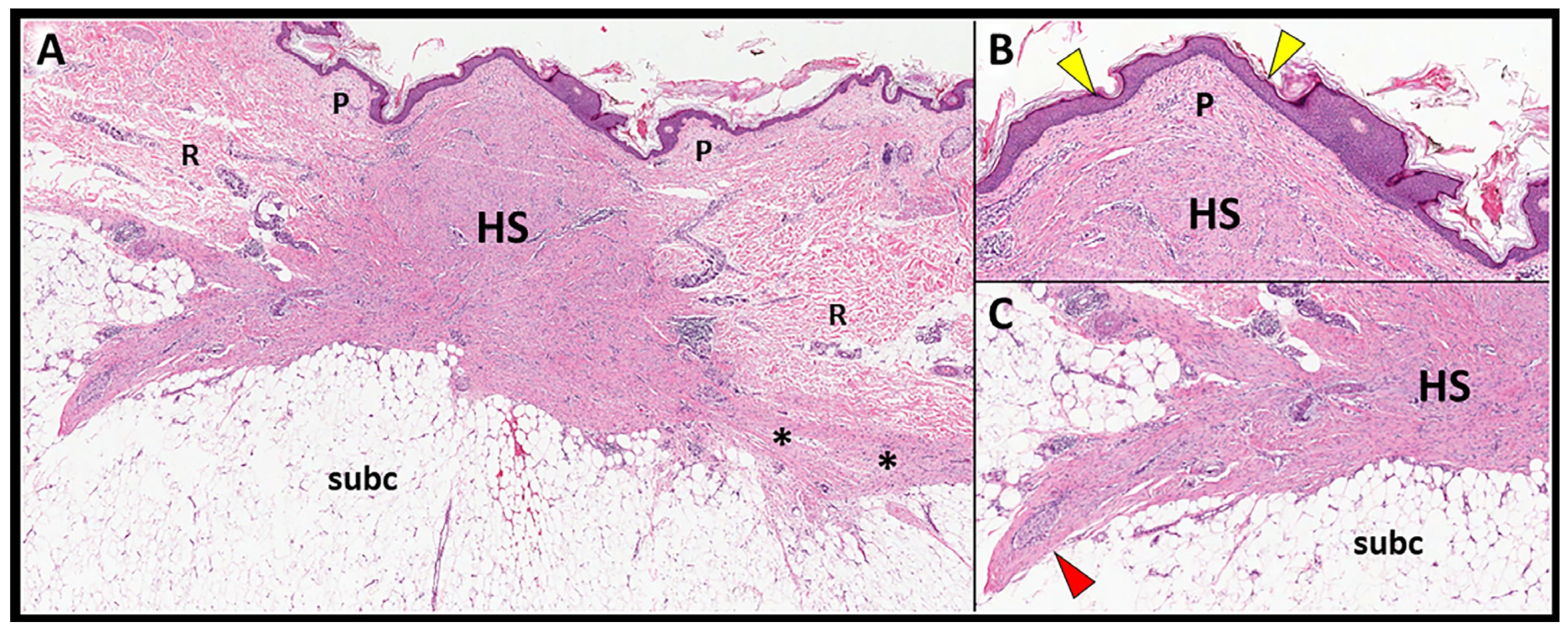
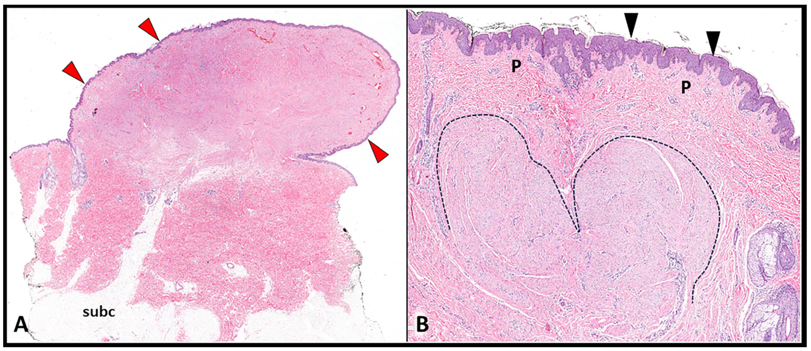
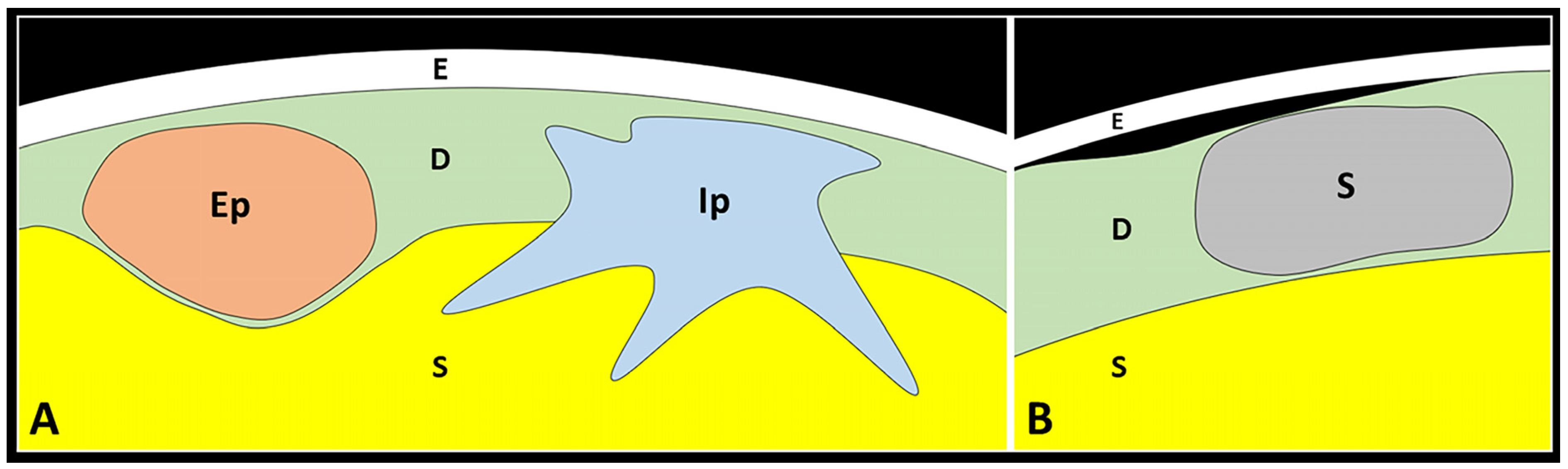
3.1. Histopathology of SSs
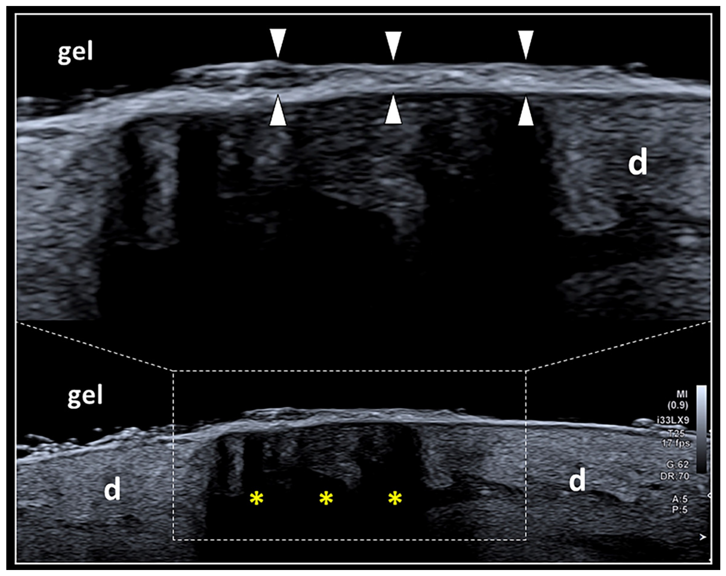
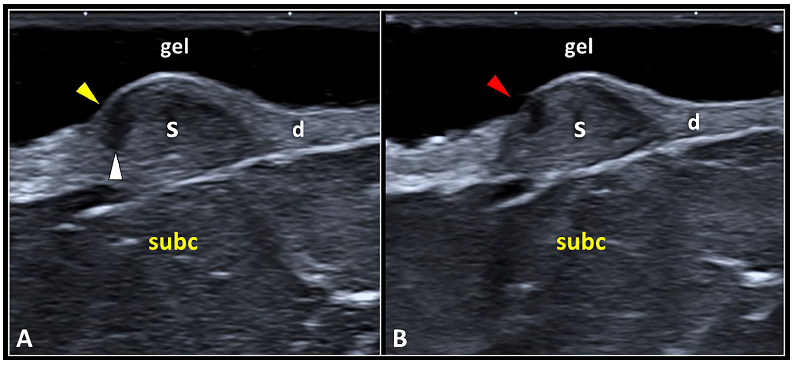
3.2. Sonography of SSs
3.2.1. Epidermis
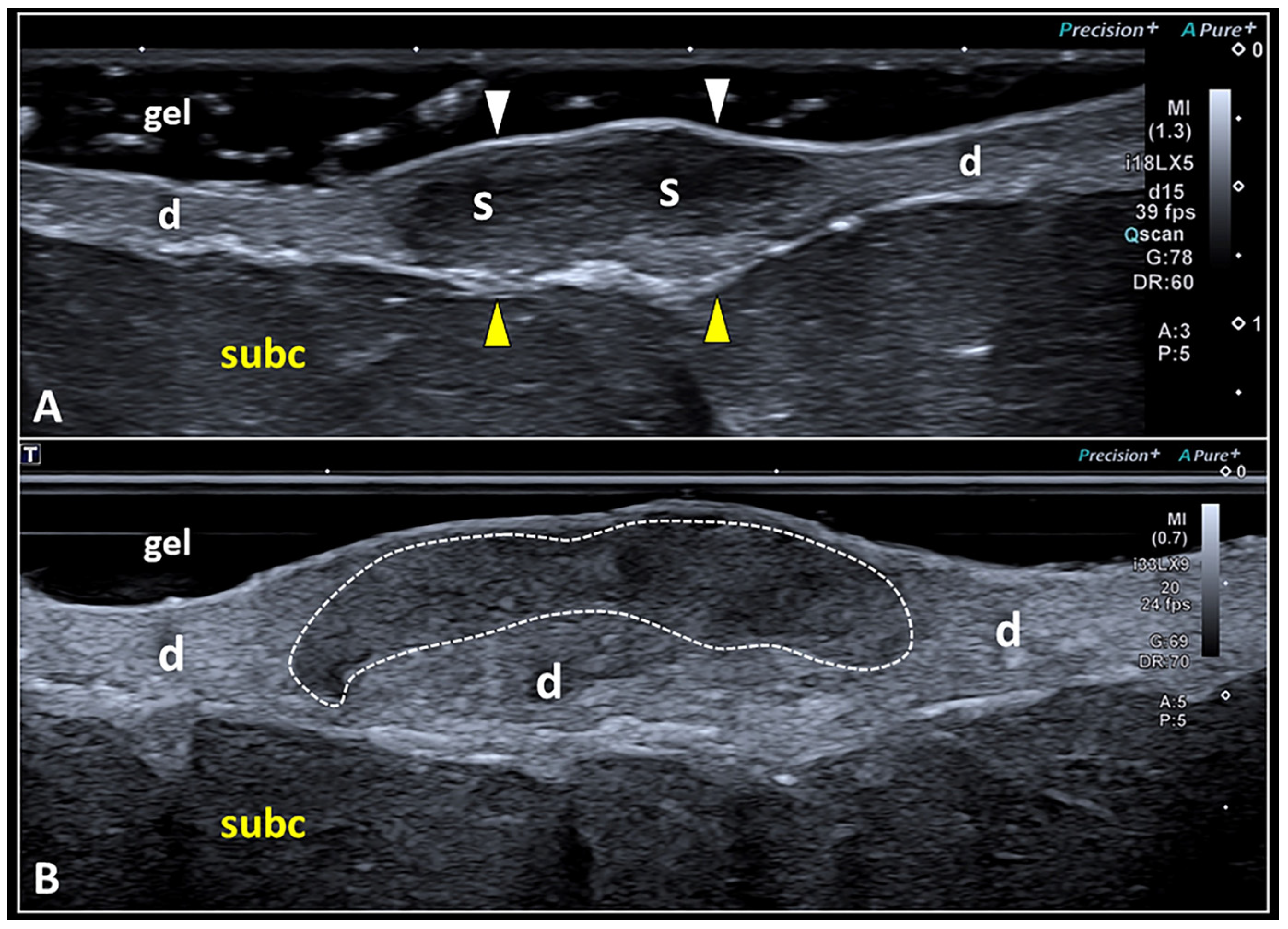
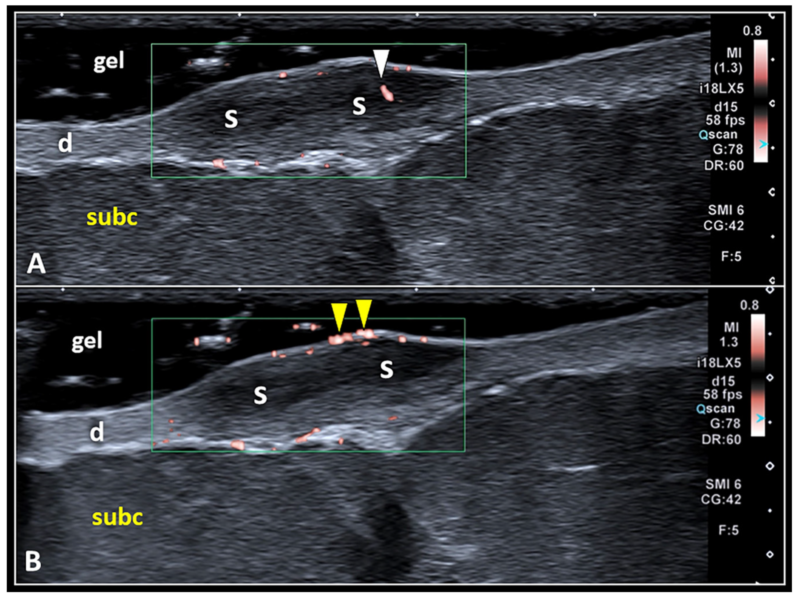
3.2.2. Dermis
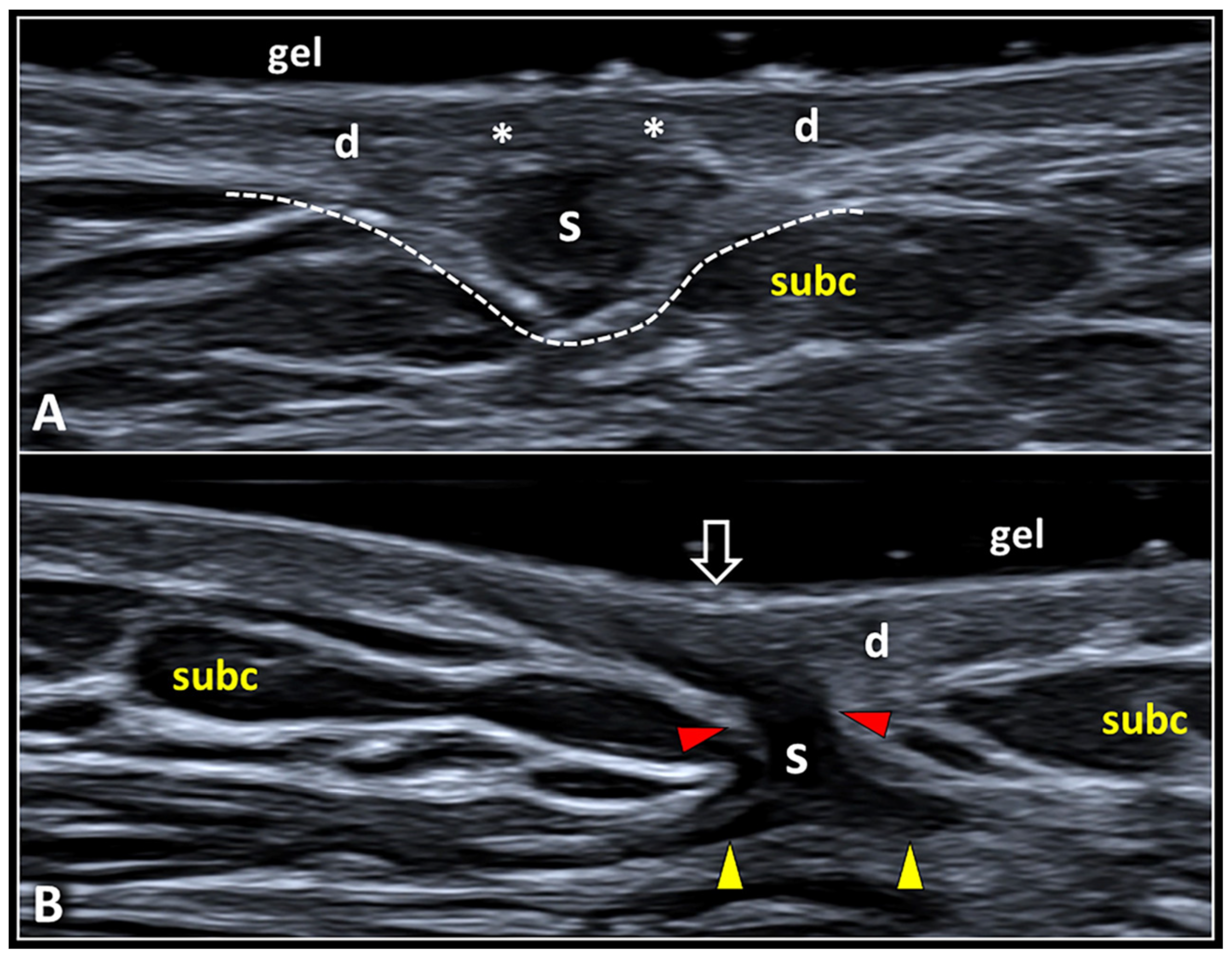
3.2.3. Dermo-Hypodermal Interface and Subcutaneous Tissue
4. Conclusions
Author Contributions
Funding
Institutional Review Board Statement
Informed Consent Statement
Data Availability Statement
Acknowledgments
Conflicts of Interest
References
- Wortsman, X. Practical applications of ultrasound in dermatology. Clin. Dermatol. 2021, 39, 605–623. [Google Scholar] [CrossRef] [Scilit] [PubMed]
- Wortsman, X. Common applications of dermatologic sonography. J. Ultrasound Med. 2012, 31, 97–111. [Google Scholar] [CrossRef] [Scilit]
- Catalano, O.; Wortsman, X. Dermatology ultrasound. imaging technique, tips and tricks, high-resolution anatomy. Ultrasound Q. 2020, 36, 321–327. [Google Scholar] [CrossRef] [Scilit]
- Wortsman, X. Ultrasound in dermatology: Why, how, and when? Semin. Ultrasound CT MRI 2013, 34, 177–195. [Google Scholar] [CrossRef] [Scilit] [PubMed]
- Ricci, V.; Ricci, C.; Tamborrini, G.; Chang, K.-V.; Mezian, K.; Zunica, F.; Naňka, O.; Kara, M.; Özçakar, L. From histology to sonography in synovitis: EURO-MUSCULUS/USPRM approach. Pathol.—Res. Pract. 2023, 241, 154273. [Google Scholar] [CrossRef] [Scilit]
- Corvino, A.; Sandomenico, F.; Corvino, F.; Campanino, M.R.; Verde, F.; Giurazza, F.; Tafuri, D.; Catalano, O. Utility of a gel stand-off pad in the detection of doppler signal on focal nodular lesions of the skin. J. Ultrasound 2020, 23, 45–53. [Google Scholar] [CrossRef] [Scilit] [PubMed]
- Bessonart, M.N.; Macedo, N.; Carmona, C. High resolution B-scan ultrasound of hypertrophic scars. Skin. Res. Technol. 2005, 11, 185–188. [Google Scholar] [CrossRef] [Scilit] [PubMed]
- Burd, A.; Huang, L. Hypertrophic response and keloid diathesis: Two very different forms of scar. Plast. Reconstr. Surg. 2005, 116, 150e–157e. [Google Scholar] [CrossRef] [Scilit]
- Limandjaja, G.C.; Niessen, F.B.; Scheper, R.J.; Gibbs, S. Hypertrophic scars and keloids: Overview of the evidence and practical guide for differentiating between these abnormal scars. Exp. Dermatol. 2021, 30, 146–161. [Google Scholar] [CrossRef] [Scilit]
- Wolfram, D.; Tzankov, A.; Pülzl, P.; Piza-Katzer, H. Hypertrophic scars and keloids—A review of their pathophysiology, risk factors, and therapeutic management. Dermatol. Surg. 2009, 35, 171–181. [Google Scholar] [CrossRef] [Scilit]
- Lobos, N.; Wortsman, X.; Valenzuela, F.; Alonso, F. Color doppler ultrasound assessment of activity in keloids. Dermatol. Surg. 2017, 43, 817–825. [Google Scholar] [CrossRef] [Scilit]
- Kanitakis, J. Anatomy, histology and immunohistochemistry of normal human skin. Eur. J. Dermatol. 2002, 12, 390–399. [Google Scholar] [PubMed]
- Murphy, G.F. Histology of the skin. In Lever’s Histopathology of the Skin, 9th ed.; Elder, D.E., Elenitsas, R., Johnson, B.L., Jr., Murphy, G.F., Eds.; Lippincott Williams & Wilkins: Philadelphia, PA, USA, 2005. [Google Scholar]
- Meigel, W.N.; Gay, S.; Weber, L. Dermal architecture and collagen type distribution. Arch. Dermatol. Res. 1977, 259, 1–10. [Google Scholar] [CrossRef] [Scilit]
- Stenn, K. Collagen heterogeneity of skin. Am. J. Dermatopathol. 1979, 1, 87–88. [Google Scholar] [CrossRef]
- Lee, J.Y.-Y.; Yang, C.-C.; Chao, S.-C.; Wong, T.-W. Histopathological differential diagnosis of keloid and hypertrophic scar. Am. J. Dermatopathol. 2004, 26, 379–384. [Google Scholar] [CrossRef] [Scilit] [PubMed]
- Ehrlich, H.P.; Desmoulière, A.; Diegelmann, R.F.; Cohen, I.K.; Compton, C.C.; Garner, W.L.; Kapanci, Y.; Gabbiani, G. Morphological and immunochemical differences between keloid and hypertrophic scar. Am. J. Pathol. 1994, 145, 105–113. [Google Scholar] [PubMed]
- Ogawa, R. Keloid and hypertrophic scars are the result of chronic inflammation in the reticular dermis. Int. J. Mol. Sci. 2017, 18, 606. [Google Scholar] [CrossRef] [Scilit]
- Sato, M. Upregulation of the Wnt/β-catenin pathway induced by transforming growth factor-β in hypertrophic scars and keloids. Acta Derm.-Venereol. 2006, 86, 300–307. [Google Scholar] [CrossRef] [Scilit]
- Verhaegen, P.D.H.M.; Van Zuijlen, P.P.M.; Pennings, N.M.; Van Marle, J.; Niessen, F.B.; Van Der Horst, C.M.A.M.; Middelkoop, E. Differences in collagen architecture between keloid, hypertrophic scar, normotrophic scar, and normal skin: An objective histopathological analysis. Wound Repair Regen. 2009, 17, 649–656. [Google Scholar] [CrossRef] [Scilit]
- Sumi, Y.; Muramatsu, H.; Hata, K.-I.; Ueda, M.; Muramatsu, T. Secretory leukocyte protease inhibitor is a novel inhibitor of fibroblast-mediated collagen gel contraction. Exp. Cell Res. 2000, 256, 203–212. [Google Scholar] [CrossRef] [Scilit]
- Andrews, J.P.; Marttala, J.; Macarak, E.; Rosenbloom, J.; Uitto, J. Keloids: The paradigm of skin fibrosis—Pathomechanisms and treatment. Matrix Biol. 2016, 51, 37–46. [Google Scholar] [CrossRef] [Scilit] [PubMed]
- Limandjaja, G.; Belien, J.; Scheper, R.; Niessen, F.; Gibbs, S. Hypertrophic and keloid scars fail to progress from the CD 34−/α-smooth muscle actin (α-SMA)+ immature scar phenotype and show gradient differences in α-SMA and p16 expression. Br. J. Dermatol. 2020, 182, 974–986. [Google Scholar] [CrossRef] [Scilit] [PubMed]
- Limandjaja, G.C.; Broek, L.J.v.D.; Breetveld, M.; Waaijman, T.; Monstrey, S.; de Boer, E.M.; Scheper, R.J.; Niessen, F.B.; Gibbs, S. Characterization of in vitro reconstructed human normotrophic, hypertrophic, and keloid scar models. Tissue Eng. Part C Methods 2018, 24, 242–253. [Google Scholar] [CrossRef] [Scilit] [PubMed]
- Yoo, M.G.; Kim, I.-H. Keloids and hypertrophic scars: Characteristic vascular structures visualized by using dermoscopy. Ann. Dermatol. 2014, 26, 603–609. [Google Scholar] [CrossRef] [Scilit] [PubMed]
- Ricci, V.; Ricci, C.; Cocco, G.; Donati, D.; Farì, G.; Mezian, K.; Naňka, O.; Özçakar, L. From histology to sonography in skin and superficial tissue disorders: EURO-MUSCULUS/USPRM approach. Pathol.—Res. Pract. 2022, 237, 154003. [Google Scholar] [CrossRef] [Scilit]
- Ricci, V.; Soylu, A.R.; Özçakar, L. Artifacts and artistic facts: A visual simulation for ultrasound training. Am. J. Phys. Med. Rehabil. 2019, 98, 521–525. [Google Scholar] [CrossRef] [Scilit] [PubMed]
- Alfageme, F.; Wortsman, X.; Catalano, O.; Roustan, G.; Crisan, M.; Crisan, D.; Gaitini, D.E.; Cerezo, E.; Badea, R. European federation of societies for ultrasound in medicine and biology (EFSUMB) position statement on dermatologic ultrasound. Ultraschall Med. 2021, 42, 39–47. [Google Scholar] [CrossRef] [Scilit]
- Kleinerman, R.; Whang, T.B.; Bard, R.L.; Marmur, E.S. Ultrasound in dermatology: Principles and applications. J. Am. Acad. Dermatol. 2012, 67, 478–487. [Google Scholar] [CrossRef] [Scilit]
- Wortsman, X.; Wortsman, J. Clinical usefulness of variable-frequency ultrasound in localized lesions of the skin. J. Am. Acad. Dermatol. 2010, 62, 247–256. [Google Scholar] [CrossRef] [Scilit]
- Yen, A.; Braverman, I.M. Ultrastructure of the human dermal microcirculation: The horizontal plexus of the papillary dermis. J. Investig. Dermatol. 1976, 66, 131–142. [Google Scholar] [CrossRef] [Scilit]
- Alaish, S.M.; Yager, D.R.; Diegelmann, R.F.; Cohen, I. Hyaluronic acid metabolism in keloid fibroblasts. J. Pediatr. Surg. 1995, 30, 949–952. [Google Scholar] [CrossRef] [Scilit]
- Babu, M.; Diegelmann, R.; Oliver, N. Fibronectin is overproduced by keloid fibroblasts during abnormal wound healing. Mol. Cell. Biol. 1989, 9, 1642–1650. [Google Scholar] [CrossRef] [Scilit]
- Ricci, V.; Ricci, C.; Gervasoni, F.; Giulio, C.; Farì, G.; Andreoli, A.; Özçakar, L. From physical to ultrasound examination in lymphedema: A novel dynamic approach. J. Ultrasound 2022, 25, 757–763. [Google Scholar] [CrossRef] [Scilit]
- Ricci, V.; Ricci, C.; Gervasoni, F.; Andreoli, A.; Özçakar, L. From histo-anatomy to sonography in lymphedema: EURO-MUSCULUS/USPRM approach. Eur. J. Phys. Rehabil. Med. 2021, 58, 108–117. [Google Scholar] [CrossRef] [Scilit] [PubMed]
- Di Santolo, M.S.; Sagnelli, M.; Mancini, M.; Scalvenzi, M.; Delfino, M.; Schonauer, F.; Molea, G.; Ayala, F.; Salvatore, M. High-resolution color-doppler ultrasound for the study of skin growths. Arch. Dermatol. Res. 2015, 307, 559–566, Erratum in Arch. Dermatol. Res. 2015, 307, 567. [Google Scholar] [CrossRef] [Scilit] [PubMed]
- Corvino, A.; Varelli, C.; Cocco, G.; Corvino, F.; Catalano, O. Seeing the unseen with superb microvascular imaging: Ultrasound depiction of normal dermis vessels. J. Clin. Ultrasound 2022, 50, 121–127. [Google Scholar] [CrossRef] [Scilit] [PubMed]
- Avram, A.S.; Avram, M.M.; James, W.D. Subcutaneous fat in normal and diseased states: 2. Anatomy and physiology of white and brown adipose tissue. J. Am. Acad. Dermatol. 2005, 53, 671–683. [Google Scholar] [CrossRef] [Scilit] [PubMed]
- Sbarbati, A.; Accorsi, D.; Benati, D.; Marchetti, L.; Orsini, G.; Rigotti, G.; Panettiere, P. Subcutaneous adipose tissue classification. Eur. J. Histochem. 2010, 54, e48. [Google Scholar] [CrossRef] [Scilit]




| Histological Compartment | Ultrasound Findings |
|---|---|
| Epidermis | Continuous, interrupted, hyper-reflective § +/− dermo-epidermal dissociation |
| Dermis | Thickness, echogenicity, and shape * of the scar tissue +/− vascular signals on CD/PD |
| DHI | Continuous, interrupted, retracted |
| Subcutaneous tissue | Infiltrative pattern, compressive pattern |
| Hypertrophic Scar | Keloid |
|---|---|
| High incidence | Low incidence |
| No association with skin color | Association with darker skin |
| Confined within original wound boundaries | Extension beyond the original wound boundaries * |
| Usually <1 cm thick/wide | Variable size |
| Associated with contracture | Not associated with contracture |
| Expansive Pattern | Infiltrative Pattern |
|---|---|
| Scar flat or globular in shape | Scar irregular in shape * |
| Compression of the SUBC | Infiltration of the SUBC |
| Continuous DHI | Interrupted DHI |
| Absence of retractions | Retraction of surrounding tissues |
Disclaimer/Publisher’s Note: The statements, opinions and data contained in all publications are solely those of the individual author(s) and contributor(s) and not of MDPI and/or the editor(s). MDPI and/or the editor(s) disclaim responsibility for any injury to people or property resulting from any ideas, methods, instructions or products referred to in the content. |
© 2023 by the authors. Licensee MDPI, Basel, Switzerland. This article is an open access article distributed under the terms and conditions of the Creative Commons Attribution (CC BY) license (https://creativecommons.org/licenses/by/4.0/).
Share and Cite
Ricci, V.; Cocco, G.; Donati, D.; Farì, G.; Chang, K.-V.; Özçakar, L. From Histopathology to High-Resolution Ultrasound Imaging of Skin Scars. Diagnostics 2023, 13, 3629. https://doi.org/10.3390/diagnostics13243629
Ricci V, Cocco G, Donati D, Farì G, Chang K-V, Özçakar L. From Histopathology to High-Resolution Ultrasound Imaging of Skin Scars. Diagnostics. 2023; 13(24):3629. https://doi.org/10.3390/diagnostics13243629
Chicago/Turabian StyleRicci, Vincenzo, Giulio Cocco, Danilo Donati, Giacomo Farì, Ke-Vin Chang, and Levent Özçakar. 2023. "From Histopathology to High-Resolution Ultrasound Imaging of Skin Scars" Diagnostics 13, no. 24: 3629. https://doi.org/10.3390/diagnostics13243629
APA StyleRicci, V., Cocco, G., Donati, D., Farì, G., Chang, K.-V., & Özçakar, L. (2023). From Histopathology to High-Resolution Ultrasound Imaging of Skin Scars. Diagnostics, 13(24), 3629. https://doi.org/10.3390/diagnostics13243629