The Effect of Set Up Position on EMG Amplitude, Lumbar Spine Kinetics, and Total Force Output During Maximal Isometric Conventional-Stance Deadlifts


Abstract
:1. Introduction
2. Materials and Methods
2.1. Research Design
2.2. Participants
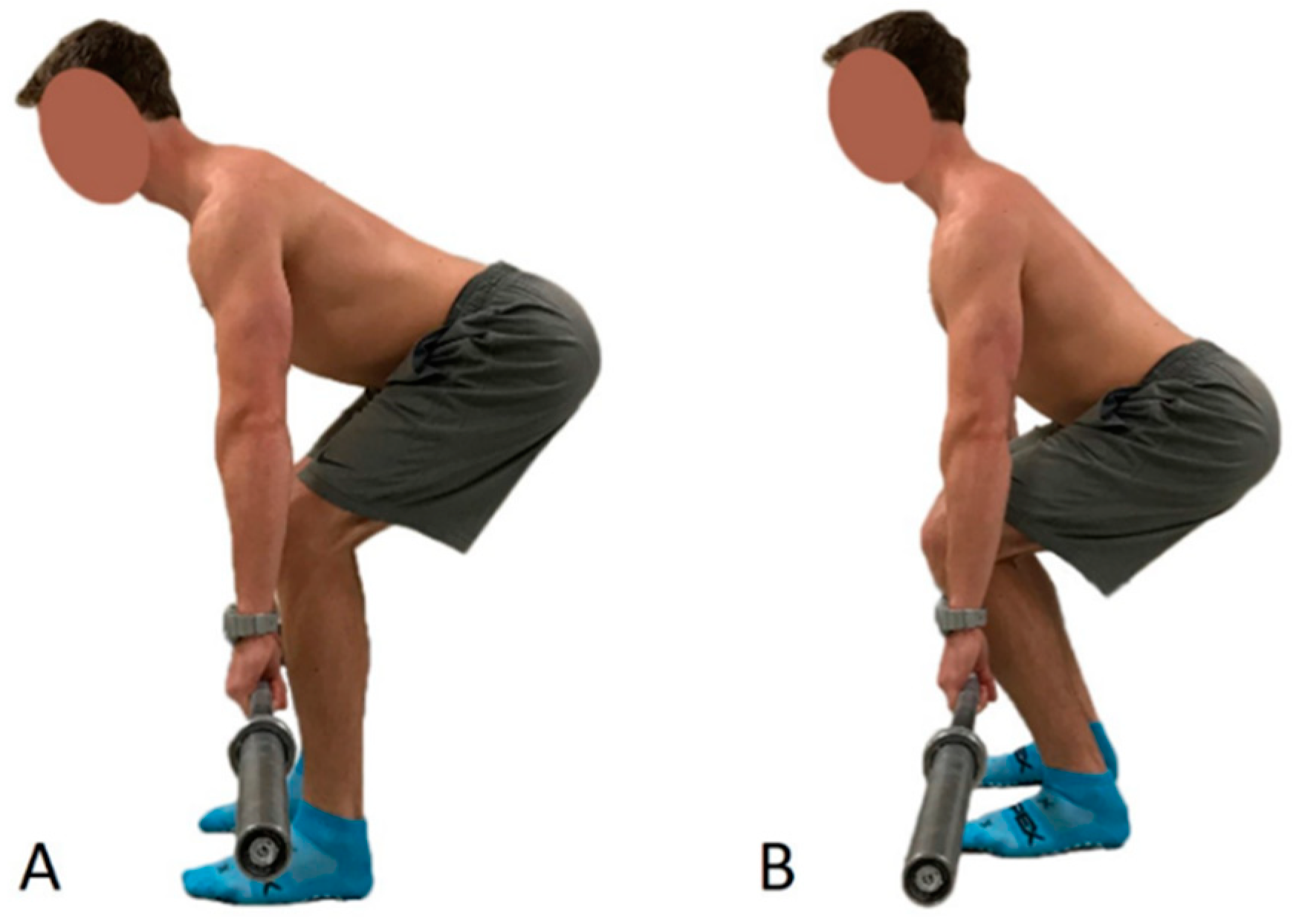
2.3. Procedure
2.3.1. Landmarking
2.3.2. Warm-Up
2.3.3. Electromyography and Motion Capture Marker Placement
2.3.4. Equipment for Data Collection
2.4. Data Analysis
2.5. Statistical Analyses
3. Results
3.1. Angles
3.2. Electromyography
3.3. Moments
3.4. Ground Reaction Force
3.5. Lumbar Shear and Compression Force
4. Discussion
5. Conclusions
Practical Applications
Author Contributions
Funding
Acknowledgments
Conflicts of Interest
References
- Hales, M. Improving the deadlift: Understanding biomechanical constraints and physiological adaptations to resistance exercise. Strength Cond. J. 2010, 32, 44–51. [Google Scholar] [CrossRef]
- Escamilla, R.F.; Francisco, A.C.; Kayes, A.V.; Speer, K.P.; Moorman, C.T. An electromyographic analysis of sumo and conventional style deadlifts. Med. Sci. Sports Exerc. 2002, 34, 682–688. [Google Scholar] [PubMed]
- Faigenbaum, A.D.; Myer, G.D. Resistance training among young athletes: Safety, efficacy and injury prevention effects. Br. J. Sports Med. 2010, 44, 56–63. [Google Scholar] [CrossRef] [PubMed]
- Hancock, S.; Wyatt, F.; Kilgore, J.L. Variation in barbell position relative to shoulder and foot anatomical landmarks alters movement efficiency. Int. J. Exerc. Sci. 2012, 5, 183–195. [Google Scholar]
- Hamlyn, N.; Behm, D.G.; Young, W.B. Trunk muscle activation during dynamic weight-training exercises and isometric instability activities. J. Strength Cond. Res. 2007, 21, 1108–1112. [Google Scholar] [PubMed]
- SENIAM Project. Surface ElectroMyoGraphy for the Non-Invasive Assessment of Muscles. Sensor Locations. 2017. Available online: http://www.seniam.org/ (accessed on 28 June 2017).
- Cholewicki, J.; McGill, S.M.; Norman, R.W. Lumbar spine loads during the lifting of extremely heavy weights. Med. Sci. Sports Exerc. 1991, 23, 1179–1186. [Google Scholar] [CrossRef] [PubMed]
- van Dieën, J.H.; de Looze, M.P. Sensitivity of single-equivalent trunk extensor muscle models to anatomical and functional assumptions. J. Biomech. 1999, 32, 195–198. [Google Scholar] [CrossRef]
- Potvin, J.R.; Norman, R.W.; McGill, S.M. Reduction in anterior shear forces on the L4/L5 disc by the lumbar musculature. Clin. Biomech. 1991, 6, 88–96. [Google Scholar] [CrossRef]
- Swinton, P.A.; Stewart, A.; Agouris, I.; Keogh, J.W.; Lloyd, R. A biomechanical analysis of straight and hexagonal barbell deadlifts using submaximal loads. J. Strength Cond. Res. 2011, 25, 2000–2009. [Google Scholar] [CrossRef] [PubMed]
- Buseck, M.; Schipplein, O.D.; Andersson, G.B.J.; Andriacchi, T.P. Influence of dynamic factors and external loads on the moment at the lumbar spine in lifting. Spine 1987, 13, 918–921. [Google Scholar] [CrossRef]
- Bird, S.P.; Barrington-Higgs, B. Exploring the deadlift. Strength Cond. J. 2010, 32, 46–51. [Google Scholar] [CrossRef]
- Wallden, M. The neutral spine principle. J. Bodyw. Mov. Ther. 2009, 13, 350–361. [Google Scholar] [CrossRef] [PubMed]
- Cholewicki, J.; McGill, S. Lumbar posterior ligament involvement during extremely heavy lifts estimated from fluoroscopic measurements. J. Biomech. 1992, 25, 17–28. [Google Scholar] [CrossRef]
- Camara, K.D.; Coburn, J.W.; Dunnick, D.D.; Brown, L.E.; Galpin, A.J.; Costa, P.B. An examination of muscle activation and power characteristics while performing the deadlift exercise with straight and hexagonal barbells. J. Strength Cond. Res. 2016, 30, 1183–1188. [Google Scholar] [CrossRef] [PubMed]
- Branch, T.P.; Hunter, R.; Donath, M. Dynamic EMG analysis of anterior cruciate deficient legs with and without bracing during cutting. Am. J. Sports Med. 1989, 17, 35–41. [Google Scholar] [CrossRef] [PubMed]
- Arjmand, N.; Shirazi-Adl, A. Biomechanics of Changes in Lumbar Posture in Static Lifting. Spine 2005, 30, 2637–2648. [Google Scholar] [CrossRef] [PubMed]


| Characteristic | Mean (Standard Deviation) |
|---|---|
| Years of experience (years) | 6.05 (3.35) |
| Mass (kg) | 88.73 (22.16) |
| Body Mass Index (kg/m2) | 29.73 (7.00) |
| Standing height (cm) | 172.48 (10.87) |
| Arm length (cm) | 52.34 (3.57) |
| Femur length (cm) | 38.42 (4.22) |
| Tibia length (cm) | 40.99 (3.06) |
| Torso length (cm) | 42.67 (2.90) |
| Reported deadlift 1RM (kg) | 179.85 (63.06) |
| # | Foot Angle (°) | Tibia Angle (°) | Knee Angle (°) | Hip Angle (°) | Pelvis Angle (°) | Lumbar Angle (°) | Torso Angle (°) | |||||||
|---|---|---|---|---|---|---|---|---|---|---|---|---|---|---|
| FBDL | CBDL | FBDL | CBDL | FBDL | CBDL | FBDL | CBDL | FBDL | CBDL | FBDL | CBDL | FBDL | CBDL | |
| 2 | 15.5 | 12.10 | 22.26 | 8.16 | 64.36 | 47.50 | 87.69 | 90.42 | 45.71 | 50.87 | 11.23 | 12.17 | 71.16 | 78.88 |
| 3 | 9.34 | 8.30 | 20.52 | 7.11 | 60.40 | 43.22 | 71.28 | 70.21 | 29.72 | 33.52 | 24.42 | 25.23 | 72.37 | 77.57 |
| 5 | 11.38 | 6.51 | 27.02 | 12.48 | 81.96 | 57.80 | 67.10 | 67.48 | 12.46 | 21.98 | 25.07 | 27.31 | 65.15 | 80.32 |
| 6 | 16.52 | 13.13 | 23.76 | 12.89 | 72.77 | 55.53 | 61.00 | 58.13 | 11.71 | 15.25 | 22.27 | 24.22 | 70.78 | 76.64 |
| 8 | 5.04 | 3.71 | 15.12 | 5.01 | 65.40 | 39.67 | 87.65 | 87.07 | 34.06 | 46.31 | 38.68 | 40.98 | 72.10 | 85.57 |
| 9 | 12.89 | 15.23 | 12.30 | 6.81 | 71.28 | 58.55 | 90.07 | 89.20 | 31.04 | 37.31 | 6.91 | 7.72 | 61.28 | 68.80 |
| 10 | 12.49 | 11.52 | 13.50 | 8.35 | 66.40 | 62.65 | 99.13 | 105.66 | 45.65 | 49.06 | 22.10 | 23.83 | 65.73 | 71.79 |
| 11 | 13.82 | 13.63 | 21.84 | 7.70 | 66.43 | 45.42 | 63.48 | 60.46 | 18.17 | 21.41 | 37.26 | 38.65 | 74.05 | 80.90 |
| Mean ± SD | 12.12 ± 3.64 | 10.52 ± 3.96 | 19.54 ± 5.29 | 8.56 * ± 2.75 | 68.63 ± 6.63 | 51.29 * ± 8.38 | 78.4 ± 14.35 | 78.70 ± 16.83 | 28.57 ± 13.49 | 34.36 * ± 13.77 | 23.49 ± 11.03 | 25.01 ± 11.42 | 69.0 ± 4.46 | 77.56 * ± 5.29 |
| # | Thoracic Erector Spinae (V) | Latissimus Dorsi (V) | Lower Lumbar Erector Spinae (V) | Upper Lumbar Erector Spinae (V) | Gluteus Maximus (V) | Biceps Femoris (V) | Vastus Lateralis (V) | |||||||
|---|---|---|---|---|---|---|---|---|---|---|---|---|---|---|
| FBDL | CBDL | FBDL | CBDL | FBDL | CBDL | FBDL | CBDL | FBDL | CBDL | FBDL | CBDL | FBDL | CBDL | |
| 2 | 2.40 × 10⁻¹ | 2.82 × 10⁻¹ | 1.00 × 10⁻¹ | 1.21 × 10⁻¹ | 1.14 × 10⁻¹ | 1.26 × 10⁻¹ | 1.42 × 10⁻¹ | 1.45 × 10⁻¹ | 2.11 × 10⁻² | 2.18 × 10⁻² | 5.22 × 10⁻² | 8.09 × 10⁻² | 4.95 × 10⁻² | 2.77 × 10⁻² |
| 3 | 5.80 × 10⁻² | 5.40 × 10⁻² | 2.93 × 10⁻² | 2.70 × 10⁻² | 5.33 × 10⁻² | 6.57 × 10⁻² | 1.06 × 10⁻¹ | 1.22 × 10⁻¹ | 5.86 × 10⁻³ | 4.83 × 10⁻³ | 1.68 × 10⁻² | 2.95 × 10⁻² | 4.61 × 10⁻² | 2.79 × 10⁻² |
| 5 | 7.79 × 10⁻² | 1.01 × 10⁻¹ | 2.57 × 10⁻² | 5.34 × 10⁻² | 7.82 × 10⁻² | 6.92 × 10⁻² | 1.06 × 10⁻¹ | 1.34 × 10⁻¹ | 2.40 × 10⁻² | 1.54 × 10⁻² | 1.40 × 10⁻² | 2.60 × 10⁻² | 7.20 × 10⁻² | 4.56 × 10⁻² |
| 6 | 9.98 × 10⁻² | 1.07 × 10⁻¹ | 1.16 × 10⁻¹ | 2.77 × 10⁻² | 3.99 × 10⁻² | 3.96 × 10⁻² | 1.13 × 10⁻¹ | 1.32 × 10⁻¹ | 1.57 × 10⁻² | 1.56 × 10⁻² | 2.62 × 10⁻² | 4.12 × 10⁻² | 4.47 × 10⁻² | 4.38 × 10⁻² |
| 8 | 1.02 × 10⁻¹ | 9.54 × 10⁻² | 2.42 × 10⁻² | 2.15 × 10⁻² | 8.86 × 10⁻³ | 7.97 × 10⁻³ | 3.45 × 10⁻² | 3.46 × 10⁻² | 1.74 × 10⁻³ | 1.95 × 10⁻³ | 1.34 × 10⁻² | 7.49 × 10⁻³ | 9.91 × 10⁻³ | 8.01 × 10⁻³ |
| 9 | 9.18 × 10⁻² | 9.89 × 10⁻² | 1.79 × 10⁻¹ | 1.94 × 10⁻¹ | 66.14 × 10⁻² | 6.59 × 10⁻² | 1.09 × 10⁻¹ | 1.18 × 10⁻¹ | 1.62 × 10⁻² | 1.69 × 10⁻² | 1.86 × 10⁻² | 3.48 × 10⁻² | 4.48 × 10⁻² | 4.13 × 10⁻² |
| 10 | 9.43 × 10⁻² | 8.74 × 10⁻² | 1.45 × 10⁻¹ | 1.28 × 10⁻¹ | 4.59 × 10⁻² | 4.72 × 10⁻² | 1.24 × 10⁻¹ | 1.26 × 10⁻¹ | 2.10 × 10⁻² | 1.45 × 10⁻² | 2.58 × 10⁻² | 2.53 × 10⁻² | 5.31 × 10⁻² | 4.19 × 10⁻² |
| 11 | 1.49 × 10⁻² | 1.89 × 10⁻² | 1.07 × 10⁻² | 1.15 × 10⁻² | 3.40 × 10⁻³ | 4.49 × 10⁻³ | 1.04 × 10⁻² | 1.20 × 10⁻² | 1.60 × 10⁻³ | 2.02 × 10⁻³ | 3.97 × 10⁻³ | 4.51 × 10⁻³ | 7.09 × 10⁻³ | 6.21 × 10⁻³ |
| Mean ± SD | 9.73 × 10⁻² ± 6.46 × 10⁻² | 1.06 × 10⁻¹ ± 7.73 × 10⁻² | 7.88 × 10⁻² ± 6.45 × 10⁻² | 7.30 × 10⁻² ± 6.67 × 10⁻² | 5.06 × 10⁻² ± 3.58 × 10⁻² | 5.32 × 10⁻² ± 3.87 × 10⁻² | 9.30 × 10⁻² ± 4.5 × 10⁻² | 1.03 * × 10−1 ± 5.03 × 10⁻² | 1.34 × 10⁻² ± 9.07 × 10⁻³ | 1.16 × 10⁻² ± 7.58 × 10⁻³ | 2.14 × 10⁻² ± 1.44 × 10⁻² | 3.12* × 10−2 ± 2.37 × 10⁻² | 4.09 × 10⁻² ± 2.19 × 10⁻² | 3.03 * × 10−2 ± 1.49 × 10⁻² |
| # | Knee Moment (N·m) | Hip Moment (N·m) | L5-S1 Moment (N·m) | |||
|---|---|---|---|---|---|---|
| FBDL | CBDL | FBDL | CBDL | FBDL | CBDL | |
| 2 | 36.40 | 0.73 | 282.62 | 288.36 | 683.03 | 670.74 |
| 3 | 31.58 | 8.17 | 131.72 | 141.35 | 422.20 | 413.23 |
| 5 | 313.63 | 85.66 | 206.37 | 239.30 | 621.85 | 682.57 |
| 6 | 86.89 | 48.29 | 264.81 | 282.90 | 779.92 | 837.33 |
| 8 | 49.11 | 6.93 | 177.25 | 171.42 | 507.91 | 463.69 |
| 9 | 1.89 | 13.53 | 184.81 | 189.69 | 398.45 | 415.36 |
| 10 | 8.09 | 13.94 | 202.60 | 190.56 | 391.01 | 344.87 |
| 11 | 59.15 | 25.81 | 148.91 | 169.29 | 472.70 | 528.18 |
| Mean ± SD | 50.59 ± 42.59 | 22.00 * ± 31.52 | 200.13 ± 52.68 | 209.12 ± 54.71 | 534.63 ± 144.50 | 544.50 ± 169.59 |
| # | Total GRF (N) | Horizontal GRF (N) | L5-S1 Shear Force (N) | L5-S1 Compression Force (N) | ||||
|---|---|---|---|---|---|---|---|---|
| FBDL | CBDL | FBDL | CBDL | FBDL | CBDL | FBDL | CBDL | |
| 2 | 2702.79 | 2597.55 | −28.75 | −31.14 | 2965.16 | 2911.84 | 16,816.28 | 16,513.85 |
| 3 | 1590.88 | 1524.73 | 8.85 | 0.99 | 1832.86 | 1793.90 | 10,394.65 | 10,173.70 |
| 5 | 2068.55 | 2271.96 | 12.96 | 15.26 | 2699.56 | 2963.19 | 15,309.99 | 16,805.08 |
| 6 | 2525.44 | 2621.69 | 6.24 | −4.00 | 3385.79 | 3635.03 | 19,201.76 | 20,615.28 |
| 8 | 1948.59 | 1904.42 | −31.67 | −44.04 | 2204.93 | 2012.96 | 12,504.79 | 11,416.06 |
| 9 | 1526.00 | 1557.32 | 41.06 | 10.29 | 1729.75 | 1803.15 | 9809.89 | 10,226.15 |
| 10 | 1516.33 | 1495.29 | 3.06 | 14.01 | 1697.45 | 1497.14 | 9626.71 | 8490.69 |
| 11 | 1951.34 | 2088.81 | −22.90 | 23.19 | 20.52 | 2292.95 | 11,637.94 | 13,003.94 |
| Mean ± SD | 1978.74 ± 447.54 | 2007.71 ± 464.56 | −2.16 ± 24.78 | −7.73 ± 22.52 | 2363.77 ± 736.20 | 2320.95 ± 627.30 | 13,162.75 ±3357.57 | 13,405.59 ± 4175.22 |
© 2018 by the authors. Licensee MDPI, Basel, Switzerland. This article is an open access article distributed under the terms and conditions of the Creative Commons Attribution (CC BY) license (http://creativecommons.org/licenses/by/4.0/).
Share and Cite
Edington, C.; Greening, C.; Kmet, N.; Philipenko, N.; Purves, L.; Stevens, J.; Lanovaz, J.; Butcher, S. The Effect of Set Up Position on EMG Amplitude, Lumbar Spine Kinetics, and Total Force Output During Maximal Isometric Conventional-Stance Deadlifts. Sports 2018, 6, 90. https://doi.org/10.3390/sports6030090
Edington C, Greening C, Kmet N, Philipenko N, Purves L, Stevens J, Lanovaz J, Butcher S. The Effect of Set Up Position on EMG Amplitude, Lumbar Spine Kinetics, and Total Force Output During Maximal Isometric Conventional-Stance Deadlifts. Sports. 2018; 6(3):90. https://doi.org/10.3390/sports6030090
Chicago/Turabian StyleEdington, Corey, Cassandra Greening, Nick Kmet, Nadia Philipenko, Lindsay Purves, Jared Stevens, Joel Lanovaz, and Scotty Butcher. 2018. "The Effect of Set Up Position on EMG Amplitude, Lumbar Spine Kinetics, and Total Force Output During Maximal Isometric Conventional-Stance Deadlifts" Sports 6, no. 3: 90. https://doi.org/10.3390/sports6030090