Thermal Stability of Ru–Al Multilayered Thin Films on Inconel 617

Institute of Materials Engineering, National Taiwan Ocean University, Keelung 20224, Taiwan
*
Author to whom correspondence should be addressed.
Metals 2018, 8(7), 514; https://doi.org/10.3390/met8070514
Submission received: 7 June 2018
/
Revised: 28 June 2018
/
Accepted: 2 July 2018
/
Published: 4 July 2018
(This article belongs to the Special Issue Metal and Metal-Oxide Film Deposition)
Abstract
:Ru-riched and equiatomic Ru–Al multilayered thin films were fabricated on Si and Inconel 617 substrates. These thin films exhibited a multilayered structure that is caused by stacking cyclical gradient concentration through cosputtering. X-ray diffraction analysis indicated that the as-deposited Ru–Al multilayers comprised Ru and RuAl phases. Oxidation that is caused by annealing atmospheres and elements diffused from substrates was investigated. The results indicated that the inward diffusion of O at 600 °C in a 1% O2–99% Ar atmosphere was restricted by the formation of an amorphous Al-oxide sublayer, and inward diffusion of O at 800 °C in air was limited by the formation of a crystalline Al2O3 scale. Additionally, the outward diffusion of elements from Inconel 617 penetrated the unoxidized parts of the 800 °C–annealed Ru–Al multilayers.
1. Introduction
Inconel 617, which is a Ni-based superalloy, is widely used in metal components that must withstand temperatures above 800 °C [1,2]. Thermal barrier coatings (TBCs) are employed for high-temperature applications to provide thermal and oxidation protection to metal components [3,4]. Y2O3-stabilized ZrO2 (YSZ) has been used as a TBC for gas turbine blades and vanes [5,6,7,8,9]. Because O can penetrate YSZ, aluminide bond coats (BCs) have been used to combine YSZ and Ni-based superalloys. These BCs behave as diffusion barriers after forming thermally grown oxides (TGO), such as α-Al2O3. Therefore, a typical TBC/TGO/BC/superalloy assembly is a common material structure in jet engine components. RuAl exhibits excellent oxidation resistance, thermodynamic stability, and strength at high temperatures, as well as excellent ductility at room temperature [10,11]. Moreover, RuAl and Al2O3 possess similar coefficients of thermal expansion [12]. Accordingly, Ru-modified aluminides have been used as BCs for thermal barrier systems [13,14]. RuAl thin films that were fabricated by sputtering have also been considered for use as working layers for glass molding dies at temperatures above 600 °C [15,16] and for metallization on surface acoustic wave devices that are annealed at 800 °C under high vacuum conditions [17,18,19,20]. Therefore, it is important to understand the thermal stability of Ru–Al thin films at high temperatures. In a previous study [21], the oxidation behavior of Ru0.63Al0.37 multilayered thin films prepared on Si substrates was investigated in a low-oxygen-content atmosphere of 1% O2–99% Ar, and the films exhibited internal and external oxidation at 400–600 and 700–800 °C, respectively. A 1% O2–99% Ar atmosphere has also been used as an oxidation-accelerating atmosphere to evaluate the performance of protective coatings on glass molding dies [22]. In the present study, the oxidation resistance of Ru0.48Al0.52 multilayered thin films in 1% O2–99% Ar atmosphere at 600 °C was evaluated. Subsequently, the thermal stability of the Ru0.63Al0.37 and Ru0.48Al0.52 thin films that were prepared on Inconel 617 substrates in air at 800 °C was investigated.
2. Materials and Methods
Ru–Al multilayered thin films with an interlayer were fabricated through magnetron cosputtering onto silicon and Inconel 617 substrates with dimensions of 20 × 20 × 0.525 mm3 and 20 × 20 × 3 mm3, respectively. Ti and Ru interlayers were deposited to improve the adhesion strength of Ru–Al thin films on Si and Inconel 617 substrates, respectively. Pure metal targets of Ru (99.95%), Al (99.999%), and Ti (99.995%) with diameters of 50.8 mm each were adopted as source materials for sputtering (Figure 1). The sputter guns were inclined to focus plasma on the circular track of the substrate holder, which resulted in cyclical gradient concentration deposition [21,23,24]. The cosputtering processes for fabricating multilayered thin films were described in detail in a previous study [23]. Ru and Al were cosputtered onto the interlayers using various powers, while the substrate holder was rotated at 1 rpm and kept at 400 °C during sputtering. After sputtering Ru–Al deposits for 35 min, the sputter power of Al target was turned off for an extra substrate holder revolution to fabricate a Ru layer on the surface for protective purposes [21]. The Ru–Al thin films that were deposited on Si and Inconel 617 were further annealed at 600 °C in 1% O2–99% Ar and at 800 °C in air, respectively.
Chemical composition analyses were conducted using a field emission electron probe microanalyzer (FE-EPMA, JXA-8500F, JEOL, Akishima, Japan) at a 12-kV accelerating voltage on the surface. Surface morphology and thickness measurement of the thin films were performed by using a field emission scanning electron microscope (FE-SEM, S4800, Hitachi, Tokyo, Japan) at a 15-kV accelerating voltage. A conventional X-ray diffractometer (XRD, X’Pert PRO MPD, PANalytical, Almelo, The Netherlands) with Cu Kα radiation was used to identify the thin film phases using a grazing incidence technique at an incidence angle of 1°. The accelerating voltage and the current of XRD in this study were applied for 45 kV and 40 mA, respectively. The nanostructure of the thin films and scales was further examined using transmission electron microscopy (TEM, JEM-2010F, JEOL, Tokyo, Japan) at a 200-kV accelerating voltage. TEM samples were prepared by applying a focused ion beam system (FEI Nova 200, Hillsboro, OR, USA) at an accelerating voltage of 30 kV with a gallium ion source. A Pt layer was deposited to protect the free surface during sample preparation. An energy dispersive spectrometry (EDS, Inca x-sight, Oxford Instruments, Tokyo, Japan), equipped with the TEM was used to determine local chemical compositions qualitatively. The residual stress of the films prepared on Si substrates, as measured by the curvature method was calculated using Stoney’s equation [25].
3. Results and Discussion
3.1. As-Deposited Ru–Al Thin Films
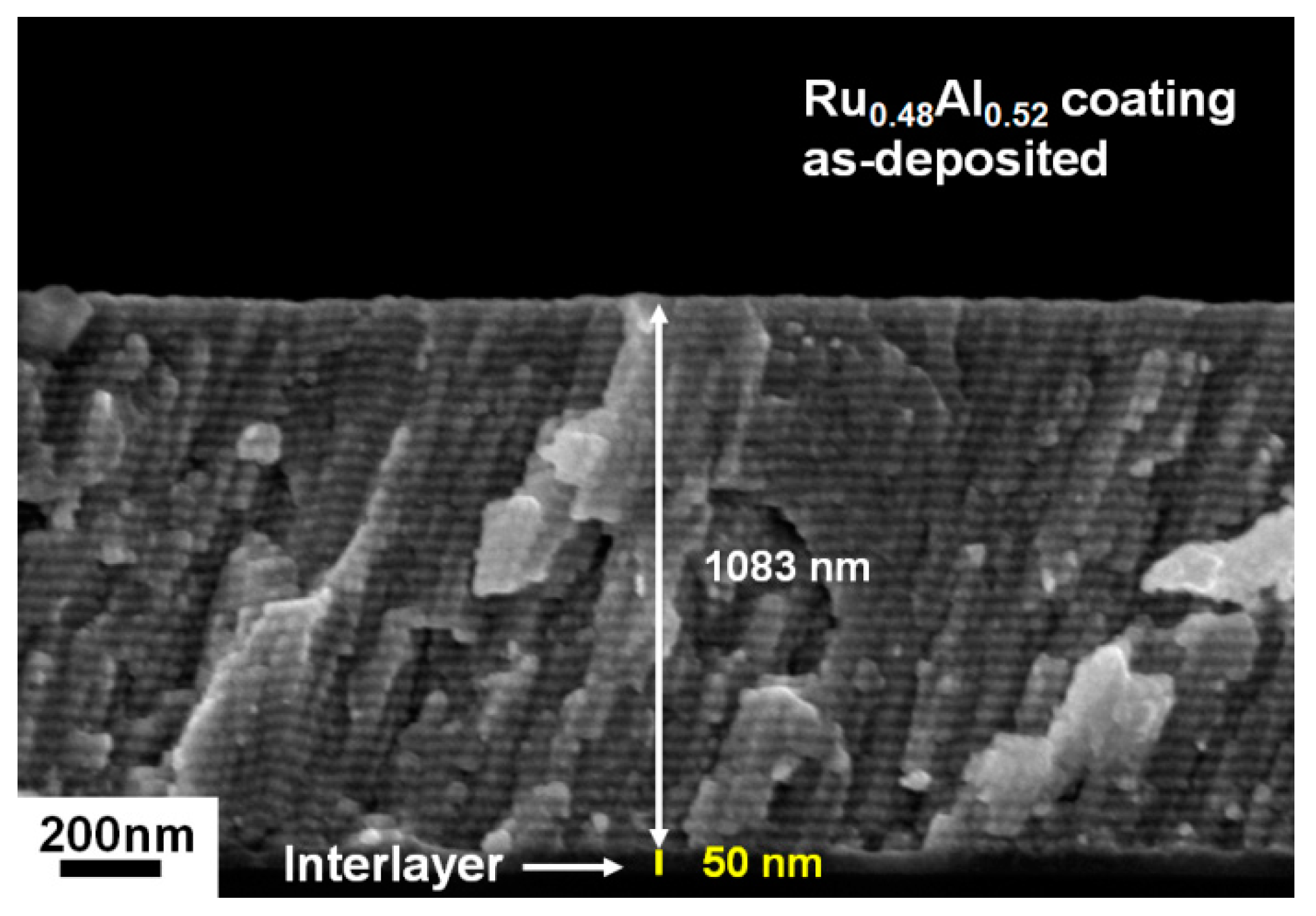
Table 1 lists the chemical compositions of the as-deposited Ru–Al thin films that were prepared on Si substrates using various sputter powers and a substrate holder rotation speed of 1 rpm. The thin films are denoted in the table as Ru0.89Al0.11, Ru0.63Al0.37, and Ru0.48Al0.52. Figure 2 presents the XRD patterns of the as-deposited Ru–Al thin films. The Ru0.89Al0.11 thin films exhibited a hexagonal Ru [ICDD 00-006-0663] phase, whereas the Ru0.63Al0.37 and Ru0.48Al0.52 thin films exhibited a mixture of cubic RuAl [ICDD 00-029-1404] and Ru phases. The peaks at the two-theta angle of approximately 52° were caused by the Si substrate [26]. The reflections of the Ti interlayers were not observed because they were of low intensity. Figure 3 depicts the XRD pattern of the Ru0.48Al0.52/Ti/Si samples that were captured using a Bragg–Brentano scan. The scan indicated RuAl and Ru phases accompanied by a Ti phase. Figure 4 presents a cross-sectional SEM image of the as-deposited Ru0.48Al0.52 thin films. The films exhibited a columnar and multilayered structure due to cyclical gradient concentration deposition. The thickness of the Ru0.48Al0.52 thin film was 1083 nm. Because the number of revolutions of the substrate holder was 35, the multilayered structure of the Ru0.48Al0.52 thin film had a stacking period of 31 nm. The Ru0.89Al0.11 and Ru0.63Al0.37 thin films both exhibited stacking periods of 37 nm (Table 1).
3.2. Ru0.48Al0.52 Thin Films Annealed in 1% O2–99% Ar at 600 °C
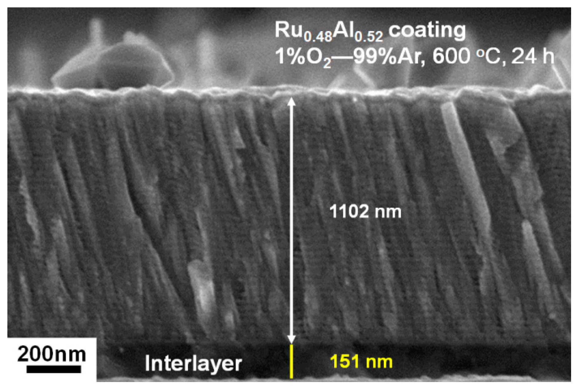
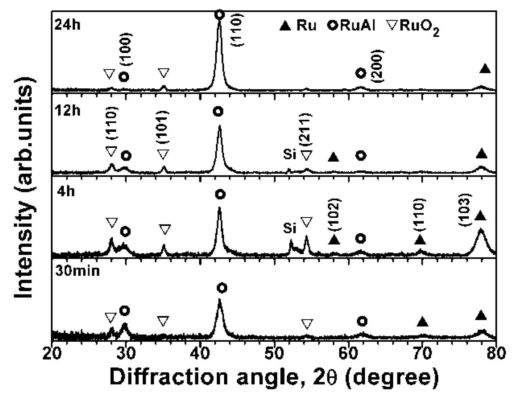
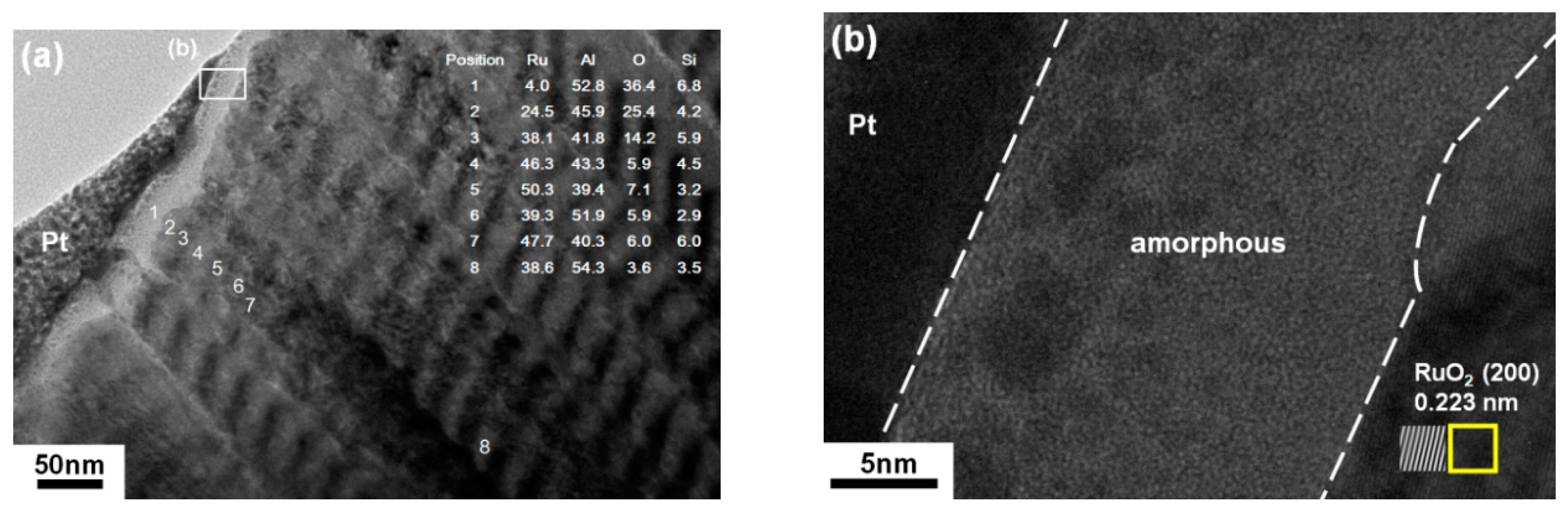
Figure 5 presents a cross-sectional SEM image of the Ru0.48Al0.52 thin films that were annealed in 1% O2–99% Ar at 600 °C for 24 h. The surface oxide scales were not evident and the laminated layers were maintained. The thickness of the films increased slightly from 1083 to 1102 nm, whereas the Ti interlayer increased from 50 to 151 nm, implying the interdiffusion of Ti and Si. Figure 6 exhibits the XRD patterns of the annealed Ru0.48Al0.52 thin films. RuO2 reflections [ICDD 00-040-1290] were observed after annealing for 30 min. Ru and RuAl phases were observed even after annealing for up to 24 h. Figure 7a exhibits the cross-sectional TEM image of the 24 h-annealed Ru0.48Al0.52 thin films, in which the multilayered structure was maintained. The EDS results qualitatively indicated that the surface scale was Al-oxide (Position 1), and the O content exhibited a higher level of 14–25 at.% for the first stacking periods (Positions 2 and 3). The columnar structure had a width of 50 nm. The original Ru toplayer disappeared; this may be attributed to the oxidation of Ru to the higher valance states of RuO3 or RuO4, which are volatile [20,27,28]. Because the standard Gibbs free energies of RuO3 and RuO4 at 600 °C are −16.908 and−28.003 kJ/(mol of O2) [29], respectively, and the atmosphere was constructed by constantly flowing O2–Ar mixed gases into a tube furnace, the formation of these volatile oxides was possible. In a previous study [22], partial Re atoms in IrRe films formed volatile Re2O7 and escaped after annealing in 1% O2–99% Ar at 600 °C for 500 min. The EDS results also indicated that Positions 5 and 7 were Ru-enriched black sublayers, whereas Positions 4, 6, and 8 were Al-enriched gray sublayers. The columnar boundaries may have provided oxygen diffusion paths [21,30] in the early oxidation stage. High-resolution TEM imaging indicated that the surface Al-oxide scale was amorphous, and the lattice fringes of RuO2 were observed beneath the surface oxide layer (Figure 7b). The amorphous Al-oxide sublayer restricted oxidation at 600 °C in 1% O2–99% Ar. The oxidation of the Ru0.48Al0.52 thin films at 600 °C in 1% O2–99% Ar was similar to that of the Ru0.63Al0.37 thin films that were reported previously [21]. The oxidation depth of Ru0.63Al0.37 thin films after annealing for 24 h was approximately the outmost two stacking periods. By contrast, the Ru0.89Al0.11 thin films detached after they were annealed in 1% O2–99% Ar at 600 °C for 30 min. Because the as-deposited Ru0.89Al0.11, Ru0.63Al0.37, and Ru0.48Al0.52 thin films exhibited similar residual stress levels of 1.57 ± 0.16, 1.43 ± 0.16, and 1.61 ± 0.29 GPa, respectively, the detachment of the Ru0.89Al0.11 films was attributed to a high oxide–metal–volume ratio of 2.32 for RuO2/Ru. This value was determined using an XRD database.
3.3. Ru–Al Thin Films Annealed in Air at 800 °C
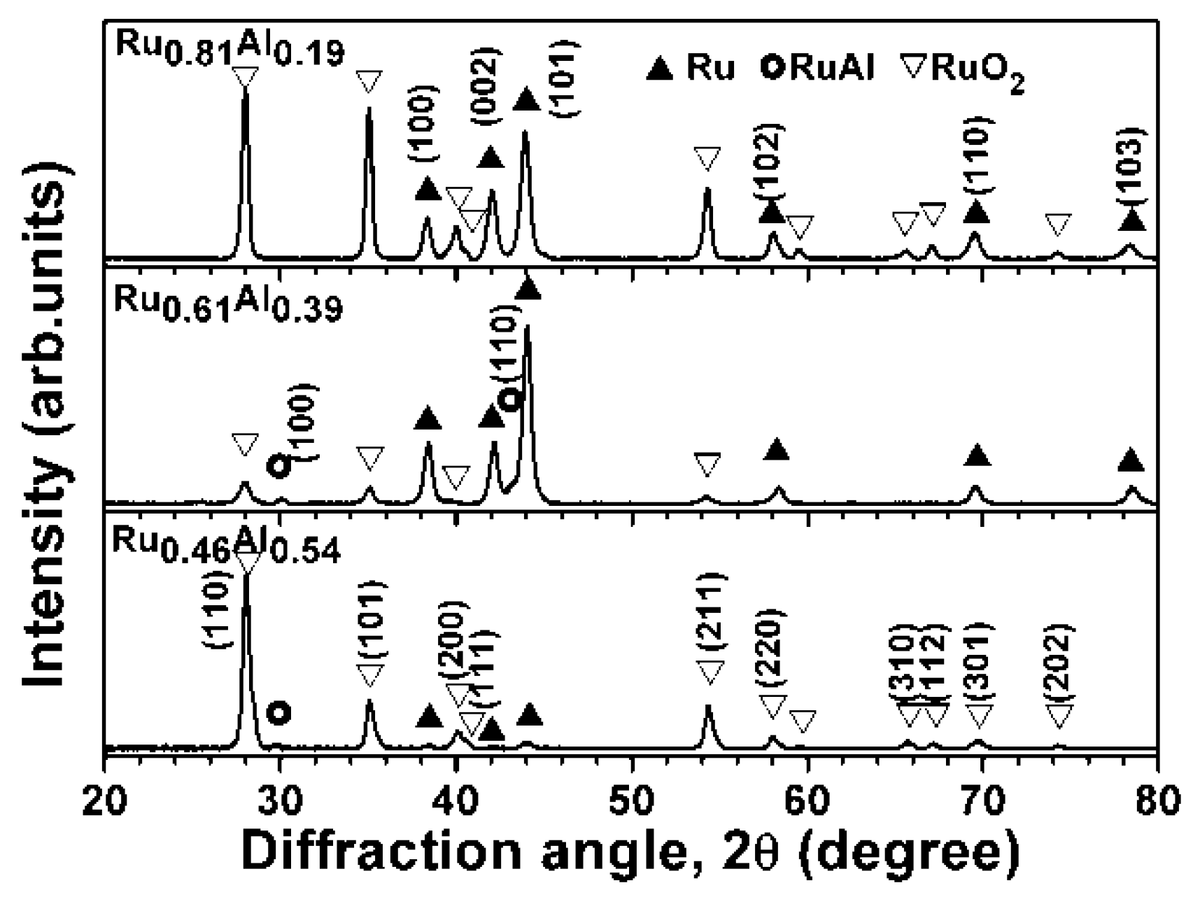
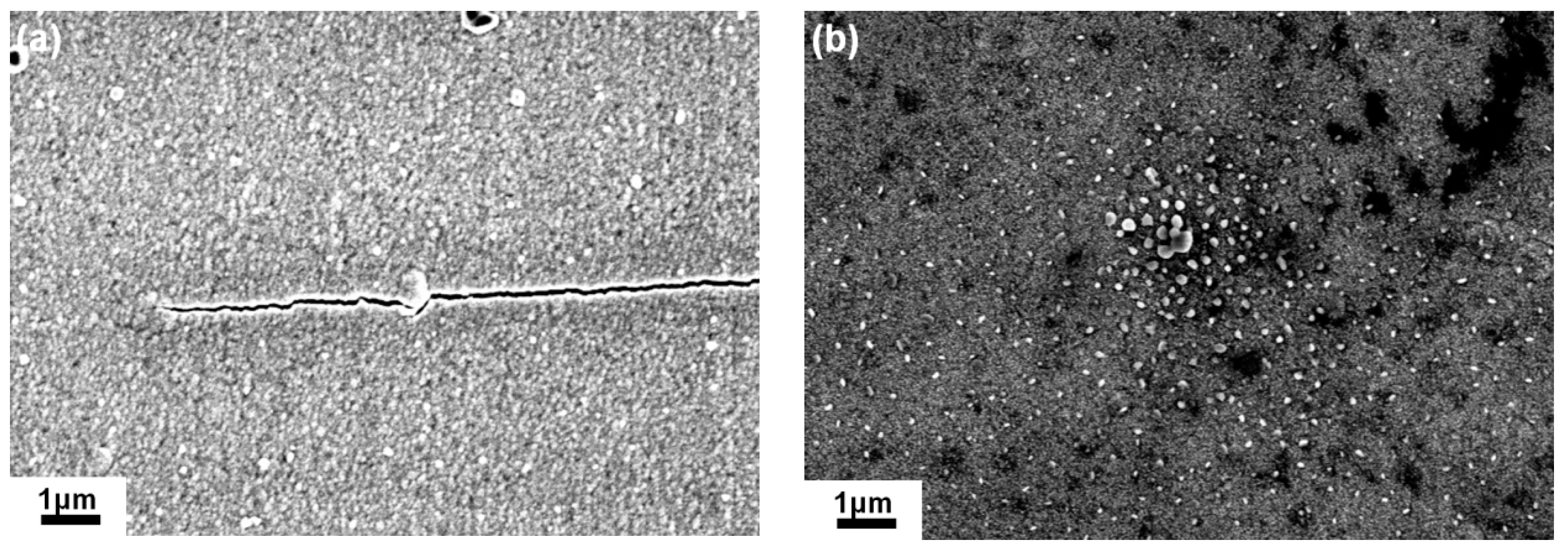
Table 2 presents the chemical compositions of the Ru–Al thin films deposited on Inconel 617 substrates with a Ru interlayer. These thin films were denoted as Ru0.81Al0.19, Ru0.61Al0.39, and Ru0.46Al0.54. Figure 8 depicts XRD patterns of the Ru–Al thin films that were prepared on Inconel 617 substrates with a Ru interlayer. These patterns were similar to those of the Ru–Al thin films prepared on Si substrates with a Ti interlayer (Figure 2). The Ru0.81Al0.19 thin films exhibited a Ru phase, and the Ru0.61Al0.39 and Ru0.46Al0.54 thin films exhibited a mixture of RuAl and Ru phases. Because both the Ti and Ru interlayers have hexagonal phases, the crystalline phases of Ru–Al thin films deposited on the two interlayers were the same. The reflections of the Inconel 617 substrates were not observed due to low intensity and overlapping with reflections of Ru and RuAl phases. The XRD patterns of an Inconel 617 substrate and the Ru0.46Al0.54/Ru/Inconel samples under a Bragg–Brentano scan are presented in Figure 3. Figure 9 exhibits the XRD patterns of the Ru–Al thin films after they were annealed in air at 800 °C for 30 min. Ru and RuO2 dominated the crystalline phases, whereas RuAl became a minor phase. No Al2O3 reflections were evident; however, Al should be preferentially oxidized, implying that the Al-oxide should be X-ray amorphous. Part of the annealed Ru0.81Al0.19 thin films detached after annealing. This phenomenon was similar to that of the 1% O2–99% Ar, 600 °C, and 30 min-annealed Ru0.89Al0.11 thin films prepared on Si substrates. Figure 10 illustrates the surface morphologies of the Ru0.61Al0.39 and Ru0.46Al0.54 thin films after they were annealed in air at 800 °C for 30 min. No spallation was evident, but cracks and small granular oxide particles were observed on the surface of the annealed Ru0.61Al0.39 films, whereas the annealed Ru0.46Al0.54 films only exhibited oxide particles.
Figure 11 presents the cross-sectional TEM image of the Ru0.61Al0.39 thin films after annealing in air at 800 °C for 30 min. The image indicated a reaction region of 373 nm, an unoxidized thin film of 1094 nm, and an interlayer of 207 nm. The reaction region was restricted to the seven outermost stacking periods of the multilayered structure, which consisted of an oxidized region (five stacking periods) and a black region (two stacking periods). The EDS results at Positions 1 and 2 indicated a Ru–Al–O region that was attributed to the inward diffusion of O and the formation of nonprotective oxide accompanied by ruthenium oxide evaporation and crack development [16,31]. The EDS results indicated that the oxidation front (Position 3) consisted of Al-oxide, whereas a black region beneath the oxide scale exhibited a Ru-enriched composition (Position 4), which was attributed to the outward diffusion of Al at high temperatures [18,31,32]. Therefore, the EDS analysis on Position 3 indicated high Al content. The unoxidized thin film comprised large grains across several stacking periods. The EDS results indicated that elements Ni, Cr, Co, and Mo diffused from Inconel 617 across the Ru interlayer and into the Ru0.61Al0.39 thin films. Figure 12 presents the cross-sectional TEM image of the Ru0.46Al0.54 thin films after annealing in air at 800 °C for 30 min. The image shows an oxide scale of 366 nm and an unoxidized thin film of 965 nm. The oxide scale exhibited a high O level of 46–55 at.% at Positions 1–3, whereas Position 4 beneath the oxide scale exhibited a low O level of 3 at.%. A continuous Al-oxide sublayer formed at the original fifth laminated period (Position 3) and restricted the subsequent inward diffusion of O. The EDS results indicated that the Al/(Ru + Al) ratios at Positions 4–6 remained at 0.53–0.58, which was closed to the corresponding values of the as-deposited Ru0.46Al0.54 thin films. Additionally, four periods that were close to the thin-film/interlayer interface surrounding Position 6 thickened, which was attributed to the outward diffusion of Ni, Cr, and Co from Inconel 617, as identified by EDS.
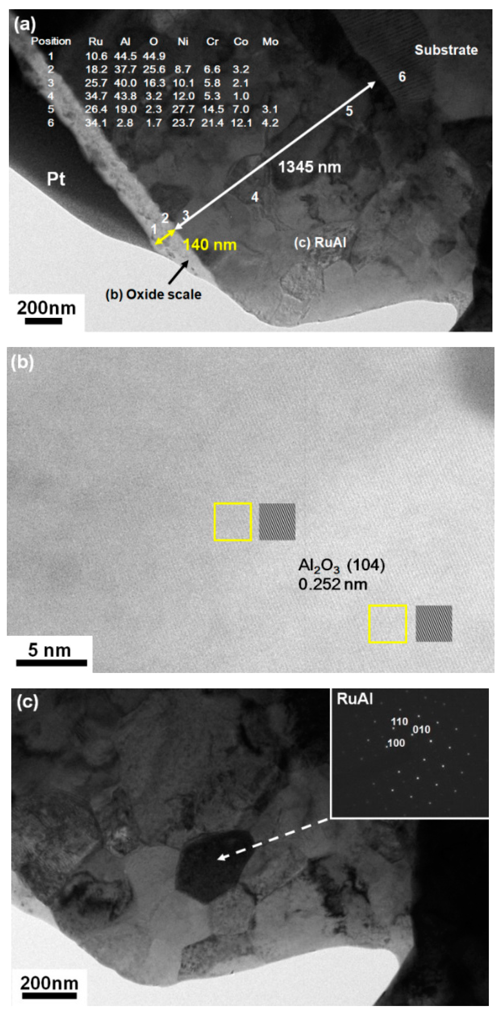
Figure 13 presents the XRD patterns of the Ru0.46Al0.54/Ru/Inconel 617 sample after annealing in air at 800 °C for 4 h. The patterns exhibited reflections of RuAl, RuO2, γ-Al2O3 [ICDD 00-050-0741], and α-Al2O3 [ICDD 00-046-1212] phases. Figure 14a depicts the cross-sectional TEM image of the Ru0.46Al0.54 thin films after annealing in air at 800 °C for 4 h; this image exhibited an oxide scale of 140 nm and an inner part of 1345 nm. The inner part comprised an interdiffused film, interlayer, and substrate. The oxide scale exhibited a high O level of 45 at.% at Position 1, for which a high-resolution TEM image exhibited lattice fringes of crystalline α-Al2O3 (Figure 14b). The depth of the oxide scale of the 4 h-annealed Ru0.46Al0.54 thin films appeared to be less than that of the 30 min-annealed films (Figure 12), which implied that the outmost oxidized part of the films volatilized during further oxidation, and only the Al2O3-dominant oxide scale remained. Figure 14c represents the RuAl grains of 200 nm that were beneath the oxide scale.
4. Conclusions
Ru–Al multilayered thin films that were stacked with cyclical gradient concentration were fabricated through cosputtering. The oxidation depth of Ru0.48Al0.52 thin films that were annealed in a 1% O2–99% Ar atmosphere at 600 °C for 24 h was restricted at the outmost two stacking periods, which behaved similar to that of the Ru0.63Al0.37 thin films and was attributed to the formation of amorphous Al-oxide sublayers restricting the inward diffusion of O. Additionally, the oxidation fronts to five stacking periods of the Ru0.46Al0.54 and Ru0.61Al0.39 thin films after they were annealed in air at 800 °C for 30 min. The Al-oxide sublayers remained amorphous. In the interior part of the Ru0.61Al0.39 films, (i.e., the unoxidized portions) the multilayered structure transformed into large grains, accompanied by outward diffusion of Inconel 617 substrate elements. By contrast, the unoxidized part of the Ru0.46Al0.54 films maintained its multilayer structure after 30 min of annealing, and the diffusion of elements from the substrate was limited. An extending annealing time of 4 h resulted in the formation of a crystalline α-Al2O3-dominated oxide scale on the surface of the Ru0.46Al0.54 films, and the original Ru on the surface region formed volatile oxides. Additionally, beneath the α-Al2O3 oxide scale, the structure transformed into large grains of RuAl phase accompanied by the outward diffusion of substrate elements. Modification of the Ru–Al multilayered thin films by introducing a third element to form a diffusion barrier for constitutive elements of Inconel 617 is a major concern that should be addressed in future research.
Author Contributions
Conceptualization, Project Administration, and Writing-Original Draft Preparation, Y.-I.C.; Investigation, Z.-T.Z. and J.-W.J.
Funding
This research was funded by the Ministry of Science and Technology, Taiwan. The grant number is 106-2221-E-019-022-MY3.
Acknowledgments
The support of Inconel 617 materials from Wu Kai at National Taiwan Ocean University is greatly acknowledged.
Conflicts of Interest
The authors declare no conflict of interest.
References
- Rahman, M.d.S.; Priyadarshan, G.; Raja, K.S.; Nesbitt, C.; Misra, M. Characterization of high temperature deformation behavior of INCONEL 617. Mech. Mater. 2009, 41, 261–270. [Google Scholar] [CrossRef]
- Gariboldi, E.; Cabibbo, M.; Spigarelli, S.; Ripamonti, D. Investigation on precipitation phenomena of Ni–22Cr–12Co–9Mo alloy aged and crept at high temperature. Int. J. Press. Vessel. Pip. 2008, 85, 63–71. [Google Scholar] [CrossRef]
- DeMasi-Marcin, J.T.; Gupta, D.K. Protective coatings in the gas turbine engine. Surf. Coat. Technol. 1994, 68–69, 1–9. [Google Scholar] [CrossRef]
- Vaßen, R.; Jarligo, M.O.; Steinke, T.; Mack, D.E.; Stöver, D. Overview on advanced thermal barrier coatings. Surf. Coat. Technol. 2010, 205, 938–942. [Google Scholar] [CrossRef]
- Miller, R.A. Current status of thermal barrier coatings—An overview. Surf. Coat. Technol. 1987, 30, 1–11. [Google Scholar] [CrossRef]
- Evans, A.G.; Mumm, D.R.; Hutchinson, J.W.; Meier, G.H.; Pettit, F.S. Mechanisms controlling the durability of thermal barrier coatings. Prog. Mater. Sci. 2001, 46, 505–553. [Google Scholar] [CrossRef]
- Aygun, A.; Vasiliev, A.L.; Padture, N.P.; Ma, X. Novel thermal barrier coatings that are resistant to high-temperature attack by glassy deposits. Acta Mater. 2007, 55, 6734–6745. [Google Scholar] [CrossRef]
- Huang, J.; Wang, W.; Lu, X.; Hu, D.; Feng, Z.; Guo, T. Effect of particle size on the thermal shock resistance of plasma-sprayed YSZ coatings. Coatings 2017, 7, 150. [Google Scholar] [CrossRef]
- Hu, N.; Khan, M.; Wang, Y.; Song, X.; Lin, C.; Chang, C.; Zeng, Y. Effect of Microstructure on the Thermal Conductivity of Plasma Sprayed Y2O3 Stabilized Zirconia (8% YSZ). Coatings 2017, 7, 198. [Google Scholar] [CrossRef]
- Mücklich, F.; Ilić, N. RuAl and its alloys. Part I. Structure physical properties, microstructure and processing. Intermetallics 2005, 13, 5–21. [Google Scholar] [CrossRef]
- Mücklich, F.; Ilić, N.; Wol, K. RuAl and its alloys, part II: Mechanical properties, environmental resistance and applications. Intermetallics 2008, 16, 593–608. [Google Scholar] [CrossRef]
- Tryon, B.; Pollock, T.M.; Gigliotti, M.F.X.; Hemker, K. Thermal expansion behavior of ruthenium aluminides. Scr. Mater. 2014, 50, 845–848. [Google Scholar] [CrossRef]
- Tryon, B.; Feng, Q.; Wellman, R.G.; Murphy, K.S.; Yang, J.; Levi, C.G.; Nicholls, J.R.; Pollock, T.M. Multilayered ruthenium-modified bond coats for thermal barrier coatings. Metall. Mater. Trans. A 2006, 37A, 3347–3358. [Google Scholar] [CrossRef]
- Wang, Y.; Guo, H.B.; Peng, H.; Peng, L.Q.; Gong, S.K. Diffusion barrier behaviors of (Ru,Ni)Al/NiAl coatings on Ni-based superalloy substrate. Intermetallics 2011, 19, 191–195. [Google Scholar] [CrossRef]
- Guitar, M.A.; Woll, K.; Ramos-Moore, E.; Mücklich, F. Study of grain growth and thermal stability of nanocrystalline RuAl thin films deposited by magnetron sputtering. Thin Solid Films 2013, 527, 1–8. [Google Scholar] [CrossRef]
- Guitar, M.A.; Ramos-Moore, E.; Mücklich, F. The influence of impurities on the formation of protective aluminum oxides on RuAl thin films. J. Alloys Compd. 2014, 594, 165–170. [Google Scholar] [CrossRef]
- Seifert, M.; Menzel, S.B.; Rane, G.K.; Hoffmann, M.; Gemming, T. RuAl thin films on high–temperature piezoelectric substrates. Mater. Res. Express 2015, 2, 085001. [Google Scholar] [CrossRef]
- Seifert, M.; Rane, G.K.; Menzel, S.B.; Gemming, T. TEM studies on the changes of the composition in LGS and CTGS substrates covered with a RuAl metallization and on the phase formation within the RuAl film after heat treatment at 600 and 800 °C. J. Alloys Compd. 2016, 664, 510–517. [Google Scholar] [CrossRef]
- Seifert, M.; Rane, G.K.; Menzel, S.B.; Gemming, T. The influence of barrier layers (SiO2, Al2O3, W) on the phase formation and stability of RuAl thin films on LGS and CTGS substrates for surface acoustic wave technology. J. Alloys Compd. 2016, 688, 228–240. [Google Scholar] [CrossRef]
- Seifert, M.; Rane, G.K.; Oswald, S.; Menzel, S.B.; Gemming, T. The influence of the composition of Ru100–xAlx (x = 50, 55, 60, 67) thin films on their thermal stability. Materials 2017, 10, 277. [Google Scholar] [CrossRef] [PubMed]
- Chen, Y.I.; Zheng, Z.T.; Kai, W.; Huang, Y.R. Oxidation behavior of Ru–Al multilayer coatings. Appl. Surf. Sci. 2017, 406, 1–7. [Google Scholar] [CrossRef]
- Liu, S.C.; Chen, Y.I.; Tsai, H.Y.; Lin, K.C.; Chen, Y.H. Thermal stability of Ir–Re coatings annealed in oxygen-containing atmospheres. Surf. Coat. Technol. 2013, 237, 105–111. [Google Scholar] [CrossRef]
- Chen, Y.I. Laminated structure in internally oxidized Ru–Ta coatings. Thin Solid Films 2012, 524, 205–210. [Google Scholar] [CrossRef]
- Chen, Y.I.; Lu, T.S.; Zheng, Z.T. Internally oxidized Ru–Zr multilayer coatings. Coatings 2017, 7, 46. [Google Scholar] [CrossRef]
- Janssen, G.C.A.M.; Abdalla, M.M.; van Keulen, F.; Pujada, B.R.; van Venrooy, B. Celebrating the 100th anniversary of the Stoney equation for film stress: Developments from polycrystalline steel strips to single crystal silicon wafers. Thin Solid Films 2009, 517, 1858–1867. [Google Scholar] [CrossRef]
- Deng, Y.L.; Lee, J.W.; Lou, B.S.; Duh, J.G.; Chu, J.P.; Jang, J.S.C. The fabrication and property evaluation of Zr–Ti–B–Si thin film metallic glass materials. Surf. Coat. Technol. 2014, 259, 115–122. [Google Scholar] [CrossRef]
- Bell, W.E.; Tagami, M. High-temperature chemistry of the ruthenium–oxygen system. J. Phys. Chem. 1963, 67, 2432–2436. [Google Scholar] [CrossRef]
- Huang, J.H.; Chen, J.S. Material characteristics and electrical property of reactively sputtered RuO thin films. Thin Solid Films 2001, 382, 139–145. [Google Scholar] [CrossRef]
- Barin, I. Thermochemical Data of Pure Substances, 3rd ed.; VCH: New York, NY, USA, 1995. [Google Scholar]
- Chen, Y.I.; Chu, H.N.; Kai, W. Internal oxidation of laminated Nb–Ru coatings. Appl. Surf. Sci. 2016, 389, 477–483. [Google Scholar] [CrossRef]
- Bellina, P.J.; Catanoiu, A.; Morales, F.M.; Rühle, M. Formation of discontinuous Al2O3 layers during high-temperature oxidation of RuAl alloys. J. Mater. Res. 2006, 21, 276–286. [Google Scholar] [CrossRef]
- Soldera, F.; Ilić, N.; Brännström, S.; Barrientos, I.; Gobran, H.; Mücklich, F. Formation of Al2O3 scales on single-phase RuAl produced by reactive sintering. Oxid. Met. 2003, 59, 529–542. [Google Scholar] [CrossRef]
Figure 1.
(a) Schematic of the cosputtering equipment and (b) substrate holder and sample positions related to sputter targets.
Figure 1.
(a) Schematic of the cosputtering equipment and (b) substrate holder and sample positions related to sputter targets.
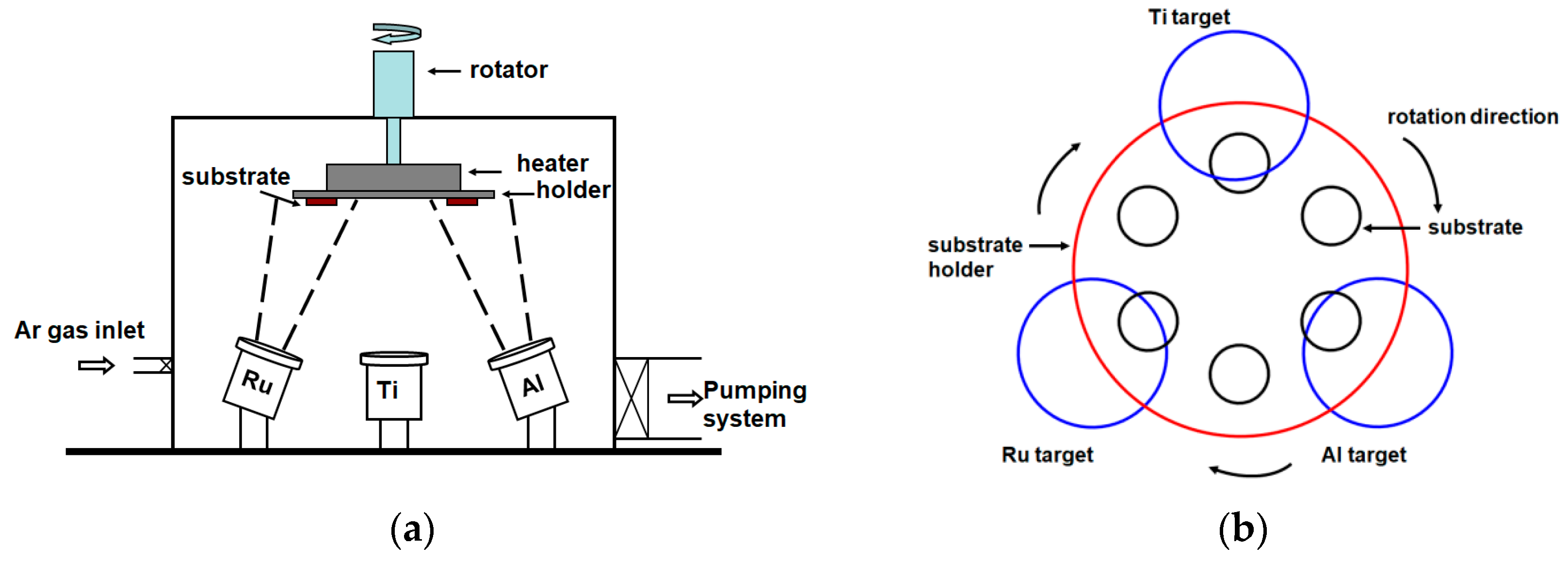
Figure 2.
X-ray diffractometer (XRD) patterns of as-deposited Ru–Al thin films.
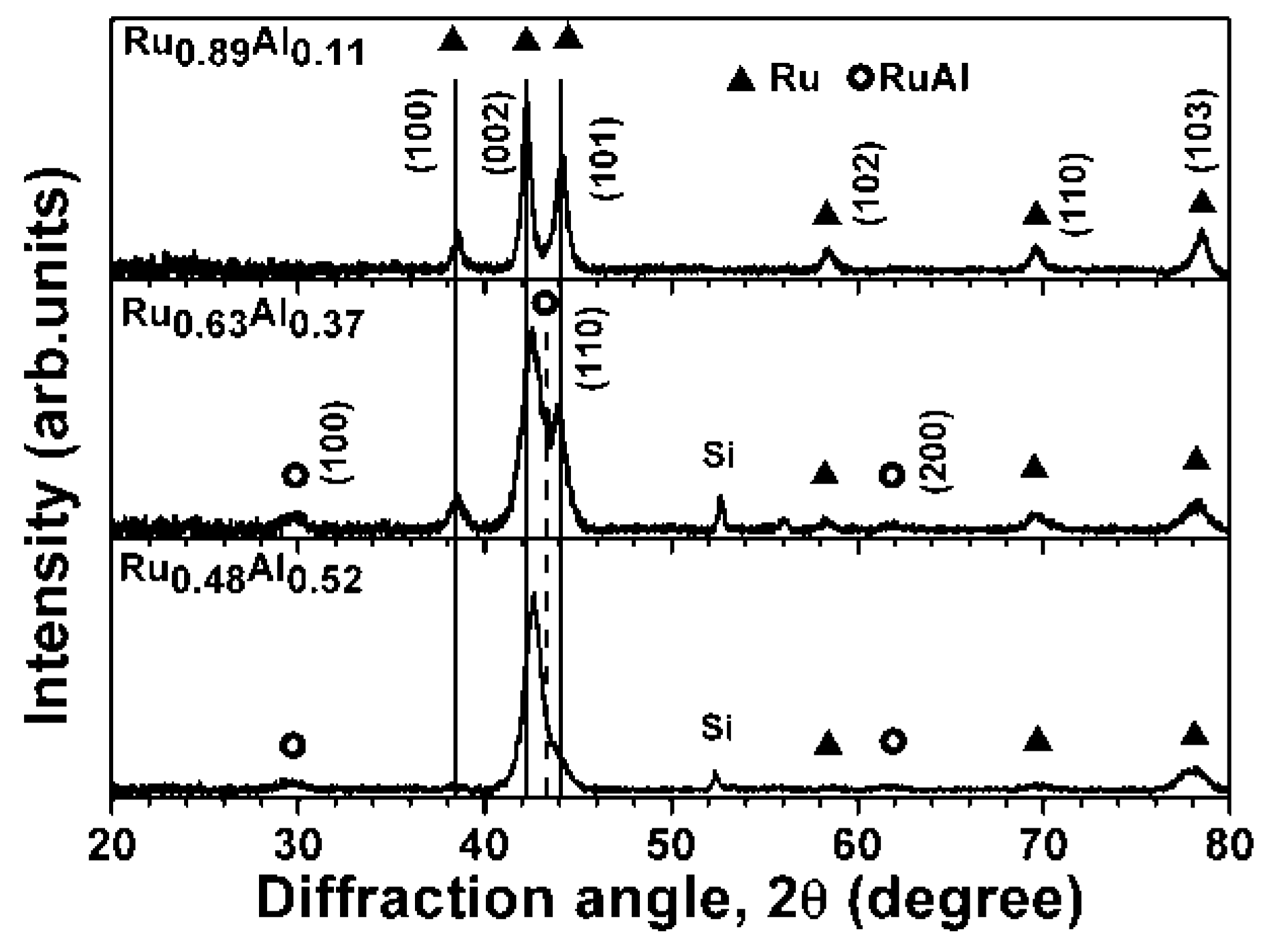
Figure 3.
Bragg–Brentano scan of XRD patterns from the Inconel substrate, Ru0.46Al0.54/Ru/Inconel, and Ru0.48Al0.52/Ti/Si samples.
Figure 3.
Bragg–Brentano scan of XRD patterns from the Inconel substrate, Ru0.46Al0.54/Ru/Inconel, and Ru0.48Al0.52/Ti/Si samples.

Figure 4.
Cross-sectional SEM image of the as-deposited Ru0.48Al0.52 thin films.

Figure 5.
Cross-sectional SEM image of the Ru0.48Al0.52 thin films annealed in 1% O2–99% Ar at 600 °C for 24 h.
Figure 5.
Cross-sectional SEM image of the Ru0.48Al0.52 thin films annealed in 1% O2–99% Ar at 600 °C for 24 h.

Figure 6.
XRD patterns of the Ru0.48Al0.52 thin films annealed in 1% O2–99% Ar at 600 °C for 0.5–24 h.
Figure 6.
XRD patterns of the Ru0.48Al0.52 thin films annealed in 1% O2–99% Ar at 600 °C for 0.5–24 h.

Figure 7.
(a) Cross-sectional transmission electron microscopy (TEM) image and (b) high-resolution TEM image of the Ru0.48Al0.52 thin films annealed in 1% O2–99% Ar at 600 °C for 24 h.
Figure 7.
(a) Cross-sectional transmission electron microscopy (TEM) image and (b) high-resolution TEM image of the Ru0.48Al0.52 thin films annealed in 1% O2–99% Ar at 600 °C for 24 h.

Figure 8.
XRD patterns of the as-deposited Ru–Al thin films prepared on Inconel 617 substrates with a Ru interlayer.
Figure 8.
XRD patterns of the as-deposited Ru–Al thin films prepared on Inconel 617 substrates with a Ru interlayer.

Figure 9.
XRD patterns of the Ru–Al thin films prepared on Inconel 617 substrates with a Ru interlayer and annealed in air at 800 °C for 30 min.
Figure 9.
XRD patterns of the Ru–Al thin films prepared on Inconel 617 substrates with a Ru interlayer and annealed in air at 800 °C for 30 min.

Figure 10.
Surface morphologies of the (a) Ru0.61Al0.39 and (b) Ru0.46Al0.54 thin films after they were annealed in air at 800 °C for 30 min.
Figure 10.
Surface morphologies of the (a) Ru0.61Al0.39 and (b) Ru0.46Al0.54 thin films after they were annealed in air at 800 °C for 30 min.

Figure 11.
Cross-sectional TEM image and energy dispersive spectrometry (EDS) results for the Ru0.61Al0.39/Ru/Inconel 617 sample after annealing in air at 800 °C for 30 min.
Figure 11.
Cross-sectional TEM image and energy dispersive spectrometry (EDS) results for the Ru0.61Al0.39/Ru/Inconel 617 sample after annealing in air at 800 °C for 30 min.
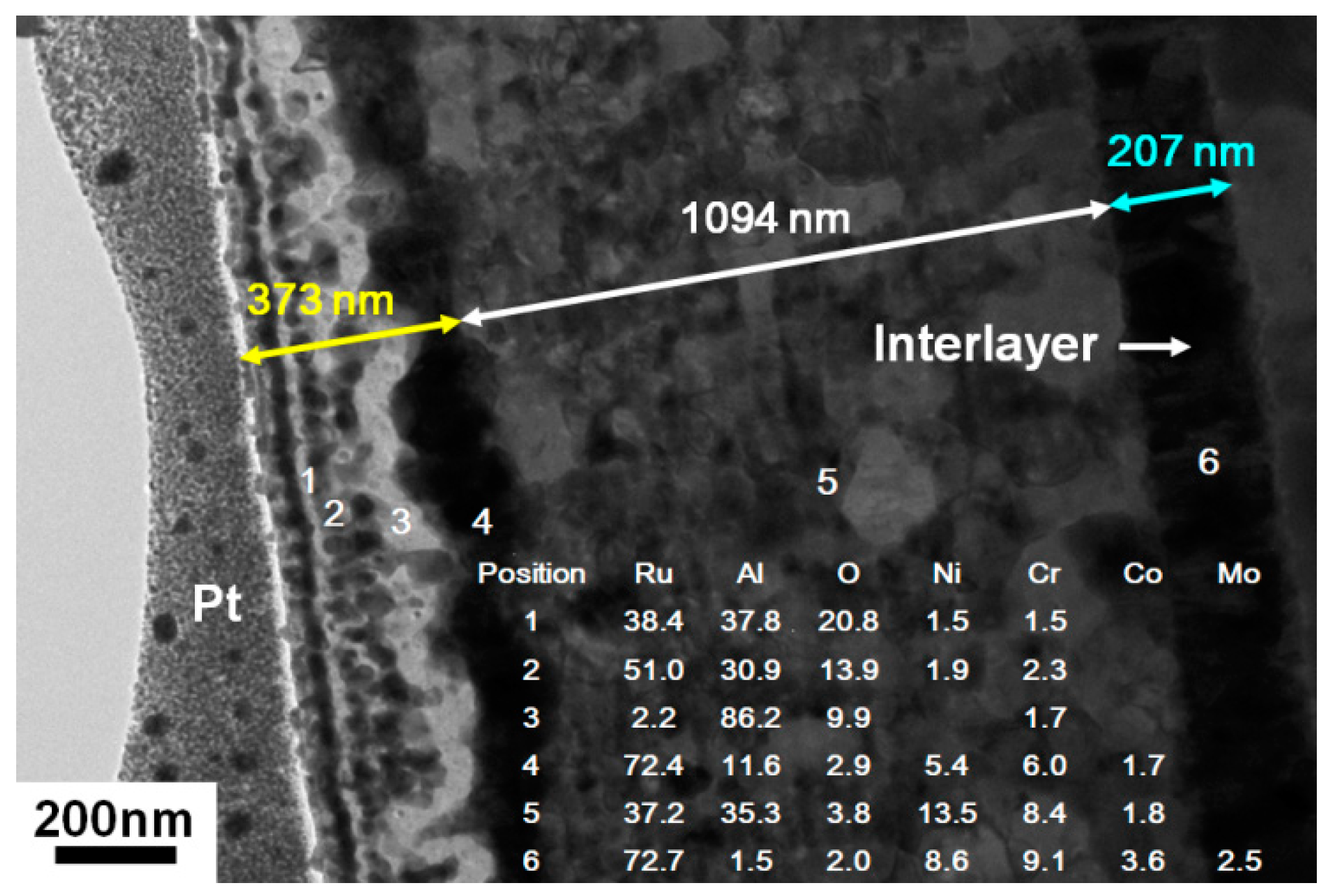
Figure 12.
Cross-sectional TEM image and EDS results for the Ru0.46Al0.54/Ru/Inconel 617 sample after annealing in air at 800 °C for 30 min.
Figure 12.
Cross-sectional TEM image and EDS results for the Ru0.46Al0.54/Ru/Inconel 617 sample after annealing in air at 800 °C for 30 min.
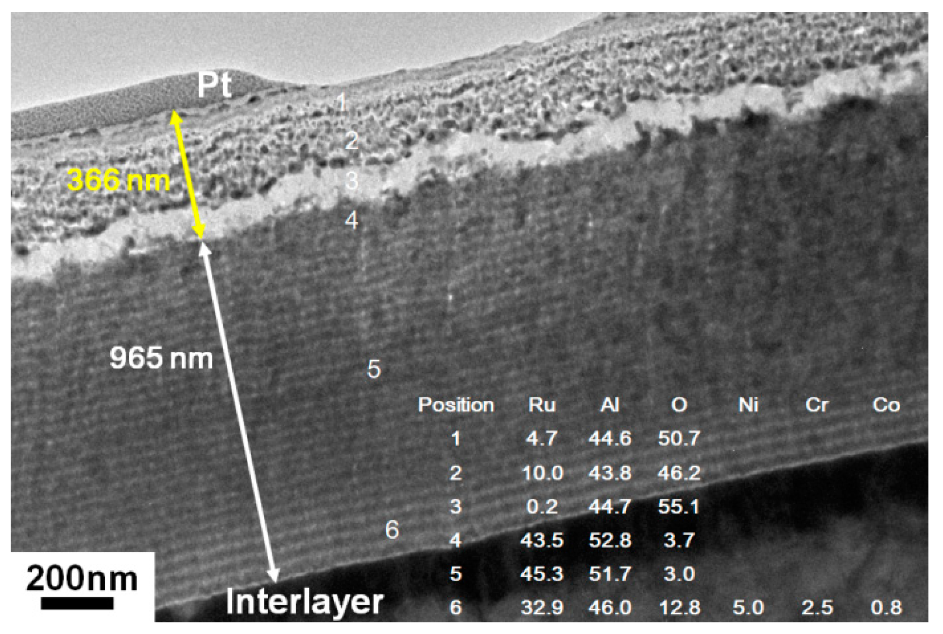
Figure 13.
XRD pattern of the Ru0.46Al0.54/Ru/Inconel 617 sample after annealing in air at 800 °C for 4 h.
Figure 13.
XRD pattern of the Ru0.46Al0.54/Ru/Inconel 617 sample after annealing in air at 800 °C for 4 h.

Figure 14.
(a) Cross-sectional TEM image of the Ru0.46Al0.54 thin films annealed in air at 800 °C for 4 h, (b) high-resolution TEM image of the surface oxide scale, and (c) a RuAl grain beneath the oxide scale.
Figure 14.
(a) Cross-sectional TEM image of the Ru0.46Al0.54 thin films annealed in air at 800 °C for 4 h, (b) high-resolution TEM image of the surface oxide scale, and (c) a RuAl grain beneath the oxide scale.


Table 1.
Chemical compositions, thicknesses, and stacking periods of the as-deposited Ru–Al thin films.
Table 1.
Chemical compositions, thicknesses, and stacking periods of the as-deposited Ru–Al thin films.
| Sample | Sputter Power (W) | Chemical Composition (at.%) | Thickness (nm) | Period | ||||
|---|---|---|---|---|---|---|---|---|
| WRu | WAl | Ru | Al | O | Film | Interlayer | (nm) | |
| Ru0.89Al0.11 | 200 | 100 | 86.69 ± 0.39 | 10.89 ± 0.02 | 2.42 ± 0.37 | 1305 | 50 | 37 |
| Ru0.63Al0.37 | 150 | 150 | 59.33 ± 0.32 | 34.75 ± 0.32 | 5.92 ± 0.03 | 1312 | 50 | 37 |
| Ru0.48Al0.52 | 100 | 200 | 47.35 ± 0.39 | 52.06 ± 0.34 | 0.59 ± 0.14 | 1083 | 50 | 31 |
Table 2.
Chemical compositions of Ru–Al thin films deposited on Inconel 617 with a Ru interlayer.
| Sample | Sputter Power (W) | Chemical Composition (at.%) | |||
|---|---|---|---|---|---|
| WRu | WAl | Ru | Al | O | |
| as-deposited | |||||
| Ru0.81Al0.19 | 200 | 100 | 80.02 ± 0.58 | 19.16 ± 0.24 | 0.82 ± 0.78 |
| Ru0.61Al0.39 | 150 | 150 | 57.16 ± 0.20 | 37.16 ± 0.06 | 5.68 ± 0.15 |
| Ru0.46Al0.54 | 100 | 200 | 44.80 ± 0.03 | 52.54 ± 0.15 | 2.66 ± 0.13 |
© 2018 by the authors. Licensee MDPI, Basel, Switzerland. This article is an open access article distributed under the terms and conditions of the Creative Commons Attribution (CC BY) license (http://creativecommons.org/licenses/by/4.0/).
Share and Cite
MDPI and ACS Style
Chen, Y.-I.; Zheng, Z.-T.; Jhang, J.-W. Thermal Stability of Ru–Al Multilayered Thin Films on Inconel 617. Metals 2018, 8, 514. https://doi.org/10.3390/met8070514
AMA Style
Chen Y-I, Zheng Z-T, Jhang J-W. Thermal Stability of Ru–Al Multilayered Thin Films on Inconel 617. Metals. 2018; 8(7):514. https://doi.org/10.3390/met8070514
Chicago/Turabian StyleChen, Yung-I, Zhi-Ting Zheng, and Jia-Wei Jhang. 2018. "Thermal Stability of Ru–Al Multilayered Thin Films on Inconel 617" Metals 8, no. 7: 514. https://doi.org/10.3390/met8070514
Note that from the first issue of 2016, this journal uses article numbers instead of page numbers. See further details here.