Article

Influence of Monovalent Cations on the Efficiency of Ferrous Ion Oxidation, Total Iron Precipitation, and Adsorptive Removal of Cr(VI) and As(III) in Simulated Acid Mine Drainage with Inoculation of Acidithiobacillus ferrooxidans

Department of Environmental Engineering, School of Information and Safety Engineering, Zhongnan University of Economics and Law, Wuhan 430073, China
*
Author to whom correspondence should be addressed.
Metals 2018, 8(8), 596; https://doi.org/10.3390/met8080596
Submission received: 21 June 2018 / Revised: 24 July 2018 / Accepted: 24 July 2018 / Published: 31 July 2018

Abstract

:
Acid mine drainage is highly acidic and contains large quantities of Fe and heavy metal elements. Thus, it is important to promote the transformation of Fe into secondary iron minerals that exhibit strong heavy-metal removal abilities. Using simulated acid mine drainage, this work analyzes the influence of monovalent cations (K+, NH4+, and Na+) on the Fe2+ oxidation and total Fe deposition efficiencies, as well as the phases of secondary iron minerals in an Acidithiobacillus ferrooxidans system. It also compares the Cr(VI) (K2Cr2O7) and As(III) (As2O3) removal efficiencies of different schwertmannites. The results indicated that high concentrations of monovalent cations (NH4+ ≥ 320 mmol/L, and Na+ ≥ 1600 mmol/L) inhibited the biological oxidation of Fe2+. Moreover, the mineralizing abilities of the three cations differed (K+ > NH4+ > Na+), with cumulative Fe deposition efficiencies of 58.7%, 28.1%, and 18.6%, respectively [n(M) = 53.3 mmol/L, cultivation time = 96 h]. Additionally, at initial Cr(VI) and As(III) concentrations of 10 and 1 mg/L, respectively, the Cr(VI) and As(III) removal efficiencies exhibited by schwertmannites acquired by the three mineralization systems differed [n(Na) = 53.3 > n(NH4) = 53.3 > n(K) = 0.8 mmol/L]. Overall, the analytical results suggested that the removal efficiency of toxic elements was mainly influenced by the apparent structure, particle size, and specific surface area of schwertmannite.

Graphical Abstract

1. Introduction

Acid mine drainage (AMD) is highly acidic and contains large quantities of Fe ions (Fe2+ and Fe3+) and heavy metal elements [1,2,3,4,5,6]. It not only corrodes the pipes in mine wells and other related equipment, but also heavily pollutes surface water and land resources [7,8]. At present, lime neutralization has been widely adopted owing to its convenience and speed. This process has been reported to raise the pH level and remove Fe and SO42− ions from the AMD system through the formation of Fe(OH)3, Fe(OH)2, and CaSO4. However, this method has serious disadvantages with respect to the higher operating costs and difficulty of dewatering the waste residue [9,10,11]. Therefore, it is of great significance to develop an environmentally-friendly and efficient way to remove Fe ions and toxic elements present in the AMD.
Acidithiobacillus ferrooxidans (A. ferrooxidans), one of the most common acidophilic Fe-oxidizing microorganisms, has been reported to catalyze the oxidation of Fe2+ to Fe3+. During the Fe2+ oxidation process, secondary iron minerals (such as schwertmannite and jarosite) are usually generated due to the facilitated subsequent Fe3+ hydrolysis. Reactions (1)–(3) present the schwertmannite and jarosite formation processes [12,13,14,15]:
Bioreaction: 4Fe2+ + O2 + 4H+ → 4Fe3+ + 2H2O
Chemical reaction 1: 8Fe3+ + 14H2O + SO42− → Fe8O8(OH)6SO4 (schwertmannite) + 22H+
Chemical reaction 2: M+ + 3Fe3+ + 2SO42− + 6H2O → MFe3(SO4)2(OH)6 (jarosite) + 6H+
M+ represents K+, NH4+, Na+, and H3O+. In addition, secondary iron minerals can also be synthesized directly through chemical methods. For example, under acidic conditions, schwertmannite can be obtained by iron salt (such as FeCl3, Fe(NO3)3, Fe2(SO4)3) hydrolysis or ferrous salt oxidation (FeSO4-H2O2) [16,17,18]. Also, jarosite is usually formed from the direct hydrolysis of Fe3+ under high temperature. Generally, schwertmannite and jarosite are commonly found in acidic Fe2+-, Fe3+- and SO42−-rich environments. Previous studies confirm that these minerals are ideal adsorbents, exhibiting relatively strong heavy-metal adsorption and co-precipitation properties [19,20,21]. For example, Liao [22] discovered that schwertmannite can hold or passivate As(III) or As(V) much stronger than other iron oxides (saturated adsorption capacity for As(III) = 114 mg/g). Moreover, Wang et al. [23] determined that jarosite could efficiently adsorb and remove Cr(VI)-bearing drainage (Cr(VI) percentage removal = 70%~85%). In addition, Asta et al. [24] reported that in extremely acidic environments (pH = 1.5~2.5), chemosynthetic jarosite successfully adsorbed As(V) at a capacity of 21 mg/g. These reports suggest that the transformation of Fe to secondary iron minerals in an acid sulfate environment could play an important role in the treatment of AMD.
Usually, the formation of secondary iron minerals is influenced by a variety of factors, including the acidity of the solution, reaction time, reaction temperature, concentration of Fe2+, and the variety or concentration of monovalent cations [25,26]. According to Reactions (1)–(3), the biosynthesis of schwertmannite and jarosite involves a process whereby Fe2+ ions are oxidized to Fe3+ that in turn form Fe8O8(OH)6SO4 or MFe3(SO4)2(OH)6. In this process, the supply rate of Fe3+ as an intermediate product determines the formation of secondary iron minerals. In an acidic system abundant with Fe and SO42−, the total iron precipitation (secondary iron minerals formation) and phases of the secondary iron minerals mediated by A. ferrooxidans are mainly influenced by two factors; the nature of M and the Fe/M ratio. Notably, existing research indicates that a rise in the K+ concentration favors jarosite formation but deters schwertmannite formation [27,28,29]. Bai et al. [30] also studied the biosynthesis of secondary iron minerals in a K+, NH4+, and Na+ coexistence system. However, they only acquired a schwertmannite/jarosite mixture and did not observe the formation of ammonium or sodium jarosite. This result indicated a difference in the ability of these three iron species to catalyze the transformation of Fe into secondary iron minerals.
Nevertheless, it is still unclear how the initial monovalent cations impact the efficiency of ferrous ion oxidation and total iron precipitation (secondary iron minerals formation) in A. ferrooxidans culture solutions. In previous studies, only the influence of K+ concentration has been taken into account [31], and the relative mineralizing ability of K+, NH4+, and Na+, the differences in mineral phases between the secondary iron minerals, and the removal efficiency of the secondary iron minerals with respect to heavy metal elements have not yet been reported.
Therefore, based on the biosynthesis of secondary iron minerals using A. ferrooxidans, the objectives of the present work were to analyze the influence of the K+, NH4+, and Na+ concentration on the Fe2+ oxidation efficiency, total Fe deposition efficiency, and phases of the secondary iron minerals formed. In addition, the study compares Cr(VI) and As(III) removal efficiencies of schwertmannite samples prepared under different monovalent ion conditions. We believe that the afforded results may provide an essential theoretical basis to promote the transformation of Fe to secondary iron minerals and the removal of heavy metal elements from acid sulfate environments.

2. Materials and Methods

2.1. Preparation of A. ferrooxidans Cell Suspensions

A. ferrooxidans LX5 (CGMCC No. 0727), obtained from China General Microbiological Culture Collection Center (CGMCC), was grown in a 9 K medium [32], which contained the following analytical grade salts: 3.00 g (NH4)2SO4, 0.10 g KCl, 0.50 g K2HPO4, 0.50 g MgSO4·7H2O, 0.01 g Ca(NO3)2, and 44.48 g FeSO4·7H2O in 1 L deionized water. The mixture was adjusted to pH = 2.5 using 10 mol/L H2SO4. The FeSO4 solution was filtered, sterilized, and subsequently added to the remaining autoclaved (121 °C for 15 min) medium components. Cultures of A. ferrooxidans were incubated in 500 mL Erlenmeyer flasks (Sinopharm Chemical Reagent Beijing Co., Ltd., Beijing, China), each containing 225 mL of 9 K medium and 10% (v/v) inoculum at 28 °C on a rotary shaker at 180 rev/min. The cells were harvested during the late logarithmic growth phase (~72 h after inoculation). The cultures were initially filtered (Whatman No. 4 filter paper) to remove the precipitate. Subsequently, the filtrates were centrifuged at 10,000× g for 10 min at 4 °C to precipitate the bacterial cells, and the supernatant was discarded. After twofold washing with a dilute H2SO4 solution (pH = 1.5), the cells were resuspended in a dilute H2SO4 solution (pH = 2.5). The A. ferrooxidans numbers were determined as ~3 × 10⁸ CFU/mL by the double-layer plate method [33].

2.2. Influence of Monovalent Cations on the Synthesis of Secondary Iron Minerals

The experiments on Fe2+ oxidation and Fe3+ hydroxysulfate precipitates formation with A. ferrooxidans were conducted in 500 mL conical flasks. Briefly, after the addition of Fe2+ (added as FeSO4·7H2O and designed to 8960 mg/L, Fe2+ concentration = 160 mmol/L) and monovalent cations (K+, NH4+, or Na+, added as K2SO4, (NH4)2SO4, or Na2SO4, respectively), the A. ferrooxidans cell suspension was transferred to the flasks to adjust to a cell density of 5 × 10⁷ CFU/mL. The pH values of the solutions were then changed to 3.0 using 10 mol/L H2SO4, and a fixed final volume of 250 mL was set for all mixtures. The Fe/K molar ratio in solution was designed as 3, 30, 50, and 200 through the alteration of the initial K+ concentration. Likewise, the Fe/M (M = NH4+ or Na+) molar ratio was adjusted to 0.1, 0.5, 1, and 3, respectively, according to the procedures mentioned above. The sample flasks were then incubated on a rotary shaker at 28 °C and 180 rev/min.
During the cultivation process, 1 mL liquid samples were drawn at 24 h intervals and screened using 0.22 μm filter membranes. The Fe2+ and total Fe concentrations were then determined to assess the extent of Fe2+ oxidation and Fe3+ to form the minerals, respectively. After leaving the mixtures to react for 96 h, the precipitates in the solutions were collected by filtering through a Whatman No. 4 filter paper and then washed once with deionized water. The product was then dried at 60 °C to a constant weight and identified by the methods described in Section 2.4. All experiments were performed in triplicate. Given that the abiotic oxidation of Fe2+ can hardly be initiated below pH = 4.0, a control group without the inoculation of A. ferrooxidans was not designed in this study.

2.3. Removal of Cr(VI) and As(III) by Secondary Iron Minerals

Treatment of the simulated AMD (pH = 3.0, Fe2+ concentration = 160 mmol/L) with n(Fe)/n(K) = 200, n(Fe)/n(NH4) = 3, and n(Fe)/n(Na) = 3 (Section 2.2) afforded schwertmannites. According to the results obtained from the elemental analysis, the n(Fe)/n(S) molar ratios of schwertmannite acquired under the different treatment processes [n(Fe)/n(K) = 200, n(Fe)/n(NH4) = 3, and n(Fe)/n(Na) = 3] were 6.08, 5.67, and 4.68, respectively. Therefore, the corresponding chemical formulas can be expressed as Fe8O8(OH)5.36(SO4)1.32, Fe8O8(OH)5.18(SO4)1.41, and Fe8O8(OH)4.58(SO4)1.71, respectively. To compare the Cr(VI) and As(III) adsorption removal efficiencies of these three schwertmannites, experiments were conducted by adding 0.05 g synthesized schwertmannite to 250 mL triangular flasks, each containing 100 mL of a solution of 10 mg/L Cr(VI) (prepared from K2Cr2O7) or 1 mg/L As(III) (prepared from As2O3). The suspension pH was adjusted to 3.0 by adding 1 mol/L HNO3. All triangular flasks were shaken in a reciprocating shaker at 28 °C and 180 rev/min. After 6 h, the solution was filtered through 0.22-μm membranes and the Cr(VI) and As(III) concentrations in the liquid phases were examined. Each treatment process was conducted in triplicates. The efficiencies of Cr(VI) and As(III) removal for each treatment process were subsequently calculated from the obtained results.

2.4. Analytical Procedures

The solution pH was measured using a pHS-3C model digital pH-meter with a resolution of 0.01 pH unit. Fe2+ and total Fe concentrations were determined using the 1, 10-phenanthroline method. The mineral phase and morphology of the precipitate were determined by power X-ray Diffraction (XRD) (Bruker D8A25, Bruker Corporation, Karlsruhe, Germany) and Field Emission Scanning Electron Microcopy (FESEM) (SU8010, Hitachi Limited, Tokyo, Japan). The specific surface area of the precipitate was determined using the Brunauer, Emmett & Teller (BET) method by adsorption of N2 gas at liquid N2 temperature using an automatic specific surface and porosity analyzer (TriStar II 3020, Micromeritics Instrument Corp, Norcross, GA, USA). The As(III) and Cr(VI) contents in solution were determined by Atomic Fluorescence Spectroscopy (AFS) (AFS-9730, Beijing Haiguang Instrument Co., Ltd., Beijing, China) and Inductively Coupled Plasma-Atomic Emission Spectrometry (ICP-AES) (Agilent 7700e, Agilent Technologies, Santa Clara, CA, USA) with a detection limit of 0.01 μg/L. All experimental was analyzed using the SAS 9.2 software. The data shown in the figures was presented as mean values with standard deviations to indicate the reproducibility and reliability of the results.

3. Results and Discussion

3.1. Oxidation of Fe2+ during the Biogenic Formation of Secondary Iron Minerals

Figure 1 illustrates the influence of the monovalent cation content on the oxidation of Fe2+ by A. ferrooxidans at a Fe2+ concentration of 160 mmol/L. As a result, we found that the cationic influence differed within the concentration range allowed by the experiment. For n(Fe)/n(K) ratios ranging between 3 and 200, no difference in the Fe2+ oxidation was observed for the different treatment processes. Specifically, Fe2+ always oxidized rapidly, and the average Fe2+ oxidation efficiencies were 21.9%, 83.8%, and 100% for cultivation times of 24, 48, and 72 h, respectively. However, at extremely high concentrations, both NH4+ and Na+ inhibited the biological oxidation of Fe2+.
Csonka [34] reported that the capacity of organisms to respond to fluctuations in their osmotic environments is an important physiological process that determines their ability to thrive in a variety of habitats. We expect that this occurs possibly because the high-concentration salt causes a change in the osmotic pressure of the microbial cells, thus influencing the bio-oxidation activity of Fe2+. Using Na+ as an example, when the n(Fe)/n(Na) ratio was in the range of 0.5~3 (Na+ = 53.3~320 mmol/L), Fe2+ was completely oxidized after 72 h. This indicated that A. ferrooxidans still exhibited strong oxidation activity. However, when the Na+ concentration was raised further so that the n(Fe)/n(Na) ratio became 0.1 (Na+ = 1600 mmol/L), the activity of A. ferrooxidans was inhibited. These results are in good agreement with those of Wang et al. [35] who have also found no effect on the activity of A. ferrooxidans within the 81.6~326.5 mmol/L concentration range of Na+, with the growth curves of A. ferrooxidans being essentially identical. Yet, when the concentration of Na+ was increased to 653 mmol/L, the growth of A. ferrooxidans was inhibited. Moreover, Wu [36] reported that microorganisms grow well in Na+ solutions of 5.0~8.5 g/L (Na+ = 85.5~145.3 mmol/L). However, when the concentration of Na+ exceeds 20 g/L (Na+ = 341.9 mmol/L), most of the freshwater microorganisms are generally considered unable to survive. According to our previous study, the critical threshold of A. ferrooxidans tolerance to NH4+ is within the range of 80~160 mmol/L, and the critical threshold of the completely suppressed concentration is between 160 and 320 mmol/L [37]. Furthermore, Suzuki [38] studied the oxidation of elemental sulfur by Thiobacillus thiooxidans in the presence of different concentrations of various anions (sulfate, phosphate, chloride, nitrate, and fluoride) and cations (potassium, sodium, lithium, rubidium, and cesium). The reported results agree with the expected response to osmotic pressure of this acidophilic bacterium. Therefore, this confirming that the variety and concentration of monovalent cations exhibit considerable influence on the biological oxidation of Fe2+ in AMD. Specifically, a high concentration of monovalent cations inhibits the oxidizing ability of A. ferrooxidans.

3.2. Total Fe Deposition Efficiency during the Biogenic Formation of Secondary Iron Minerals

Reportedly, secondary iron minerals are generated during the Fe2+ oxidation process by A. ferrooxidans, because of the facilitated subsequent Fe3+ hydrolysis [12,13,14,18]. Figure 2 compares the time-dependent trend of the total Fe deposition efficiencies in the different treatment processes. Both Figure 1 and Figure 2 indicate that the total Fe deposition efficiency can be improved if the concentration of the externally sourced cations is kept within an appropriate range that does not inhibit the oxidation activity of A. ferrooxidans. For example, the variation trend in the total Fe deposition efficiency was nearly identical at n(Fe)/n(K) ratios of 200, 50, and 30. Moreover, after 24, 48, 72, and 96 h, the average cumulative Fe deposition efficiencies were found to be 2.4%, 10.3%, 21.1%, and 25.9%, respectively. This indicated that the secondary iron minerals forming ability was rather poor at a K+ concentration in the range of 0.8~5.3 mmol/L. However, when the concentration of K+ was raised [n(Fe)/n(K) ratio = 3], the total Fe deposition efficiency increased to 58.0% in 96 h, nearly doubling the total Fe deposition efficiency compared to the result obtained at an n(Fe)/n(K) ratio of 30. As demonstrated in Figure 2, the cumulative process of Fe deposition with respect to n(Fe)/n(K) mainly occurred between 24 and 72 h. Within this time range, the biological oxidation speed of Fe2+ was very high and Fe3+ ions were supplied rapidly. This changes the driving force of secondary iron mineral formation in the environment and accelerates their formation to some extent [39]. According to the experimental results, the three types of cations exhibited different mineralizing abilities in the order K+ > NH4+ > Na+. In particular, at an n(Fe)/n(M) ratio of 3 and a simulated AMD cultivated for 96 h, the cumulative Fe deposition efficiencies were 58.0%, 28.1%, and 18.6%, respectively.

3.3. Changes in the Mineral Phases during the Biogenic Formation of Secondary Iron Minerals

Figure 3 presents the XRD spectra of the acquired secondary iron minerals. Compared to standard schwertmannite and jarosite spectra, the XRD results presented herein indicated that the K+, NH4+, and Na+ concentrations exhibited a major influence on the variety of biogenic secondary iron minerals. Thus, a rise in the K+ concentration was beneficial to the biosynthesis of jarosite (Figure 3a). In fact, as the K+ concentration increased, the jarosite diffraction peak became more intense while the schwertmannite peak diminished. At an initial Fe2+ concentration of 160 mmol/L, only a treatment process with n(Fe)/n(K) = 3 afforded jarosite. At n(Fe)/n(K) = 200, only schwertmannite was generated after Fe2+ was oxidized and hydrolyzed. In the remaining treatments, mixtures of schwertmannite and jarosite were generated.
According to the experimental results, the jarosite-forming ability of NH4+ was far more inferior to that of K+ (Figure 3b). Ammonium jarosite formation required a higher concentration of NH4+ at the same amount of Fe2+. Moreover, ammonium jarosite was generated only by the treatment process where n(Fe)/n(NH4) = 0.1, whereas schwertmannite was only generated by the treatment process where n(Fe)/n(NH4) = 3.0. In contrast, Na+-mediated secondary iron minerals including both schwertmannite and sodium jarosite were not generated until the n(Fe)/n(Na) ratio reached a value of 0.1, at which the diffraction peak of schwertmannite was still present. Furthermore, the monovalent cations differed in terms of their jarosite-forming ability, specifically, K+ > NH4+ > Na+, which was consistent with previous reports by Gramp et al. [28] and Bai et al. [30]. This result indicated a difference in the abilities of these three ion species to catalyze the transformation of Fe into ferric hydroxysulfate minerals.

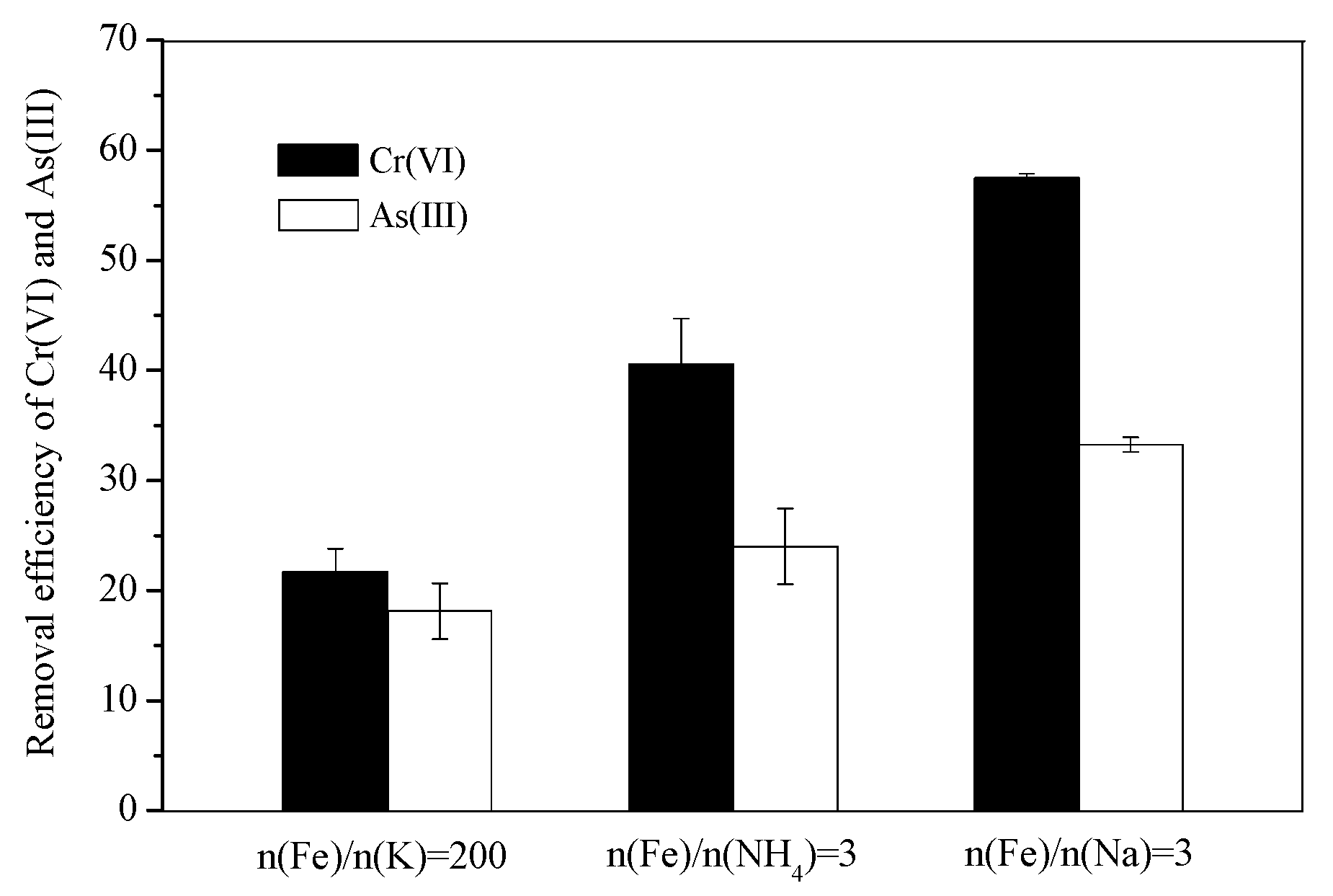
3.4. Comparison of the Cr(VI) and As(III) Removal Efficiency

To compare the secondary iron mineral removal efficiency of Cr(VI) and As(III) present in the AMD, we selected schwertmannite, which was acquired under the three mineralization systems [n(Fe)/n(K) = 200, n(Fe)/n(NH4) = 3, and n(Fe)/n(Na) = 3], according to XRD analysis. As shown in Figure 4, the removal efficiencies of Cr(VI) and As(III) from AMD (pH = 3.0) by biogenic schwertmannite generated from n(Fe)/n(K) = 200, n(Fe)/n(NH4) = 3, and n(Fe)/n(Na) = 3 systems were determined to be 57.5%, 40.6%, 21.7% and 33.2%, 24.0%, 18.1%, respectively. Thus, the removal efficiency of toxic elements was in the order of n(Fe)/n(Na) = 3 > n(Fe)/n(NH4) = 3 > n(Fe)/n(K) = 200.
Cr(VI) and As(III) are influenced by the acidity or alkalinity of solution: the transformation process between various forms are shown as follows:
Cr2O72− + H2O ↔ 2HCrO4
HCrO4 ↔ CrO42− + H+
H3AsO3 ↔H+ + H2AsO3
H2AsO3↔H+ + HAsO32−
HAsO32− ↔H+ + AsO33−
When solution pH below 2.2, along with the increase in solution acidity, the predominant Cr(VI) species is H2Cr2O7, which is not conducive to the adsorption of schwertmannite. In the pH range from 2.2 to 5.9, HCrO4 can complexed with Fe3+ in the schwertmannite structure [40]. When solution pH is above 5.9, CrO42− not only can be exchanged with SO42− in the surface structure of schwertmannite, but it also can be exchanged with SO42− through ligand exchange into the internal structure of the mineral, so that better Cr(VI) removal efficiency can be achieved [40,41]. In addition, considering the transformation of As(III), it has been reported that at a high solution acidity, As(III) is mainly in the form of H3AsO3, which makes it difficult to replace the HSO4 and SO42− groups in the schwertmannite structure, thus affecting the removal efficiency of As(III) [42]. In this study, at pH = 3 and an initial As(III) concentration of 1 mg/L, the removal efficiency of As(III) was only in the range of 18.1%~33.2% under the action of three different schwertmannites (0.5 g/L). The As(III) adsorption efficiencies obtained herein were in accordance with the study of Liao et al. [22], who reported that the amount of As(III) sorbed by schwertmannite (0.25 g/L) was only about 20% at Ph = 3.0, while the maximum As(III) removal (98%) occurs at about pH 7~9.
The influence of the type and concentration of monovalent cations on the variety of secondary iron minerals and the critical mineralization value of schwertmannite and jarosite was previously studied [27,28,29,30]. However, a comparison of the heavy metal removal capacity of different biogenic secondary iron minerals mediated by monovalent cations has not yet been reported. In order to determine why the three types of schwertmannite differ in terms of heavy metal adsorption and removal ability, a number of comprehensive tests, including SEM, average particle size and specific surface area measurements were conducted.
Figure 5 displays the SEM images of biogenic schwertmannite under different treatment processes. Obviously, the different types of schwertmannite were all of a standard spherical shape, but their structures differed. For the schwertmannite acquired by the treatment process where n(Fe)/n(K) = 200, the particle surface was slightly coarse with an average particle size of ~1.72 μm and a specific surface area of 9.72 m2/g. However, for the schwertmannite acquired by the treatment process where n(Fe)/n(NH4) = 3, the mineral particles were wool ball- or sponge-shaped, and the particle surface was very coarse and contained a large quantity of cellular structures. In this case, the average particle size was ~1.61 μm and the specific surface area was 16.43 m2/g. Surprisingly, for the schwertmannite acquired by the treatment process where n(Fe)/n(Na) = 3, the apparent structure differed significantly from that of the two other types of schwertmannite. Specifically, the mineral particles were sea urchin- or chestnut shell-shaped, and the particle surface was full of needle-shaped burrs, the pores were very large and deep. In this case, the average particle size was ~1.47 μm and the specific surface area was 22.15 m2/g. Numerous studies have also reported that most schwertmannite species formed in an acid mine environment are sea urchin-shaped with a particle diameter ranging between 300 and 500 nm and a particle surface covered in needle-shaped burrs with a length in the range of 60–90 nm [43,44,45].
According to XRD analysis, the secondary iron minerals acquired under the three mineralization systems [n(Fe)/n(K) = 200, n(Fe)/n(NH4) = 3, and n(Fe)/n(Na) = 3] were all schwertmannite, which indicates that the monovalent cations did not participate in the mineralization reaction. We speculate that the large differences in the specific surface areas of the three schwertmannites were related to the concentration of H+ released during the formation of schwertmannite. According to the synthesis reaction of schwertmannite [Reaction (2)], the higher mineralization degree of Fe3+ in the solution would induce a higher release of H+, thereby leading to a gradual diffusion and dissolution of the previously synthesized minerals [17,46]. As can be seen from Figure 2, the cumulative Fe deposition efficiencies in the three mineralization systems [n(Fe)/n(K) = 200, n(Fe)/n(NH4) = 3, and n(Fe)/n(Na) = 3] were 29.6%, 28.1%, and 18.6%, respectively. This indicates that the degree of H+ release in the three systems was in the following order: n(Fe)/n(K) = 200 > n(Fe)/n(NH4) = 3 > n(Fe)/n(Na) = 3. Since the low acidity leads to the dissolution of the needle-shaped burrs of the previously formed schwertmannite in the systems of n(Fe)/n(K) = 200 and n(Fe)/n(NH4) = 3, the specific surface area of the schwertmannite was reduced. According to Liu et al. [47], who investigated the stability of schwertmannite in in acid environments, the dissolution efficiency of schwertmannite in a system with pH = 3.0 is only 3.34% after a 72 h oscillation, while the dissolution efficiency in a pH = 2.0 system is as high as 61.46% for the same amount of oscillation time. Notably, the Cr(VI) and As(III) removal efficiencies change significantly upon varying the schwertmannite specific surface area. Therefore, in this study, schwertmannites with small specific surface areas exhibited poor removal efficiencies of Cr(VI) and As(III).
It is well known that a small mineral particle size denotes a large specific surface area and a strong adsorption power [48]. Thus, the apparent structure, particle size, and specific surface area can reasonably explain why the three types of schwertmannite differ in terms of their Cr(VI) and As(III) adsorption and removal capacities.

4. Conclusions

The concentration of monovalent cations exhibited considerable influence on the biological oxidation of Fe2+. Specifically, we found that a high concentration of monovalent cations inhibited the oxidizing ability of A. ferrooxidans, and that these cations exhibited different mineralizing abilities (K+ > NH4+ > Na+). For example, at an initial pH value of 3.0 and an n(Fe)/n(M) = 3, the cumulative Fe deposition efficiencies for the three ions were 58.0%, 28.1%, and 18.6%, respectively, after the simulated AMD was cultivated for 96 h. In addition, the removal efficiency of Cr(VI) and As(III) contained in the acid drainage for schwertmannite acquired under the three mineralization systems were different: n(Fe)/n(Na) = 3 > n(Fe)/n(NH4) = 3 > n(Fe)/n(K) = 200. The results of this study may help provide insight to the transformation of Fe to secondary iron minerals and the immobilization of heavy metals and metalloids in acid sulfate environments.

Author Contributions

Y.S. conceived and designed this study, conducted the data collation and analysis and wrote the manuscript. H.W. performed the experiments, conducted the data collation and analysis and wrote the manuscript. J.Y. and Y.C. contributed to the revision of the manuscript. All authors have read and approved the final manuscript.

Funding

This research was funded by the Natural Science Foundation of Hubei Province, China (2016CFB289).

Acknowledgments

The authors would like to thank the Solid Waste Research Institute of Nanjing Agricultural University for kindly providing A. ferrooxidans LX5 (CGMCC No. 0727).

Conflicts of Interest

The authors declare no conflict of interest.

References

  1. Cravotta, C.A.; Trahan, M.K. Limestone drains to increase pH and remove dissolved metals from acidic mine drainage. Appl. Geochem. 1999, 14, 581–606. [Google Scholar] [CrossRef]
  2. Peppas, A.; Komnitsas, K.; Halikia, I. Use of organic covers for acid mine drainage control. Miner. Eng. 2000, 13, 563–574. [Google Scholar] [CrossRef]
  3. Sahoo, P.K.; Bhattacharyya, P.; Tripathy, S.; Equeenuddin, S.M.; Panigrahi, M.K. Influence of different forms of acidities on soil microbiological properties and enzyme activities at an acid mine drainage contaminated site. J. Hazard. Mater. 2010, 179, 966–975. [Google Scholar] [CrossRef] [PubMed]
  4. Vhahangwele, M. A novel technology for neutralizing acidity and attenuating toxic chemical species from acid mine drainage using cryptocrystalline magnesite tailings. J. Water Process Eng. 2016, 10, 67–77. [Google Scholar]
  5. Wei, X.; Wolfe, F.A. Minerals and mine drainage. Water Environ. Res. 2013, 85, 1515–1547. [Google Scholar] [CrossRef]
  6. Wu, Z.L.; Zou, L.C.; Chen, J.H.; Lai, X.K.; Zhu, Y.G. Column bioleaching characteristic of copper and iron from Zijinshan sulfide ores by acid mine drainage. Int. J. Miner. Process 2016, 149, 18–24. [Google Scholar] [CrossRef]
  7. Akcil, A.; Koldas, S. Acid mine drainage (AMD): Causes, treatment and case studies. J. Clean. Prod. 2006, 14, 1139–1145. [Google Scholar] [CrossRef]
  8. España, J.S.; Pamo, E.L.; Santofimia, E.; Aduvire, O.; Reyes, J.; Barettino, D. Acid mine drainage in the Iberian Pyrite Belt (Odiel river watershed, Huelva, SW Spain): Geochemistry, mineralogy and environmental implications. Appl. Geochem. 2005, 20, 1320–1356. [Google Scholar] [CrossRef]
  9. Lee, W.C.; Lee, S.W.; Yun, S.T.; Lee, P.K.; Hwang, Y.S.; Kim, S.O. A novel method of utilizing permeable reactive kiddle (PRK) for the remediation of acid mine drainage. J. Hazard. Mater. 2016, 301, 332–341. [Google Scholar] [CrossRef] [PubMed]
  10. Meschke, K.; Herdegen, V.; Aubel, T.; Janneck, E.; Repke, J.U. Treatment of opencast lignite mining induced acid mine drainage (AMD) using a rotating microfiltration system. J. Environ. Chem. Eng. 2015, 4, 2848–2856. [Google Scholar] [CrossRef]
  11. Song, Y.W.; Wang, M.; Liang, J.R.; Zhou, L.X. High-rate precipitation of iron as jarosite by using a combination process of electrolytic reduction and biological oxidation. Hydrometallurgy 2014, 143, 23–27. [Google Scholar] [CrossRef]
  12. Diao, Z.; Shi, T.; Wang, S.; Huang, X.; Zhang, T.; Tang, Y.; Zhang, X.; Qiu, R. Silane-based coatings on the pyrite for remediation of acid mine drainage. Water Res. 2013, 47, 4391–4402. [Google Scholar] [CrossRef] [PubMed]
  13. Singer, P.C.; Stumm, W. Acidic mine drainage: The rate-determining step. Science 1970, 167, 1121–1123. [Google Scholar] [CrossRef] [PubMed]
  14. Valente, T.; Grande, J.A.; De, I.T.M.L.; Santisteban, M.; Cerón, J.C. Mineralogy and environmental relevance of AMD-precipitates from the Tharsis mines, Iberian Pyrite Belt (SW, Spain). Appl. Geochem. 2013, 39, 11–25. [Google Scholar] [CrossRef]
  15. Zhu, J.Y.; Gan, M.; Zhang, D.; Hu, Y.H.; Chai, L.Y. The nature of schwertmannite and jarosite mediated by two strains of Acidithiobacillus ferrooxidans with different ferrous oxidation ability. Mater. Sci. Eng. C 2013, 33, 2679–2685. [Google Scholar] [CrossRef] [PubMed]
  16. Regenspurg, S.; Brand, A.; Peiffer, S. Formation and stability of schwertmannite in acid mining lakes. Geochim. Cosmochim. Acta 2004, 68, 1185–1197. [Google Scholar] [CrossRef]
  17. Loan, M.; Richmond, W.R.; Parkinson, G.M. On the crystal growth of nanoscale schwertmannite. J. Cryst. Growth 2005, 275, 1875–1881. [Google Scholar] [CrossRef]
  18. Barham, R.J. Schwertmannite: A unique mineral, contains a replaceable ligand, transforms to jarosites, hematites, and/or basic iron sulfate. J. Mater. Res. 1997, 12, 2751–2758. [Google Scholar] [CrossRef]
  19. Min, G.; Sun, S.J.; Zheng, Z.H.; Tang, H.J.; Sheng, J.R.; Zhu, J.Y.; Liu, X.X. Adsorption of Cr(VI) and Cu(II) by AlPO4 modified biosynthetic schwertmannite. Appl. Surf. Sci. 2015, 356, 986–997. [Google Scholar]
  20. Mihone, K.M.; Hana, F.; Sanda, R.; Lidija, C. Assessment of metal risks from different depths of jarosite tailing waste of Trepça Zinc Industry, Kosovo based on BCR procedure. J. Geochem. Explor. 2015, 148, 161–168. [Google Scholar]
  21. Zhang, S.L.; Jia, S.Y.; Yu, B.; Liu, Y.; Wu, S.H.; Han, X. Sulfidization of As(V)-containing schwertmannite and its impact on arsenic mobilization. Chem. Geol. 2016, 420, 270–279. [Google Scholar] [CrossRef]
  22. Liao, Y.H.; Liang, J.R.; Zhou, L.X. Adsorptive removal of As(III) by biogenic schwertmannite from simulated As-contaminated groundwater. Chemosphere 2011, 83, 295–301. [Google Scholar] [CrossRef] [PubMed]
  23. Wang, C.Q.; Ma, S.F.; Lu, A.H. A preliminary study on the Cr(VI) removing from wastewater by precipitation of jarosite group minerals. Bull. Miner. Petrol. Geochem. 2006, 25, 335–338. [Google Scholar]
  24. Asta, M.P.; Cama, J.; Martínez, M.; Giménez, J. Arsenic removal by goethite and jarosite in acidic conditions and its environmental implications. J. Hazard. Mater. 2009, 171, 965–972. [Google Scholar] [CrossRef] [PubMed]
  25. Dutrizac, J.E.; Kaiman, S. Synthesis and properties of jarosite-type compounds. Can. Mineral. 1976, 14, 151–158. [Google Scholar]
  26. Dutrizac, J.E. The effectiveness of jarosite species for precipitating sodium jarosite. JOM 1999, 51, 30–32. [Google Scholar] [CrossRef]
  27. Bigham, J.M.; Schwertmann, U.; Traina, S.J.; Winland, R.L.; Wolf, M. Schwertmannite and the chemical modeling of iron in acid sulfate waters. Geochim. Cosmochim. Acta 1996, 60, 2111–2121. [Google Scholar] [CrossRef]
  28. Gramp, J.P.; Jones, F.S.; Bigham, J.M.; Tuovinen, O.H. Monovalent cation concentrations determine the types of Fe(III) hydroxysulfate precipitates formed in bioleach solutions. Hydrometallurgy 2008, 94, 29–33. [Google Scholar] [CrossRef]
  29. Liao, Y.H.; Zhou, L.X.; Liang, J.R.; Xiong, H.X. Biosynthesis of schwertmannite by Acidithiobacillus ferrooxidans cell suspensions under different pH condition. Mater. Sci. Eng. C 2009, 29, 211–215. [Google Scholar] [CrossRef]
  30. Bai, S.Y.; Liang, J.R.; Zhou, L.X. Effects of monovalent cation and dissolved organic matter on the formation of biogenic secondary iron minerals in bioleaching system. Acta Mineral. Sinica 2011, 31, 118–125. [Google Scholar]
  31. Bai, S.Y.; Liang, J.R.; Zhou, L.X. Effects of iron/potassium molar ratio on mass of biogenic Fe(III) hydroxysulfate precipitates in the FeSO4-K2SO4-H2O system and their environmental implications. Acta Sci. Circumst. 2015, 35, 476–483. [Google Scholar]
  32. Silverman, M.P.; Lundgren, D.G. Studies on the chemoautotrophic iron bacterium Ferrobacillus ferrooxidans. І. An improved medium and a harvesting procedure for securing high cell yields. J. Bacteriol. 1959, 77, 642–647. [Google Scholar] [PubMed]
  33. Wang, S.M.; Zhou, L.X. A renovated approach for increasing colony count efficiency of Thiobacillus ferrooxidans and Thiobacillus thiooxidans: Double-layer plates. Acta Sci. Circumst. 2005, 25, 1418–1420. [Google Scholar]
  34. Csonka, L.N. Physiological and genetic responses of bacteria to osmotic stress. Microbiol. Rev. 1989, 53, 121–147. [Google Scholar] [PubMed]
  35. Wang, Q.L.; Qiu, G.Z.; Yu, R.L.; Hu, E.M.; Zhang, H.C.; Xiong, X. Effects of coexisting ions on activity of Thiobacillus ferrooxidans in in-situ leaching of uranium. China Min. Mag. 2015, 24, 116–119. [Google Scholar]
  36. Wu, Q.T. Study on treatment technique of preserved szechuan pickle wastewater with high salinity. Master’s Thesis, Chongqing University, Chongqing, China, 2007. [Google Scholar]
  37. Song, Y.W.; Wang, H.R.; Cao, Y.X.; Zhu, Y.H.; Zhou, L.X. Effect of NH4+ on the formation of secondary iron minerals and the removal of heavy metals. China Environ. Sci. 2018, 38, 2116–2123. [Google Scholar]
  38. Suzuki, I.; Lee, D.; Mackay, B.; Harahuc, L.; Oh, J.K. Effect of various ions, pH, and osmotic pressure on oxidation of elemental sulfur by Thiobacillus thiooxidans. Appl. Environ. Microbiol. 1999, 65, 5163–5171. [Google Scholar] [PubMed]
  39. Sasaki, K.; Konno, H. Morphology of jarosite-group compounds precipitated from biologically and chemically oxidized Fe ions. Can. Mineral. 2000, 38, 45–56. [Google Scholar] [CrossRef]
  40. Regenspurg, S.; Peiffer, S. Arsenate and chromate incorporation in schwertmannite. Appl. Geochem. 2005, 20, 1226–1239. [Google Scholar] [CrossRef]
  41. Bigham, J.M.; Schwertmann, U.; Carlson, L. A poorly crystallized xyhydoxysulfate of iron formed by bacterial oxidation of Fe(II) in acid mine waters. Geochim. Cosmochim. Acta 1990, 54, 2743–2758. [Google Scholar] [CrossRef]
  42. Duquesne, K.; Lebrun, S. Immobilization of arsensite and ferric iron by Acidithiobacillus ferrooxidans and its relevance to acid mine drainage. Appl. Environ. Microbiol. 2003, 69, 6165–6173. [Google Scholar] [CrossRef] [PubMed]
  43. Jönsson, J.; Persson, P.; Sjöberg, S.; Lövgren, L. Schwertmannite precipitated from acid mine drainage: Phase transformation, sulfate release and surface properties. Appl. Geochem. 2005, 20, 179–191. [Google Scholar] [CrossRef]
  44. Šubrt, J.; Boháček, J.; Štengl, V.; Grygar, T.; Bezdička, P. Uniform particles with a large surface area formed by hydrolysis of Fe2(SO4)3 with urea. Mater. Res. Bull. 1999, 34, 905–914. [Google Scholar] [CrossRef]
  45. Sun, H.F.; Zhao, F.H.; Cong, Z.Y.; Yue, M.; Ren, D.Y. The mineral schwertmannite found in China and its characteristics. Acta Mineral. Sinica 2006, 26, 38–42. [Google Scholar]
  46. Dold, B. Dissolution kinetics of schwertmannite and ferrihydrite in oxidized mine samples and their detection by differential X-ray diffraction (DXRD). Appl. Geochem. 2003, 18, 1531–1540. [Google Scholar] [CrossRef]
  47. Liu, F.W.; Bu, Y.S.; Tian, G.J.; Cui, C.H.; Zhou, L.X. Influence of temperature and pH on dissolution behavior of biogenic schwertmannite in acidic environment and the adsorption of Cu2+. Acta Sci. Circumst. 2013, 33, 2445–2451. [Google Scholar]
  48. Liu, F.W.; Zhou, J.; Zhang, S.S.; Liu, L.L.; Zhou, L.X.; Fan, W.H. Schwertmannite synthesis through ferrous ion chemical oxidation under different H2O2 supply rates and its removal efficiency for arsenic from contaminated groundwater. PLoS ONE 2015, 10, e0138891. [Google Scholar] [CrossRef] [PubMed]
Figure 1. Effect of n(Fe)/n(M) (M: K, NH4, Na) on the Fe2+ oxidation facilitated by A. ferrooxidans.
Metals 08 00596 g001
Figure 2. Effect of n(Fe)/n(M) (M: K, NH4, Na) on the total Fe precipitation efficiency.
Metals 08 00596 g002
Figure 3. XRD patterns of precipitates formed through Fe2+ oxidation by A. ferrooxidans under different n(Fe)/n(M) ratios (M: K, NH4, Na): (a) n(Fe)/n(K); (b) n(Fe)/n(NH4); (c) n(Fe)/n(Na).
Metals 08 00596 g003
Figure 4. Removal efficiency of Cr(VI) and As(III) in the solution over 0.5 g/L schwertmannite harvested from different biosynthesis systems: n(Fe)/n(K) = 200, n(Fe)/n(NH4) = 3, n(Fe)/n(Na) = 3.
Metals 08 00596 g004
Figure 5. SEM images of schwertmannite under different treatments: (a) n(Fe)/n(K) = 200; (b) n(Fe)/n(NH4) = 3; (c) n(Fe)/n(Na) = 3.
Metals 08 00596 g005aMetals 08 00596 g005b

