Inhibition of Adherence of Mycobacterium avium to Plumbing Surface Biofilms of Methylobacterium spp.


1
Department of Clinical Microbiology, Fundación Jiménez Díaz Hospital, 28040 Madrid, Spain
2
Department of Civil and Environmental Engineering, Virginia Tech, Blacksburg, VA 24061, USA
3
Department of Biological Sciences, Virginia Tech, Blacksburg, VA 24061, USA
*
Author to whom correspondence should be addressed.
Pathogens 2017, 6(3), 42; https://doi.org/10.3390/pathogens6030042
Submission received: 1 August 2017
/
Revised: 6 September 2017
/
Accepted: 11 September 2017
/
Published: 14 September 2017
Abstract
:Both Mycobacterium spp. and Methylobacterium spp. are opportunistic premise plumbing pathogens that are found on pipe surfaces in households. However, examination of data published in prior microbiological surveys indicates that Methylobacterium spp. and Mycobacterium spp. tend not to coexist in the same household plumbing biofilms. That evidence led us to test the hypothesis that Methylobacterium spp. in biofilms could inhibit the adherence of Mycobacterium avium. Measurements of adherence of M. avium cells to stainless steel coupons using both culture and PCR-based methods showed that the presence of Methylobacterium spp. biofilms substantially reduced M. avium adherence and vice versa. That inhibition of M. avium adherence was not reduced by UV-irradiation, cyanide/azide exposure, or autoclaving of the Methylobacterium spp. biofilms. Further, there was no evidence of the production of anti-mycobacterial compounds by biofilm-grown Methylobacterium spp. cells. The results add to understanding of the role of microbial interactions in biofilms as a driving force in the proliferation or inhibition of opportunistic pathogens in premise plumbing, and provide a potential new avenue by which M. avium exposures may be reduced for at-risk individuals.
1. Introduction
Nontuberculous mycobacteria (NTM) are opportunistic human pathogens whose source of infection is the environment [1]. Mycobacterium species are found in drinking water distribution systems [2], hospitals [3], and household plumbing [4], and cause life-threatening pulmonary infections [5] that are difficult to treat [6]. The most common species associated with pulmonary infection in the United States is Mycobacterium avium [5,6].
The incidence of NTM disease in the United States and Canada is rising [7,8]. In Toronto (Canada), NTM disease incidence has risen from 1.5 to 9.0 per 100,000 over the period 1997–2003 [7]. Similarly, NTM disease is increasing in the United States, based on reports of NTM lung disease in hospitalized persons [8]. A major contributor to this increase is the fact that elderly, slender women, lacking any of the classic risk factors for NTM disease, have a greater tendency than the general population to develop NTM pulmonary disease [9,10,11]. It follows that as the population of the United States continues to age—25% of the US population will be over 60 years by 2025 [12]—the incidence of NTM pulmonary disease will continue to increase. Further, as NTM-infected patients are subject to reemergence of infection or reinfection by other environmental NTM [13], it is of value to identify measures to reduce NTM exposure.
Recently, it was shown that the DNA fingerprints of M. avium isolates recovered from both the biofilm and water from an M. avium-infected pulmonary patient’s shower were related to the patient’s M. avium isolate [14]. That study was followed by a report demonstrating the widespread presence and high numbers of Mycobacterium spp. and M. avium in showerhead biofilms across the United States [15]. Although not highlighted by the authors, examination of that data indicated a potentially important pattern; namely, the presence of a high proportion of pink-pigmented Methylobacterium spp. were associated with reduced numbers of Mycobacterium spp. and the presence of a high proportion of Mycobacterium spp. with a low proportion of Methylobacterium spp. [15]. Identical results were observed by cultivation of showerhead biofilms in households in Philadelphia, Pennsylvania [16].
Like M. avium and other NTM, Methylobacterium spp. are normal inhabitants of drinking water distribution systems [17,18,19,20,21] and plumbing in buildings, including hospitals [22,23]. Further, a substantial proportion of Methylobacterium spp. isolates are chlorine-resistant [24], form biofilms [25,26], and belong to the group of amoeba-resisting bacteria in drinking water [27]. Household plumbing is also a habitat, as Methylobacterium spp. have been shown to be abundant amongst DNA clones recovered from shower curtains [28].
In this study, it was hypothesized that the presence of the pink-pigmented Methylobacterium spp. will be associated with the absence of Mycobacterium spp., and that the presence of Mycobacterium spp. will be associated with the absence of Methylobacterium spp. Laboratory experiments were performed to identify the basis for the exclusion of M. avium by Methylobacterium spp. Exclusion of M. avium by Methylobacterium spp. could provide a new approach for limiting the exposure of at-risk individuals to M. avium and other NTM.
2. Results
2.1. Adherence Measurements
Stainless steel coupons, held in paddles in the CDC Biofilm Reactor, were exposed to suspensions of two different consortia of water-acclimated Methylobacterium spp. cells, or normal tap water, for 21 days at room temperature, to produce biofilms. The paddles and coupons with biofilms, and a control paddle and coupons lacking any biofilm (control), were washed twice in sterile tap water, and then placed in a suspension of water-acclimated M. avium cells (~105 CFU/mL) in the CDC Biofilm Reactor. Immediately and after 1, 2, 3, and 6 h exposure, paddles and coupons were removed, coupons aseptically removed from the paddles, placed in 5 mL of sterile tap water, adherent cells suspended by vortexing, and the number of adherent M. avium cells measured as colony-forming units.
2.2. Methylobacterium Extorquens Adherence
Given that cells of M. extorquens and other Methylobacterium spp. aggregate spontaneously in broth media [21], an indicator of high hydrophobicity, it was hypothesized that, like M. avium, Methylobacterium spp. would readily adhere to surfaces and form biofilms. To test this, the adherence of water-acclimated cells of the M. extorquens strain to stainless steel coupons was measured in the presence and absence of Blacksburg tap water biofilms. The results (Table 1) demonstrated that M. extorquens cells readily adhered to the coupons, and that the presence of an existing tap water biofilm increased the extent of adherence. Adherence was apparently quite rapid, as a substantial number of M. extorquens cells adhered immediately (time 0) after exposing the M. extorquens cells to coupons with biofilms (Table 1). Approximately 5 min was required for removal of coupons and their transfer to centrifuge tubes, thus, the time 0 samples allowed for 5 min of adherence.
2.3. Methylobacterium spp. Inhibition of M. avium Adherence by Colony Count
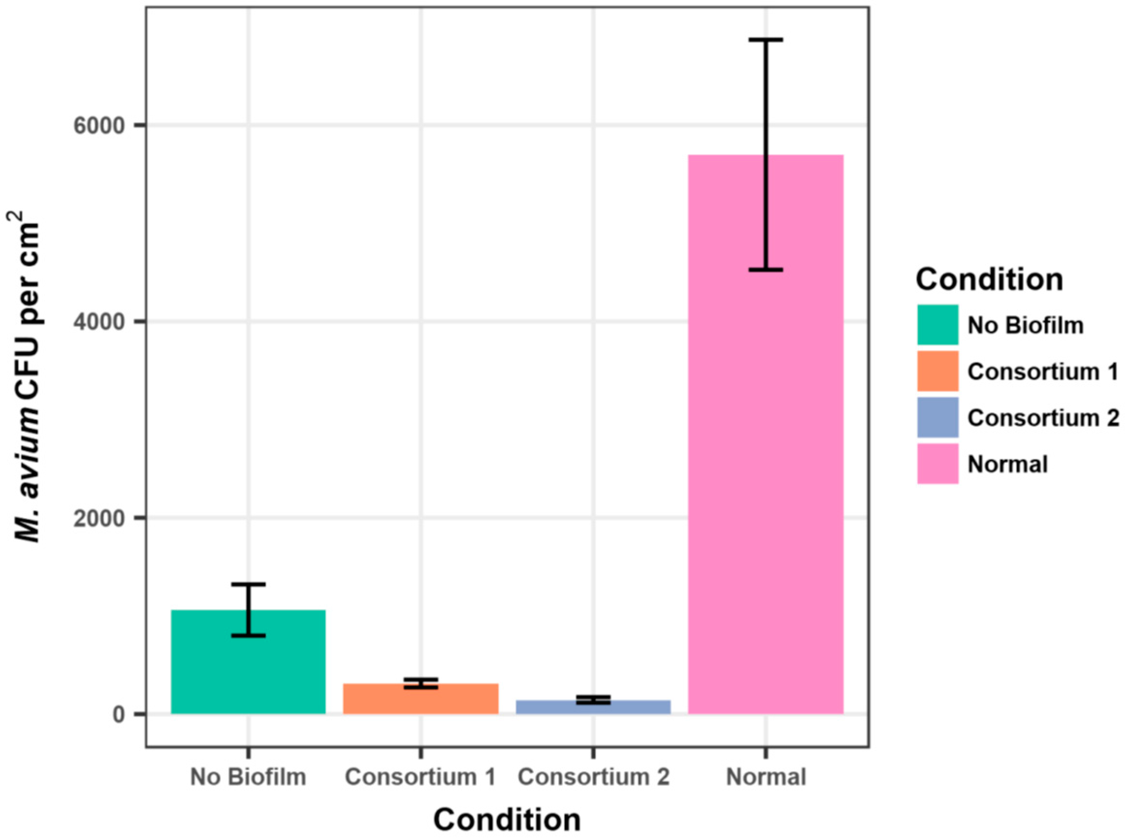
The presence of established 21-day biofilms, composed of either Consortium 1 (Methylobacterium spp.) or Consortium 2 (Methylobacterium spp. and Deinococcus grandis), significantly reduced (ANOVA < 0.01) the adherence of M. avium strain A5 cells (Figure 1 and Supplementary Materials Table S1). The normal microbial biofilm produced by 21 days incubation of coupons in non-sterile Blacksburg tap water significantly (ANOVA p < 0.05) increased the adherence of the M. avium cells (Figure 1, Supplementary Materials Table S1), as noted for M. extorquens (Table 1). The results illustrated in Figure 1 reflect 6 h exposure to the M. avium suspension. Measurements of adherent M. avium cells at 0, 1, 2, and 3 h showed the same results, namely that Methylobacterium spp. consortium 1 and 2 inhibited the adherence of M. avium to the coupons (Supplementary Materials Table S1).
As these results came from short term adherence measurements (0 to 6 h), the conditions would have been unlikely to have had sufficient time to induce a viable, but nonculturable (VBNC) state in M. avium. Specifically, the loss of culturability of M. avium in biofilms requires at least one day [29]. To rule out the possibility that methylobacterial biofilms bound M. avium cells more tightly, and thereby, reduced suspension of adherent cells (thus reducing apparent adherence), coupons that had been treated to recover adherent mycobacteria were placed on M7H10 agar medium and incubated at 37 °C. Low numbers of M. avium colonies were observed (<10 colonies/coupon), even against the background of methylobacterial colonies that were low because of the relatively high incubation temperature. Such conditions would have also induced resuscitation of any VBNC mycobacterial cells over the course of incubation for 7 days on laboratory medium. We conclude that the reduced number of M. avium colonies in methylobacterial biofilms was not due to immediate induction of the VBNC state in mycobacterial cells.
As pink-pigmented yeast and cocci were recovered from patient household samples [16] and consortium 2 contained a strain of Deinococcus grandis, the adherence of M. avium strain A5 to stainless steel coupons was measured in the presence and absence of a yeast isolate 32-14 and the D. grandis strain JM-1-1. Neither the yeast, nor D. grandis biofilms reduced the adherence of M. avium strain A5 (Table 2). In fact, D. grandis biofilms appeared to have increased numbers of adherent M. avium cells (Table 2). In conclusion, the data support the hypothesis that the inhibition of M. avium adherence by Consortium 2 (Figure 1) was solely due to the Methylobacterium spp. cells.
2.4. Methylobacterium spp. Inhibition of M. avium Adherence by qPCR
One possible explanation for the reduced number of M. avium cells on Methylobacterium spp. biofilms (Figure 1, Supplementary Materials Table S1) could be that adherent M. avium cells lost their culturability and were unable to form colonies. To rule out that possibility, M. avium counts were measured by qPCR. Following suspension of biofilm-adherent cells, DNA was isolated, and M. avium gene copies enumerated by a sensitive and specific qPCR method [30]. The data, reported as M. avium gene copies/µL of isolated DNA, show that the number of gene copies on Methylobacterium spp. biofilms were below the limit of quantification (i.e., 3.95 × 10⁶ copies/cm2) immediately and after 1 and 3 h (Table 3). These results show that the reduction of M. avium counts on Methylobacterium spp. biofilms reflected by colony counts were not an artifact due to loss of culturability.
2.5. Inhibition of M. avium Adherence by Methylobacterium spp. Biofilms Does Not Require Methylobacterium spp. Viability
Three approaches were taken to measure whether viability of a Methylobacterium spp. biofilm was required for the inhibition of M. avium adherence: (1) poisoning with 10 mM cyanide and 10 mM azide (CN/AZ); (2) killing with ultraviolet-irradiation (UV); and (3) killing by autoclaving (Auto). The results demonstrated that Methylobacterium spp. biofilms whose survival was reduced to <100 CFU/cm2 by azide/cyanide-exposure, UV-irradiation, or autoclaving, still inhibited M. avium adherence (Table 4). The differences between the untreated control versus poisoned, versus UV-irradiated, or versus autoclaved Methylobacterium spp. biofilms on adherent M. avium colony counts were not significant (ANOVA > 0.05). In spite of the variation in the inhibition percentages, the data suggest that viability is not required for the inhibition of M. avium adherence to Methylobacterium spp. biofilms.
2.6. Methylobacterium spp. Biofilms Do Not Kill Adherent M. avium
To measure whether Methylobacterium spp. biofilms killed adherent M. avium cells, M. avium cells were exposed to a Methylobacterium spp. biofilm for 6 h to ensure adherence. Then, the coupons were removed from the M. avium suspension, washed, and placed in a CDC Biofilm Reactor containing sterile Blacksburg tap water. Immediately and daily (to 3 days), coupons were removed and the number of M. avium colony-forming units measured. Measurement of survival (CFU/cm2) of M. avium strain A5 cells adhering to Methylobacterium spp. biofilms showed no decrease over time; in fact, numbers increased (Table 5). That data rules out the possibility that cells of M. avium did adhere to Methylobacterium spp. biofilms, but were killed.
2.7. M. avium Strain A5 Biofilms Inhibit the Adherence of Methylobacterium spp. Cells to Stainless Steel
As only Mycobacterium spp. were present when Methylobacterium spp. were absent from biofilms of showerheads [15] or NTM patient household plumbing [16], the adherence of Methylobacterium spp. consortia to M. avium strain A5 biofilms was measured. The results show that 7-day M. avium biofilms reduced adherence of Methylobacterium spp. Consortium 1 cells, but only reduced the number of adherent cells of Consortium 2 after 3 h exposure to the Methylobacterium spp. suspension (Table 6).
3. Discussion
The data are consistent with the observations that Methylobacterium spp. and M. avium seldom are present in the same plumbing biofilm samples [15,16]. Further, the data support the hypothesis that the physical presence of either Methylobacterium spp. or M. avium in biofilms inhibits the adherence of the other species. Inspection of the Tables and Figures shows that there was substantial variation in the standard deviations of the average CFU/cm2 values. In part, this is due to the hydrophobicity-driven aggregation of the M. avium strain [31]. Colony counts of aggregates are subject to wide variation, as a sample may or may not contain aggregates with the same number of cells, and an aggregate can yield different colony counts depending upon the efficacy of spreading suspensions on agar media. For the experiments reported here, care was taken to ensure that suspensions were spread to dryness on 3-day agar media. Further, the data in each table present the results of the same experiment, using the same starting suspension of water-acclimated cells of M. avium to ensure that the different surfaces (e.g., present or absence of biofilm) could be made evident. At present, there are no alternatives to using aggregating mycobacterial strains as non-aggregating, hydrophilic derivatives are seldom isolated from patients and drinking water [1], and are thus unrepresentative. Likewise, aggregation appears to be a shared characteristic of Methylobacterium [21]. The use of detergents to produce uniform suspensions of cells is discouraged, as it alters surface hydrophobicity, and therefore, the natural behavior of these waterborne bacteria.
We conclude that the presence of an established Methylobacterium spp. biofilm substantially reduces the adherence and biofilm formation by M. avium cells. The inhibition in M. avium adherence did not require the viability of the Methylobacterium spp. cells, suggesting that the physical presence of Methylobacterium spp. cells is sufficient. It is possible that the first to attach may govern the further development of the biofilm microbial population. The presence of a normal, established microbial biofilm actually increased adherence of mycobacteria and methylobacteria. There was no demonstration of production of anti-mycobacterial activity by Methylobacterium spp. in biofilms, as the CFU/cm2 of M. avium cells adhering to Methylobacterium spp. biofilms did not decrease over time. That observation also rules out the possibility that nutrient competition is responsible, as Methylobacterium spp. are adapted to thrive at low nutrient concentrations [32].
Here, we describe a specific interaction between M. avium and Methylobacterium spp. in drinking water biofilms. Although these studies were focused on the interaction between M. avium and Methylobacterium spp., it is likely that other examples of inhibition of adherence will be found between other microorganisms. These observations contribute to an emerging understanding of drinking water biofilm microbiomes, and their importance in governing establishment and virulence of opportunistic pathogens [33]. The findings are particularly significant to those estimated 30,000 individuals in the United States with pulmonary mycobacterial disease [7,8], as they are innately susceptible to continued mycobacterial infections [13]. Specifically, the ecological interaction identified here in which M. avium adherence to biofilms is inhibited by Methylobacterium spp. could potentially be exploited as a strategy to limit adherence and biofilm formation by M. avium and possibly other Mycobacterium species, and reduce exposure of individuals to these opportunistic premise plumbing pathogens. This is in line with a “probiotic” framework recently suggested by Wang et al. [34]. Rather than suggest the “inoculation” of household plumbing with Methylobacterium spp. cells, we are investigating the possible inhibition of M. avium adherence by cellular fractions of Methylobacterium spp.
4. Materials and Methods
4.1. Mycobacterium avium, Methylobacterium spp., Deinococcus grandis, and Yeast Isolates
Mycobacterium avium strain A5 is a plasmid-free clinical isolate [35]. The Methylobacterium spp. isolates were obtained from culture collections or pink-pigmented isolates recovered from showers and identified on the basis of 16S rRNA sequence (Table 7. The D. grandis isolate was included in Consortium 2, as it was pink-pigmented like Methylobacterium and recovered from a shower curtain. In addition, pink-pigmented yeast, strain P32-14, isolated from a shower curtain was included to rule out the possibility that it was responsible for the inhibition of M. avium adherence.
4.2. Preparation of M. avium Strain A5 and Methylobacterium spp. for Adherence Measurements
M. avium strain A5 was grown in 20 mL of Middlebrook 7H9 broth containing 0.5% (vol/vol) glycerol and 10% (vol/vol) oleic acid–albumin (M7H10) to mid log phase at 37 °C with aeration (60 rpm). Methylobacterium spp. strains were grown separately in 20 mL of Nutrient Broth (BD, Sparks, MD) to mid log phase at 30 °C with aeration. Following growth, cells of both M. avium and Methylobacterium spp. strains were collected by centrifugation (5000× g for 20 min), the supernatant medium discarded, and the cells were suspended in 20 mL of autoclaved Blacksburg tap water containing 0.05 mg humic acid/mL (Aldrich, St. Louis, MO, USA). Humic acid was added to provide a nutrient source that is common in drinking water. The suspensions were incubated at room temperature with aeration (60 rpm) for 7 days to acclimate to tap water. Two consortia of Methylobacterium spp. strains (Table 1) were prepared by mixing equal volumes of the water-acclimated suspensions.
4.3. CDC Reactor and Preparation for Adherence and Biofilm Measurements
CDC Biofilm Reactors (BioSurface Technologies Corp., Bozeman, MT, USA) were employed to measure adherence and biofilm formation on stainless steel coupons [36]. Stainless steel coupons were used as stainless steel pipes are present in household plumbing, relatively resistant to surface changes, and M. avium adherence is relatively high; though not as high as galvanized surfaces that are subject to surface changes over time [37]. Before use, the stainless steel coupons were thoroughly scrubbed and washed in detergent, rinsed, and dried, then soaked in 2 M HCl for 2 h, rinsed, dried, and placed into paddles of the CDC Biofilm Reactor.
4.4. Recovery and Enumeration of Adherent Cells by Colony Count
Paddles with coupons were removed from the CDC Biofilm Reactor and rinsed gently by immersion in sterile tap water twice. Coupons were aseptically removed from a paddle, and each coupon placed in 10 mL of sterile Butterfield buffer (per liter: 0.4 g KH2PO4, 1 gm peptone, and 20 mL Tween 80) contained in a 50 mL screw cap centrifuge tube. Each coupon was gently swirled to remove unattached cells, and the washed coupon drained and transferred to a second tube containing 10 mL fresh Butterfield buffer, and vortexed for 60 s. The undiluted and 10-, 100-, and 1000-fold diluted (Butterfield buffer) suspensions were spread (0.1 mL) in triplicate on either on M7H10 agar (M. avium) or R2A agar (Methylobacterium spp.), and incubated at either 37 °C (M. avium) or 30 °C (Methylobacterium spp.), and colonies with the appropriate pigmentation and morphology counted.
4.5. Recovery and Enumeration of Adherent Cells by qPCR
To complement the enumeration of adherent M. avium cells by colony count, qPCR was employed to measure numbers of adherent M. avium cells on Methylobacterium spp. biofilms. Paddles with coupons were removed from the CDC Biofilm Reactor and rinsed gently by immersion in sterile tap water twice. Coupons were aseptically removed from a paddle, and each coupon placed in a second tube containing 2 mL sterile tap water, and vortexed for 60 s. Following suspension of biofilm-adherent cells, an aliquot of 200 μL suspension was subject to DNA extraction using SPIN Kit (MP Biomedicals) according to the manufacturer’s instruction. Quantitative polymerase chain reaction (qPCR) was applied to measure the gene numbers of M. avium using previously established protocol [30]. The data was reported as M. avium gene copies/cm2.
4.6. Establishment of a Blacksburg Tap Water, Methylobacterium spp., Yeast, D. grandis, or M. avium Biofilms
The following 300 mL suspensions were added to separate sterile CDC Biofilm Reactor with paddles and coupons: (1) non-sterile Blacksburg tap water (normal biofilm flora); (2) a water-acclimated suspension of 105 CFU Methylobacterium spp. Consortium 1 or 2/mL; (3) a water-acclimated suspension of 105 CFU Philadelphia yeast isolate P32-14/mL; (4) a water-acclimated suspension of 105 D. grandis strain JM-1-1/mL, or a water-acclimated suspension of 105 M. avium strain A5 (for measurement of M. extorquens strain ATCC 43645 adherence to M. avium biofilms). The individual CDC reactors were incubated at room temperature for 21 days.
4.7. Measurement of M. avium Adherence to Stainless Steel in the Absence or Presence of A Methylobacterium spp. Biofilm
Following 21 days incubation to establish biofilms, paddles and coupons were removed, rinsed, and placed in a suspension containing 105 CFU M. avium strain A5/mL. Immediately, and at 1, 3, and 6 h incubation at room temperature, a paddle with 3 coupons was removed and the number of adherent M. avium CFU measured, as described above.
4.8. Measurement of M. extorquens Adherence to Stainless Steel in the Absence or Presence of an M. avium Biofilm
For measurement of M. extorquens adherence, M. avium biofilm paddles and coupons were placed in a water-acclimated 105 CFU M. extorquens ATCC strain 43,645/mL suspension. Results are expressed as M. avium or M. extorquens CFU/cm2 at each time point.
4.9. Is Methylobacterium spp. Viability Required for Inhibition of M. avium Adherence?
Three approaches were selected to reduce the viability of Methylobacterium spp. cells in biofilms: UV-irradiation, cyanide/azide-exposure, and autoclaving. Biofilms of Consortia 1 and 2 were established by incubation of the CDC reactor with stainless steel coupons. After 7 days incubation at room temperature, paddles and coupons were removed and separately washed in sterile tap water. Biofilms on coupons with consortium 1 or 2 biofilms were: (1) exposed to 1.5 mJ for 30 min and turned over for exposure of the opposite side for 30 min; (2) exposed to a 10 mM sodium azide (NaN3) and 10 mM potassium cyanide (KCN) solution for 60 min at room temperature; or (3) autoclaved in sterile water (15 min at 15 psi). After the three individual exposures and rinsing, the biofilms failed to yield viable Methylobacterium spp. CFU; survival <100/cm2. The exposed and unexposed (control) paddles with the coupons were washed by gentle immersion in sterile Blacksburg tap water, and placed in a CDC reactor containing a 105 CFU M. avium strain A5/mL. M. avium adherence was measured immediately, and 2 and 3 h after placement of the coupons in the M. avium strain A5 suspension. Results are expressed as M. avium CFU/cm2 at 0, 2, and 3 h.
4.10. Measurement of Survival of Adherent M. avium by Methylobacterium spp. Biofilms
Based on the observation that a high frequency of anti-Legionella pneumophila-producing bacteria are present in biofilms [38], the survival of adherent M. avium cells in Methylobacterium spp. biofilms was measured. Biofilms of Consortia 1 and 2 were established by incubation of the CDC reactor with stainless steel coupons. After 7 days incubation at room temperature, paddles and coupons were removed and separately washed in sterile tap water. Paddles with Methylobacterium spp. biofilms were placed in a CDC Biofilm Reactor containing 105 CFU M. avium strain A5/mL, and incubated for 6 h at room temperature to permit adherence of M. avium cells. The paddles were removed and gently rinsed in sterile Blacksburg tap water, and inserted into a sterilized CDC reactor containing only sterile Blacksburg tap water. Immediately and at daily intervals to 3 days, the number of adherent M. avium CFU/cm2 were measured as described above.
Supplementary Materials
The following are available online at www.mdpi.com/ 2076-0817/6/3/42/s1, Table S1. Adherence of M. avium strain A5 to stainless steel coupons in the presence and absence of established normal microbial and Methylobacterium spp. biofilms measured by colony counts, Table S2. Effect of 10 mM azide and 10 mM cyanide exposure to Methylobacterium spp. biofilms on adherence of M. avium strain A5, Table S3. Effect of ultraviolet irradiation of Methylobacterium spp. biofilms on adherence of M. avium strain A5.
Acknowledgments
Mari Carmen Muñoz Egea’s research in the Falkinham lab was supported by a grant from the Sociedad Española de Enfermedades Infecciosas y Microbiología Clínica (SEIMC). A portion of these studies was supported by U.S. National Science Foundation CBET award #1033498 and the Alfred P. Sloan Foundation Microbiology of the Built Environment grant to Amy Pruden and a Virginia Tech Institute for Critical Technology and Applied Science (ICTAS) Center for the Science and Engineering of the Exposome (SEE).
Author Contributions
Mari Carmen Muñoz Egea devised and performed the cyanide/azide and UV-killing experiments, Pan Ji devised and performed the qPCR measurements of adherent M. avium cells, Amy Pruden was responsible for the sequencing and identification of the Methylobacterium spp. and Deinococcus grandis strains, and Joseph O. Falkinham, III devised the experimental approaches.
Conflicts of Interest
The authors declare no conflict of interest.
References
- Falkinham, J.O., III. Nontuberculous mycobacteria in the environment. Clin. Chest Med. 2002, 23, 529–551. [Google Scholar] [CrossRef]
- Falkinham, J.O., III; Norton, C.D.; LeChevallier, M.W. Factors influencing numbers of Mycobacterium avium, Mycobacterium intracellulare, and other mycobacteria in drinking water distribution systems. Appl. Environ. Microbiol. 2001, 67, 1225–1231. [Google Scholar] [CrossRef] [PubMed]
- Du Moulin, G.C.; Stottmeier, K.D.; Pelletier, P.A.; Tsang, A.Y.; Hedley-Whyte, J. Concentration of Mycobacterium avium by hospital water systems. J. Am. Med. Assoc. 1988, 260, 1599–1601. [Google Scholar] [CrossRef]
- Falkinham, J.O., III. Nontuberculous mycobacteria from household plumbing of patients with nontuberculous pulmonary disease. Emerg. Infect. Dis. 2011, 17, 419–424. [Google Scholar] [CrossRef] [PubMed]
- Marras, T.K.; Daley, C.L. Epidemiology of human pulmonary infection with nontuberculous mycobacteria. Clin. Chest Med. 2002, 23, 553–567. [Google Scholar] [CrossRef]
- Griffith, D.; Aksamit, T.; Brown-Elliott, B.; Catanzaro, A.; Daley, C.; Gordin, F.; Holland, S.; Horsburgh, R.; Huitt, G.; Iademarco, M.; et al. An official ATS/IDSA statement: Diagnosis, treatment, and prevention of nontuberculous mycobacterial diseases. Am. J. Respir. Crit. Care Med. 2007, 175, 367–416. [Google Scholar] [CrossRef] [PubMed]
- Marras, T.K.; Chedore, P.; Ying, A.M.; Jamieson, F. Isolation prevalence of pulmonary non-tuberculous mycobacteria in Ontario, 1997–2003. Thorax 2007, 62, 661–666. [Google Scholar] [CrossRef] [PubMed]
- Billinger, M.E.; Olivier, K.N.; Viboud, C.; Montes de Oca, R.; Steiner, C.; Holland, S.M.; Prevots, D.R. Nontuberculous mycobacteria-associated lung disease in hospitalized persons, United States, 1998–2005. Emerg. Infect. Dis. 2009, 15, 1562–1569. [Google Scholar] [CrossRef] [PubMed]
- Prince, D.S.; Peterson, D.D.; Steiner, R.M.; Gottlieb, J.E.; Scott, R.; Israel, H.L.; Figueroa, W.G.; Fish, J.E. Infection with Mycobacterium avium complex in patients without predisposing conditions. N. Engl. J. Med. 1989, 321, 863–868. [Google Scholar] [CrossRef] [PubMed]
- Reich, J.M.; Johnson, R.E. Mycobacterium avium complex pulmonary disease. Incidence, presentation, and response to therapy in a community setting. Am. Rev. Respir. Dis. 1991, 143, 1381–1385. [Google Scholar] [CrossRef] [PubMed]
- Kennedy, T.P.; Weber, D.J. Nontuberculous mycobacteria. An underappreciated cause of geriatric lung disease. Am. J. Respir. Crit. Care Med. 1994, 149, 1654–1658. [Google Scholar] [CrossRef] [PubMed]
- United Nations Population Division. World Population Aging: 1950–2050; United Nations: New York, NY, USA, 2002. [Google Scholar]
- Wallace, R.J., Jr.; Zhang, Y.; Brown-Elliott, B.A.; Yakrus, M.A.; Wilson, R.W.; Mann, L.; Couch, L.; Girard, W.M.; Griffith, D.E. Repeat positive cultures in Mycobacterium intracellulare lung disease after macrolide therapy represent new infections in patients with bronchiectasis. J. Infect. Dis. 2002, 186, 266–273. [Google Scholar] [CrossRef] [PubMed]
- Falkinham, J.O., III; Iseman, M.D.; De Haas, P.; van Soolingen, D. Mycobacterium avium in a shower linked to pulmonary disease. J. Water Health 2008, 6, 209–213. [Google Scholar] [PubMed]
- Feazel, L.M.; Baumgartner, L.K.; Peterson, K.L.; Frank, D.N.; Harris, J.K.; Pace, N.R. Opportunistic pathogens enriched in showerhead biofilms. Proc. Natl. Acad. Sci. USA 2009, 106, 16393–16399. [Google Scholar] [CrossRef] [PubMed]
- Falkinham, J.O., III; Williams, M.D.; Kwait, R.; Lande, L. Methylobacterium spp. as an indicator for the presence or absence of Mycobacterium spp. Int. J. Mycobacteriol. 2016, 5, 240–243. [Google Scholar] [CrossRef] [PubMed]
- Ultee, A.; Souvatzi, N.; Maniadi, K.; König, H. Identification of the culturable and nonculturable bacterial population in ground water of a municipal water supply in Germany. J. Appl. Microbiol. 2004, 96, 560–568. [Google Scholar] [CrossRef] [PubMed]
- Gallego, V.; García, M.T.; Ventosa, A. Methylobacterium hispanicum sp. nov. and Methylobacterium aquaticum sp. nov., isolated from drinking water. Int. J. Syst. Evol. Microbiol. 2005, 55, 281–287. [Google Scholar] [CrossRef] [PubMed]
- Gallego, V.; García, M.T.; Ventosa, A. Methylobacterium isbiliense sp. nov. isolated from the drinking water system of Sevilla, Spain. Int. J. Syst. Evol. Microbiol. 2005, 55, 2333–2337. [Google Scholar] [CrossRef] [PubMed]
- Gallego, V.; García, M.T.; Ventosa, A. Methylobacterium variabile isolated from an aquatic environment. Int. J. Syst. Evol. Microbiol. 2005, 55, 1429–1433. [Google Scholar] [CrossRef] [PubMed]
- Gallego, V.; García, M.T.; Ventosa, A. Methylobacterium adhaesivum sp. nov., isolated from drinking water. Int. J. Syst. Evol. Microbiol. 2006, 56, 339–342. [Google Scholar] [CrossRef] [PubMed]
- Gilardi, G.L.; Faur, Y.C. Pseudomonas mesophilica and an unnamed taxon, clinical isolates of pink-pigmented oxidative bacteria. J. Clin. Microbiol. 1984, 20, 626–629. [Google Scholar] [PubMed]
- Kressel, A.B.; Kidd, F. Pseudo-outbreak of Mycobacterium chelonae and Methylobacterium mesophilicum caused by contamination of an automated endoscopy washer. Infect. Control Hosp. Epidemiol. 2001, 22, 414–418. [Google Scholar] [CrossRef] [PubMed]
- Hirashi, A.; Furuhata, K.; Matsumoto, A.; Koike, K.A.; Fukuyama, M.; Tabuchi, K. Phenotypic and genetic diversity of chlorine-resistant Methylobacterium strains isolated from various environments. Appl. Environ. Microbiol. 1995, 61, 2099–2107. [Google Scholar]
- Simões, L.C.; Simões, M.; Viera, M.J. Biofilm interactions between distinct bacterial genera isolated from drinking water. Appl. Environ. Microbiol. 2007, 73, 6192–6200. [Google Scholar] [CrossRef] [PubMed] [Green Version]
- Ramalingam, B.; Sekar, R.; Boxall, J.B.; Biggs, C.A. Aggregation and biofilm formation of bacteria isolated from domestic drinking water. Water Sci. Technol. Water Supply 2013, 13, 1016–1023. [Google Scholar] [CrossRef]
- Thomas, V.; Herrera-Rimann, K.; Blanc, D.S.; Greub, G. Biodiversity of amoebae and amoeba-resisting bacteria in a hospital water network. Appl. Environ. Microbiol. 2006, 72, 2428–2438. [Google Scholar] [CrossRef] [PubMed]
- Kelley, S.T.; Theisen, U.; Angenent, L.T.; St Amand, A.; Pace, N.R. Molecular analysis of shower curtain biofilm microbes. Appl. Environ. Microbiol. 2004, 70, 4187–4192. [Google Scholar] [CrossRef] [PubMed]
- Nikitushkin, V.D.; Demina, G.R.; Shleeva, M.O.; Kaprelyants, A.S. Peptidoglycan fragments stimulate resuscitation of “non-culturable” mycobacteria. Antonie Leeuwenhoek 2013, 103, 37–46. [Google Scholar] [CrossRef] [PubMed]
- Wang, H.; Edwards, M.; Falkinham, J.O., III; Pruden, A. Molecular survey of the occurrence of Legionella spp. Mycobacterium spp., Pseudomonas aeruginosa, and amoeba hosts in two chloraminated drinking water distribution systems. Appl. Environ. Microbiol. 2012, 78, 6285–6294. [Google Scholar] [CrossRef] [PubMed]
- Parker, B.C.; Ford, M.A.; Gruft, H.; Falkinham, J.O., III. Epidemiology of infection by nontuberculous mycobacteria. IV. Preferential aerosolization of Mycobacterium intracellulare from natural waters. Am. Rev. Respir. Dis. 1983, 128, 652–656. [Google Scholar] [PubMed]
- Yano, T.; Kubota, H.; Hanai, J.; Hitomi, J.; Tokuda, H. Stress tolerance of Methylobacterium biofilms in bathrooms. Microbes Environ. 2013, 28, 87–95. [Google Scholar] [CrossRef]
- Percival, S.L.; Walker, J.T. Potable water and biofilms: A review of the public health implications. Biofouling 1999, 14, 99–115. [Google Scholar] [CrossRef]
- Wang, H.; Edwards, M.; Falkinham, J.O., III; Pruden, A. Probiotic approach to pathogen control in premise plumbing systems? A review. Environ. Sci. Technol. 2013, 47, 10117–10128. [Google Scholar] [CrossRef] [PubMed]
- Beggs, M.L.; Crawford, J.T.; Eisenach, K.D. Isolation and sequencing of the replication region of the Mycobacterium avium plasmid pLR7. J. Bacteriol. 1995, 177, 4836–4840. [Google Scholar] [CrossRef] [PubMed]
- Goeres, D.M.; Loetterle, L.R.; Hamilton, H.A.; Murga, R.; Kirby, D.W.; Donlan, R.M. Statistical assessment of a laboratory method for growing biofilms. Microbiology 2005, 151, 757–762. [Google Scholar] [CrossRef] [PubMed]
- Mullis, S.N.; Falkinham, J.O., III. Adherence and biofilm formation of Mycobacterium avium, Mycobacterium intracellulare and Mycobacterium abscessus to household plumbing materials. J. Appl. Microbiol. 2013, 115, 908–914. [Google Scholar] [CrossRef] [PubMed]
- Guerrieri, E.; Bondi, M.; de Niederhäusern, S.; Borella, P.; Messi, P. Effect of bacterial interference on biofilm development by Legionella pneumophila. Curr. Microbiol. 2008, 57, 532–536. [Google Scholar] [CrossRef] [PubMed]
Figure 1.
Adherence of M. avium strain A5 to stainless steel coupons after 6 h in the presence and absence of established normal tap water and Methylobacterium spp. biofilms measured by colony counts (±standard error).
Figure 1.
Adherence of M. avium strain A5 to stainless steel coupons after 6 h in the presence and absence of established normal tap water and Methylobacterium spp. biofilms measured by colony counts (±standard error).


Table 1.
Adherence of Methylobacterium extorquens strain ATCC 43645 to stainless steel coupons in the presence and absence of an established tap water microbial biofilm 1,2.
Table 1.
Adherence of Methylobacterium extorquens strain ATCC 43645 to stainless steel coupons in the presence and absence of an established tap water microbial biofilm 1,2.
| Hours | M. extorquens CFU/cm2 No Biofilm | M. extorquens CFU/cm2 21 d Tap Water Biofilm |
|---|---|---|
| 0 | 19 ± 13 | 5200 ± 120 (<0.05) |
| 1 | 190 ± 80 | 2300 ± 250 (<0.05) |
| 2 | 680 ± 270 | 6900 ± 260 (<0.05) |
| 3 | 1500 ± 450 | 3700 ± 240 (<0.05) |
| 6 | 5700 ± 1200 | 7200 ± 160 (NS) |
1 Average number of CFU/cm2 ± standard deviation adhering to each coupon type of triplicate measurements from two independent experiments. 2 (Statistical significance, ANOVA) compared to no biofilm. NS = not significant.
Table 2.
Mycobacterium avium strain A5 Adherence to Biofilms of Deinococcus grandis strain JM-1-1 and Yeast strain 32-14 1.
Table 2.
Mycobacterium avium strain A5 Adherence to Biofilms of Deinococcus grandis strain JM-1-1 and Yeast strain 32-14 1.
| Hours | No Biofilm | D. grandis Biofilm | Yeast Biofilm |
|---|---|---|---|
| 0 | 831 ± 390 | 1060 ± 240 | 1065 ± 300 |
| 1 | 482 ± 220 | 1570 ± 700 | 640 ± 640 |
| 2 | 890 ± 470 | 2810 ± 1,500 | 850 ± 900 |
| 3 | 2032 ± 700 | 4700 ± 1,700 | 1550 ± 350 |
| 6 | 1780 ± 450 | 3350 ± 1,200 | 1990 ± 210 |
1 Average number of CFU/cm2 ± standard deviation adhering to each coupon type of triplicate measurements from two independent experiments.
Table 3.
Adherence of M. avium strain A5 to stainless steel coupons in the presence and absence of Methylobacterium spp. biofilms measured by qPCR 1,2.
Table 3.
Adherence of M. avium strain A5 to stainless steel coupons in the presence and absence of Methylobacterium spp. biofilms measured by qPCR 1,2.
| Hours | Methylobacterium Consortium I |
|---|---|
| 0 | 0 (non-detectable) |
| 1 | 0 (non-detectable) |
| 2 | <3.95 × 10⁶ (3.95 × 10⁶ ± ×3.16 × 10⁶) |
| 3 | 0 (non-detectable) |
| 6 | 6.83 × 10⁶ ± 7.75 × 10⁶ |
1 Average number of M. avium gene copies/cm2 (±standard deviation) adhering to each coupon type of triplicate measurements from two independent experiments.
Table 4.
Percent inhibition of 2 h M. avium adherence to untreated control, 10 mM azide/10 mM cyanide-exposed, 1.5 J UV-irradiated, and autoclaved biofilms of Methylobacterium spp. Consortia 1,2.
Table 4.
Percent inhibition of 2 h M. avium adherence to untreated control, 10 mM azide/10 mM cyanide-exposed, 1.5 J UV-irradiated, and autoclaved biofilms of Methylobacterium spp. Consortia 1,2.
| Treatment | Consortium 1 | Consortium 2 |
|---|---|---|
| Control | 100% | 100% |
| CN/AZ | 99 ± 9 | 116 ± 12 |
| UV | 114 ± 87 | 113 ± 47 |
| Autoclaved | 146 ± 31 | 99 ± 54 |
1 Differences between Treatments not significant (ANOVA > 0.05). 2 Raw data in Supplementary Tables S2 and S3.
Table 5.
Survival of M. avium strain A5 cells adhering to Consortium 1 and 2 biofilms 1.
| Days | Consortium 1 | Consortium 2 |
|---|---|---|
| 0 (Initial) | 930 ± 670 | 150 ± 150 |
| 1 | 1300 ± 600 | 330 ± 53 |
| 2 | 1800 ± 900 | 570 ± 470 |
| 3 | 4600 ± 2100 | 610 ± 430 |
1 Average number of CFU/cm2 ± standard deviation adhering to each coupon type of triplicate measurements from two independent experiments.
Table 6.
M. avium strain A5 biofilms inhibit the adherence of Methylobacterium spp. cells to stainless steel 1.
Table 6.
M. avium strain A5 biofilms inhibit the adherence of Methylobacterium spp. cells to stainless steel 1.
| Hours | Consortium 1 No Biofilm | Consortium 1 M. avium Biofilm | Consortium 2 No Biofilm | Consortium 2 M. avium Biofilm |
|---|---|---|---|---|
| 0 | <20 | <20 | <20 | <20 |
| 1 | 59 ± 24 | 39 ± 31 | 49 ± 22 | 103 ± 49 |
| 2 | 118 ± 78 | 59 ± 14 | 74 ± 16 | 89 ± 10 |
| 3 | 92 ± 65 | 30 ± 22 | 127 ± 41 | 64 ± 8 |
1 Average number of CFU/cm2 ± standard deviation adhering to each coupon type of triplicate measurements from two independent experiments.
Table 7.
Methylobacterium spp. strains.
| Consortium | Species | Strain | Source |
|---|---|---|---|
| 1 | M. extorquens | ATCC 43645 | Soil, Japan |
| 1 | M. aquaticum | NCIMB 14006 | Drinking Water, Seville |
| 1 | M. adhaesivum | NCIMB 14625 | Drinking Water, Seville |
| 1 | M. isbiliense | NCIMB 14626 | Drinking Water, Seville |
| 1 | M. variable | NCIMB 14628 | Drinking Water, Seville |
| 2 | M. hispanicum | JM-5 | Shower Curtain, USA |
| 2 | M. hispanicum | JM-8 | Shower Curtain, USA |
© 2017 by the authors. Licensee MDPI, Basel, Switzerland. This article is an open access article distributed under the terms and conditions of the Creative Commons Attribution (CC BY) license (http://creativecommons.org/licenses/by/4.0/).
Share and Cite
MDPI and ACS Style
Muñoz Egea, M.C.; Ji, P.; Pruden, A.; Falkinham III, J.O. Inhibition of Adherence of Mycobacterium avium to Plumbing Surface Biofilms of Methylobacterium spp. Pathogens 2017, 6, 42. https://doi.org/10.3390/pathogens6030042
AMA Style
Muñoz Egea MC, Ji P, Pruden A, Falkinham III JO. Inhibition of Adherence of Mycobacterium avium to Plumbing Surface Biofilms of Methylobacterium spp. Pathogens. 2017; 6(3):42. https://doi.org/10.3390/pathogens6030042
Chicago/Turabian StyleMuñoz Egea, Mari Carmen, Pan Ji, Amy Pruden, and Joseph O. Falkinham III. 2017. "Inhibition of Adherence of Mycobacterium avium to Plumbing Surface Biofilms of Methylobacterium spp." Pathogens 6, no. 3: 42. https://doi.org/10.3390/pathogens6030042
Note that from the first issue of 2016, this journal uses article numbers instead of page numbers. See further details here.