Article

Association and Expression of Virulence from Plasmids of the Group B Strain in Pseudomonas syringae pv. eriobotryae

1
Agricultural Genetics Institute, Pham Van Dong Street, Hanoi 122300, Vietnam
2
Division of Development Technology, Graduate School for International Development and Cooperation (IDEC), Hiroshima University, Higashi Hiroshima 739-8529, Japan
*
Author to whom correspondence should be addressed.
Pathogens 2018, 7(2), 41; https://doi.org/10.3390/pathogens7020041
Submission received: 5 February 2018 / Revised: 10 April 2018 / Accepted: 12 April 2018 / Published: 14 April 2018
(This article belongs to the Section Plant Pathogens)

Abstract

:
Pseudomonas syringae pv. eriobotryae causes serious stem canker in loquat (Eriobotrya japonica) trees. This study was conducted to determine whether plasmids are involved with its virulence. The strain NAE89, which belonged to the B group, harbored two plasmids at approximately 6.2 and 50 Mdal that caused stem canker and halo leaf spots on loquat plants. Following digestion with BamHI and ligation into the BamHI cloning site of the broad range host cosmid pLAFR3, four DNA fragments at 3.8, 6.6, 12.3, and 22.8 kb were generated. Although the plasmid-encoded virulence gene psvA was undigested with the BamHI, the halo leaf spot gene may be adjacent to the psvA gene was digested. A pLAFR3 cosmid clone was introduced into the non-pathogenic PE0 and NAE89-1 strains by triparental matings and the pathogenicity was recovered. As a result, the pLAFR3 cosmid clone was introduced into the largest size DNA fragment of 22.8 kb and determined to be the causal agent of canker on the stem of the loquat. This study revealed that the psvA gene, previously found in the 50 Mdal plasmid, was also observed in the 22.8 kb DNA fragment.

1. Introduction

Pseudomonas syringae pv. eriobotryae is a pathogenic bacterium that causes stem canker in loquat trees. The pathogen attacks all plant parts, causing weak growth and reduction of commercial value of fruits [1,2]. Damage from this disease has been documented in Argentina, Australia, China, Japan, New Zealand, and the US [3,4,5,6,7,8,9]. The stem canker bacterium has been classified into three groups—A, B, and C—based on their pigment production and pathogenicity to loquat leaves [10]. The A and B strains do not produce pigment. Unlike group A strains, group B strains are pathogenic to mesophyll. The group C strains produce dark brown pigment, but are not pathogenic to mesophyll [11,12]. However, all of the three bacterium groups cause pathogenicity to the stems of loquat plants. In particular, the group B strains cause both stem canker and halo leaf spot.
The strain NAE6 was found to belong to the A group of Pseudomonas syringae pv. eriobotryae (Ps. pv. eriobotryae). While it contained the 25, 52, and 60 Mdal plasmids, the 52 Mdal plasmid appeared to be required for virulence [13]. A pLAFR3 cosmid clone, which contained a 23 kb inserted DNA derived from the 52 Mdal plasmid, was designated the pVIR6 and reintroduced into a cured virulent PE0 strain with the helper plasmid pRK2014 [13]. The conjugates that received pVKR6 regained virulence. It was proposed that the 52 Mdal plasmid possessed the virulence gene(s) [13]. In the group A strains, a loss of the 85 Mdal plasmid was associated with the disappearance of virulence. Attempts to reintroduce the 85 Mdal plasmid into a cured strain have not been successful [13].
Several studies were conducted to determine the involvement and expression of plasmids with regards to virulence gene(s) such as the halo bright pathogen of bean, Pseudomonas savastanoi pv. phaseolicola, which consists of nine races, eight of which harbored large plasmids of about 150 kb. The pAV511 resulted in the loss of virulence in bean cultivars [14]. A similar native plasmid for curing of an approximately 82 kb native plasmid by heat shock (32 °C) from a strain of Ps. syringae pv. eriobotryae, the causal agent of stem canker in the loquat, led to a failure of pathogenicity in the host plant [13]. A single gene, psvA, identified by Hiehata et al. [15] and named Pse-a, was capable of restoring pathogenicity [13,15]. The gene, with a low % G + C and potential hrpL promoter sequence, showed no similarity to other genes in the databases. An exception was a small portion of the N-terminus of the putative protein products, which had a similarity to AvrA [16]. Many plant pathogens, particularly the soft-rot pathogen Erwinia carotovora and some pathovars of Xanthomonas campestris, secreted extracellular enzymes that were specific to chromosomal genes [17]. The others included the pathogen Burkholderia cepacia and some plant strains cause bulb-rot of the onion (Allium cepa) [17].
The production of an Endopolygalacturonase strain was found to be due to the gene, pehA, which was located on a 200 kb plasmid [18]. A potential vir gene, designated virPphA, restored virulence to bean when introduced into the cured race 7 strain RW60 [18]. However, it conferred a virulence to certain soybean (Glycine max) cultivars. Given the common regulatory features of virPphA and the avr genes in Ps. syringae, implied that they might be the virulence genes [18]. A gene showing dual avr/vir gene behavior was isolated from the pPATH plasmid of Erwinia herbicola pv. gypsophilae [19].
It was reported that the resistance to loquat canker (group A) controlled by a single dominant gene, designated as Pse-a [15]. The wild species bronze loquat (Eriobotrya deflexa) and was mapped [12] to reveal the presence of Pse-a, with the constructed linkage group spanned by 69.4 cM and had an average marker density of 2.6 cM [12]. Resistance to the group C bacteria was controlled by a recessive gene Pse-c [20], and mapped [2]. Hiehata et al. [21] screened 52 loquat cultivars for resistance to groups A, B, and C. There were 25 resistant cultivars, whereas 22 were susceptible to group A. Most of these observations exhibited similar responses to group B, thus it was suggested that the resistance of the gene Pse-a might be associated with groups A and B [21]. However, further pathogenic analyses on group B have not been carried out.
Although much attention has been focused on the A and C strain groups of the Ps. pv. eriobotryae as mentioned above, little information was available on the B strain group, which uniquely caused both stem canker and halo leaf spot (Supplement 1). In addition, the involvement of the pathogenic gene(s) involved in the B strain groups has remained unknown. Hence, this study was conducted to investigate the pathogenic expression of the plasmids in the B strain group.

2. Results

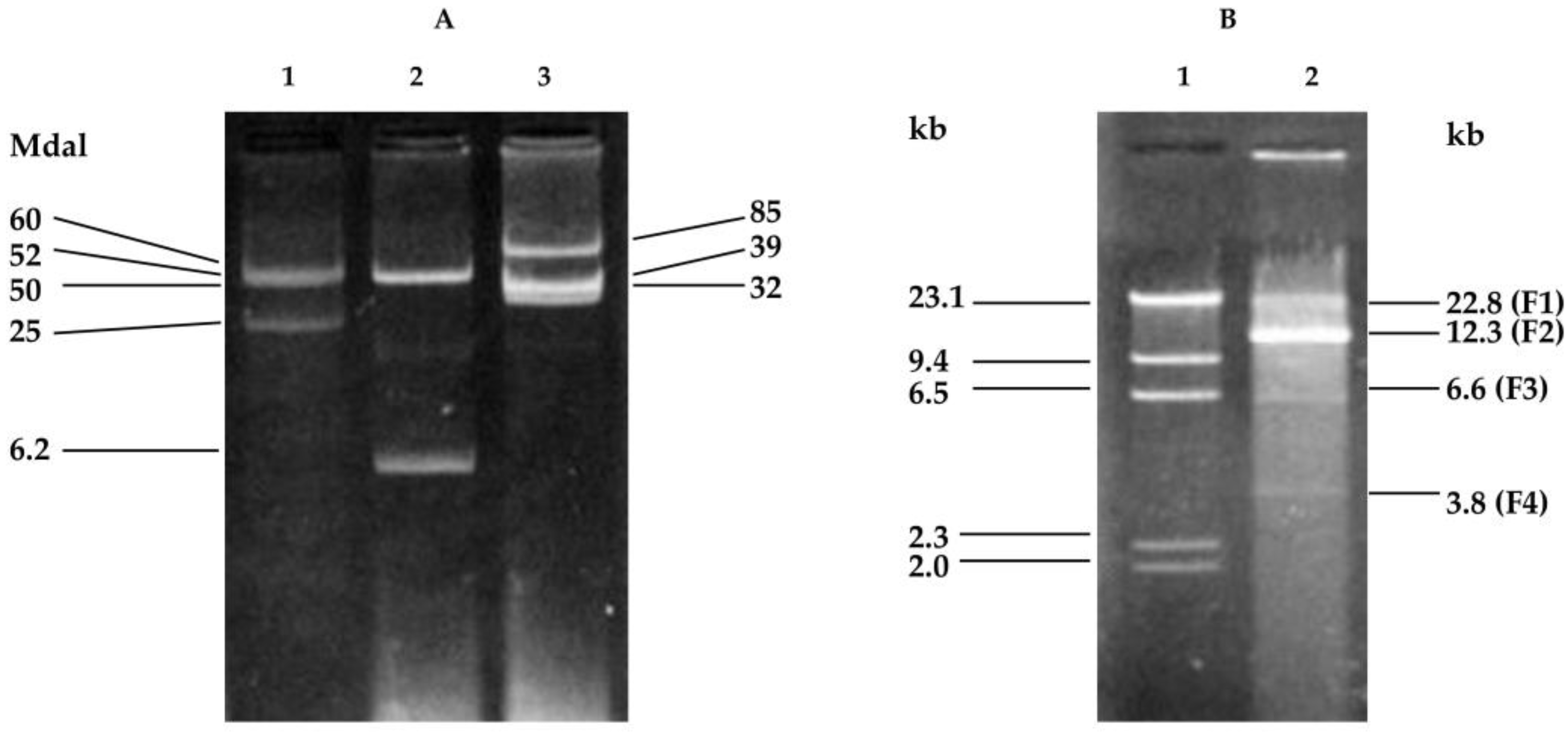
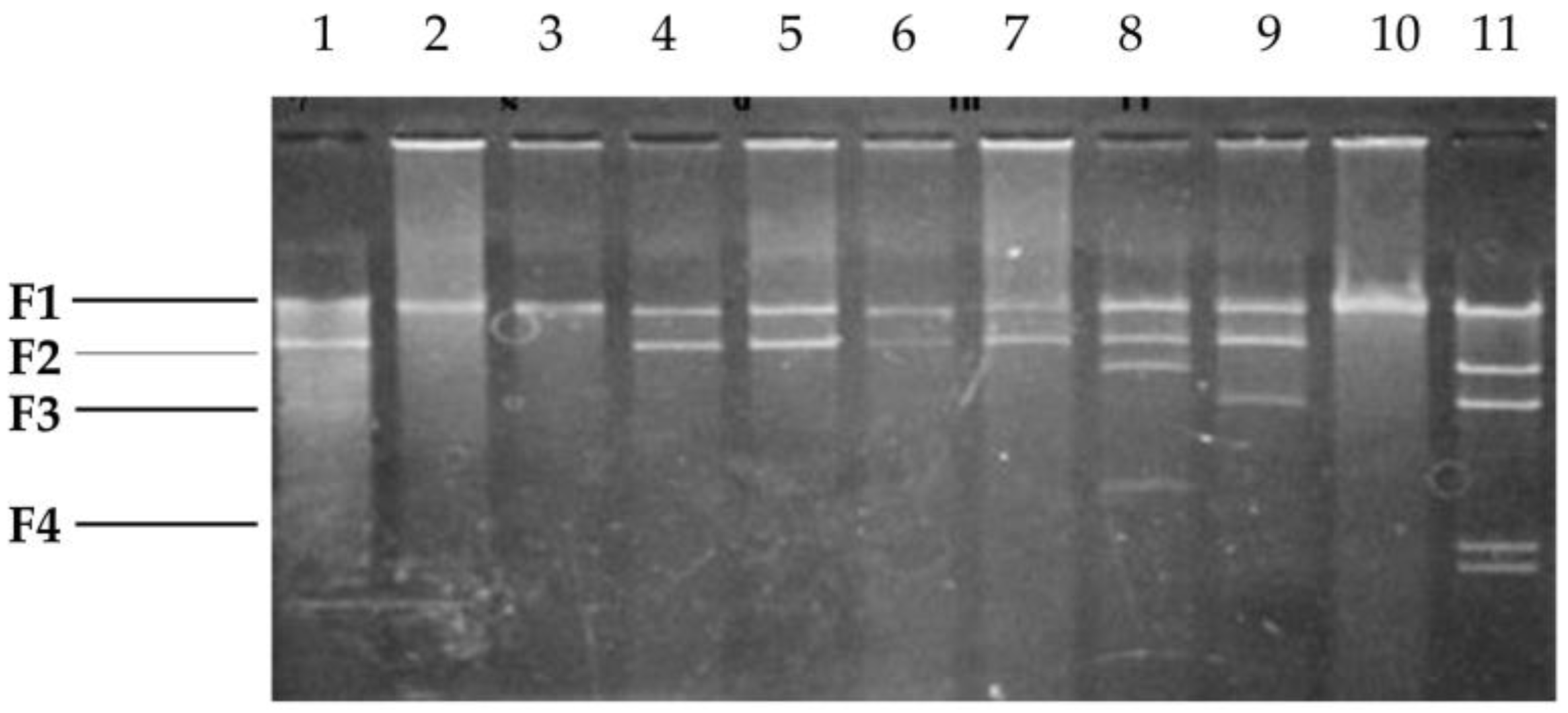
2.1. Cloning Fragments from Plasmid NAE89

Plasmids of the NAE89 strain were extracted following a method described in Kado and Liu [22], and used for agarose gel electrophoresis. Similar plasmid groups were extracted, including the NAE6 bacteria (A group) and NAE57 bacteria (C group). The NAE89 strains harbored two kinds of plasmids at 6.2 Mdal and 50 Mdal (Figure 1A). It was known that these strains belong to a group with similar plasmid patterns and had a high possibility of being borne by the plasmid [13].
It was assumed that the pathogenic gene, psvA associated with the establishment of canker disease, and the gene(s) involved in halo leaf spot formation, implicated that the plasmids of the NAE89 strain existed. Although the psvA gene was not cut by BamHI, the plasmids of the NAE89 strain were extracted for the first time, cut by BamHI, and used for agarose gel electrophoresis.
Consequently, four DNA fragments—22.8 kb (F1), 12.3 kb (F2), 6.6 kb (F3), and 3.8 kb (F4)—were produced (Figure 1B). The fragment of λ/HindIII was used as a standard and its molecular weight used to calculate the size of each DNA fragment. After ligation via in vitro packaging, the plasmids of the pLAFR3 and NAE89 strains were digested with BamHI and introduced into the E. coli strain DH5. All plasmids of the 200 obtained clones were examined (data not shown) based on their patterns and molecular weights. Their sizes became larger than pPLAFR3 in each plasmid, and the cloned pLAFR3 plasmids were identified (Figure 2). Furthermore, the plasmids derived from those clones were digested with BamHI and agarose gel electrophoresis was performed. Investigations were undertaken on the clones of the BamHI fragments of the observed NAE89 plasmid.
Results showed that pLAFC1, pLAFC2, and pLAFC9 were inserted into the F1 fragment at 22.8 kb. Whilst pLAFC3, pLAFC4, pLAFC5, and pLAFC6 were inserted into the F2 fragment at 12.3 kb, pLAFC8 was inserted into the F2 and F3 fragments, pLAFC7 was inserted into the F2 and F4 fragments, and a fragment of 9.4 kb was inserted into the BamHI site of pLAFR3 (Figure 2). In addition, although the fragment of 9.4 kb was also newly inserted into pLAFC7, the F3 and F4 fragments were joined together.
Four NAE89 plasmid fragments digested with BamHI were inserted. PE0 and NAE89-1, a bacterial strain non-pathogenic to the loquat, were introduced into NAE89 via triparental matings. Consequently, the inserted PE0 caused one deletion, and all fragment sizes become smaller. In the case of NAE89-1, the deletion occurred in two strains, while most were completely introduced. The result of a deletion varied between pLAFRC1 and pLAFRC2, in which F1 was the longest fragment inserted into PE0 (data not shown).

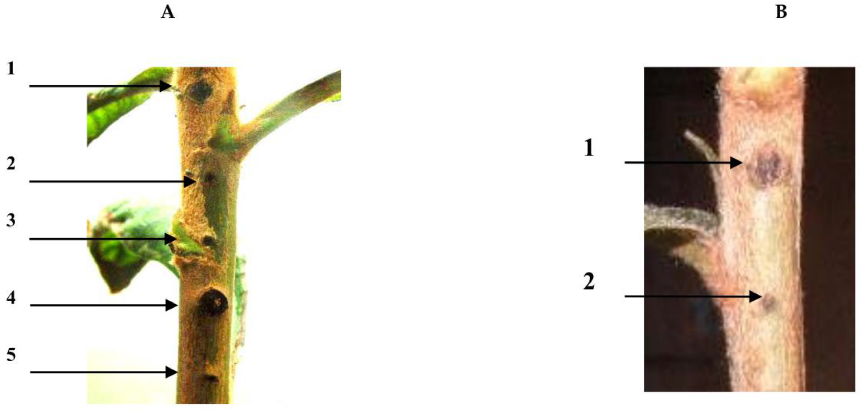
2.2. Pathogenic Experiment

The PE0 bacteria of the loquat did not carry the code for the psvA gene. As a result, the PE0 bacteria did not exhibit canker symptoms. Therefore, the NAE89 was a mutation stock that expressed a loss of halo leaf symptoms during its formation. The PE0 and NAE89-1 were introduced into pLAFR3, in which each BamHI fragment of the NAE89 plasmid was inserted by triparental matings. pLAFC1 and pLAFC2 from the F1 fragment with the largest size were inserted, and showed pathogenicity in the loquat stem. Other inserted fragments (F2–F4) did not display pathogenicity in the loquat stem (Figure 3A,B).

2.3. Southern Hybridization

The pathogenic examination revealed that the F1 fragment of the largest size showed pathogenicity. To investigate whether the pathogenic gene psvA existed, a Southern hybridization was performed using the pathogenic psvA (canker bacteria of the loquat) as a probe. Consequently, the F1 fragment hybridized with the largest size was formed, and the existence of the psvA gene(s) became clear (Figure 4B, lane 1). In addition, the 50 Mdal plasmid of the NAE89 strain and a hybrid were formed. The F1 fragment of the NAE89 obviously had a 50 Mdal origin (Figure 4A, lane 2). Of which, the 50 Mdal band of the plasmid was thin (unclear), and the hybrid was formed in the position of chromosomal DNA. This was part of the closed circular 50 Mdal fractured plasmid, and considered at a similar position as the linear chromosome using the agarose gel electrophoresis method.
However, the NAE6 strain was the 52 Mdal plasmid, from which the existence of the psvA gene was identified and hybrids were formed (Figure 4, lane 3). The pathogenicity was exhibited through a partial deletion when pLAFC1 and pLAFC2 were introduced into PE0 by triparental matings. It was proposed that there were deleted parts other than caused by the psvA gene(s), and the deletion plasmids introduced into PE0 were investigated by Southern hybridization. Each of these plasmids formed a hybrid with psvA, which indicated the existence of the psvA (Figure 5).

3. Discussion

This study showed that the strains of the group A harbored three plasmids at 25, 52, and 60 Mdal; the group B strains were at 6.2 and 50 Mdal; and the group C strains were at 32, 39, and 85 Mdal (Figure 1A). The present study showed that the psvA gene existed in the 50 Mdal plasmid of the NAE89 strain, which involved in stem canker formation. An attempt was made to detect a phenotypic marker that was encoded on the 52 Mdal plasmid in a similar manner [23]. By this study, it was found that the group B strain bacterium caused stem canker and halo leaf spots of loquat trees and harbored 6.2 and 50 Mdal gene fragments.
Kamiunten [16] reported that the pLAFR3 cosmid clone, pVIR6, which contained a virulence gene in the 23 kb insert DNA region. This virulence gene originated from the 52 Mdal plasmid fragment of the group A strain Ps. syringae pv. eriobotryae. The deletion analyses of pVIR6 determined that about 7 kb of the inserted DNA restored pathogenicity to an avirulent PE0 strain. The plasmid in which the deletion occurred was denoted to be the pKPN35. A 6961 bp inserted to the DNA of pKPN35 was sequenced, and four possible open reading frames were found in tandem [16]. Of which, ORF1 and ORF4 exerted no significant homology to known genes. The ORF2 had an amino acid sequence similar to the transposase of the IS5 of E. coli. A recombinant plasmid pNSF1 containing only the ORF3 region restored pathogenicity to the avirulent PE0 strain. However, an ORF3 mutant of pNSF1, which was constructed by deleting a 580 bp BssHII segment from ORF3, failed to restore virulence. Consequently, the ORF3 was identified as a virulent psvA gene [16].
In this experiment, it was found that the BamHI fragment of the plasmid NAE89 strain, belonging to group B contained a known psvA virulence gene. The BamHI cutting site and the halo leaf spot gene(s) being adjacent to the psvA gene of the cosmid vector pLAFR3 were also examined. Furthermore, the NAE89 strain plasmid produced four fragments (F1 fragment, 22.8 kb; F2 fragment, 12.3 kb; F3 fragment, 6.6 kb; F4 fragment, 3.8 kb). Following the result of investigating among 200 clones (data not shown), it was possible to obtain the E. coli DH5 into the four digested BamHI fragments. Hence, each of BamHI fragments (F1–F4) was introduced into NAE89-1 via triparental matings with PE0. When being inoculated (Figure 3), the F1 fragment of the largest size showed causal canker symptoms on the stems.
This study was the first to provide evidence that the psvA gene associated with the 50 Mdal plasmid, and proposed that the 50 Mdal plasmid of the group B strain might play an important role in the development of canker symptoms on loquat stems and pathogenic activities. These findings provide useful information for the clarification of the halo leaf spot gene(s). Further trials with sequences should be carried out to re-estimate the positions of the observed fragments (F1–F4). In addition, the involvement of the psvA gene in the group B strain should be further investigated.

4. Materials and Methods

4.1. Bacterial Strains, Plasmids, Culture Conditions, and Chemicals

The bacterial strains and studied plasmids were listed in Table 1. The Pseudomonas syringae pv. eriobotryae strains were kindly supplied by Nagasaki Fruit-Tree Experiment Station, Nagasaki Prefecture, Japan. They were originally isolated from loquat stem cankers collected from various locations in Japan, and used as a probe in this study. The bacteria were ordinarily cultured in yeast extract–peptone (YP) medium (yeast extract 5 g, polypetone 10 g, NaCl 5 g, glucose 1 g, 1000 mL distilled water, pH 7.0) at 25 °C, following a method described in Hasebe et al. [24]. For growth on solid medium, 1.5% agar was added. Escherichia coli strains were grown in similar medium at 37 °C. HindIII-digested λ DNA was used as markers of standard molecular weight.

4.2. Plant Inoculation

Young yellow-green loquats were selected and washed in 70% ethanol to disinfect them of bacteria or fungi. Bacteria grown on YP agar (1.5%) medium for 24 h were inoculated into one-year-old loquat stems using a needle. The inoculated plants were placed in polyethylene bags for 12 h to keep the humidity high, then they were transferred to a growth chamber at 25 °C with a 12 h photoperiod. The symptom of halo spot formation developments on the stems was observed at 60 days after inoculation.

4.3. General DNA Manipulation and Clone Plasmid

Plasmid extraction, restriction enzyme digestion, DNA ligation, plasmid clone, and agarose gel electrophoresis were carried out using standard procedures [22,25]. Plasmids were introduced into the E. coli by transformation and into Ps. syringae pv. eriobotryae by triparental matings with pRK2013 as a conjugative plasmid.

4.4. Southern Hybridization Experiments

The isolation of total genomic DNA was performed following a method previously described by Kamiunten [13]. The cosmid vector pLAFR3 was digested with BamHI to convert to a single linear fragment and electrophoresed. The linearized pLAFR3 was then isolated from the agarose gel with an Easytrap Kit (Takara Biochemicals, Tokyo, Japan) and used as probe DNA to detect the integrated NAE6 into chromosomal DNA. To identify the virulence expression, the gene psvA (ORF3) was used as a hybridization probe. The labelling of probe DNA with horseradish peroxidase, hybridization and detection by exposure on autoradiography film were done using an ECL direct nucleic acid labelling and detection kit (Amersham, London, UK) (GE Healthcare Life Sciences, Little Chalfont, UK).

Supplementary Materials

Supplementary File 1

Acknowledgments

The authors thank Hiroshi Kamiunten for his support and guidance in this research.

Author Contributions

T.D.K. conducted the experiments. T.D.X. wrote and revised the manuscript. Both authors had checked and approved the final version of the manuscript.

Conflicts of Interest

The authors declare no conflict of interests.

References

  1. Morita, A. Effect of the inoculation of Pseudomonas syringae pv. eriobotryae at seedling stage upon its growth and fruit-productivity. Ann. Phytopathol. Soc. Jpn. 1991, 57, 629–633. [Google Scholar]
  2. Fukuda, S.; Ishimoto, K.; Terakami, S.; Yamamoto, T.; Hiehata, N. Genetic mapping of the loquat canker resistance gene pse-c in loquat (Eriobotrya japonica). Sci. Hort. 2016, 200, 19–24. [Google Scholar] [CrossRef]
  3. Alippi, A.M.; Alippi, H.E. Stem canker of loquat: A new disease in Argentina. Rev. Argent. Microbiol. 1990, 22, 155–158. [Google Scholar] [PubMed]
  4. Lai, M.; McCartney, W.O.; Morin, C.W. Canker of loquat caused by Pseudomonas sp. Phytopathology 1971, 61, 248–249. [Google Scholar] [CrossRef]
  5. Lai, M.; Morin, C.W.; Weigle, C.G. Two strains of Pseudomonas eriobotryae isolated for loquat cankers in California. Phytopathology 1972, 62, 310–313. [Google Scholar] [CrossRef]
  6. Lin, S.; Sharpe, R.H.; Janick, J. Loquat: Botany and Horticulture. Hort. Rev. 1999, 23, 233–276. [Google Scholar]
  7. McRae, E.M.; Hale, C.N. New plant disease record in New Zealand: Loquat canker. N. Z. J. Agric. Res. 1986, 29, 547–550. [Google Scholar] [CrossRef]
  8. Mukoo, H. Studies on the causal bacteria of the loquat canker (1). Bull. Natl. Inst. Agric. Sci. 1952, C-1, 1–190. [Google Scholar]
  9. Wimalajeewa, D.L.S.; Pascoe, I.G.; Jones, D.L. Bacterial stem canker of loquat. Australas. Plant Pathol. 1978, 7. [Google Scholar] [CrossRef]
  10. Morita, A. Studies on the loquat canker caused by Pseudomonas eriobotryae (Takimoto) Dowson. II: Grouping of the bacterial isolates on the basis of their pigment producibility and pathogenicity. Ann. Phytopathol. Soc. Jpn. 1978, 44, 6–13. [Google Scholar] [CrossRef]
  11. Morita, A. Studies on physiology and ecology of loquat canker. J. Gen. Plant Pathol. 2001, 67, 244–247. [Google Scholar] [CrossRef]
  12. Fukuta, S.; Ishimoto, K.; Sato, S.; Terakami, S.; Yamamoto, T.; Hiehata, N. Genetic mapping of the loquat canker resistance locus in bronze loquat (Eriobotrya deflexa). Tree Genet. Genomes 2014, 10, 875–883. [Google Scholar] [CrossRef]
  13. Kamiunten, H. Involvement of a plasmid in the expression of virulence in Pseudomonas syringae pv. eriobotryae. Ann. Phytopathol. Soc. Jpn. 1995, 61, 376–380. [Google Scholar] [CrossRef]
  14. Jackson, R.W.; Athanassopoulos, E.; Tsiamis, G.; Mansfield, W.J.; Arnold, L.D.; Gibbon, J.M.; Murillo, J.; Taylor, D.J.; Vivian, A. Identification of a pathogenicity island, which contains genes for virulence and a virulence, on a large native plasmid in the bean pathogen Pseudomonas syringae pathovar phaseolicola. Proc. Natl. Acad. Sci. USA 1999, 96, 10875–10880. [Google Scholar] [CrossRef] [PubMed]
  15. Hiehata, N.; Sato, Y.; Fukuda, S.; Terai, O. Inheritance of resistance to loquat canker (Pseudomonas syringae pv. eriobotryae, Group A) in loquat (Eriobotrya japonica). J. Jpn. Soc. Hort. Sci. 2002, 71, 255–261. [Google Scholar] [CrossRef]
  16. Kamiunten, H. Isolation and characterization of virulence gene psvA on a plasmid of Pseudomonas syringae pv. eriobotryae. Ann. Phytopathol. Soc. Jpn. 1999, 65, 501–509. [Google Scholar] [CrossRef]
  17. Alan, V.; Jesús, M.; Robert, W.J. The roles of plasmids in phytopathogenic bacteria: mobile arsenal? Microbiology 2001, 147, 763–780. [Google Scholar]
  18. Gonzalez, C.F.; Pettit, E.A.; Valadez, V.A.; Provin, E.M. Mobilization, cloning and sequence determination of plasmid encoded polygalacturonase from a phytopathogenic Burkholderia Pseudomonas cepacia. Mol. Plant Microbe Interact. 1997, 10, 840–851. [Google Scholar] [CrossRef] [PubMed]
  19. Ezra, D.; Barash, I.; Valinsky, L.; Manulis, S. The dual function in virulence and host-range restriction of a gene isolated from the pPATHEhg plasmid of Erwinia herbicola pv. gypsophilae. Mol. Plant Microbe Interact. 2000, 13, 683–692. [Google Scholar] [CrossRef] [PubMed]
  20. Hiehata, N.; Sato, Y.; Fukuda, S.; Tominaga, Y.; Terai, O.; Nesumi, H. Inheritance of resistance to loquat canker (Group C) in progenies derived from ‘Shiromogi’ loquat. J. Am. Soc. Hort. Sci. 2012, 137, 152–156. [Google Scholar]
  21. Hiehata, N.; Fukuda, S.; Terai, O. Evaluation of loquat canker resistance in new loquat cultivars released in Japan and introduced from foreign countries. Bull. Nagasaki. Fruit Tree Exp. Stat. 2007, 10, 14–21. [Google Scholar]
  22. Kado, C.I.; Liu, S.T. Rapid procedure for detection and isolation of large and small plasmids. J. Bacteriol. 1981, 145, 1365–1373. [Google Scholar] [PubMed]
  23. Kamiunten, H. Loss of a plasmid in Pseudomonas syringae pv. eriobotryae is correlated with a change of symptoms. Ann. Phytopathol. Soc. Jpn. 1990, 56, 645–650. [Google Scholar] [CrossRef]
  24. Hasebe, A.; Tsushima, S.; Lida, S. Isolation and characterization of IS1416 from Pseudomonas glumae, a new member of the IS3 family. Plasmid 1998, 39, 196–204. [Google Scholar] [CrossRef] [PubMed]
  25. Perbal, B. A Practical Guide to Molecular Cloning, 2nd ed.; John Wiley & Sons: New York, NY, USA, 1988; pp. 396–399. [Google Scholar]
Figure 1. (A) Agarose gel electrophoresis of the plasmids from Ps. pv. eriobotryae; Lanes 1: 60, 52, and 25 Mdal plasmids of NAE6 (group A); Lanes: 50, 6.2 Mdal plasmids of NAE89 (group B); Lanes 3: 85, 39, 32 plasmids of NAE57 (group C); (B) Agarose gel electrophoresis of NAE89 plasmids digested with BamHI. Lane 1: λ/HindIII, 23.1, 9.4, 6.5, 2.3, and 2.0 kb. Lane 2: NAE89/BamHI with 22.8 (F1), 12.3 (F2), 6.6 (F3), and 3.8 kb (F4).
Pathogens 07 00041 g001
Figure 2. Size of DNA fragment of the pLAFR3 cosmid clone. Lane 1: NEA89 plasmid/BamHI (F1–F4); Lane 2: pLAFC1/BamHI (F1); Lane 3: pLAFC2/BamHI (F1); Lane 4: pLAFC3/BamHI (F2); Lane 5: pLAFC4/BamHI (F2); Lane 6: pLAFC5/BamHI (F2); Lane 7: pLAFC6/BamHI (F2); Lane 8: pLAFC7/BamHI (F2, F4); Lane 9: pLAFC8/BamHI (F2, F3); Lane 10: pLAFC9/BamHI (F1); Lane 11: standard of λ/HindIII.
Pathogens 07 00041 g002
Figure 3. Pathogenic expression of each cloned plasmid inoculated to loquat stem. (A) Virulent expression: 1. PE0-TM (C2); 2. PE0-TM (C5); 3. PE0-TM (C8); 4. NAE6; 5. PE0-TM (C7); (B) Invirulent expression (controls): 1. PE0-TM (C1) (PE0 without insertion), (2) distilled water; TM: treatment.
Pathogens 07 00041 g003
Figure 4. Southern hybridization of the NAE89 plasmid from the P. syringae pathovar using the virulence gene psvA as a probe. (A) Ethidium bromide gel. Lane 1: NAE89 was digested with BamHI, lane 2: NAE89, lane 3: NAE6, lane 4: λ/HindIII as a standard; (B) Southern blot of gel in panel. The F1 fragment with the largest size was formed from hybridization.
Pathogens 07 00041 g004
Figure 5. Southern hybridization showing that pathogenicity was recovered by a non-pathogenic strain in the triparental mating trial; (A) 1% agarose gel; (B) Southern blot of gel; Lane 1: NAE6; Lane 2: PE0-TM (C2) a; Lane 3: PE0-TM (C1) a; Lane 4: PE0-TM (C1) b; psvA used as a probe; TM (treatment).
Pathogens 07 00041 g005
Table 1. Bacterial strains and plasmids
Strains or PlasmidsRelevant PropertiesSource or Reference
Pseudomonas syringae pv. eriobotryae Nagasaki Fruit-Tree Experiment Station, Japan
NAE6Wild-type virulence in group A[13]
PE0Plasmid lost NAE 6[13]
NAE89Wild-type virulence in group BNippongene
NAE89-1The variant of lost halo gene formationNippongene
NAE57Wild-type in the C groupNippongene
E. coli DH5E. coli bacteria and bacterium strainNippongene
pET-3a (psvA)Vector inserted with BamHI to psvA used as a probe in this study, isolated from a bacteria lesion of the strain group AStratagene
pRK2013Helper plasmidThis study
pLAFR3Cosmid vectorThis study

