Article

Physiological and Genomic Characterization of a Novel Obligately Chemolithoautotrophic, Sulfur-Oxidizing Bacterium of Genus Thiomicrorhabdus Isolated from a Coastal Sediment

1
Key Laboratory of Marine Genetic Resources, Third Institute of Oceanography, Ministry of Natural Resources, Xiamen 361005, China
2
State Key Laboratory Breeding Base of Marine Genetic Resources, Xiamen 361005, China
3
School of Marine Sciences, China University of Geosciences, Beijing 100083, China
4
School of Marine Biology, Xiamen Ocean Vocational College, Xiamen 361005, China
*
Authors to whom correspondence should be addressed.
These authors contributed equally to this work.
Microorganisms 2023, 11(10), 2569; https://doi.org/10.3390/microorganisms11102569
Submission received: 13 September 2023 / Revised: 8 October 2023 / Accepted: 13 October 2023 / Published: 15 October 2023
(This article belongs to the Special Issue Microbial Communities in Changing Aquatic Environments)

Abstract

:
Thiomicrorhabdus species, belonging to the family Piscirickettsiaceae in the phylum Pseudomonadotav are usually detected in various sulfur-rich marine environments. However, only a few bacteria of Thiomicrorhabdus have been isolated, and their ecological roles and environmental adaptations still require further understanding. Here, we report a novel strain, XGS-01T, isolated from a coastal sediment, which belongs to genus Thiomicrorhabdus and is most closely related to Thiomicrorhabdus hydrogeniphila MAS2T, with a sequence similarity of 97.8%. Phenotypic characterization showed that XGS-01T is a mesophilic, sulfur-oxidizing, obligate chemolithoautotrophy, with carbon dioxide as its sole carbon source and oxygen as its sole electron acceptor. During thiosulfate oxidation, strain XGS-01T can produce extracellular sulfur of elemental α-S8, as confirmed via scanning electron microscopy and Raman spectromicroscopy. Polyphasic taxonomy results indicate that strain XGS-01T represents a novel species of the genus Thiomicrorhabdus, named Thiomicrorhabdus lithotrophica sp. nov. Genomic analysis confirmed that XGS-01T performed thiosulfate oxidation through a sox multienzyme complex, and harbored fcc and sqr genes for sulfide oxidation. Comparative genomics analysis among five available genomes from Thiomicrorhabdus species revealed that carbon fixation via the oxidation of reduced-sulfur compounds coupled with oxygen reduction is conserved metabolic pathways among members of genus Thiomicrorhabdus.

1. Introduction

The genus Thiomicrorhabdus belonging to the family Piscirickettsiaceae of the phylum Pseudomonadota (formerly named Proteobacteria) was first proposed based on phylogenetic analyses, including the 16S rRNA gene and 53 ribosomal protein sequences, by Boden et al. [1]. Members of genus Thiomicrorhabdus were distributed in various habitats, including deep-sea hydrothermal vents, continental shelf sediments, and intertidal mud flats [2,3,4,5]. Thiomicrorhabdus species are strict chemolithoautotrophs and have diverse energy metabolic pathways [6,7]. They generally oxidize reduced sulfur compounds to fix CO2 with oxygen or nitrate as electron acceptors and synthesize the primary productivity of ecosystems in the process [2,3,5].
At the time of writing, the genus Thiomicrorhabdus has 12 reported species with validly published names. Thiomicrorhabdus frisia JB-A2T is the first validly published species for the genus Thiomicrorhabdus and is isolated from marine sediments along the Wadden Sea [5]. Subsequently, Thiomicrorhabdus chilensis Ch-1T was isolated from the coastal shelf of Chilean [2]. In 2005, Knittel et al. identified the first cryophilic sulfur-oxidizing bacteria of this genus, Thiomicrorhabdus psychrophile SVAL-DT and Thiomicrorhabdus arctica SVAL-ET [3]. Thiomicrorhabdus hydrogeniphila MAS2T was isolated from a tank containing coastal seawater from Tokyo Bay [8]. Thiomicrorhabdus aquaedulcis HaS4T was isolated from the water of Lake Harutori in Japan [9]. Thiomicrorhabdus indica 13–15AT were isolated from the deep-sea hydrothermal vent environment of Southwest Indian Ocean [6]. Thiomicrorhabdus sediminis G1T and Thiomicrorhabdus xiamenensis G2T were isolated from sediment samples collected from the coast of Xiamen [7]. Recently, Thiomicrorhabdus heinhorstiae [10], Thiomicrorhabdus cannonii [10], and Thiomicrorhabdus immobilis [11] were discovered one after another.
Although genus Thiomicrorhabdus have been detected from worldwide marine systems, its physiological diversity and ecological role have not yet been made fully explicit due to the lack of cultivated strains. In this study, we isolated a new strain, named XGS-01T, from the coastal sediment of Xiamen, China. We characterized the bacterium as a novel species of genus Thiomicrorhabdus using a polyphasic taxonomic approach; meanwhile, we compared genomic analyses among members of the genus Thiomicrorhabdus to gain insights into the ecological roles and environmental adaptation mechanisms underlying their widespread distribution. Furthermore, we determined the characteristics of extracellular sulfur produced by Thiomicrorhabdus bacteria.

2. Materials and Methods

2.1. Enrichment and Isolation

Samples were collected in February 2021 from coastal sediments at Xiamen, Fujian, China (118°12′ E, 24°29′ N), and kept cold in dark during transportation to the laboratory. Samples were stored at 4 °C before further processing. A total of 1 g of sediment samples were transferred to 10 mL of modified MMJS medium [12] containing NaCl (15 g/L), CaCl2·2H2O (0.14 g/L), NH4Cl (0.25 g/L), MgCl2·6H2O (4.18 g/L), MgSO4·7H2O (3.4 g/L), KCl (0.33 g/L), K2HPO4 (0.14 g/L), NiCl2·6H2O (0.5 mg/L), Fe(NH4)2(SO4)2·6H2O (0.01 g/L), Na2SeO3·5H2O (0.5 mg/L), NaHCO3 (1 g/L), Wolfe trace elements 10 mL/L, and Wolfe vitamin 1 mL/L [13]. Before autoclaving, materials, except the vitamin solution, Na2S2O3·5H2O and NaHCO3, were dissolved, and the pH value of the medium was adjusted to 7.0. After autoclaving, the filter-sterilized vitamin solution, Na2S2O3·5H2O and NaHCO3, were added into the medium. Then, 10 mL medium was anaerobically dispensed into 50 mL serum bottles with gas purging and sealed with a butyl-rubber stopper under a gas phase of 76% N2/20% CO2/4% O2 (200 KPa) [14]. After 3 days of incubation at 28 °C, the cell growth was determined under the light microscope (Eclipse 80i, Nikon, Tokyo, Japan). Strain XGS-01T was obtained from the highest dilution via the dilution-to-extinction technique, and the purity was confirmed via microscopic examination and 16S rRNA gene sequencing.

2.2. DNA Extraction and Genomic Analyses

The genomic DNA of strain XGS-01T was extracted according to a previously reported method [15]. The complete genome of strain XGS-01T was sequenced using SMRT technology performed by Shanghai Majorbio Bio-pharm Technology Co., Ltd. (Shanghai, China). The sequencing was performed on a Pacific Biosciences (PacBio) sequencing platform via single-molecule real-time (SMRT) technology. The sequenced reads were filtered, and high-quality paired-end reads were assembled to reconstruct the circulating genome by using SOAPdenovo2 software. The G+C content of chromosomal DNA was determined from the complete genome sequences.

2.3. 16S rRNA Gene Phylogeny

The 16S rRNA gene was PCR-amplified and sequenced with 27F (5′-AGAGTTGATCMTGCGCTCAG-3′) and 1492R (3′-TACGYTACGTCTTACGACT-5′) primers. The almost full-length 16S rRNA gene sequence of strain XGS-01T was analyzed using the gapped-blast search algorithm [16]. The gene sequences of the strains were edited by DNAMAN and used to identify the 16S rRNA sequence similarities and phylogenetic neighbors by NCBI (https://www.ncbi.nlm.nih.gov/, accessed on 14 May 2023) and the Ez Biocloud server (https://www.ezbiocloud.net, accessed on 21 May 2023) [17]. The 16S rRNA sequences with taxa closely related to the strain XGS-01T were selected for downloading. The phylogenetic tree was constructed using MEGA 11.0 software [18] with three methods—the neighbor-joining (NJ), maximum-likelihood (ML), and maximum-parsimony (MP) methods [19]—for the target sequences and all downloaded gene sequences. Genetic distance was calculated using the Kimura two-parameter model [20]. Bootstrap analysis was calculated based on 1000 replications.

2.4. Morphology, Physiology, and Chemotaxonomic Analysis

Morphological observations of the novel isolated XGS-01T was performed via a transmission electron microscopy (JEM-1400, JEOL, Tokyo, Japan) with cultures grown in MMJS liquid medium at 32 °C for 1 day. The Gram stain test was conducted with a Gram-staining kit (Hangzhou Tianhe Microbiological Reagent Co., Ltd., Hangzhou, China).
The growth of strain XGS-01T was measured via direct cell counting using a phase contrast microscope (Eclipse 80i, Nikon). Triplicate cultures were examined under each condition, and cultures were grown in MMJS medium to determine the physiological characterization of strain XGS-01T. The growth temperature was tested at 0, 4, 10, 15, 20, 25, 28, 30, 35, 37, 45, and 55 °C. The pH range for growth was determined as from pH 4.5 to 9.0 at increments of 0.5 pH units using 30 mM hydrogen phosphate–citrate buffer (pH 4–5), MES (pH 5–6), PIPES (pH 6–7), HEPES (pH 7–7.5), and Tris (pH 8–9.5) at 30 °C. The salt concentration for growth was investigated by using MMJS medium supplemented with 0, 170, 340, 510, 680, 850, 1020, 1190, 1360, 1530, and 1700 mM NaCl. The effect of O2 on growth was measured at 30 °C by setting different O2 content gradients (0, 1, 2, 4, 6, 8, and 10% at 200 kPa and 21% at 100 kPa), and 10 mM nitrate was added as a potential electron acceptor for oxygen absence condition. After determining the optimal growth conditions, subsequent cultivation will use the optimal growth conditions.
The potential electron donors were investigated using sulfite (5 mM), thiocyanate (5 mM), tetrathionate (5 mM), elemental sulfur (1% w/v), or sodium sulfide (50 and 100 μM) to replace thiosulfate in MMJS medium, and the gas phase is 76% N2/20% CO2/4% O2 (200 kPa). The utilization of hydrogen was also examined with 76% H2/20% CO2/4% O2 (200 kPa) in the absence of thiosulfate. As to other electron acceptors, nitric acid (0.1%, w/v), nitrite (0.1 and 0.01%, w/v), sulfite (0.01%, w/v), and elemental sulfur (1%, w/v) were tested in MMJS medium with sodium thiosulfate as the electron donor. Heterotrophic growth was examined in MMJS medium by replacing NaHCO3 with potential organic carbon sources under the gas phase of 98% N2/2% O2 (200 kPa). The organic carbon sources including 20 amino acids (methionine, glutamine, glycine, alanine, valine, lysine, arginine, leucine, isoleucine, phenylalanine, proline, tyrosine, cysteine, threonine, tryptophan, serine, aspartic acid, and histidine) at a final concentration of 5 mM; 0.02% (w/v) glucose, sucrose, galactose, lactose, fructose, maltose and alginate; 5 mM formate, acetate, propionate, citrate, succinate, pyruvate; 0.1% (w/v) peptone, yeast extract, tryptone, starch; and a gas phase of 96% N2/4% O2 (200 kPa). In an attempt to determine the alternative energy source, these organic compounds were used as an energy source to replace thiosulfate in MMJS medium with the gas phase of 78% N2/20% CO2/2% O2 (200 kPa). To detect the utilization of nitrogen sources for XGS-01T, NH4Cl (5 mM), NaNO3 (5 mM), NaNO2 (5 mM), nitrilotriacetic acid (5 mM), and N2 were added to the MMJS medium without NH4Cl. The gas phase was 78% H2/20% CO2/4% O2 (200 kPa) and 78% N2/20% CO2/4% O2 (200 kPa). To detect antibiotic resistance, bacterial growth was detected by adding 0.1 g/L kanamycin, rifampicin, chloramphenicol, ampicillin, and streptomycin in MMJS medium, and other conditions were the same as the culture conditions.
Fatty acids of whole cells grown in the MMJS medium at 28 °C for 24 h were saponified, methylated, and extracted using the standard midi protocol (Sherlock Microbial Identification System, version 6.0B). Using the same method, the cellular fatty acid composition of T. sediminis G1T and T. xiamenensis G2T was also determined in parallel with that of strain XGS-01T in this study.

2.5. The Determination of Extracellular Sulfur via Scanning Electron Microscopy and Raman Spectromicroscopy

In order to determine the characteristics of extracellular sulfur produced by strain XGS-01T, strain XGS-01T was incubated at 32 °C for 12 h, and subsequently, the extracellular sulfur was collected by using 0.2 μm filter membrane and rinsed it with phosphate buffer solution (PBS) before centrifugation, repeating three times to remove residual culture medium on the surface of elemental sulfur. The collected elemental sulfur sample was determined using scanning electron microscopy (SEM) (ZEISS Sigma FE-SEM, Carl Zeiss, Jena, Germany), Raman spectromicroscopy (Horiba LabRAM HR-evolution, Longjumeau, France), and X-ray diffraction analysis (HyPix-400, Rigaku Ultima IV, Tokyo, Japan).

3. Results and Discussion

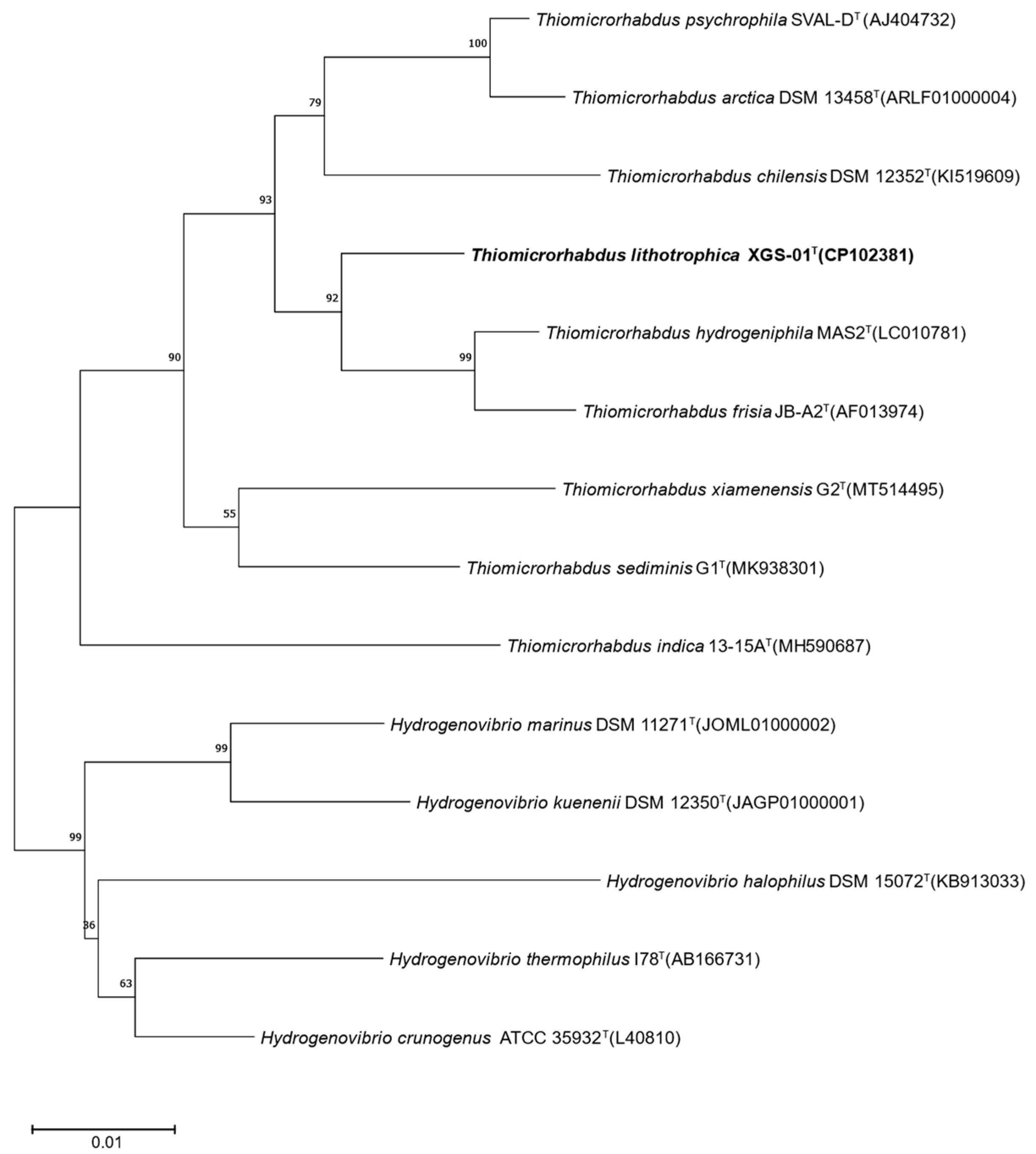
3.1. Phylogenetic Analysis Based on 16S rRNA Gene

The phylogenetic tree was constructed based on the NJ method for the complete 16S rRNA gene sequence (1553 bp) of XGS-01T, showing that strain XGS-01T was clustered with Thiomicrorhabdus species (Figure 1). The phylogenetic tree based on the ML method (Figure S1) and the MP method (Figure S2) also supports this topology. These results indicated that strain XGS-01T belonged to genus Thiomicrorhabdus. BLAST comparison showed that strain XGS-01T had the highest sequence similarity with T. hydrogeniphila MAS2T [21], with 97.8% similarity, followed by T. psychrophila SVAL-DT (97.4% similarity) [3] and T. frisia JB-A2T (97.3% similarity) [5]. This value is lower than the threshold criterion for prokaryotic species delineation, which is 98.65% for 16S rRNA gene similarity, indicating that strain XGS-01T represents a potential new species within the genus Thiomicrorhabdus.

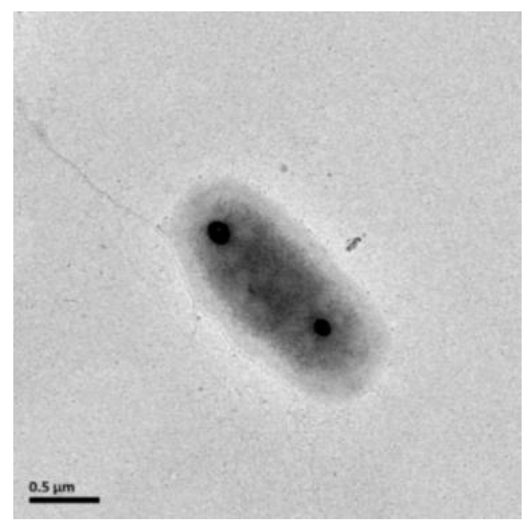
3.2. Morphology, Physiology, and Chemotaxonomic Analysis

Strain XGS-01T was rod-shaped, about 0.7–0.8 μm wide and 1.6–1.8 μm long, and had non-flagella (Figure 2), which was similar to other strains in the genus Thiomicrorhabdus. Cells were Gram stain negative. Strain XGS-01T could utilize a variety of reduced sulfur compounds as electron donors, including thiosulfate, sulfide, sulfur, and tetrathionate, but not sulfite. The strain XGS-01T cannot use hydrogen as an electron donor, which differs from that of its closely related species strain MAS2T. Oxygen is the sole electron acceptor for strain XGS-01T. In addition, strain XGS-01T could not utilize all tested organic matters as carbon sources and energy sources. These results indicate that strain XGS-01T is an obligate chemolithoautotrophic sulfur-oxidizing bacterium (Table 1). For the utilization of nitrogen sources, strain XGS-01T is able to utilize both inorganic nitrogen sources, such as ammonium, nitrogen and nitrate, and organic nitrogen, such as aminotriacetic acid. Strain XGS-01T could not use nitrite as the nitrogen source. The result is significantly different from strain MAS2T, which utilizes ammonium as the nitrogen source. Kanamycin, rifampicin, chloramphenicol, ampicillin, and streptomycin could inhibit the growth of strain XGS-01T, which was basically consistent with other strain types of this genus. The major fatty acids of strain XGS-01T are C18:1 (50.14%), C16:0 (15.97%), and C16:1 (6.90%). There are significant differences between strain XGS-01T and the closely related species, T. hydrogeniphila MAS2T. The proportion of fatty acid C16:1 in strain XGS-01T is 6.90%, whereas it is the main fatty acid in strain MAS2T, accounting for 44.40%. In addition, some fatty acids detected in strain XGS-01T were not found in strain MAS2T. The profiles of fatty acids in strain XGS-01T were also different from those of strains G1T and G2T, such as C18:0 (Table 2).

3.3. Genome Features and Central Metabolism among Genus Thiomicrorhabdus

The genome size of strain XGS-01T was 2.5 Mb, and the G+C content of strain XGS-01T was 39.1 mol%, which is similar to those of T. hydrogeniphila MAS2T (39.6 mol%) [8] and T. frisia. JB-A2T (39.6 mol%) [5] and lower than that of T. chilensis DSM 12352T (49.9 mol%) [2]. The average nucleotide identity (ANI) values between strain XGS-01T and other species were below the thresholds for species delineation (95–96%) [22], indicating that strain XGS-01T represents a novel species of the genus Thiomicrorhabdus. None of them have plasmids.
Central metabolic pathways, including sulfur oxidation, nitrogen, and carbon metabolism, were further analyzed among members of genus Thiomicrorhabdus, including the complete genome of strain XGS-01T obtained in this study, and T. frisia Kp2T, T. arctica DSM 13458T, T. chilensis DSM 12352T, and Thiomicrorhabdus sp. Milos-T2T, genomes of which are currently available in the NCBI database. The genome of strain XGS-01T contains a complete set of soxABCDXYZ genes encoding the Sox multienzyme complex, which performs thiosulfate oxidation. The gene cluster is ubiquitously present in all species of the genus Thiomicrorhabdus. This result shows that thiosulfate oxidation performed via a Sox multienzyme complex is highly conserved among Thiomicrorhabdus species. Strain XGS-01T has sqr and fcc genes for sulfide oxidation, which were also found in other species, except T. arctica SVAL-ET, which lacks the fcc gene (Table 3). There are no complete sulfate-reducing genes and dsr genes in the genome of strain XGS-01T. All genes encoding the carboxyl oxygenase (Rubisco) and ribulose diphosphate (RuBP), essential for carbon fixation, are present in the genome of strain XGS-01T. In addition, strain XGS-01T possesses two carbonic anhydrases, including α-type and γ-type. There are some differences in carbonic anhydrase among these species; for example, T. frisia JB-A2T, T. arctica DSM 134585T, and Thiomicrorhabdus sp. Milos-T2T have α-type, β-type, and γ-type, whereas T. chilensis DSM 12352T has α-type and γ-type carbonic anhydrase. Strain XGS-01T has an incomplete nitrogen metabolism pathway. The genome has no genes involved in the denitrification pathway. The genes encoding nitrate reductase and nitrite reductase are absent in the genome of strain XGS-01T. The incomplete nitrogen metabolism pathway was also found in the other species, except T. frisia JB-A2T.

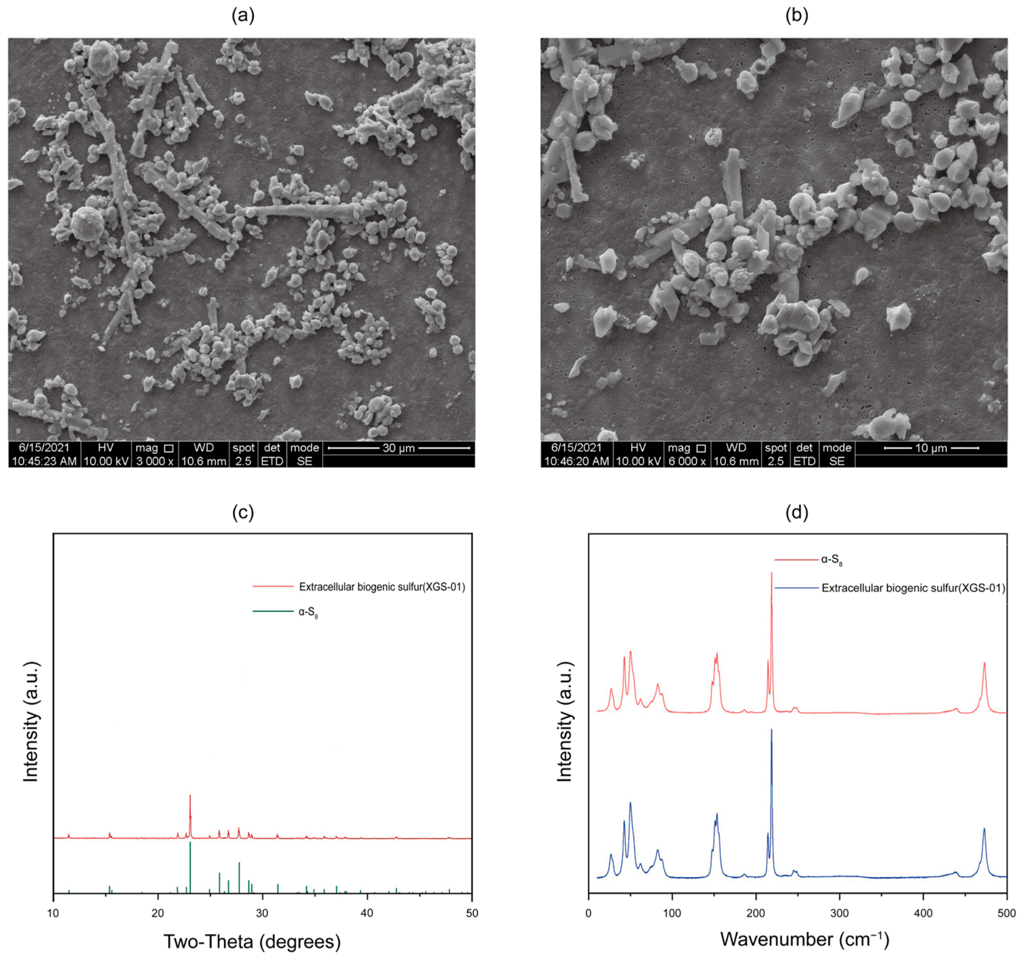
3.4. Characterization of Extracellular Sulfur Produced by Thiomicrorhabdus Species with SEM, XRD, and Raman Spectroscopy

Elemental sulfur was present in the culture during the mid-exponential growth phase when strain XGS-01T was incubated with thiosulfate and oxygen. Previous studies showed that the accumulation of elemental sulfur as an intermediate metabolite of thiosulfate oxidation only occurred in microorganisms lacking both SoxCD and dsr operons [23,24,25]. In this study, strain XGS-01T can not only oxidize thiosulfate to produce sulfate through the Sox system; it can also secrete extracellular sulfur during this process despite the presence of SoxCD genes in its genomes. The reason for extracellular sulfur production may be the inhibition of SoxCD. The expression of SoxCD usually depends on environmental pH, and the optimal pH value for the sulfite dehydrogenase (SoxCD) activity is 8.0 [26]. During the thiosulfate oxidation process of strain XGS-01T, the pH of medium will gradually decrease due to the production of metabolites, such as sulfate, and then inhibit the expression of SoxCD.
The extracellular sulfur was further characterized via SEM, XRD, and Raman spectroscopy. Morphological observations under SEM showed sulfur particles in two shapes: rod and globule. The length of rod-shaped sulfur can reach up to 30 μm, and some rod-shaped sulfur has short branches. The diameter of sulfur globules ranges from several hundred nanometers to 8 um, mostly 2–3 μm (Figure 3a,b). The XRD patterns of biogenic S0 matched well with the diffraction patterns of α-S8 in the database (Figure 3c). In addition, the Raman spectroscopy of S0 particles also showed the presence of α-S8 compared with a commercial S0 standard (Figure 3d). These results suggest that the elemental sulfur produced by strain XGS-01T are aggregates of nanocrystalline α-S8, which is similar to most currently reported extracellular sulfur types, such as Allochromatium vinosum, Chlorobaculum tempidum, and Sulfurimonas hydrogeniphila [27,28,29]. The produced a-S8 has been found in very diverse environments, such as marine sediments, water columns, sulfidic caves, and hydrothermal vents, and the pattern is the most thermodynamically stable form of elemental sulfur at ambient temperature and pressure [23,24,25]. The crystalline sulfur could serve as an important intermediate in the biogeochemical sulfur cycle and could be further consumed by a wide diversity of microorganisms, such as sulfur oxidizers, sulfur reducers, or sulfur disproportionators.

4. Conclusions

A novel Thiomicrorhabuds species was characterized from coastal sediments, which contributed to our understanding of the ecological role and environmental adaptation of Thiomicrorhabuds bacteria in marine environments. The result of comparisons of the complete 16S rRNA gene sequence showed that the sequence of strain XGS-01T was at least 2.2% different from other Thiomicrorhabdus members described previously. Pairwise ANI values between strain XGS-01T and other species of the genus Thiomicrorhabdus were 70.0–72.0%. According to the generally recognized criteria for delineating bacterial species, strains with a 16S rRNA gene sequence dissimilarity of greater than 2% and ANI values of less than 95–96% are considered to belong to separate species. Thus, strain XGS-01T does not belong to any presently described species. Although strain XGS-01T shared the highest 16S rRNA gene sequence similarity to T. hydrogeniphila MAS2T, many of the physiological characteristics of strain XGS-01T are different from strain MAS2T (Table 1). The growth conditions and maximum growth rate differed between the two strains. The utilization patterns of electron donors were also different in that strain XGS-01T could not use hydrogen as sole energy source, whereas strain MAS2T could. In addition, strain XGS-01T is able to utilize diverse inorganic nitrogen and organic nitrogen, whereas strain MAS2T only uses ammonium as a nitrogen source. There are also significant differences between strain XGS-01T and MAS2T in the composition of fatty acids. These results clearly indicate that strain XGS-01T can be differentiated from T. hydrogeniphila MAS2T at the species level. Correspondingly, genomic analysis confirmed that strain XGS-01T performed thiosulfate oxidation through a sox multienzyme complex and harbored fcc and sqr genes to catalyze sulfide oxidation. Comparative genomics further revealed that carbon fixation via the oxidation of reduced sulfur compounds coupled with oxygen reduction is conserved metabolic pathways among members of genus Thiomicrohabdus, indicating they play an important role in the sulfur oxidation process in aerobic marine environments. In addition, our study used strain XGS-01T as the model with which to first characterize the extracellular sulfur produced by genus Thiomicrorhabdus. The produced a-S8, as the most thermodynamically stable form of elemental sulfur, could serve as an important intermediate in the biogeochemical sulfur cycle and could be further consumed by a wide diversity of microorganisms. Our results provide new insight into the potential ecological significance of extracellular S0 produced by Thiomicrorhabdus in coastal marine ecosystems.

Description of Thiomicrorhabdus lithotrophica sp. nov

Thiomicrorhabdus lithotrophica XGS-01T.nov. (li.tho.tro’phi.ca. Gr. n. lithos, stone; N.L. fem. adj. trophica (from Gr. fem.adj. trophik), nursing, tending, or feeding; N.L. fem. adj. lithotrophica, inorganic-substrate-consuming.)
Cells are Gram-stain negative, rod-shaped (1.6–1.8 µm × 0.7–0.8 µm), motile, and have non-flagella. They are grown at 4–45 °C (optimum temperature, 30 °C), pH 5.0–9.0 (optimum pH 7.0), and 170–850 mM NaCl (optimum 510 mM). Strictly aerobic chemoautotrophic growth occurred with reduced sulfur compounds such as sulfide, thiosulfate, tetrasulfate, and elemental sulfur as electron donors and molecular oxygen as the only electron acceptor. Ammonium, nitrogen, nitrate, and aminotriacetic acid are utilized as nitrogen sources. It is sensitive to chloramphenicol (0.1 g/L), kanamycin (0.1 g/L), ampicillin (0.1 g/L), streptomycin (0.1 g/L), and vancomycin (0.1 g/L).
Type strain XGS-01T (=MCCC 1A18865T) was isolated from the coastal sediments of Xiamen, China. The GenBank accession number for the genome sequence of Thiomicrorhabdus lithotrophica XGS-01T is CP102381, and the 16S rRNA sequence number is OQ978225.

Supplementary Materials

The following supporting information can be downloaded at https://www.mdpi.com/article/10.3390/microorganisms11102569/s1: Figure S1: Maximum-likelihood phylogenetic tree based on 16S rRNA gene sequences; Figure S2: Maximum-Parsimony phylogenetic tree based on 16S rRNA gene sequences.

Author Contributions

Conceptualization, Y.G., H.Z. and L.J.; Data curation, Y.G. and H.Z.; Methodology, Y.G., H.Z. and J.W.; Resources, Z.S., S.W. and R.C.; Supervision, R.W. and R.C.; Writing—original draft, H.Z.; Writing—review and editing, L.J. and R.W. All authors have read and agreed to the published version of the manuscript.

Funding

This work was financially supported by the National Natural Science Foundation of China (No. 42176134), Scientific Research Foundation of Third Institute of Oceanography (No. 2019021), and National Natural Science Foundation of China (No. 42006088).

Institutional Review Board Statement

The present study does not include any experimental work with humans or vertebrates.

Informed Consent Statement

Not applicable.

Data Availability Statement

The GenBank accession numbers for the 16S rRNA gene sequence and the complete genome sequence of Thiomicrorhabdus lithotrophica XGS-01T are OQ978225 and CP102381, respectively.

Conflicts of Interest

The authors declare no conflict of interest.

References

  1. Boden, R.; Scott, K.M.; Williams, J.; Russel, S.; Antonen, K.; Rae, A.W.; Hutt, L.P. An evaluation of Thiomicrospira, Hydrogenovibrio and Thioalkalimicrobium: Reclassification of four species of Thiomicrospira to each Thiomicrorhabdus gen. nov. and Hydrogenovibrio, and reclassification of all four species of Thioalkalimicrobium to Thiomicrospira. Int. J. Syst. Evol. Microbiol. 2017, 67, 1140–1151. [Google Scholar] [CrossRef] [PubMed]
  2. Brinkhoff, T.; Muyzer, G.; Wirsen, C.O.; Kuever, J. Thiomicrospira chilensis sp. nov., a mesophilic obligately chemolithoautotrophic sulfuroxidizing bacterium isolated from a Thioploca mat. Int. J. Syst. Bacteriol. 1999, 49 Pt 2, 875–879. [Google Scholar] [CrossRef] [PubMed]
  3. Knittel, K.; Kuever, J.; Meyerdierks, A.; Meinke, R.; Amann, R.; Brinkhoff, T. Thiomicrospira arctica sp. nov. and Thiomicrospira psychrophila sp. nov., psychrophilic, obligately chemolithoautotrophic, sulfur-oxidizing bacteria isolated from marine Arctic sediments. Int. J. Syst. Evol. Microbiol. 2005, 55, 781–786. [Google Scholar] [CrossRef]
  4. Scott, K.M.; Williams, J.; Porter, C.M.B.; Russel, S.; Harmer, T.L.; Paul, J.H.; Antonen, K.M.; Bridges, M.K.; Camper, G.J.; Campla, C.K.; et al. Genomes of ubiquitous marine and hypersaline Hydrogenovibrio, Thiomicrorhabdus and Thiomicrospira spp. encode a diversity of mechanisms to sustain chemolithoautotrophy in heterogeneous environments. Environ. Microbiol. 2018, 20, 2686–2708. [Google Scholar] [CrossRef]
  5. Brinkhoff, T.; Muyzer, G.; Wirsen, C.O.; Kuever, J. Thiomicrospira kuenenii sp. nov. and Thiomicrospira frisia sp. nov., two mesophilic obligately chemolithoautotrophic sulfur-oxidizing bacteria isolated from an intertidal mud flat. Int. J. Syst. Bacteriol. 1999, 49 Pt 2, 385–392. [Google Scholar] [CrossRef]
  6. Liu, X.; Jiang, L.; Hu, Q.; Lyu, J.; Shao, Z. Thiomicrorhabdus indica sp. nov., an obligately chemolithoautotrophic, sulfur-oxidizing bacterium isolated from a deep-sea hydrothermal vent environment. Int. J. Syst. Evol. Microbiol. 2020, 70, 234–239. [Google Scholar] [CrossRef] [PubMed]
  7. Xuewen, L.; Baoping, C.; Qiliang, L.; Zongze, S.; Lijing, J. Thiomicrorhabdus sediminis sp. nov. and Thiomicrorhabdus xiamenensis sp. nov., novel sulfur-oxidizing bacteria isolated from coastal sediments and an emended description of the genus Thiomicrorhabdus. Int. J. Syst. Evol. Microbiol. 2021, 71, 004660. [Google Scholar] [CrossRef]
  8. Watsuji, T.O.; Hada, E.; Miyazaki, M.; Ichimura, M.; Takai, K. Thiomicrospira hydrogeniphila sp. nov., an aerobic, hydrogen- and sulfur-oxidizing chemolithoautotroph isolated from a seawater tank containing a block of beef tallow. Int. J. Syst. Evol. Microbiol. 2016, 66, 3688–3693. [Google Scholar] [CrossRef]
  9. Kojima, H.; Fukui, M. Thiomicrorhabdus aquaedulcis sp. nov., a sulfur-oxidizing bacterium isolated from lake water. Int. J. Syst. Evol. Microbiol. 2019, 69, 2849–2853. [Google Scholar] [CrossRef]
  10. Tatum, U.; Grayson, S.C.; Hunter, G.; Sheila, P.; Rebecca, P.; Robert, W.; Clare, D.; Madison, D.; James, B.; Rich, B.; et al. Thiomicrorhabdus heinhorstiae sp. nov. and Thiomicrorhabdus cannonii sp. nov.: Novel sulphur-oxidizing chemolithoautotrophs isolated from the chemocline of Hospital Hole, an anchialine sinkhole in Spring Hill, Florida, USA. Int. J. Syst. Evol. Microbiol. 2022, 72, 005233. [Google Scholar] [CrossRef]
  11. Kojima, H.; Mochizuki, J.; Kanda, M.; Watanabe, T.; Fukui, M. Thiomicrorhabdus immobilis sp. nov., a mesophilic sulfur-oxidizing bacterium isolated from sediment of a brackish lake in northern Japan. Arch. Microbiol. 2022, 204, 605. [Google Scholar] [CrossRef] [PubMed]
  12. Takai, K.; Inagaki, F.; Nakagawa, S.; Hirayama, H.; Nunoura, T.; Sako, Y.; Nelson, K.H.; Horikoshi, K. Isolation and phylogenetic diversity of members of previously uncultivated epsilon-Proteobacteria in deep-sea hydrothermal fields. FEMS Microbiol. Lett. 2003, 218, 167–174. [Google Scholar] [CrossRef] [PubMed]
  13. Balch, W.E.; Fox, G.E.; Magrum, L.J.; Woese, C.R.; Wolfe, R. Methanogens: Reevaluation of a unique biological group. Microbiol. Rev. 1979, 43, 260–296. [Google Scholar] [CrossRef]
  14. Jiang, L.; Lyu, J.; Shao, Z. Sulfur Metabolism of Hydrogenovibrio thermophilus Strain S5 and Its Adaptations to Deep-Sea Hydrothermal Vent Environment. Front. Microbiol. 2017, 8, 2513. [Google Scholar] [CrossRef] [PubMed]
  15. Jiang, L.; Zheng, Y.; Peng, X.; Zhou, H.; Zhang, C.; Xiao, X.; Wang, F. Vertical distribution and diversity of sulfate-reducing prokaryotes in the Pearl River estuarine sediments, Southern China. FEMS Microbiol. Ecol. 2009, 70, 93–106. [Google Scholar] [CrossRef]
  16. Altschul, S.F.; Madden, T.L.; Schäffer, A.A.; Zhang, J.; Zhang, Z.; Miller, W.; Lipman, D.J. Gapped BLAST and PSI-BLAST: A new generation of protein database search programs. Nucleic Acids Res. 1997, 25, 3389–3402. [Google Scholar] [CrossRef]
  17. Kim, O.S.; Cho, Y.J.; Lee, K.; Yoon, S.H.; Kim, M.; Na, H.; Park, S.C.; Joen, Y.S.; Lee, J.-H.; Yi, H. Introducing EzTaxon-e: A prokaryotic 16S rRNA gene sequence database with phylotypes that represent uncultured species. Int. J. Syst. Evol. Microbiol. 2012, 62 Pt 3, 716–721. [Google Scholar] [CrossRef]
  18. Sudhir, K.; Glen, S.; Koichiro, T. MEGA7: Molecular Evolutionary Genetics Analysis Version 7.0 for Bigger Datasets. Mol. Biol. Evol. 2016, 33, 1870–1874. [Google Scholar] [CrossRef]
  19. Naruya, S.; Masatoshi, N. The neighbor-joining method: A new method for reconstructing phylogenetic trees. Mol. Biol. Evol. 1987, 4, 406–425. [Google Scholar] [CrossRef]
  20. Kimura, M. A simple method for estimating evolutionary rates of base substitutions through comparative studies of nucleotide sequences. J. Mol. Evol. 1980, 16, 111–120. [Google Scholar] [CrossRef]
  21. Boden, R.; Scott, K.M.; Rae, A.W.; Hutt, L.P. Reclassification of Thiomicrospira hydrogeniphila (Watsuji et al. 2016) to Thiomicrorhabdus hydrogeniphila comb. nov., with emended description of Thiomicrorhabdus (Boden et al., 2017). Int. J. Syst. Evol. Microbiol. 2017, 67, 4205–4209. [Google Scholar] [CrossRef] [PubMed]
  22. Meier-Kolthoff, J.P.; Klenk, H.P.; Göker, M. Taxonomic use of DNA G+ C content and DNA–DNA hybridization in the genomic age. Int. J. Syst. Evol. Microbiol. 2014, 64 Pt 2, 352–356. [Google Scholar] [CrossRef] [PubMed]
  23. Marnocha, C.L.; Sabanayagam, C.R.; Modla, S.; Powell, D.H.; Henri, P.A.; Steele, A.S.; Hanson, T.E.; Webb, S.M.; Chan, C.S. Insights into the Mineralogy and Surface Chemistry of Extracellular Biogenic S(0) Globules Produced by Chlorobaculum tepidum. Front. Microbiol. 2019, 10, 271. [Google Scholar] [CrossRef]
  24. Pickering, I.J.; George, G.N.; Yu, E.Y.; Brune, D.C.; Tuschak, C.; Overmann, J.; Beatty, T.; Prince, R.C. Analysis of sulfur biochemistry of sulfur bacteria using X-ray absorption spectroscopy. Biochemistry 2001, 40, 8138–8145. [Google Scholar] [CrossRef] [PubMed]
  25. Wang, S.; Jiang, L.; Hu, Q.; Cui, L.; Zhu, B.; Fu, X.; Lai, Q.; Shao, Z.; Yang, S. Characterization of Sulfurimonas hydrogeniphila sp. nov., a Novel Bacterium Predominant in Deep-Sea Hydrothermal Vents and Comparative Genomic Analyses of the Genus Sulfurimonas. Front. Microbiol. 2021, 12, 626705. [Google Scholar] [CrossRef]
  26. Friedrich, C.G.; Bardischewsky, F.; Rother, D.; Quentmeier, A.; Fischer, J. Prokaryotic sulfur oxidation. Curr. Opin. Microbiol. 2005, 8, 253–259. [Google Scholar] [CrossRef]
  27. Jerez, C.A.; Albar, J.P.; Paradela, A.; Beard, S. Growth of Acidithiobacillus ferrooxidans ATCC 23270 in Thiosulfate under Oxygen-Limiting Conditions Generates Extracellular Sulfur Globules by Means of a Secreted Tetrathionate Hydrolase. Front. Microbiol. 2011, 2, 79. [Google Scholar] [CrossRef]
  28. Javor, B.J.; Wilmot, D.B.; Vetter, R.D. pH-Dependent metabolism of thiosulfate and sulfur globules in the chemolithotrophic marine bacterium Thiomicrospira crunogena. Arch. Microbiol. 1990, 154, 231–238. [Google Scholar] [CrossRef]
  29. Houghton, J.L.; Foustoukos, D.I.; Flynn, T.M.; Vetriani, C.; Bradley, A.S.; Fike, D.A. Thiosulfate oxidation by Thiomicrospira thermophila: Metabolic flexibility in response to ambient geochemistry. Environ. Microbiol. 2016, 18, 3057–3072. [Google Scholar] [CrossRef]
Figure 1. Neighbor-joining phylogenetic tree based on 16S rRNA gene sequences showing the relationship between strain XGS-01T and members of the genus Thiomicrorhabdus. Bootstrap values (>50%) based on 1000 replicates are shown at branch nodes. Bar, 0.01 substitutions per nucleotide position.
Microorganisms 11 02569 g001
Figure 2. Transmission electron microscopy of a cell of strain XGS-01T.
Microorganisms 11 02569 g002
Figure 3. The morphology and structure of extracellular sulfur. (a,b) Extracellular sulfur morphology under scanning electron microscopy; (c) XRD pattern of extracellular sulfur; (d) Raman spectroscopy of extracellular sulfur.
Microorganisms 11 02569 g003
Table 1. Comparison of characteristics of strain XGS-01T and other species within the genus Thiomicrorhabdus. Strains: 1, T. lithotrophica XGS-01T; 2, T. hydrogeniphila MAS2T; 3, T. frisia JB-A2T; 4, T. psychrophila SVAL-DT; 5, T. arctica DSM 13458T. +, positive; −, negative.
Characteristic12345
ShapeRodRodRodRodRod
G+C content (mol%)39.139.639.642.542.2
Maximum growth rate (h−1)0.150.400.450.200.14
Temperature range (°C)4.0–45.02.0–40.03.5–39.0−2.0–20.8−2.0–20.8
Optimal temperature (°C)30.030.025.0–32.014.6–15.411.5–13.2
Optimal Na+ concentration (mM)510270470250250
pH range5.0–9.05.0–8.04.2–8.56.5–9.06.5–9.0
Optimal pH7.06.06.57.5–8.57.3–8.0
Electron donor:
Thiosulfate+++++
Sulfide+++++
Elemental sulfur+++++
Sulfite
Tetrathionate+++++
Hydrogen+
Table 2. Cellular fatty acid composition of strain XGS-01T, T. hydrogeniphila MAS2T, T. sediminis G1T, and T. xiamenensis G2T. Strain: 1, XGS-01T; 2, T. hydrogeniphila MAS2T; 3, T. sediminis G1T; 4, T. xiamenensis G2T. Values are percentages of total fatty acids. ND, Not detected.
Fatty Acid1234
Saturated:
C9:00.23ND1.112.85
C11:00.06NDND1.01
C12:00.431.001.631.08
C14:01.380.803.612.90
C16:015.9716.9027.0325.65
C17:0NDND1.341.18
C18:05.911.9033.7929.42
C19:00.17NDND2.24
Unsaturated:
C16:16.9044.403.603.17
C17:10.81ND4.873.17
C18:150.1430.409.217.47
C18:20.56ND3.602.59
C18:30.42ND1.321.27
Hydroxy:
C8:0 3OH0.25ND0.680.57
C12:0 3OH5.01ND1.763.03
C18:1 2OH0.49ND4.25ND
Table 3. Genome comparison of five strains. Strain 1, T. lithotrophica XGS-01T; 2, T. frisia JB-A2T; 3, T. arctica DSM 13458T; 4, T. chilensis DSM 12352T; 5, T. sp. Milos-T2T. +, positive; −, negative.
Characteristic12345
Genome size (Mb)2.52.72.62.42.6
GC content (%)39.139.642.249.938.0
ANI71.170.170.471.5
Gene count22922526233722852382
rRNA operons93323
tRNA count4745454346
SoxABCDXYZABCDXYZABCDXYZABCDXYZABCDXYZ
Fcc++++
Sqr+++++
TCA Cycle+++++
Pentose phosphate pathway+++++
Glycolysis/Gluconeogenesis+++++
Carboxysome+++++
Carbonic anhydrase1α-class,
1γ-class
1α-class,
1γ-class,
1β-class
1α-class,
1γ-class,
1β-class
1α-class,
2γ-class
1α-class,
1γ-class,
1β-class
Nitrate reductase +
Nitrite reductase ++
Disclaimer/Publisher’s Note: The statements, opinions and data contained in all publications are solely those of the individual author(s) and contributor(s) and not of MDPI and/or the editor(s). MDPI and/or the editor(s) disclaim responsibility for any injury to people or property resulting from any ideas, methods, instructions or products referred to in the content.

