Article

Genomic Characteristics and Functional Analysis of Brucella sp. Strain WY7 Isolated from Antarctic Krill

1
State Key Laboratory for Biocontrol and Southern Marine Science and Engineering Guangdong Laboratory (Zhuhai), School of Marine Sciences, Sun Yat-sen University, 135 Xingang Road West, Guangzhou 510275, China
2
Key Laboratory of the East China Sea and Oceanic Fishery Resources Exploitation, Ministry of Agriculture, East China Sea Fisheries Research Institute, Shanghai 116023, China
3
Guangdong Province Key Laboratory for Aquatic Economic Animals, and Guangdong Provincial Key Laboratory of Marine Resources and Coastal Engineering, Sun Yat-sen University, 135 Xingang Road West, Guangzhou 510275, China
*
Author to whom correspondence should be addressed.
These authors contributed equally to this work.
Microorganisms 2023, 11(9), 2281; https://doi.org/10.3390/microorganisms11092281
Submission received: 16 July 2023 / Revised: 7 September 2023 / Accepted: 8 September 2023 / Published: 11 September 2023

Abstract

:
Antarctic krill (Euphausia superba) is a key species of the Antarctic ecosystem whose unique ecological status and great development potential have attracted extensive attention. However, the genomic characteristics and potential biological functions of the symbiotic microorganisms of Antarctic krill remain unknown. In this study, we cultured and identified a strain of Brucella sp. WY7 from Antarctic krill using whole-genome sequencing and assembly, functional annotation, and comparative genomics analysis. First, based on 16S rDNA sequence alignment and phylogenetic tree analysis, we identified strain WY7 as Brucella. The assembled genome of strain WY7 revealed that it has two chromosomes and a plasmid, with a total genome length of 4,698,850 bp and an average G + C content of 57.18%. The DNA—DNA hybridization value and average nucleotide identity value of strain WY7 and Brucella anthropi ATCC® 49188TM, a type strain isolated from human clinical specimens, were 94.8% and 99.07%, respectively, indicating that strain WY7 is closely related to Brucella anthropi. Genomic island prediction showed that the strain has 60 genomic islands, which may produce HigB and VapC toxins. AntiSMASH analysis results showed that strain WY7 might produce many secondary metabolites, such as terpenes, siderophores and ectoine. Moreover, the genome contains genes involved in the degradation of aromatic compounds, suggesting that strain WY7 can use aromatic compounds in its metabolism. Our work will help to understand the genomic characteristics and metabolic potential of bacterial strains isolated from Antarctic krill, thereby revealing their roles in Antarctic krill and marine ecosystems.

1. Introduction

The Antarctic krill is endemic in the Southern Ocean, serving as a direct energy link between the ocean’s primary producers (phytoplankton) and higher trophic levels. Krill comprises the largest population of a multicellular wild animal species on Earth [1] and plays a critical role in marine biogeochemical cycles that affect climate and ocean productivity. Antarctic krill has a unique living environment, and a large number of symbiotic microorganisms colonize its body surface, digestive tract, and other organs [2]. These symbiotic microorganisms may be closely related to important life activities such as metabolism, immunity, environmental adaptation, and host defence [3,4]. This also makes the extreme marine microorganisms in the Antarctic a natural resource pool of interest. Cui X et al. [5] and Wang Y et al. [6] isolated three phyla of culturable bacteria from Antarctic krill, including Firmicutes, Proteobacteria and Actinobacteria, and showed their affiliation with twelve genera, including Bacillus, Delftia, Psychrobacter, Acinetobacter and Pseudoalteromonas. An increasing number of active secondary metabolites have been found from different groups of Antarctic microorganisms, showing antibacterial, antitumour, antiviral, antioxidant, immunomodulatory and other biological activities [7,8]. However, current research on Antarctic krill mainly focuses on fishing, storage, and the processing of products [9,10], and there are few reports on the species diversity of symbiotic bacteria in Antarctic krill and their possible biological functions.
The genus Brucella, an α-2 subclass of Proteobacteria, is rod-shaped, aerobic, gram-negative, nonpigmented, free-living, and capable of producing acids from several carbohydrates and degrading nitrates and nitrites, taking advantage of abundant surrounding flagellar motility [11]. Brucella was first discovered in 1988 when Holmes et al. [11] isolated Brucella anthropi from human clinical specimens, mostly blood cultures. At present, Brucella has been found in a variety of environments, including water, soil, plants and animals. According to some published studies, Brucella can degrade hydrocarbons such as petroleum under extreme conditions [12]. Brucella strains that can efficiently degrade oil with crude oil as the sole carbon source were isolated from seabed sediments in the oil-polluted area of Xingang, Dalian [13]. A strain of Brucella found in the deep sea can produce new annamycin antibiotics, trienocycline H and I [14], which may have strong antitumour activity. It has also been reported that under anaerobic conditions, Brucella can produce hydrocarbons and act as biosurfactants (BSFs) [15]. The genus Brucella is known to catabolize aromatic compounds [16]. Wang X et al. [17] isolated a strain of Brucella anthropi from saline soil contaminated by polycyclic aromatic hydrocarbons (PAHs) that could degrade PAHs with 5% NaCl. In conclusion, previous studies have shown that Brucella has important application value in the degradation of aromatic hydrocarbons and the production of new antibiotics, biosurfactants and so on.
In the present study, the strain Brucella sp. WY7 from Antarctic krill was isolated and identified with whole-genome sequencing and assembly, functional annotation, and comparative genomics analysis.

2. Materials and Methods

2.1. Bacterial Isolation and Transmission Electron Microscopy Observations

Antarctic krill samples (58–60° W, 62–64° S) were obtained from the East China Sea Fisheries Research Institute (Shanghai, China) in November 2016 [6]. The samples were washed three times with sterile seawater to remove excess sediment and microorganisms. Three krill from the same site were ground in a mortar as a sample. The ground samples were diluted with approximately 1 mL of sterile water, collected in a 2 mL sterile centrifuge tube, and centrifuged at 3500 rpm for 5–10 min. A small amount of supernatant was obtained using the inoculation loop and applied to agar-mixed LB medium. The inoculated medium was cultured at 10 °C until colonies were visible. The colonies were randomly isolated from the medium, collected and expanded approximately three times under the same conditions. A total of 86 colonies were obtained, and the strains were stored in liquid medium containing 20% glycerol in a refrigerator at −8 °C for subsequent use. The shape of the isolated bacteria was observed by transmission electron microscopy. A small number of colonies were picked by the inoculation loop and put into a 1.5 mL centrifuge tube. Then, sterile ultrapure water was added, mixed, and centrifuged at low speed, and the cleaning was repeated 3 times. Ten microlitres of bacterial solution were adsorbed on the copper wire for observation and negatively stained with a 2% sodium phosphotungstate solution. Finally, the shape of the bacteria was observed by transmission electron microscopy.

2.2. DNA Extraction, PCR Amplification and Phylogenetic Tree Construction

Genomic DNA was extracted from purified and cultured strains using a bacterial DNA extraction kit (Omega, Washington, DC, USA). Using the extracted DNA of the strain as a template, the sequence of the 8–1510 bp region in bacterial 16S rDNA was amplified with universal primers 27F (5′-AGAGTTTGATCCTGGCTCA-3′) and 1492R (5′-GGTTACCTTGTTACGACTT-3′) (Tsingke, Beijing, China), and then NCBI BLAST was used for comparison. The length of the amplified fragment was approximately 1500 bp. The ClustalW program in MEGA-X was used to analyse the phylogeny [18,19,20].

2.3. Genome Assembly

The PacBio Sequel platform was used for single-molecule real-time (SMRT) sequencing [21]. PacBio sequencing was carried out by OE Biotech Co., Ltd. (Shanghai, China). Based on third-generation sequencing data, the genome was assembled from scratch. Falcon [22] was used for preliminary assembly, and GenomicConsensus v0.8.0 was used for correction. The SPRAI (single-pass read accuracy improver)-corrected sequence was used as auxiliary data for Circulator [23] cyclization processing, and the cyclized bacterial genome was obtained. The genome information of the bacteria was uploaded to NCBI, and accession numbers are CP049796–CP049798 for Brucella sp.

2.4. Genomic Bioinformatics Analysis

The software Prodigal (Prokaryotic Dynamic Programming Genefinding Algorithm) v2.6.3 [24] was used for coding-gene prediction. RepeatMasker v4.0.7 [25] was used to predict the repeat sequence. tRNAscan-SE v1.3.1 [26] was used to predict tRNA, RNAmmer v1.2 [27] was used to predict rRNA, and Rfam v10.0 [28] was used to predict sRNA. The preceding software parameters were default.
A genome circle map was generated using the Proksee server. The dDDH (digital DNA—DNA hybridization) values were calculated using the Type (Strain) Genome Server [29]. The JSpeciesWS online service [30] was used to calculate the ANIb (average nucleotide identity) value between genomes. Annotation was carried out using RAST (Rapid Annotation using Subsystem Technology) server 2.0 [31]. The RAST subsystem is a collection of functionally related protein families. RAST is an automated annotation server for microbial genomes built on the framework provided by the SEED system. The KEGG (Kyoto Encyclopedia of Genes and Genomes) database [32] was used for annotation. Briefly, the comparison results were compared with the KEGG database, and then KOBAS v3.0 (http://kobas.cbi.pku.edu.cn/kobas-2.1.1/kobas-3.0.3.tar.gz, accessed on 15 July 2023) [33] was used to convert the comparison results into KO and pathway annotations. CAZy (Carbohydrate Active Enzymes Database) [34] was used for annotation. The database contains families of enzymes that catalyse carbohydrate degradation, modification, and biosynthesis. Genomic islands (GIs) were predicted using IslandViewer 4 [35]. A genomic island is a kind of gene cluster that can be transferred horizontally in the genome of microorganisms. Genome islands can be predicted by differences in the genome composition of different bacterial species, mainly by SIGI-HMM, IslandPath-DIMOB and other methods. The AntiSMASH server [36] was used to predict biosynthesis-related gene clusters and perform secondary metabolic analysis.
The OrthoVenn2 server [37] was used for genomic homology cluster analysis. Venn diagrams and histograms were used to describe the comparative genomic analysis of the genome of this bacterium and its related species, showing the number and function of homologous and nonhomologous gene clusters, thus analysing the functional characteristics of this bacterium.

3. Results

3.1. Taxonomic Identification of 16S rDNA and Morphological Characteristics

The 16S rDNA sequence alignment using NCBI BLAST showed that the sequence similarity between the isolated strain WY7 and Brucella anthropi ATCC® 49188TM was more than 99%. To further determine the phylogenetic status of the strain, a phylogenetic tree was constructed based on the 16S rDNA sequence. The results showed that the strain was closely related to Brucella anthropi ATCC® 49188TM isolated from clinical human blood [8] and Brucella cytisi ESC1 isolated from nodules of Cytisus scoparius growing in Spanish soil [38], both of which were in an independent branch (Figure 1A). Transmission electron microscopy observation showed that strain Brucella sp. WY7 was a bacterium with parallel sides and round ends, often appeared singly, and the size of a single cell was approximately 1.15–1.40 μm long and 0.70–0.85 μm wide (Figure 1B). These results suggested that strain WY7 might belong to the genus Brucella.

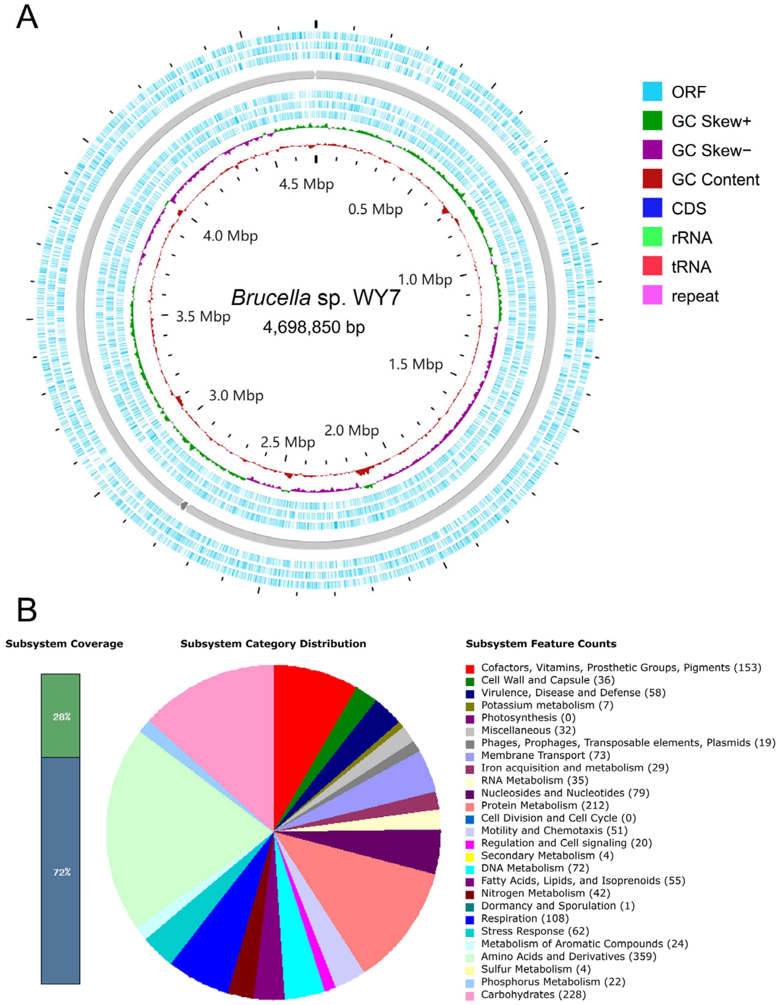
3.2. Genomic Characteristics

To obtain the genomic characteristics of strain WY7, the whole genome assembly was investigated. The assembled genome showed that strain WY7 had two chromosomes and one plasmid. As shown in Table 1, the genome length of chromosome 1 was 2,785,871 bp with 57.61% G + C content, containing 2647 genes. The genome length of chromosome 2 was 1,893,993 bp with 56.57% G + C content, containing 1718 genes. The plasmid genome length was 18,986 bp, with a G + C content of 55.17%, containing 24 genes. The total length of the genome was 4,698,850 bp, and there were three contigs with an average G + C content of 57.18%. A total of 4389 genes were identified (Figure 2A, Table 1).
To further obtain the taxonomic characteristics of strain WY7, the dDDH value and ANIb value between strain WY7 and the reference strains were calculated. The results showed that the dDDH values of strain WY7 with the type strains Brucella anthropi ATCC® 49188TM and Brucella anthropi NCTC® 12168TM were both 94.8% (Table 2), which were higher than the threshold of species definition by 70%. The ANIb values of strain WY7 and its close relatives Brucella anthropi ATCC® 49188TM and Brucella anthropi NCTC® 12168TM were as high as 99.07% (Table 3), higher than the threshold of 95% for defining species. These results indicated that strain WY7 is closely related to Brucella anthropi.

3.3. RAST Quick Genome Annotation

The functional distribution of strain WY7 was preliminarily obtained by genome annotation with RAST. According to the SEED system of RAST, genes were assigned to subsystems, which could be divided into 27 categories. The genome annotation information showed that RAST divided them into 339 subsystems; only 28% (1256 genes) were annotated in the subsystem, and the other 72% of the genome was not assigned to the RAST subsystem. Among them, Amino Acids and Derivatives (359 genes), Carbohydrates (228 genes), Protein Metabolism (212 genes), Cofactors, Vitamins, Prosthetic Groups, Pigments (153 genes), Respiration (108 genes) and other functions accounted for the majority (Figure 2B). The above results showed that strain WY7 was rich in amino acids and their derivatives, carbohydrates and protein metabolism.
Notably, genes related to Cell Division and Cell Cycle were not found in strain WY7, but a gene related to Dormancy and Sporulation was found. The genome also lacked genes related to Photosynthesis, suggesting that the bacterium was not a photosynthetic bacterium.

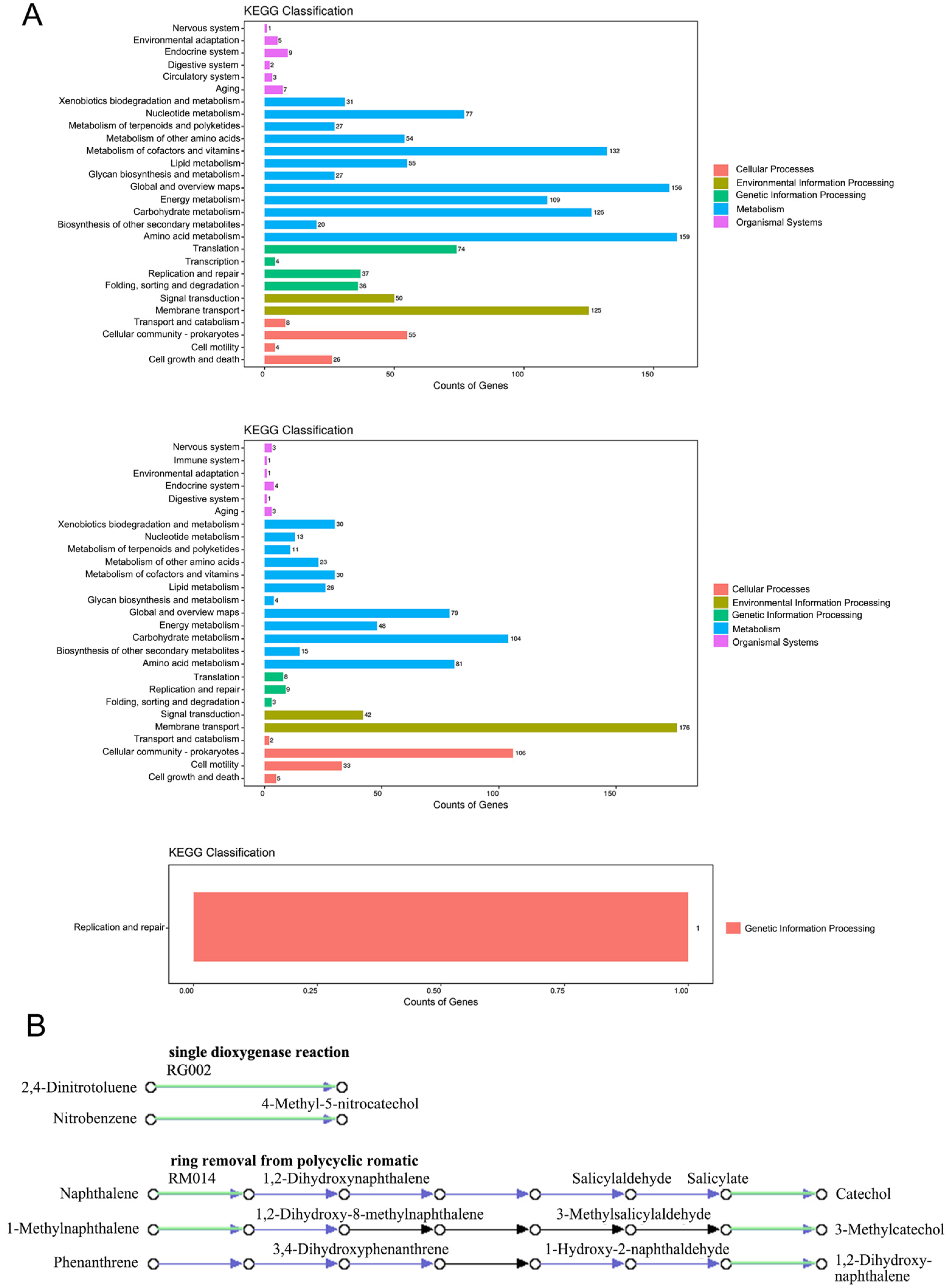
3.4. KEGG Database Annotation

The genome function was annotated using the KEGG database, and the approximate functional distribution of strain WY7 was obtained. Gene functions were divided into five categories by the KEGG database: Cellular Process, Environmental Information Processing, Genetic Information Processing, Metabolism, and Organismal Systems, and then each category was further subdivided into subcategories (Figure 3A, Table 4). Among them, 1419 genes were annotated on chromosome 1, 861 genes were annotated on chromosome 2, and only one gene was annotated on the plasmid, whose function was gene replication and repair.
On chromosome 1, amino acid metabolism (159 genes), global and overview maps (156 genes), metabolism of cofactors and vitamins (132 genes), carbohydrate metabolism (126 genes), membrane transport (125 genes), and energy metabolism (109 genes) were the dominant functional categories. On chromosome 2, membrane transport (176 genes), cellular community—prokaryotes (106 genes), carbohydrate metabolism (104 genes) and other functional categories dominated. A total of 40 genes in WY7 were associated with organismal systems, mainly involving the endocrine system, aging, environmental adaptation, nervous system, circulatory system, and digestive system. The results showed that strain WY7 has strong carbohydrate metabolism and membrane transport capacity and might also have an impact on the organizational systems of its host, thus providing great potential for environmental adaptation and the degradation, transformation, and utilization of complex carbohydrates.
In-depth analysis of the metabolic pathways of strain WY7 revealed genes related to aromatic hydrocarbon degradation pathways, such as naphthalene 1,2-dioxygenase ferredoxin reductase component (gene 0212 and gene 2319), protocatechuate 3,4-dioxygenase (gene 0594 and gene 0595), and salicylate hydroxylase (gene 0845). In addition, we mapped the relevant pathways of aromatic hydrocarbons (e.g., salicylate, 2,4-dinitrotoluene, nitrobenzene, naphthalene, and 1-methylnaphthalene) that suggest the isolate may be involved in degradation (Figure 3B). The above results indicated that strain WY7 has potential for application in the process of aromatic hydrocarbon metabolism.

3.5. CAZy Database Annotation

To explore the characteristics of the carbohydrate-active enzyme family of WY7, the predicted genes were annotated using the CAZy database (Figure 4A). Twenty-one genes encoding glycosyl transferases (GTs), 18 genes encoding glycoside hydrolases (GHs), 11 genes encoding carbohydrate esterases (CEs) and one gene encoding carbohydrate binding modules (CBMs) were identified on chromosome 1. Fourteen genes encoding GT, seven genes encoding GH and five genes encoding CE were identified on chromosome 2. Only one gene encoding GH was identified in the plasmid. These observations indicated that glycosyl transferases and glycoside hydrolases accounted for the majority in strain WY7, providing a basis for the formation, transfer and further metabolism of monosaccharides, polysaccharides, and glycosides.

3.6. Genomic Islands Prediction

To investigate more potential special functions of Brucella sp. WY7, 25 genomic islands on chromosome 1, 35 genomic islands on chromosome 2, and their location in the genome of the strain were predicted by IslandViewer4 (Figure 4B). The 25 genomic islands of chromosome 1 were composed of 484 genes, with gene distributions ranging from 295,164 to 2,494,506 bp. Among them, 221 genes expressed hypothetical nonfunctional proteins, eight genes expressed mobile element proteins, five genes located on the same genomic island expressed anti-restriction proteins, and two genes located on the same genomic island expressed VapC toxin proteins. The 35 genomic islands of chromosome 2 were composed of 464 genes, which ranged from 216,887 to 1,878,563 bp. Seven genes were found to express mobile element proteins and two HigB toxin genes were found to be located on the same genomic island. These results suggested that the strain had gene clusters transferred horizontally and that some gene clusters could express toxin proteins involved in the regulation of bacterial growth under specific environments.

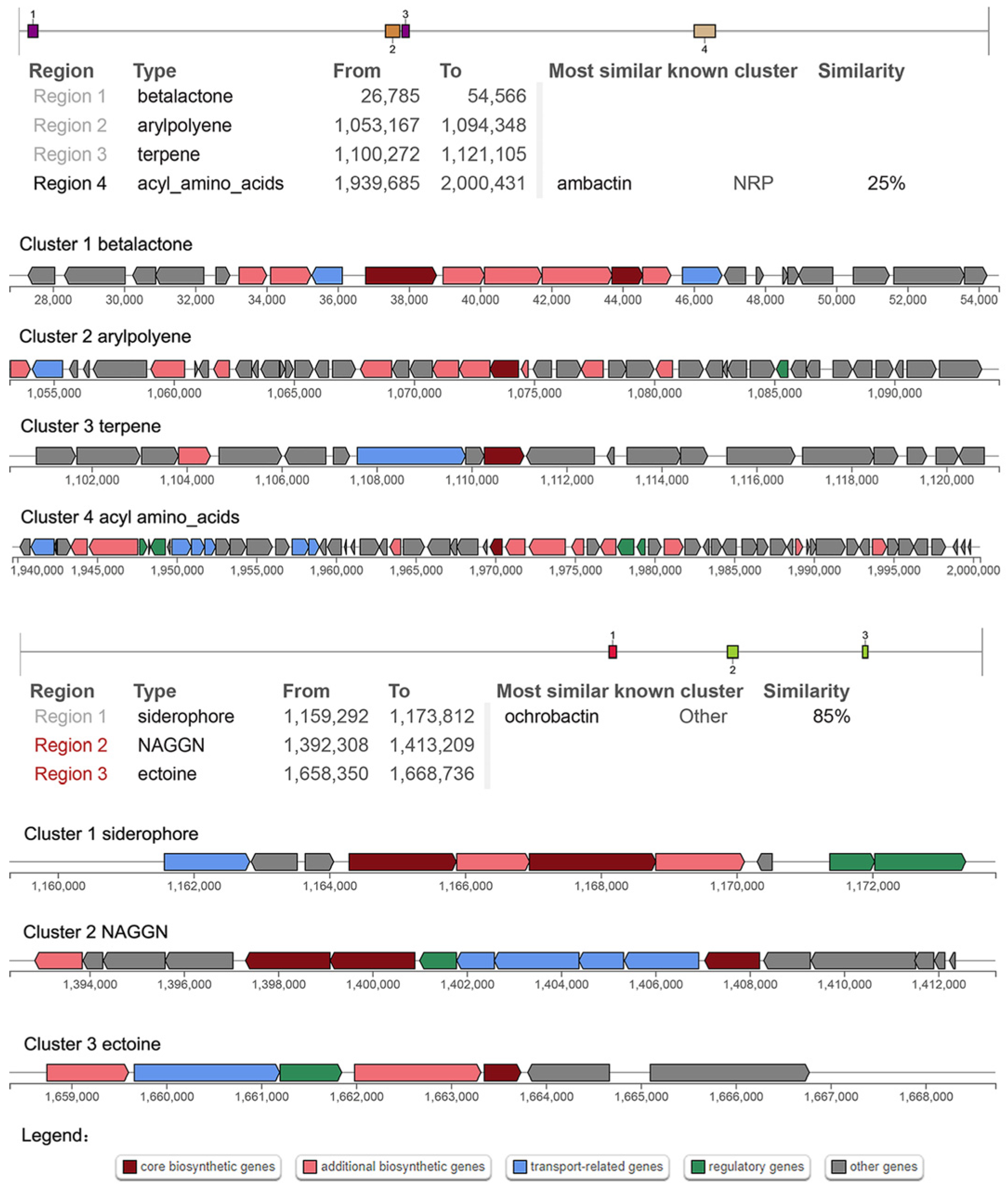
3.7. Secondary Metabolites

To gain further insight into the potential functions of WY7, secondary metabolites were predicted by antiSMASH. Gene screening for secondary metabolites on chromosome 1 revealed four distinct gene clusters (Figure 5). The secondary metabolites were batalactone, arylpolyene, terpene and acyl amino acids. Gene Clusters 1, 2 and 3 could not identify the known homologous gene cluster, and gene Cluster 4 (nucleotide 1,939,685–2,000,431) had 25% similarity with the gene cluster NRP producing ambactin.
Gene screening of secondary metabolites on chromosome 2 revealed three different gene clusters (Figure 5), and the secondary metabolites were siderophore, NAGGN and ectoine. Neither Cluster 2 nor Cluster 3 identified known homologous clusters, and Cluster 1 (nucleotide 1,159,292–1,173,812) was 85% similar to a gene cluster producing Ochrobactin, which is a photoreactive amphiphilic siderophore that has been found in Brucella sp. SP18 and Vibrio sp. DS40M5 [39]. Notably, the low similarity of predicted gene clusters may represent the generation of new metabolites.

3.8. Pan-Genomic Comparative Analysis

To acquire comparative genomic characteristics of WY7, homogenous cluster analysis of the genome was performed using OrthoVenn2. This Venn diagram and histogram describe the comparative genomics of Brucella sp. WY7 and Brucella anthropi ATCC® 49188TM, Brucella anthropi PBO, Brucella anthropi NCTC® 12168TM and Brucella melitensis bv. 1 str. 16M (Figure 6). The genome in the overlapping centre was composed of 1101 homologous clusters. Most of the annotation functions of homologous clusters were involved in biological processes, such as cellular processes, primary metabolic processes, cellular aromatic compound metabolic processes, organic acid metabolic processes and macromolecule metabolic processes. Strain Brucella sp. WY7 was found to have only two nonshared gene clusters, involving processes that activate or increase the frequency, rate or extent of cellular DNA-templated transcription. The existence of nonshared gene clusters indicated that the biological process of this strain might have unique advantages over other related strains.
The specific primers (F-5′-GTCAGTCGGCGGCTTATTC-3′ and R-5′-ATATCGTCTCCTTGGCAATGTC-3′) of gene 1983 of chromosome 1 in the WY7 nonshared gene clusters were designed for PCR detection of Brucella sp. WY7 and its relatives, Brucella anthropi ATCC® 49188TM (Figure S1). The results show that only WY7 has the correct band, which means that Brucella anthropi ATCC® 49188TM does not contain this gene, and Brucella sp. WY7 we found in Antarctic krill is not completely the same as its relatives.

4. Discussion

The Antarctic Ocean has the largest proportion of unknown genes to date of all sea areas. Most reports on psychrophilic bacteria in marine ecosystems are limited to sea ice or deep-sea sediments, while the study of Antarctic heterotrophic isolated bacteria shows that the growth rate of bacteria is quite high in the temperature range of 0–20 °C [40]. The study found that the proportion of functional housekeeping genes related to cell growth and metabolism is as high as 85–90%. This advantage of housekeeping genes shows that a few key genes that determine which species are dominant are of disproportionate importance. Among these dominant groups, some genes related to secondary metabolism have been found, which has potential importance for biological exploration [40]. However, we know little about the factors that control the distribution and dominance of key microbial species, but finding these key genes may be an achievable goal. In this study, we isolated and identified the strain Brucella sp. WY7, belonging to the genus not previously discovered in Antarctica, from Antarctic krill and explored its genomic characteristics and potential functions.
Defining a new species involves two successive steps, which are 16S rDNA analysis and calculation of genomic parameters, and the minimum standards (<98.7%) proposed by Chun et al. for prokaryotic classification using genomic data [41]. The threshold values of dDDH and ANIb for species division were 70% and 95%, respectively. If the value obtained by comparing the genome of this bacterium with that of a closely related type strain is lower than the threshold value of species division, it may be a new species [30]. The ANIb and dDDH values of Brucella sp. WY7 and Brucella anthropi ATCC® 49188TM were 99.07% and 94.8%, respectively, both of which are significantly higher than the threshold of species division. This indicated that this strain may be the same species as Brucella anthropi ATCC® 49188TM and may be Brucella anthropi, which has not been found in Antarctica before. Specific primers were designed, and the grinding solution of Antarctic krill and the genomic DNA of Brucella sp.WY7 were used as templates for PCR amplification to eliminate the effects of experimental operation and environmental pollution. The results showed that the amplified product fragment was consistent with the size of the target gene, which verified the authenticity of the isolated strain (Figure S2).
Research has shown that aromatic compounds are among the most prevalent and persistent environmental pollutants [42]. The genus Brucella is known to catabolize aromatic compounds [16]. In this study, genes and pathways associated with aromatic compound metabolism were found, such as the naphthalene 1,2-dioxygenase ferredoxin reductase component gene, protocatechuate 3,4-dioxygenase gene, and salicylate hydroxylase gene. Notably, salicylate hydroxylase (E1.14.13.1) is the first enzyme in the downstream pathway of naphthalene degradation [43], and protocatechuate 3,4-dioxygenase (pcaG and pcaH) is a key enzyme that catalyses the ring-opening step of aromatic compounds in bacteria [44]. The above results indicate that strain WY7 has potential for application in the process of aromatic hydrocarbon metabolism. The potential secondary metabolites of Brucella sp. WY7 were analysed, and the gene clusters of betalactone, arylpolyene, terpene, acyl amino acids, siderophores, NAGGN and ectoine were found. The similarity between the gene cluster producing siderophore and a gene cluster producing ochrobactin was 85%. Siderophores are important bioactive substances that can transport extracellular iron ions for bacterial use [45]. Terpenoids have often been extracted from marine organisms in recent years. They have important physiological activities and are important sources for developing new drugs and studying natural products [46]. Ectoine is an osmotic regulator that helps stabilize the structure of microorganisms and enables them to adapt to extreme environments [47]. Horizontal transfer of mobile elements in microbial communities is an important mechanism by which bacterial genomes evolve and adapt to specific environmental stresses [48]. It has been reported that HigB toxin and VapC toxin act as endribosomal enzymes to specifically cleave mRNA and tRNA in bacteria, thereby blocking transcription and translation processes and inhibiting bacterial growth. These toxins often coexist with antitoxins to regulate bacterial growth and adaptation to extreme environments [49,50]. The prediction of genome islands revealed that the bacterium had 15 mobile element protein genes, which might express anti-restriction proteins and produce HigB toxin and VapC toxin. It is speculated that the above genes help bacteria adapt to extreme environments.
By identifying homologous gene clusters from ancestors, comparative analyses can help clarify the relationships between different species and the evolution and adaptation of genomes [51]. The experimental strain Brucella sp. WY7 and four related strains had 4815 clusters and 1153 nonshared clusters. The distribution of core partial functions indicates that most homologous gene families encode basic bacterial metabolism, such as hydrolase activity, transferase activity, protein processing, folding and secretion, and DNA metabolism. Strain Brucella sp. WY7 had only two nonshared clusters, with functions in the positive regulation of transcription, indicating that its biological processes have some different characteristics compared with those of related strains; those characteristics, it is speculated, are related to its living environment. However, this needs to be further explored.

5. Conclusions

In this study, the whole genome of Brucella sp. WY7 was assembled, and the genomic characteristics were described by genomic analysis. This strain was identified within the genus Brucella in the class Alphaproteobacteria, which is closely related to Brucella anthropi. Genes involved in the degradation of aromatic compounds were identified, indicating the potential metabolism of an aromatic compound of Brucella sp. WY7. Through the prediction of secondary metabolites, genomic island function and comparative genomic analysis with related bacteria, the potential biological function was predicted. Our work will help to enrich the microbial resource database and provide more material sources for modern genetic engineering technology.

Supplementary Materials

The following supporting information can be downloaded at: https://www.mdpi.com/article/10.3390/microorganisms11092281/s1, Figure S1: Detection of specific primers for Brucella sp. WY7 gene1983; Figure S2: In situ identification of specific primers from the strain WY7.

Author Contributions

C.G. conceived the study. Z.F. and Y.W. performed the experiments. L.M. and L.W. provided Antarctic krill samples. Z.F., S.H. and Y.W. performed data and laboratory analyses. Z.F., Y.W. and C.G. wrote the article. J.H. and C.G. conceived the study provided Funding acquisition. All authors discussed the results and commented on the manuscript. All authors have read and agreed to the published version of the manuscript.

Funding

This work was supported by the National Key Research and Development Program of China (No. 2022YFE0203900), the Guangdong Key Research and Development Program (Nos. 2022B1111030001 and 2021B0202040002), the Guangdong Basic and Applied Basic Research Foundation (No. 2021A1515010647), Guangdong Laboratory for Lingnan Modern Agriculture (No. NZ2021018), the China Agriculture Research System (No. CARS-46), Innovation Group Project of Southern Marine Science and Engineering Guangdong Laboratory (Zhuhai) (No. 311021006), and Guangdong Provincial Special Fund for Modern Agriculture Industry Technology Innovation Teams (No. 2022KJ143).

Data Availability Statement

The authors confirm that the data supporting the findings of this study are available within the article and its Supplementary Materials.

Conflicts of Interest

The authors declare no conflict of interest.

References

  1. Meyer, B.; Kawaguchi, S. Antarctic marine life under pressure. Science 2022, 378, 230. [Google Scholar] [CrossRef] [PubMed]
  2. Paul, V.J.; Ritson-Williams, R.; Sharp, K. Marine chemical ecology in benthic environments. Nat. Prod. Rep. 2011, 28, 345–387. [Google Scholar] [CrossRef] [PubMed]
  3. Wang, C.; Tian, X.; Yang, Q.; Lu, Y.; Ma, L.; Huang, H.; Fan, C. Diversity of secondary metabolites from two Antarctic microbes Rhodococcus sp. NJ-008 and Pseudomonas sp. NJ-011. Open J. Mar. Sci. 2014, 2014, 48424. [Google Scholar]
  4. Yang, Q. Taxonomic Identification and Bioactivity Screening of the Symbiotic Bacteria Strains of Euphausia superba from Antarctic Ocean. Appl. Mech. Mater. 2013, 295, 173–177. [Google Scholar] [CrossRef]
  5. Cui, X.; Zhu, G.; Liu, H.; Jiang, G.; Wang, Y.; Zhu, W. Diversity and function of the Antarctic krill microorganisms from Euphausia superba. Sci. Rep. 2016, 6, 36496. [Google Scholar] [CrossRef]
  6. Wang, Y.; Ma, L.; He, J.; He, Z.; Wang, M.; Liu, Z.; Li, Z.; Wang, L.; Weng, S.; Guo, C.; et al. Environmental risk characteristics of bacterial antibiotic resistome in Antarctic krill. Ecotoxicol. Environ. Saf. 2022, 232, 113289. [Google Scholar] [CrossRef]
  7. Brinkmeyer, R.; Knittel, K.; Jürgens, J.; Weyland, H.; Amann, R.; Helmke, E. Diversity and structure of bacterial communities in Arctic versus Antarctic pack ice. Appl. Environ. Microbiol. 2003, 69, 6610–6619. [Google Scholar] [CrossRef]
  8. Chain, P.S.; Lang, D.M.; Comerci, D.J.; Malfatti, S.A.; Vergez, L.M.; Shin, M.; Ugalde, R.A.; Garcia, E.; Tolmasky, M.E. Genome of Ochrobactrum anthropi ATCC 49188 T, a versatile opportunistic pathogen and symbiont of several eukaryotic hosts. J. Bacteriol. 2011, 193, 4274–4275. [Google Scholar] [CrossRef] [PubMed]
  9. Klein, E.S.; Watters, G.M. Comparing feedback and spatial approaches to advance ecosystem-based fisheries management in a changing Antarctic. PLoS ONE 2020, 15, e0231954. [Google Scholar] [CrossRef] [PubMed]
  10. Sun, R.; Liu, X.; Yu, Y.; Miao, J.; Leng, K.; Gao, H. Preparation process optimization, structural characterization and in vitro digestion stability analysis of Antarctic krill (Euphausia superba) peptides-zinc chelate. Food Chem. 2021, 340, 128056. [Google Scholar] [CrossRef]
  11. Holmes, B.; Popoff, M.; Kiredjian, M.; Kersters, K. Ochrobactrum anthropi gen. nov., sp. nov. from human clinical specimens and previously known as Group Vd. Int. J. Syst. Bacteriol. 1988, 38, 406–416. [Google Scholar] [CrossRef]
  12. Al-Mur, B.A.; Pugazhendi, A.; Jamal, M.T. Application of integrated extremophilic (halo-alkalo-thermophilic) bacterial consortium in the degradation of petroleum hydrocarbons and treatment of petroleum refinery wastewater under extreme condition. J. Hazard. Mater. 2021, 413, 125351. [Google Scholar] [CrossRef]
  13. Chen, Y.; Liu, Q.; Yu, J.; Chen, C. Isolation and identification of marine petroleum degradable bacterium AH07 and its degradation performance characteristics. J. Microbiol. 2020, 40, 32–36. [Google Scholar] [CrossRef]
  14. Fan, Y.; Wang, C.; Wang, L.; Chairoungdua, A.; Piyachaturawat, P.; Fu, P.; Zhu, W. New ansamycins from the deep-sea-derived bacterium Ochrobactrum sp. OUCMDZ-2164. Mar. Drugs 2018, 16, 282. [Google Scholar] [CrossRef] [PubMed]
  15. Domingues, P.M.; Oliveira, V.; Serafim, L.S.; Gomes NC, M.; Cunha, Â. Biosurfactant production in sub-oxic conditions detected in hydrocarbon-degrading isolates from marine and estuarine sediments. Int. J. Environ. Res. Public Health 2020, 17, 1746. [Google Scholar] [CrossRef] [PubMed]
  16. Nayak, T.; Panda, A.N.; Kumari, K.; Adhya, T.K.; Raina, V. Comparative genomics of a paddy field bacterial isolate Ochrobactrum sp. CPD-03: Analysis of chlorpyrifos degradation potential. Indian J. Microbiol. 2020, 60, 325–333. [Google Scholar] [CrossRef]
  17. Wang, X.; Jin, D.; Zhou, L.; Zhang, Z. Draft genome sequence of Ochrobactrum anthropi strain W13P3, a halotolerant polycyclic aromatic hydrocarbon-degrading bacterium. Genome Announc. 2015, 3, e00867-15. [Google Scholar] [CrossRef] [PubMed]
  18. Thompson, J.D.; Gibson, T.J.; Plewniak, F.; Jeanmougin, F.; Higgins, D.G. The CLUSTAL_X windows interface: Fexible strategies for multiple sequence alignment aided by quality analysis tools. Nucleic Acids Res. 1997, 25, 4876–4882. [Google Scholar] [CrossRef]
  19. Kumar, S.; Stecher, G.; Li, M.; Knyaz, C.; Tamura, K. MEGA X: Molecular evolutionary genetics analysis across computing platforms. Mol. Biol. Evol. 2018, 35, 1547–1549. [Google Scholar] [CrossRef]
  20. Felsenstein, J. Confdence limits on phylogenies: An approach using the bootstrap. Evolution 1985, 39, 783–791. [Google Scholar] [CrossRef]
  21. Chin, C.S.; Alexander, D.H.; Marks, P.; Klammer, A.A.; Drake, J.; Heiner, C.; Clum, A.; Copeland, A.; Huddleston, J.; Eichler, E.E.; et al. Nonhybrid, finished microbial genome assemblies from long-read SMRT sequencing data. Nat. Methods 2013, 10, 563–569. [Google Scholar] [CrossRef] [PubMed]
  22. Chin, C.S.; Peluso, P.; Sedlazeck, F.J.; Nattestad, M.; Concepcion, G.T.; Clum, A.; Dunn, C.; O’Malley, R.; Figueroa-Balderas, R.; Morales-Cruz, A.; et al. Phased diploid genome assembly with single-molecule real-time sequencing. Nat. Methods 2016, 13, 1050–1054. [Google Scholar] [CrossRef]
  23. Hunt, M.; De Silva, N.; Otto, T.D.; Parkhill, J.; Keane, J.A.; Harris, S.R. Circlator: Automated circularization of genome assemblies using long sequencing reads. Genome Biol. 2015, 16, 294. [Google Scholar] [CrossRef] [PubMed]
  24. Hyatt, D.; Chen, G.L.; Locascio, P.F.; Land, M.L.; Larimer, F.W.; Hauser, L.J. Prodigal: Prokaryotic gene recognition and translation initiation site identifcation. BMC Bioinform. 2010, 11, 119. [Google Scholar] [CrossRef]
  25. Chen, N. Using RepeatMasker to identify repetitive elements in genomic sequences. Curr. Protoc. Bioinform. 2004, 5, 4–10. [Google Scholar] [CrossRef]
  26. Lowe, T.M.; Eddy, S.R. tRNAscan-SE: A program for improved detection of transfer RNA genes in genomic sequence. Nucleic Acids Res. 1997, 25, 955–964. [Google Scholar] [CrossRef] [PubMed]
  27. Lagesen, K.; Hallin, P.; Rødland, E.A.; Staerfeldt, H.H.; Rognes, T.; Ussery, D.W. RNAmmer: Consistent and rapid annotation of ribosomal RNA genes. Nucleic Acids Res. 2007, 35, 3100–3108. [Google Scholar] [CrossRef] [PubMed]
  28. Griffiths-Jones, S.; Bateman, A.; Marshall, M.; Khanna, A.; Eddy, S.R. Rfam: An RNA family database. Nucleic Acids Res. 2003, 31, 439–441. [Google Scholar] [CrossRef]
  29. Meier-Kolthoff, J.P.; Göker, M. TYGS is an automated high-throughput platform for state-of-the-art genome-based taxonomy. Nat. Commun. 2019, 10, 2182. [Google Scholar] [CrossRef]
  30. Richter, M.; Rossello-Mora, R.; Oliver, G.F.; Peplies, J. JSpeciesWS: A web server for prokaryotic species circumscription based on pairwise genome comparison. Bioinformatics 2016, 32, 929–931. [Google Scholar] [CrossRef] [PubMed]
  31. Overbeek, R.; Olson, R.; Pusch, G.D.; Olsen, G.J.; Davis, J.J.; Disz, T.; Edwards, R.A.; Gerdes, S.; Parrello, B.; Shukla, M.; et al. The SEED and the rapid annotation of microbial genomes using subsystems technology(RAST). Nucleic Acids Res. 2014, 42, D206–D214. [Google Scholar] [CrossRef]
  32. Ogata, H.; Goto, S.; Sato, K.; Fujibuchi, W.; Bono, H.; Kanehisa, M. KEGG: Kyoto encyclopedia of genes and genomes. Nucleic Acids Res. 1999, 27, 29–34. [Google Scholar] [CrossRef] [PubMed]
  33. Xie, C.; Mao, X.; Huang, J.; Ding, Y.; Wu, J.; Dong, S.; Kong, L.; Gao, G.; Li, C.-Y.; Wei, L. KOBAS 2.0: A web server for annotation and identification of enriched pathways and diseases. Nucleic Acids Res. 2011, 39 (Suppl. 2), W316–W322. [Google Scholar] [CrossRef]
  34. Cantarel, B.L.; Coutinho, P.M.; Rancurel, C.; Bernard, T.; Lombard, V.; Henrissat, B. The Carbohydrate-Active EnZymes database (CAZy): An expert resource for glycogenomics. Nucleic Acids Res. 2008, 37 (Suppl. 1), D233–D238. [Google Scholar] [CrossRef] [PubMed]
  35. Bertelli, C.; Laird, M.R.; Williams, K.P.; Simon Fraser University Research Computing Group; Lau, B.Y.; Hoad, G.; Winsor, G.L.; Brinkman, F.S.L. IslandViewer 4: Expanded prediction of genomic islands for larger-scale datasets. Nucleic Acids Res. 2017, 45, W30–W35. [Google Scholar] [CrossRef] [PubMed]
  36. Blin, K.; Andreu, V.P.; Santos EL, C.; Carratore, F.D.; Lee, S.Y.; Medema, M.H.; Weber, T. The antiSMASH database version 2: A comprehensive resource on secondary metabolite biosynthetic gene clusters. Nucleic Acids Res. 2019, 47, D625–D630. [Google Scholar] [CrossRef] [PubMed]
  37. Xu, L.; Dong, Z.; Fang, L.; Luo, Y.; Wei, Z.; Guo, H.; Zhang, G.; Gu, Y.Q.; Coleman-Derr, D.; Xia, Q.; et al. OrthoVenn2: A web server for whole-genome comparison and annotation of orthologous clusters across multiple species. Nucleic Acids Res. 2019, 47, W52–W58. [Google Scholar] [CrossRef]
  38. Zurdo-Piñeiro, J.L.; Rivas, R.; Trujillo, M.E.; Vizcaíno, N.; Carrasco, J.A.; Chamber, M.; Palomares, A.; Mateos, P.F.; Martínez-Molina, E.; Velázquez, E. Ochrobactrum cytisi sp. nov., isolated from nodules of Cytisus scoparius in Spain. Int. J. Syst. Evol. Microbiol. 2007, 57 Pt 4, 784–788. [Google Scholar] [CrossRef]
  39. Martin, J.D.; Ito, Y.; Homann, V.; Haygood, M.G.; Butler, A. Structure and membrane affinity of new amphiphilic siderophores produced by Ochrobactrum sp. SP18. JBIC J. Biol. Inorg. Chem. 2006, 11, 633–641. [Google Scholar] [CrossRef]
  40. Bhatia, M.; Sharp, M.; Foght, J. Distinct bacterial communities exist beneath a high Arctic polythermal glacier. Appl. Environ. Microbiol. 2006, 72, 5838–5845. [Google Scholar] [CrossRef]
  41. Chun, J.; Oren, A.; Ventosa, A.; Christensen, H.; Arahal, D.R.; Costa, M.S.; Rooney, A.P.; Yi, H.; Xu, X.-W.; De Meyer, S.; et al. Proposed minimal standards for the use of genome data for the taxonomy of prokaryotes. Int. J. Syst. Evol. Microbiol. 2018, 68, 461–466. [Google Scholar] [CrossRef]
  42. Seo, J.-S.; Keum, Y.-S.; Li, Q.X. Bacterial degradation of aromatic compounds. Int. J. Environ. Res. Public Health 2009, 6, 278–309. [Google Scholar] [CrossRef]
  43. Zhao, H.; Liu, B.; Ma, L.; Li, Y.; Cai, B. Nucleotide seouence analysis and enzyme characterization of the Pseudomonas sp. nd6 salicylate hydroxylase gene (nahG). Acta Sci. Nat. Univ. Nankaiensis 2004, 37, 95–99. [Google Scholar]
  44. Zhang, L. The Study of the Enzymatic Properties about Protocatechuate 3,4-Dioxygenase and Its Immobilization; East China University of Science and Technology: Shanghai, China, 2017. [Google Scholar]
  45. Zhang, X.; Zhu, J.; Cai, Z.; Zhou, J. The research advance of siderophores in marine microbes. Prog. Biochem. Biophys. 2022, 49, 1658–1671. [Google Scholar] [CrossRef]
  46. Shi, Q.; Li, L.; Huo, C.; Zhang, M.; Wang, Y. Research outline of marine natural products. Chin. Tradit. Herb. Drugs 2010, 41, 1031–1047. [Google Scholar]
  47. Zhang, S.; Hu, M.; He, Y.; Dong, Z. Research progress in microbial production and application of ectoines. Acta Microbiol. Sin. 2021, 61, 2250–2263. [Google Scholar] [CrossRef]
  48. Li, H.; Zhou, L.; Wang, Y.; Top, E.M.; Zhang, Y.; Xu, H. Degradative mobile genetic elements (MGEs) and their potential use in MGE-mediated biodegradation. Chin. J. Appl. Ecol. 2011, 22, 526–536. [Google Scholar]
  49. Dong, N.; Liu, D.; Fu, Y.; Yi, Z. Amplification and bioinformatics analysis of the higB gene from Mycobacterium tuberculosis. J. Parasit. Biol. 2016, 11, 505–508. [Google Scholar] [CrossRef]
  50. Lin, S.; Wang, X.; Chen, R. Analysis and identification of seven pairs of toxin-antitoxin systems in two cyanobacterial species. J. Trop. Oceanogr. 2022, 41, 119–134. [Google Scholar] [CrossRef]
  51. Wang, Y.; Ma, L.; He, J.; Liu, Z.; Weng, S.; Wang, L.; He, J.; Guo, C. Whole genome sequencing and comparative genomic analyses of Planococcus alpniumensis MSAK28401T, a new species isolated from Antarctic krill. BMC Microbiol. 2021, 21, 288. [Google Scholar] [CrossRef]
Figure 1. (A) N-J phylogenetic tree based on the 16S rDNA sequence. Numbers at nodes indicate the levels of bootstrap support (>70%) based on 1000 resamplings. Bar, 0.01 substitutions per nucleotide position. (B) Transmission electron micrograph showing cells of strain Brucella sp. WY7. The magnification is 6000×. Bar, 500 nm.
Microorganisms 11 02281 g001
Figure 2. (A) Genome circle map of strain Brucella sp. WY7: GC content (oxblood red), GC skew curves (+/−, dark green/purple), open reading frames (ORFs, light blue), coding sequences (CDSs, dark blue), rRNAs (light green), tRNAs (vermilion), and repeats (pink). (B) RAST subsystems category distribution of annotated genes of the strain Brucella sp. WY7.
Microorganisms 11 02281 g002
Figure 3. (A) KEGG annotation statistics chart of Brucella sp. WY7. The figure shows chromosome 1, chromosome 2 and the plasmid of Brucella sp. WY7. The horizontal axis is the number of genes, and the vertical axis represents the name of the level 2 pathway. (B) Aromatic hydrocarbon degradation pathways in WY7.
Microorganisms 11 02281 g003
Figure 4. (A) CAZy annotation classification distribution map. The figure shows chromosome 1, chromosome 2 and plasmid of Brucella sp. WY7. The horizontal axis is the CAZy category, and the vertical axis is the number of genes annotated by the corresponding category. (B) The predicted genomic islands (GIs) of the strain Brucella sp. WY7. Figure shows chromosome 1 and chromosome 2. Red shows the prediction by the integrated approach; blue represents the results from IslandPath-DIMOB; orange displays genomic islands predicted via SIGI-HMM; green shows the results from IslandPick.
Microorganisms 11 02281 g004
Figure 5. AntiSMASH predicted biosynthesis-related gene clusters and secondary metabolism. The figure shows chromosome 1 and chromosome 2 of Brucella sp. WY7. Brown represents the core biosynthetic genes; pink represents the additional biosynthetic genes; blue represents the transport-related genes; green represents the regulatory genes; and grey represents other genes.
Microorganisms 11 02281 g005
Figure 6. The Venn diagram and the bar graph depict the comparative genomics among the genomes of Brucella anthropi ATCC® 49188TM (green), Brucella anthropi PBO (blue), Brucella anthropi NCTC® 12168TM (pink), Brucella melitensis bv. 1 str. 16M (yellow), and Brucella sp. WY7 (orange), showing shared and unshared orthologous gene clusters.
Microorganisms 11 02281 g006
Table 1. Genome data of Brucella sp. WY7.
AttributeChr.1Chr.2Plas.Total
Genome size (Mb)2.791.890.0194.70
DNA coding region (Mb)2.441.700.0174.16
DNA G + C (%)57.6156.5755.1757.18
DNA scaffolds1113
Total genes26471718244389
RNA gene4721068
Genes assigned to COGs2230143273669
Repeats150790229
Chr. stands for chromosome, Plas. stands for plasmid.
Table 2. The dDDH values are provided along with their confidence intervals (C.I.).
Subject StrainQuery StraindDDH (d4, in%)C.I. (d4, in%)The Origin of Subject Strain
Brucella anthropi ATCC 49188 (JAAVLS010000001.1)WY794.8[93.2–96.1]Type strain from human clinical specimens, blood cultures
Brucella anthropi NCTC 12168 (UGSA01000001.1)WY794.8[93.2–96.1]Type strain from human clinical specimens, blood cultures
Brucella lupini LUP21 (GCA 002252535.1)WY782.5[79.7–85.1]Plant root nodule samples
Brucella anthropi PBO (GCA 015326295.1)WY778.8[75.9–81.5]Plastic debris from sea coast, Qingdao, China
Brucella melitensis 16M (GCA 000007125.1)WY725.0[22.7–27.5]An infected goat
Table 3. Calculation of ANIb values between available genomes of WY7 and type strains.
SpeciesWY7Brucella anthropi NCTC 12168Brucella anthropi ATCC 49188Brucella lupini LUP21Brucella anthropi PBOBrucella melitensis 16M
WY710099.0799.0797.4997.2180.55
Brucella anthropi NCTC 1216898.9110099.7897.4796.7380.40
Brucella anthropi ATCC 4918898.7099.7710097.2696.5580.15
Brucella lupini LUP2196.6896.7796.6010096.6379.91
Brucella anthropi PBO97.1496.8996.7397.4310080.43
Brucella melitensis 16M80.9981.0080.9880.8981.07100
Table 4. Annotation function classification of Brucella sp. WY7 of the genomic KEGG database.
ClassificationNumber of Genes
Chr.1Chr.2Plas.
Cellular Processes
Cell growth and death2650
Cell motility4330
Cellular community—prokaryotes551060
Transport and catabolism820
Environmental Information Processing
Membrane transport1251760
Signal transduction50420
Genetic Information Processing
Folding, sorting and degradation3630
Replication and repair3791
Transcription400
Translation7480
Metabolism
Amino acid metabolism159810
Biosynthesis of other secondary metabolites20150
Carbohydrate metabolism1261040
Energy metabolism109480
Global and overview maps156790
Glycan biosynthesis and metabolism2740
Lipid metabolism55260
Metabolism of cofactors and vitamins132300
Metabolism of other amino acids54230
Metabolism of terpenoids and polyketides27110
Nucleotide metabolism77130
Xenobiotics biodegradation and metabolism31300
Organismal Systems
Aging730
Circulatory system300
Digestive system210
Endocrine system940
Environmental adaptation510
Immune system010
Nervous system130
Total14198611
Disclaimer/Publisher’s Note: The statements, opinions and data contained in all publications are solely those of the individual author(s) and contributor(s) and not of MDPI and/or the editor(s). MDPI and/or the editor(s) disclaim responsibility for any injury to people or property resulting from any ideas, methods, instructions or products referred to in the content.

