Article

Combined TCBS and CHROMagar Analyses Allow for Basic Identification of Vibrio vulnificus within a 48 h Incubation Period in the Coastal Baltic Sea

by
Conor Christopher Glackin
1,
Susann Dupke
2,
Thota Sharath Chandra
1,
David Riedinger
1 and
Matthias Labrenz
1,3,*
1
Leibniz Institute for Baltic Sea Research Warnemünde (IOW), Seestraße 15, 18119 Rostock, Germany
2
Robert Koch-Institute (RKI), Centre for Biological Threats and Special Pathogens, Highly Pathogenic Microorganisms, Seestraße 10, 13353 Berlin, Germany
3
Marine Research Institute, Klaipeda University, H. Manto 84, LT-92294 Klaipeda, Lithuania
*
Author to whom correspondence should be addressed.
Microorganisms 2024, 12(3), 614; https://doi.org/10.3390/microorganisms12030614
Submission received: 16 February 2024 / Revised: 12 March 2024 / Accepted: 16 March 2024 / Published: 19 March 2024
(This article belongs to the Section Environmental Microbiology)

Abstract

:
With rising infection rates in recent years, Vibrio vulnificus poses an increasing threat to public safety in the coastal brackish Baltic Sea. It is therefore important to monitor this organism and assess the V. vulnificus infection risk on a more regular basis. However, as the coastline of the Baltic Sea is 8000 km long and shared by nine nations, a convenient, fast, inexpensive, yet efficient V. vulnificus identification method is essential. We evaluated the effectiveness of a two-step agar-based approach consisting of successive Vibrio isolation and cultivation on thiosulphate-citrate-bile salt sucrose (TCBS) agar and CHROMagar™ Vibrio for V. vulnificus in comparison with V. cholerae, V. parahaemolyticus, and V. alginolyticus. Our study contains isolates from water and sediment across a broad expanse of the Baltic Sea including 13 locations and two different summers, the time of year during which Vibrio infections are usually much more frequent. Confirmation of isolate species identity was carried out using molecular analyses. The two-step agar plating method performed well across different locations and timeframes in correctly identifying V. vulnificus by more than 80%, but the sensitivity in other Vibrio species varied. Thus, our approach yielded promising results as a potential tool for early V. vulnificus detection across a broad timeframe and transect of the Baltic Sea and potentially other brackish environments.

1. Introduction

Bacteria of the genus Vibrio are ubiquitous members of marine ecosystems and occur in coastal, estuarine, brackish, and freshwaters as well as in sediments, often in association with higher organisms [1]. The genus consists of more than 130 species and around a dozen of these have been demonstrated to be human pathogens [2,3,4,5,6]. From these, Vibrio cholerae serotypes O1/O139 can cause the well-known disease cholera. Beside those two, the most important potentially pathogenic non-cholera causing Vibrio species are the ‘big four’ species, consisting of V. cholerae [different non-O1/O139 serotypes], V. vulnificus, V. parahaemolyticus, and V. alginolyticus [7]. These organisms are common pathogens present in marine and estuarine waters, sediment, or plankton and can cause infections in humans which are usually associated with the consumption of raw or undercooked shellfish or by wound infections in marine or brackish water. Their characteristic disease patterns are gastroenteritis (V. cholerae, V. vulnificus, and V. parahaemolyticus), ear infections (V. cholerae and V. alginolyticus), wound infections (V. vulnificus, V. cholerae, V. alginolyticus, and V. parahaemolyticus), or sepsis (V. vulnificus) [2]. Increasing water temperatures can lead to both the increased abundance of Vibrio spp. and the occurrence of accordant potential pathogenic Vibrio species. Importantly, and this differentiates Vibrio spp. in general from the major foodborne pathogens Salmonella, Listeria, Escherichia coli O157, and Campylobacter, it has been shown for the USA between 2006 and 2013 that it is the only group currently increasing in occurrence [8]; thus, the behaviour or appearance of members especially of Vibrio spp. change in the current environmental conditions. Consequently, with the rise in global sea surface temperatures, there is growing concern about the potential impact of these changes on Vibrio populations and the associated public health risks [9].
V. vulnificus infections of humans are often severe [>90% of all cases] and have mortality rates as high as 50%, especially in immunodeficient individuals [2,7,10]. V. vulnificus blooms have been recorded on numerous occasions [11,12,13] and it is known that its optimal growth conditions involve water temperatures exceeding around 18 °C and salinity levels ranging from 5 to 25 practical salinity units (PSU) [14]. This provides V. vulnificus with optimal growth conditions in the Baltic Sea. The Baltic Sea is a semi-enclosed marginal sea of the Atlantic located in northern Europe, with a coastline of approximately 8000 km and covering an area of 415,266 km2. Saline inflows through the North Sea produce a 2000 km long lateral surface salinity gradient throughout the whole Baltic Sea, ranging from high salinities [>25] in the transition zone of the Kattegat to low salinities [<5] in the Gulf of Bothnia [15]. The Baltic Sea faces one of the highest warming rates in marine ecosystems worldwide; thus, it is considered a high-risk environment for Vibrio infections [16]. Indeed, infection numbers have increased significantly along the Baltic coast in recent years, particularly during heatwaves [13]. During the extremely warm summers of 1994, 2003, and 2006, for instance, a plethora of reports emerged documenting Vibrio-associated wound infections linked to recreational exposure in this area and included numerous fatalities [17]. Besides tourists and travellers, the Baltic Sea has almost 30 million people living within 50 km of its coastline with an increasing susceptible risk [18]. This highlights the need to delve into the development of Vibrio monitoring tools or early warning systems for Vibrio occurrences in the Baltic Sea.
Crucial for the understanding of Vibrio spp. distribution is an accurate identification of Vibrio species that most frequently cause infections in humans, but especially of the most harmful V. vulnificus. Pinpointing a timeframe of increased risk of Vibrio infection is the next step in curbing the increasing numbers of infections and also has wide-ranging ecological and economic effects [2,19]. In the context of the Baltic Sea, where diverse Vibrio species coexist, precise as well as simple and cost-effective identification becomes more critical. Established methods, such as isolation on agar plates, already exist and have been widely used for Vibrio identification [20,21,22]. However, often these methods may lack the specificity and sensitivity required to distinguish between closely related species [21] or require higher media and labour costs [23]. TCBS was one of the first selective media used for the isolation of Vibrios [24] and is widely used to isolate Vibrio from environmental samples, including the Baltic Sea [25,26]. In contrast to other Vibrio selective media, cellobiose-polymyxin B-colistin agar and its modified formulas, modified cellobiose-polymyxin B-colistin agar and cellobiose-colistin agar, TCBS is commercially available and is less time-consuming, requiring only a boiling step [20].
Thus, the objective of this study was to test a two-plate thiosulphate-citrate-bile salt sucrose [TCBS] and CHROMagar™ cultivation approach as an easy, cheap, and efficient species-specific tool to identify potentially pathogenic Vibrio in the Baltic Sea. Specification was performed on V. cholerae, V. parahaemolyticus, V. vulnificus, and V. alginolyticus identifications, with a broader spatial scale for V. vulnificus. Cultivation-based analyses were validated by Vibrio spp. specific multiplex-PCR or genus-identifying sequencing on various spatiotemporal levels of the Baltic Sea.

2. Materials and Methods
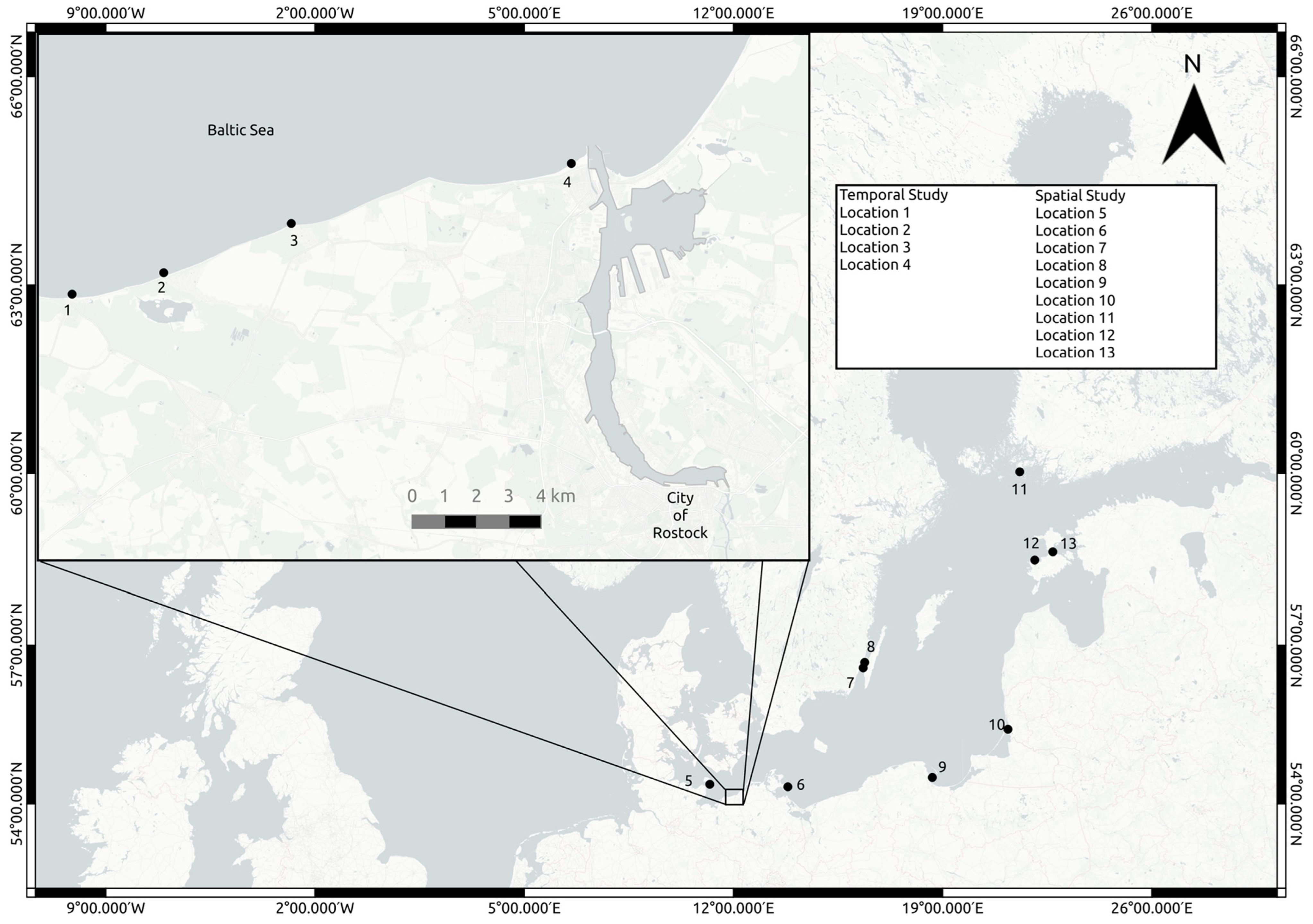
2.1. Sampling Areas

To evaluate the presence of potentially pathogenic Vibrio in the Baltic Sea based on the combined agar identification approach, two Baltic Sea monitoring campaigns took place, one in 2021 and one in 2022, incorporating a total of 13 sampling stations. A temporal study took place at four locations (Figure 1; locations 1–4, Latitude and Longitude: [54.14636° N, 11.84315° E], [54.15148° N, 11.88636° E], [54.16666° N, 11.96379° E], [54.18248° N, 12.07630° E]) on a 17 km stretch of the northern German coastline across eleven weeks (4 July 2022 to 15 September 2022) and documented V. vulnificus, V. parahaemolyticus, V. cholera, and V. alginolyticus. A spatial study focused on V. vulnificus abundance over an almost 1000 km range in the Baltic Sea. It took place from 26 July 2021 to 1 September 2021 and encompassed nine sites along the coastlines of Germany, Poland, Finland, and Estonia (Figure 1; locations 5–13, see Supplementary File S3 for coordinates).

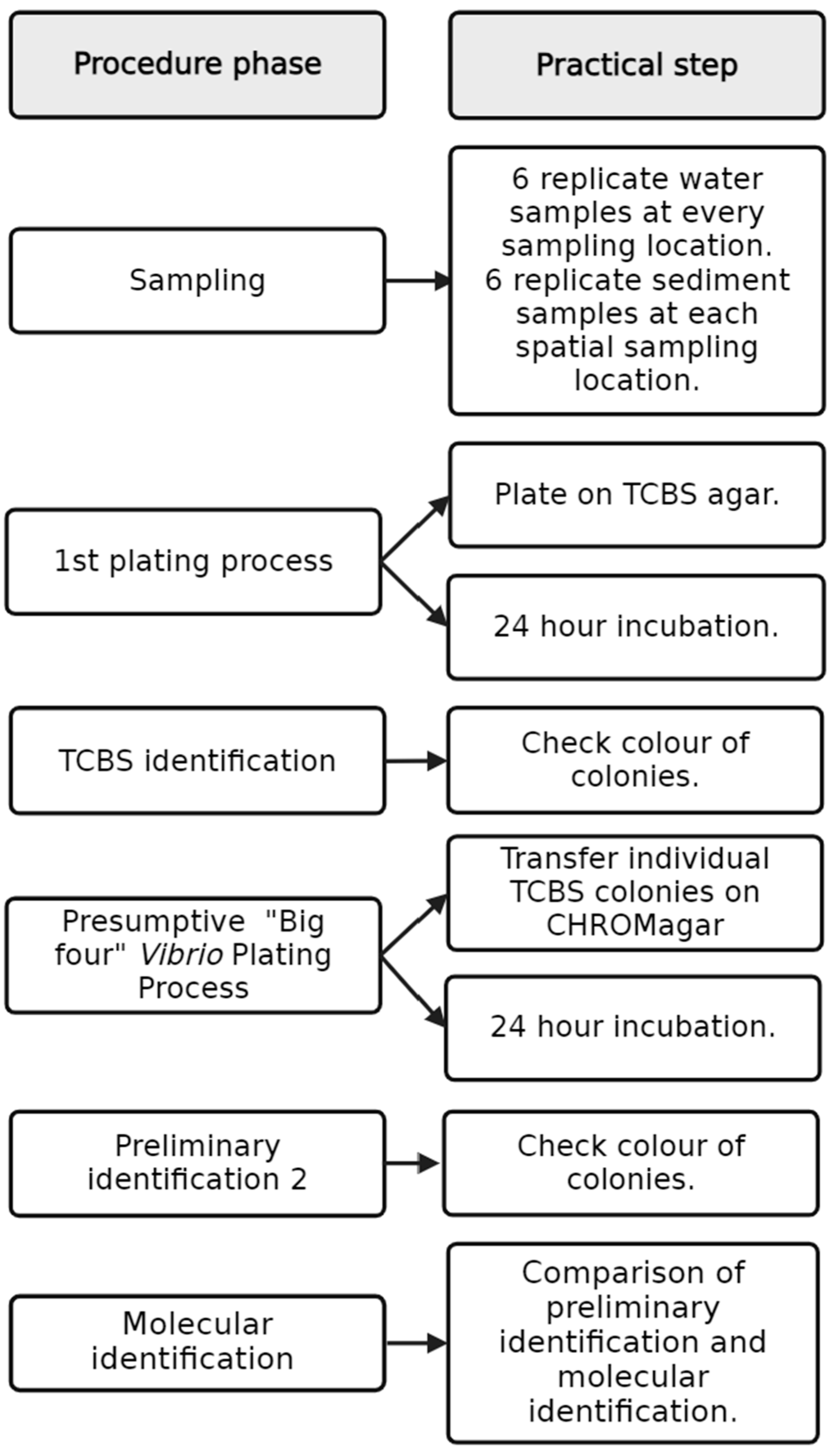
2.2. Environmental Sampling

Water samples were collected in both campaigns, whereas sediment samples were exclusively gathered during the spatial approach. The workflow from the sampling location to molecular identification and comparison is documented in Figure 2. In the temporal study, surface water (less than 50 cm from surface) was collected at a water depth of around 1 m in 6 replicates. At each station, 15 mL Falcon tubes were dipped into the water with rubber gloves. In Heiligendamm (location 1), the samples were taken 150 m from the shore and ca. 3 m depth and were taken every Tuesday at the same time for the duration of the study. In Börgerende (location 2), Nienhagen (location 3), and Warnemünde (location 4), the samples were taken on the beach at around 1 m depth each Monday and Thursday, for harmonisation always three hours after sunrise, for the duration of the study. Further information on these sampling sites can be found in Supplementary File S2. The spatial study sampling sites consisted of coastal locations along the Baltic Sea (Riedinger et al. in revision. For the spatial study, six replicate water samples were collected by SCUBA divers ca. 20 cm above the sediment with 100 mL syringes and six replicates of the top 1 cm of sediment were collected with 50 mL Falcon tubes. Environmental data of the spatial station are available in Supplementary File S3 and at IOWMeta (doi.io-warnemuende.de/10.12754/data-2023-0010).
All samples were transferred to a 4 °C cooler immediately and stored (maximally 8 h) until processing. Associated physical parameters salinity, temperature, and water depth were measured using a CTD48M (Sea & Sun Technology, Trappenkamp, Germany) during sampling at all stations.

2.3. Vibrio spp. Isolation and Culture-Based Identification

For Vibrio spp. isolation and identification, thiosulfate citrate bile sucrose (TCBS) agar (Merck KGaA, Darmstadt, Germany) as well as chromogenic agar selective for Vibrio spp. (CHROMagar™, Paris, France) were used. Agar plates were prepared according to the manufacturer’s protocols. For the isolation of the Vibrio spp. from the sediment, the overlying water in the Falcon tube was removed, and after homogenisation, a subsample of 10 g (dry-weight determined accurately after lyophilization) was taken from each sample, and in new sterile 50 mL Falcon tubes, 40 mL of double sterile filtered station water was added. Through five ultrasonic pulses of 10 s at 25% capacity at 5 s intervals from the Bandelin SONOPULS HD 2200.2 (Bandelin, Berlin, Germany), sediment-attached bacteria were detached. Following vortexing and sediment settling, water aliquots of 50, 100, or 200 μL were spread on TCBS agar in six biological replicates. For direct plating, each water sample was thoroughly shaken, and 200 μL of the sample was aseptically inoculated onto a TCBS agar plate, which was subsequently evenly spread across the surface. For indirect plating, 2 mL of each water sample was filtered through a 0.22 polycarbonate filter (Isopore™, Merck Millpore Ltd., Cork, Ireland) and this filter was placed on the agar plates. After incubation for 24 h at 37 °C, colonies were quantified and preliminarily categorised according to Table 1.
The preliminarily identified Vibrio colonies on TCBS were restreaked onto a ¼ wedge of CHROMagar™ Vibrio plates and incubated for 24 h at 37 °C. The final culture-based identification was documented according to the combined colours of colonies grown on TCBS agar and CHROMagar™ Vibrio (Table 1).
The colonies of presumptive V. cholerae, V. parahaemolyticus, V. vulnificus, and V. alginolyticus were suspended in 1 mL of Marine Broth (Roth, Germany). The mixture was left for 24 h at 37 °C. Subsequently, 200 μL of this culture was combined with 300 μL of 50% glycerol, yielding a final concentration of 30% glycerol. These isolates were shock frozen in liquid nitrogen and stored at −80 °C. For recovery, strains were re-cultured either on Columbia agar at 37 °C or in the case of no growth on Difco™ Marine Agar (BD Diagnostics, Sparks, MD, USA) at 28 °C for 24–48 h.

2.4. Molecular Identification of Vibrio spp.

Genomic DNA from Vibrio spp. colonies was extracted using the DNeasy Blood and Tissue Kit (Hilden, Germany) according to the following protocol.
A previously frozen bacterial isolate was recultivated on Columbia agar or Difco™ Marine Agar and incubated for 24–48 h. For DNA extraction, an inoculation loop full of colony material was removed by tapping five to ten single colonies. The collected material was transferred into 180 µL ATL buffer and mixed homogeneously by pipetting up and down. Then, 20 µL proteinase K was added, followed by incubation of the sample for 60 min at 56 °C and shaking at 450 rpm in a thermomixer. Subsequently, 200 µL AL buffer was added, and the sample was mixed again by pipetting up and down followed by incubation for another 10 min at 70 °C and shaking at 450 rpm. Further processing was carried out according to the manufacturer’s instructions. The purified DNA was eluted in two steps with 100 µL of elution buffer EB by centrifugation at 600 rpm for 1 min each, so that a total of 200 µL DNA eluate was generated. The DNA was stored at 4 °C.
The primer and probe sequences for the three multiplex real-time PCR systems are shown in Table 2. The localisation of the primers and probes for V. cholerae detection correspond to the sequence of the superoxide dismutase (sodB) gene of V. cholerae NCTC8457 [GenBank AAWD01000215], as well as the sequence of the cholera toxin (ctxA) gene of V. cholerae strain B [GenBank AY376267]. The localisation of the primers and probes for V. parahaemolyticus detection correspond to the sequence of the toxin regulator (toxR) gene of V. parahaemolyticus strain KP34 [GenBank DQ845170] and for V. vulnificus detection to the sequence of the cytolysin–hemolysin (vvhA) gene [GenBank AY046900]. Amplicon lengths for each primer were as follows: 145 bp for sodB, 116 bp for ctxA, 114 bp for toxR, and 118 bp for vvhA. In addition, isolates were screened for V. cholerae serogroups O1 and O139 [27].
Any bacterial isolate which could not be classified based on the PCR targeting specific markers of V. cholerae, V. vulnificus, or V. parahaemolyticus was subjected to RNA polymerase beta subunit (rpoB) sequence determination. Species identification using PCR-based amplification of the rpoB gene and analysis of the products were performed as described earlier in Tarr et al. [28] and Schirmeister et al. [29].
Finally, 16S rDNA fragment sequencing was performed on the seven bacterial isolates that could not be identified using rpoB gene sequencing. The rpoB as well as the 16S rRNA gene fragment sequences were aligned with nucleotide sequences in the GenBank database using the Basic Local Alignment Search Tool (BLAST) search algorithm.

2.5. Statistical Analyses

To test for sampling days that could be considered outliers, the Interquartile Range method was implemented in R statistical package version 4.3.2 using ggplot2:: geom_boxplot.

3. Results

3.1. Environmental Parameters

For the duration of the temporal study, the temperature ranged between 15 °C and 23 °C and salinity ranged between 9 and 17 PSU in locations 1–4. In the spatial study (locations 5–13), the temperature ranged between 19 °C and 21 °C and salinity ranged between 6 and 10 PSU. A summary of environmental parameters from the temporal study and spatial study can be found in Supplementary Files S1 and S3.

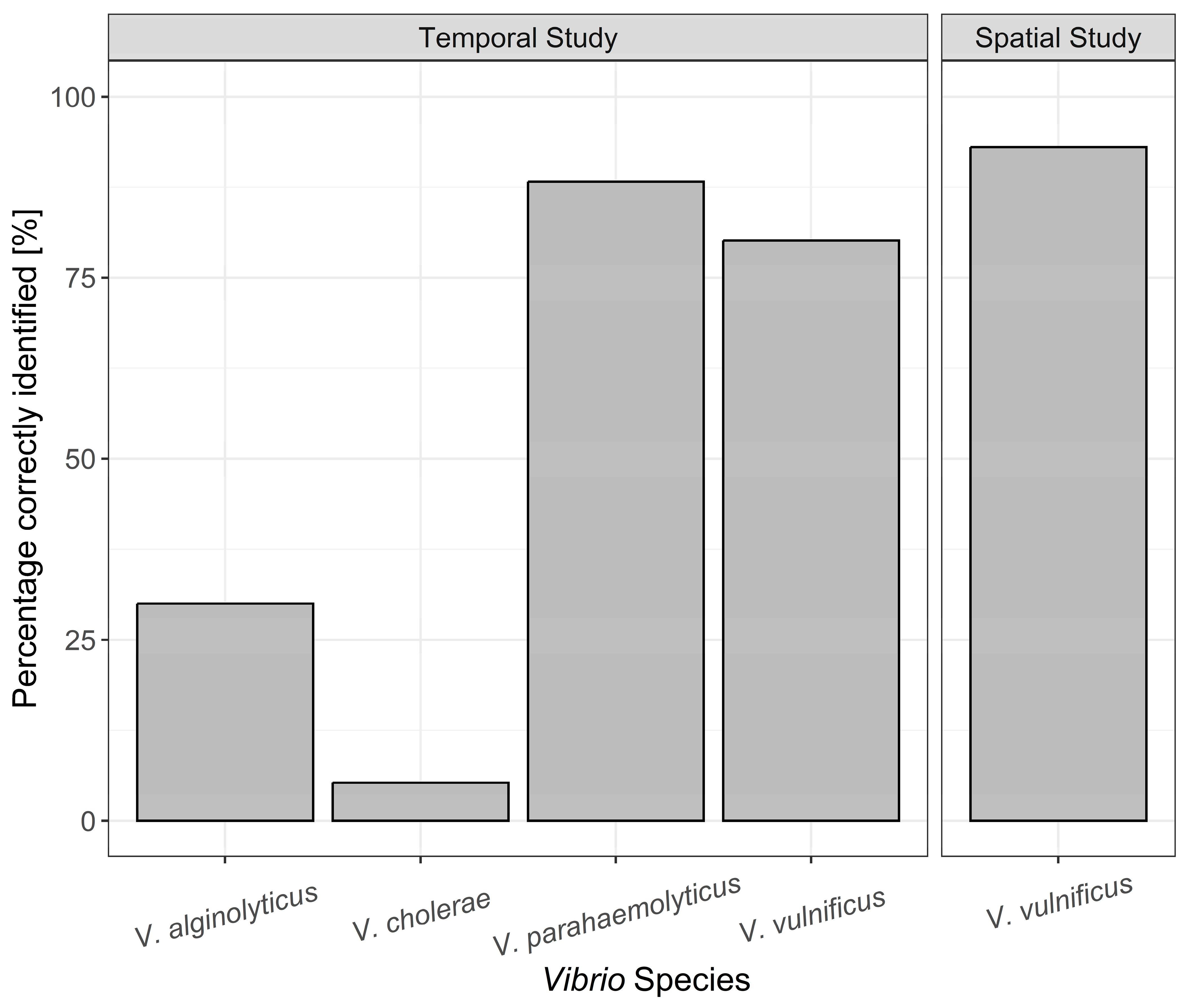
3.2. Identification of Bacterial Isolates on TCBS Agar and CHROMagar™

In the temporal study, a total of 1245 colonies were cultured and isolated on TCBS agar and transferred to CHROMagar™ Vibrio plates. Based on the combined colour code identification, 455 of these colonies (37%) were presumed to be V. parahaemolyticus whilst 214 (17%), 201 (16%), and 180 (14%) were presumed to be V. alginolyticus, V. cholerae, and V. vulnificus, respectively (Figure 3). The remaining 195 colonies (16%) were mixed cultures or unidentified using the colour code for species identification on TCBS and CHROMagar Vibrio (Figure 3). The vast majority of colonies originated from water samples taken at locations 2, 3, and 4, with 399, 477, and 315, respectively. The remaining 54 colonies were isolated from location 1 water samples which showed a considerably lower Vibrio spp. abundance.
Molecular verification of the culture-dependent Vibrio spp. identifications yielded very different values for the individual species. While V. parahaemolyticus and V. vulnificus were correctly identified with values above 80% by the two-plate TCBS agar/CHROMagar™ Vibrio approach, it was considerably lower for V. alginolyticus with 30%. This method also showed poor predictive ability with a value of 5% identification for V. cholerae (Figure 4). Of the misidentified presumptive V. cholerae colonies, 36.5 % belonged to V. aestuarianus and 51.0% to V. diazotrophicus based on molecular analyses. V. vulnificus colonies were cultured in each of the eleven weeks and were consistently isolated from samples across the three beach locations (locations 2–4).
In the spatial study from 2021, 86 colonies from eight different locations in the Baltic Sea presumed to be V. vulnificus were successfully cultured (Supplementary File S3). From these, 93% were accurately identified by the two-plate TCBS agar/CHROMagar™ Vibrio approach (Figure 4). Comparing correctly identified isolates from both the temporal study and spatial study, it became clear that V. vulnificus could be regularly identified correctly at a high level using the two-plate method. For V. parahaemolyticus, this method also showed high predictive power, correctly identifying 88% of colonies (Figure 4). In contrast, the identification level of V. alginolyticus and V. cholerae appeared to vary at an already low level (Figure 5). The number of isolates genetically identified in each location is shown in Table S1.

3.3. Highest Vibrio spp. Abundance per Day

Of the total Vibrio spp. colonies isolated, it was found that 96% were extracted from water samples taken from locations 2, 3, and 4. Further investigation highlighted that one day in particular yielded a significantly higher number of colonies at these locations (Figure 6). In total, there were 22 days in which samples were taken at each of these locations and 21 July 2022 accounted for 15% (192 colonies) of the total colonies cultured. This is a sharp rise from the total of 14 colonies cultured across all locations on the 18 July. Of the 37 presumptive V. vulnificus samples cultured on this day, 24 were identified using molecular sequencing. A total of 18 of these 24 isolates (75%) were confirmed to be V. vulnificus, demonstrating similar predictive results to the overall spatiotemporal analysis within this day.

4. Discussion

Routine monitoring for Vibrio vulnificus in the Baltic Sea is critical to provide a warning system for the public when the risk of infection is potentially high. This study examined using TCBS agar and CHROMagar™ Vibrio-based V. vulnificus identifications to achieve this, spanning across two separate spatiotemporal sampling projects. For evaluation, this agar-based method was also tested on V. cholerae, V. parahaemolyticus, and V. alginolyticus in the temporal study. The two-plate agar method proved to be accurate in identifying V. vulnificus across a broad range of locations and timeframes within the Baltic Sea, although it had contrasting results when used on other species of Vibrio.
V. vulnificus presents the largest threat for open wound infections in the Baltic Sea [30]. In total, 222 V. vulnificus samples were genetically identified across twelve locations and two different timeframes in 2021 and 2022, giving a comprehensive overview of this method of identification in the Baltic Sea. The overall correct identification of V. vulnificus was 85% and was consistent in correct identification across sampling sites (Figure 5). Thus, our study indicates that the use of TCBS agar followed by CHROMagar™ Vibrio to preliminarily identify V. vulnificus in the Baltic Sea produces applicable results.
In recent years, there have been numerous attempts to provide the accurate identification of V. vulnificus using a culture-based approach, which differ greatly in their success rate. TCBS agar alone has been shown to be not sufficiently selective enough for environmental Vibrio samples [31]. Thus, the vast majority of more recent research involves using TCBS agar, CHROMagar™ Vibrio, or Cellobiose polymyxin B colistin (CPC) agar with varying results in environmental samples. The highest performing agar approach for V. vulnificus so far was a triple plating method [23], producing a 92.8% accuracy on environmental water and oyster samples. Other comprehensive studies include a study by Froelich et al. [32] where CPC+ was used to culture presumptive V. vulnificus on agar. Results showed that the average yearly rate of samples confirmed to be V. vulnificus ranged from 0% to 45.7%. In another study by Froelich et al. [33] on oyster meat, they compared four different medium methods for presumptive V. vulnificus, resulting in correct identification rates between 44% and 81%, as confirmed by PCR. A study in the Mediterranean using TCBS and CPC found, with 3.7% and 7.6%, much lower accuracy levels of these two agar methods, respectively [34].
V. vulnificus aside, V. cholerae, V. parahaemolyticus, and V. alginolyticus are responsible for the majority of other Vibrio wound infections in the Baltic Sea [11,13]. In stark contrast to V. vulnificus, the percentage of V. cholerae correctly identified was 5%. This highlights a huge difference in identifying the two species responsible for the most Vibrio infections in the Baltic Sea using the two-plate TCBS agar/CHROMagar™ Vibrio approach method. The vast majority of the misidentified bacterial isolates were V. aestuarianus and V. diazotrophicus, reflecting that the V. cholerae colony colours using this agar method are close between the two species. In the case of V. parahaemolyticus, the TCBS agar/CHROMagar™ Vibrio yielded high correct identification results, whereas the presumptive V. alginolyticus colonies were misidentified the majority of the time, further highlighting the mixed results of this agar identification method on different Vibrio species.

Spatiotemporal Analysis

In the temporal study, 1245 different presumptive Vibrio spp. colonies were grown. In terms of overall Vibrio spp. cultivated, station 1 had considerably fewer colonies than the other three locations. The notable difference here was the water depth at the sampling areas. Although all samples were surface water, the difference in depth was around 2 m (3 m vs. 1 m) and the distance from the shoreline was 150 m for location 1 and around 10 m for locations 2–4. There are several hypotheses as to why this discrepancy occurred. Numerous environmental parameters, most notably temperature and salinity, have been associated with contributing to Vibrio spp. abundance, depending on species, habitat, and geographic location [35,36,37,38,39,40,41,42,43,44]. The similarity in both temperature and salinity in our project suggests that other environmental parameters or processes may have contributed to the change in abundance. Dissolved oxygen [45,46,47], chlorophyll [39,48,49,50], and plankton [39,51,52,53,54] have also been found to be important in the ecology of Vibrio spp. Given that location 1 was further from the shore and sediment, it is also possible that turbidity, increased nutrient loads, and increased sediment bacteria resuspension also played a role in the difference in Vibrio spp. abundance between location 1 and locations 2–4.
In the three beach locations, the temporal dynamics were similar in both overall colonies cultured and species correctly identified (Figure 4), with the 22 July showing the highest number of colonies cultivated across all locations. This day was an outlier in all three locations with regard to overall colonies cultured, highlighting the consistency in the results along the 17 km stretch of coast where the Vibrio spp. summer surveillance took place. Finding a significantly higher number of presumptive Vibrio isolates in all locations demonstrates the need to further pinpoint Vibrio blooms, given that they are present in the marine environment throughout the summer months. This once again points to the need for a simple and fast Vibrio spp. identification option. The establishment of a principal monitoring or even an early warning system for V. vulnificus in the Baltic Sea is of paramount importance due to the potential public health risks and ecological consequences associated with this pathogenic bacterium. V. vulnificus is a well-recognised human pathogen and infections have been documented in increasing numbers along the Baltic coast in recent years [30]. Tourism and economic impacts associated with the danger of infections have been described recently [5,55]. Current research suggests a correlation between sea temperature and V. vulnificus abundance and this has major implications for the Baltic Sea given that it is one of the fastest warming seas in the world [9].
This study introduces valuable insights into the suitability of early detection methods for different Vibrio species in the region. The reduction of false positive identification in culture-based methods enhances predictive power but can also significantly decrease the cost of laboratory equipment and procedures that are necessary for molecular identification of Vibrio spp. This is especially useful in areas or situations where molecular analysis is not possible or is too time consuming, and the price of cultivating colonies in this study using the two-plate TCBS agar and CHROMagar™ Vibrio approach was less than USD 3 per sample. An unexpected outcome of the agar identification was the high percentage of V. aestuarianus and V. diazotrophicus misidentified as V. cholerae and this suggests that presumptive identification varies between species and that alternative agar methods may be better in identifying these organisms. The development of improved monitoring or an early warning system for Vibrio spp. in the Baltic Sea may be produced using an amalgamation of agar methods and other environmental, chemical, and biological parameters to create predictive models.

5. Conclusions

Our study documents a quick and straightforward method of isolating presumptive V. vulnificus strains using a two-plate TCBS agar and CHROMagar™ Vibrio approach. Similar correct identification results, confirmed by molecular analyses, across various locations and timeframes in two different years suggests that this method can be used as a general marker for further research into this topic in the Baltic Sea and probably other brackish environments. Varying results documented with other Vibrio species indicate that this method is not a ‘one size fits all’ approach to Vibrio spp. identified and other agar methods may yield more consistent results.

Supplementary Materials

The following supporting information can be downloaded at https://www.mdpi.com/article/10.3390/microorganisms12030614/s1, Information on sampling sites, physical parameters, and CFU identification can be found in Supplementary File S1 and Supplementary File S2. Supplementary File S3 documents the station ID, longitude, latitude, date, sampling depth, temperature, and salinity of the corresponding isolates. Table S1. Colonies per Vibrio species from each location of isolates correctly identified on TCBS and CHROMagar and confirmed using molecular analyses. Figure S1. Data are represented as boxplots where the middle line is the median, the lower and upper hinges correspond to the first and third quartiles, the upper whisker extends from the hinge to the largest value no further than 1.5× IQR from the hinge (where IQR is the inter-quartile range), and the lower whisker extends from the hinge to the smallest value at most 1.5× IQR of the hinge. Please see function geom_boxplot in R (ggplot2).

Author Contributions

Conceptualization, C.C.G. and M.L.; formal analysis, C.C.G., D.R. and T.S.C.; investigation, C.C.G., S.D., D.R. and T.S.C.; resources, M.L. and S.D.; data curation, C.C.G., D.R. and T.S.C.; writing—original draft preparation, C.C.G.; writing—review and editing, C.C.G., M.L., D.R., S.D. and T.S.C.; visualization, C.C.G.; supervision, M.L.; project administration, M.L.; funding acquisition, M.L. All authors have read and agreed to the published version of the manuscript.

Funding

This work was funded by the German Federal Ministry of Education and Research (BMBF), in the context of Ocean Technology Campus Rostock, grant number 03ZU1107KA (OTC Genomics). It also resulted from the BiodivERsA project ‘Pathogenic Vibrio bacteria in the current and future Baltic Sea waters: mitigating the problem’ (BaltVib), funded by the European Union and the Federal Ministry of Education and Research, Germany (grant 16LC2022A).

Data Availability Statement

The original contributions presented in this study are included in the article and the associated Supplementary material. In addition, data associated with the spatial study are available at IOWMeta (doi.io-warnemuende.de/10.12754/data-2023-0010).

Acknowledgments

The authors want to thank Heike Benterbusch, Erik Zschaubitz, Clara Nietz, Lara Renner, and the rest of the OTC Genomics sampling team who made it possible to consistently and meticulously collect and process the water samples utilised in this project. We also thank Jana Michel for excellent support of the laboratory work. We are grateful to the crew and captain of the R/V Elisabeth Mann Borgese (EMB283), the research divers of the Leibniz Institute for Baltic Sea Research, the University of Rostock and the Estonian University of Life Sciences, to Jonas Nilsson for assisting with sampling in Sweden, to Victor Fernández-Juárez for supporting Vibrio cultivation, and to Adam Woźniczka, Kasia Piwosz, and Jarone Pinhassi for providing lab space.

Conflicts of Interest

The authors declare no conflicts of interest.

References

  1. Le Roux, F.; Blokesch, M. Eco-evolutionary dynamics linked to horizontal gene transfer in Vibrios. Annu. Rev. Microbiol. 2018, 72, 89–110. [Google Scholar] [CrossRef] [PubMed]
  2. Baker-Austin, C.; Oliver, J.D.; Alam, M.; Ali, A.; Waldor, M.K.; Qadri, F.; Martinez-Urtaza, J. Vibrio spp. infections. Nat. Rev. Dis. Primers 2018, 4, 8. [Google Scholar] [CrossRef] [PubMed]
  3. Potdukhe, T.V.; Caffrey, J.M.; Rothfus, M.J.; Daniel, C.E.; Swords, M.E.; Albrecht, B.B.; Jeffrey, W.H.; Waidner, L.A. Viable putative Vibrio vulnificus and parahaemolyticus in the Pensacola and Perdido Bays: Water column, sediments, and invertebrate biofilms. Front. Mar. Sci. 2021, 8, 645755. [Google Scholar] [CrossRef]
  4. Ceccarelli, D.; Amaro, C.; Romalde, J.L.; Suffredini, E.; Vezzulli, L. Vibrio species. In Food Microbiology; John Wiley & Sons, Ltd.: Hoboken, NJ, USA, 2019; pp. 347–388. [Google Scholar]
  5. Sampaio, A.; Silva, V.; Poeta, P.; Aonofriesei, F. Vibrio spp.: Life strategies, ecology, and risks in a changing environment. Diversity 2022, 14, 97. [Google Scholar] [CrossRef]
  6. Thompson, F.L.; Austin, B.; Swings, J. The Biology of Vibrios; ASM Press: Washington, DC, USA, 2006. [Google Scholar]
  7. Baker-Austin, C.; Trinanes, J.; Gonzalez-Escalona, N.; Martinez-Urtaza, J. Non-cholera Vibrios: The microbial barometer of climate change. Trends Microbiol. 2017, 25, 76–84. [Google Scholar] [CrossRef] [PubMed]
  8. Crim, S.M.; Iwamoto, M.; Huang, J.Y.; Griffin, P.M.; Gilliss, D.; Cronquist, A.B.; Cartter, M.; Tobin-D’Angelo, M.; Blythe, D.; Smith, K.; et al. Incidence and trends of infection with pathogens transmitted commonly through food--Foodborne Diseases Active Surveillance Network, 10 U.S. sites, 2006–2013. MMWR Morb. Mortal. Wkly. Rep. 2014, 63, 328–332. [Google Scholar]
  9. Baker-Austin, C.; Trinanes, J.A.; Taylor, N.G.H.; Hartnell, R.; Siitonen, A.; Martinez-Urtaza, J. Emerging Vibrio risk at high latitudes in response to ocean warming. Nat. Clim. Chang. 2013, 3, 73–77. [Google Scholar] [CrossRef]
  10. Jones, M.K.; Oliver, J.D. Vibrio vulnificus: Disease and pathogenesis. Infect. Immun. 2009, 77, 1723–1733. [Google Scholar] [CrossRef]
  11. Amato, E.; Riess, M.; Thomas-Lopez, D.; Linkevicius, M.; Pitkänen, T.; Wołkowicz, T.; Rjabinina, J.; Jernberg, C.; Hjertqvist, M.; MacDonald, E.; et al. Epidemiological and microbiological investigation of a large increase in vibriosis, northern Europe, 2018. Euro Surveill. 2022, 27, 2101088. [Google Scholar] [CrossRef]
  12. Baker-Austin, C.; Trinanes, J.A.; Salmenlinna, S.; Löfdahl, M.; Siitonen, A.; Taylor, N.G.H.; Martinez-Urtaza, J. Heat wave-associated Vibriosis, Sweden and Finland, 2014. Emerg. Infect. Dis. 2016, 22, 1216–1220. [Google Scholar] [CrossRef]
  13. Brehm, T.T.; Berneking, L.; Sena Martins, M.; Dupke, S.; Jacob, D.; Drechsel, O.; Bohnert, J.; Becker, K.; Kramer, A.; Christner, M.; et al. Heatwave-associated Vibrio infections in Germany, 2018 and 2019. Euro Surveill. 2021, 26, 2002041. [Google Scholar] [CrossRef]
  14. Oliver, J.D. The biology of Vibrio vulnificus. Microbiol. Spectr. 2015, 3. [Google Scholar] [CrossRef]
  15. Maar, M.; Møller, E.F.; Larsen, J.; Madsen, K.S.; Wan, Z.; She, J.; Jonasson, L.; Neumann, T. Ecosystem modelling across a salinity gradient from the North Sea to the Baltic Sea. Ecol. Model. 2011, 222, 1696–1711. [Google Scholar] [CrossRef]
  16. Belkin, I.M. Rapid warming of large marine ecosystems. Prog. Oceanogr. 2009, 81, 207–213. [Google Scholar] [CrossRef]
  17. Frank, C.; Littman, M.; Alpers, K.; Hallauer, J. Vibrio vulnificus wound infections after contact with the Baltic Sea, Germany. Euro Surveill. 2006, 11, E060817.1. [Google Scholar] [CrossRef]
  18. New World Encyclopedia. Baltic Sea—New World Encyclopedia, 2023UTC. Available online: https://www.newworldencyclopedia.org/p/index.php?title=Baltic_Sea&oldid=1122453 (accessed on 15 January 2024).
  19. Schütt, E.M.; Hundsdörfer, M.A.J.; von Hoyningen-Huene, A.J.E.; Lange, X.; Koschmider, A.; Oppelt, N. First steps towards a near real-time modelling system of Vibrio vulnificus in the Baltic Sea. Int. J. Environ. Res. Public Health 2023, 20, 5543. [Google Scholar] [CrossRef] [PubMed]
  20. Choopun, N.; Louis, V.; Huq, A.; Colwell, R.R. Simple procedure for rapid identification of Vibrio cholerae from the aquatic environment. Appl. Environ. Microbiol. 2002, 68, 995–998. [Google Scholar] [CrossRef] [PubMed]
  21. Di Pinto, A.; Terio, V.; Novello, L.; Tantillo, G. Comparison between thiosulphate-citrate-bile salt sucrose (TCBS) agar and CHROMagar Vibrio for isolating Vibrio parahaemolyticus. Food Control 2011, 22, 124–127. [Google Scholar] [CrossRef]
  22. Tamura, K.; Shimada, S.; Prescott, L.M. Vibrio agar: A new plating medium for isolation of Vibrio cholerae. Jpn. J. Med. Sci. Biol. 1971, 24, 125–127. [Google Scholar] [CrossRef] [PubMed]
  23. Williams, T.C.; Froelich, B.; Oliver, J.D. A new culture-based method for the improved identification of Vibrio vulnificus from environmental samples, reducing the need for molecular confirmation. J. Microbiol. Methods 2013, 93, 277–283. [Google Scholar] [CrossRef] [PubMed]
  24. Oliver, J.D. Chapter 17 Culture media for the isolation and enumeration of pathogenic Vibrio species in foods and environmental samples. In Rapid Methods in Food Microbiology; Hope, C.F.A., Adams, M.R., Eds.; Elsevier: Amsterdam, The Netherlands, 1989; pp. 249–269. ISBN 0079-6352. [Google Scholar]
  25. Gyraite, G.; Katarzyte, M.; Schernewski, G. First findings of potentially human pathogenic bacteria Vibrio in the south-eastern Baltic Sea coastal and transitional bathing waters. Mar. Pollut. Bull. 2019, 149, 110546. [Google Scholar] [CrossRef] [PubMed]
  26. Gomez-Gil, B.; Roque, A. Isolation, Enumeration, and Preservation of the Vibrionaceae. In The Biology of Vibrios; Thompson, F.L., Austin, B., Swings, J., Eds.; ASM Press: Washington, DC, USA, 2006; pp. 13–26. ISBN 9781683671787. [Google Scholar]
  27. Messelhäusser, U.; Colditz, J.; Thärigen, D.; Kleih, W.; Höller, C.; Busch, U. Detection and differentiation of Vibrio spp. in seafood and fish samples with cultural and molecular methods. Int. J. Food. Microbiol. 2010, 142, 360–364. [Google Scholar] [CrossRef] [PubMed]
  28. Tarr, C.L.; Patel, J.S.; Puhr, N.D.; Sowers, E.G.; Bopp, C.A.; Strockbine, N.A. Identification of Vibrio isolates by a multiplex PCR assay and rpoB sequence determination. J. Clin. Microbiol. 2007, 45, 134–140. [Google Scholar] [CrossRef] [PubMed]
  29. Schirmeister, F.; Dieckmann, R.; Bechlars, S.; Bier, N.; Faruque, S.M.; Strauch, E. Genetic and phenotypic analysis of Vibrio cholerae non-O1, non-O139 isolated from German and Austrian patients. Eur. J. Clin. Microbiol. Infect. Dis. 2014, 33, 767–778. [Google Scholar] [CrossRef] [PubMed]
  30. Fleischmann, S.; Herrig, I.; Wesp, J.; Stiedl, J.; Reifferscheid, G.; Strauch, E.; Alter, T.; Brennholt, N. Prevalence and distribution of potentially human pathogenic Vibrio spp. on German North and Baltic Sea coasts. Front. Cell Infect. Microbiol. 2022, 12, 846819. [Google Scholar] [CrossRef]
  31. Lotz, M.J.; Tamplin, M.L.; Rodrick, G.E. Thiosulfate-citrate-bile salts-sucrose agar and its selectivity for clinical and marine vibrio organisms. Ann. Clin. Lab. 1983, 13, 45–48. [Google Scholar]
  32. Froelich, B.A.; Weiss, M.J.; Noble, R.T. The evaluation of four recent culture-based methods for the isolation and enumeration of Vibrio vulnificus bacteria from oyster meat. J. Microbiol. Methods 2014, 97, 1–5. [Google Scholar] [CrossRef]
  33. Froelich, B.A.; Williams, T.C.; Noble, R.T.; Oliver, J.D. Apparent loss of Vibrio vulnificus from North Carolina oysters coincides with a drought-induced increase in salinity. Appl. Environ. Microbiol. 2012, 78, 3885–3889. [Google Scholar] [CrossRef]
  34. Arias, C.R.; Aznar, R.; Pujalte, M.J.; Garay, E. A comparison of strategies for the detection and recovery of Vibrio vulnificus from marine samples of the western Mediterranean coast. Syst. Appl. Microbiol. 1998, 21, 128–134. [Google Scholar] [CrossRef] [PubMed]
  35. Colwell, R.R. Global climate and infectious disease: The cholera paradigm. Science 1996, 274, 2025–2031. [Google Scholar] [CrossRef] [PubMed]
  36. DePaola, A.; Nordstrom, J.L.; Bowers, J.C.; Wells, J.G.; Cook, D.W. Seasonal abundance of total and pathogenic Vibrio parahaemolyticus in Alabama oysters. Appl. Environ. Microbiol. 2003, 69, 1521–1526. [Google Scholar] [CrossRef]
  37. Grimes, D.J.; Johnson, C.N.; Dillon, K.S.; Flowers, A.R.; Noriea, N.F.; Berutti, T. What genomic sequence information has revealed about Vibrio ecology in the ocean—A review. Microb. Ecol. 2009, 58, 447–460. [Google Scholar] [CrossRef]
  38. Huq, A.; West, P.A.; Small, E.B.; Huq, M.I.; Colwell, R.R. Influence of water temperature, salinity, and pH on survival and growth of toxigenic Vibrio cholerae serovar 01 associated with live copepods in laboratory microcosms. Appl. Environ. Microbiol. 1984, 48, 420–424. [Google Scholar] [CrossRef] [PubMed]
  39. Johnson, C.N.; Flowers, A.R.; Noriea, N.F.; Zimmerman, A.M.; Bowers, J.C.; DePaola, A.; Grimes, D.J. Relationships between environmental factors and pathogenic Vibrios in the Northern Gulf of Mexico. Appl. Environ. Microbiol. 2010, 76, 7076–7084. [Google Scholar] [CrossRef] [PubMed]
  40. Kelly, M.T. Effect of temperature and salinity on Vibrio (Beneckea) vulnificus occurrence in a Gulf Coast environment. Appl. Environ. Microbiol. 1982, 44, 820–824. [Google Scholar] [CrossRef] [PubMed]
  41. Stauder, M.; Vezzulli, L.; Pezzati, E.; Repetto, B.; Pruzzo, C. Temperature affects Vibrio cholerae O1 El Tor persistence in the aquatic environment via an enhanced expression of GbpA and MSHA adhesins. Environ. Microbiol. Rep. 2010, 2, 140–144. [Google Scholar] [CrossRef] [PubMed]
  42. Lobitz, B.; Beck, L.; Huq, A.; Wood, B.; Fuchs, G.; Faruque, A.S.; Colwell, R. Climate and infectious disease: Use of remote sensing for detection of Vibrio cholerae by indirect measurement. Proc. Natl. Acad. Sci. USA 2000, 97, 1438–1443. [Google Scholar] [CrossRef] [PubMed]
  43. Zimmerman, A.M.; DePaola, A.; Bowers, J.C.; Krantz, J.A.; Nordstrom, J.L.; Johnson, C.N.; Grimes, D.J. Variability of total and pathogenic Vibrio parahaemolyticus densities in northern Gulf of Mexico water and oysters. Appl. Environ. Microbiol. 2007, 73, 7589–7596. [Google Scholar] [CrossRef]
  44. Tamplin, M.; Rodrick, G.E.; Blake, N.J.; Cuba, T. Isolation and characterization of Vibrio vulnificus from two Florida estuaries. Appl. Environ. Microbiol. 1982, 44, 1466–1470. [Google Scholar] [CrossRef]
  45. Igbinosa, E.O.; Obi, C.L.; Okoh, A.I. Seasonal abundance and distribution of Vibrio species in the treated effluent of wastewater treatment facilities in suburban and urban communities of Eastern Cape Province, South Africa. J. Microbiol. 2011, 49, 224–232. [Google Scholar] [CrossRef]
  46. Parveen, S.; Hettiarachchi, K.A.; Bowers, J.C.; Jones, J.L.; Tamplin, M.L.; McKay, R.; Beatty, W.; Brohawn, K.; Dasilva, L.V.; DePaola, A. Seasonal distribution of total and pathogenic Vibrio parahaemolyticus in Chesapeake Bay oysters and waters. Int. J. Food Microbiol. 2008, 128, 354–361. [Google Scholar] [CrossRef]
  47. Ramirez, G.D.; Buck, G.W.; Smith, A.K.; Gordon, K.V.; Mott, J.B. Incidence of Vibrio vulnificus in estuarine waters of the south Texas Coastal Bend region. J. Appl. Microbiol. 2009, 107, 2047–2053. [Google Scholar] [CrossRef]
  48. Caburlotto, G.; Haley, B.J.; Lleò, M.M.; Huq, A.; Colwell, R.R. Serodiversity and ecological distribution of Vibrio parahaemolyticus in the Venetian Lagoon, Northeast Italy. Environ. Microbiol. Rep. 2010, 2, 151–157. [Google Scholar] [CrossRef]
  49. Deter, J.; Lozach, S.; Derrien, A.; Véron, A.; Chollet, J.; Hervio-Heath, D. Chlorophyll a might structure a community of potentially pathogenic culturable Vibrionaceae. Insights from a one-year study of water and mussels surveyed on the French Atlantic coast. Environ. Microbiol. Rep. 2010, 2, 185–191. [Google Scholar] [CrossRef] [PubMed]
  50. Julie, D.; Solen, L.; Antoine, V.; Jaufrey, C.; Annick, D.; Dominique, H.-H. Ecology of pathogenic and non-pathogenic Vibrio parahaemolyticus on the French Atlantic coast. Effects of temperature, salinity, turbidity and chlorophyll a. Environ. Microbiol. 2010, 12, 929–937. [Google Scholar] [CrossRef] [PubMed]
  51. Asplund, M.E.; Rehnstam-Holm, A.-S.; Atnur, V.; Raghunath, P.; Saravanan, V.; Härnström, K.; Collin, B.; Karunasagar, I.; Godhe, A. Water column dynamics of Vibrio in relation to phytoplankton community composition and environmental conditions in a tropical coastal area. Environ. Microbiol. 2011, 13, 2738–2751. [Google Scholar] [CrossRef] [PubMed]
  52. Martinez-Urtaza, J.; Huapaya, B.; Gavilan, R.G.; Blanco-Abad, V.; Ansede-Bermejo, J.; Cadarso-Suarez, C.; Figueiras, A.; Trinanes, J. Emergence of Asiatic Vibrio diseases in South America in phase with El Niño. Epidemiology 2008, 19, 829–837. [Google Scholar] [CrossRef] [PubMed]
  53. Rehnstam-Holm, A.S.; Godhe, A.; Härnström, K.; Raghunath, P.; Saravanan, V.; Collin, B.; Karunasagar, I. Association between phytoplankton and Vibrio spp. along the southwest coast of India: A mesocosm experiment. Aquat. Microb. Ecol. 2010, 58, 127–139. [Google Scholar] [CrossRef]
  54. Turner, J.W.; Good, B.; Cole, D.; Lipp, E.K. Plankton composition and environmental factors contribute to Vibrio seasonality. ISME J. 2009, 3, 1082–1092. [Google Scholar] [CrossRef]
  55. Novriadi, R. Vibriosis in aquaculture. Omni Akuatika 2016, 12, 1–2. [Google Scholar] [CrossRef]
Figure 1. Map of the study area in the Baltic Sea. Temporal sampling stations were 1–4 and spatial sampling stations were 5–13.
Microorganisms 12 00614 g001
Figure 2. Workflow of the spatiotemporal studies.
Microorganisms 12 00614 g002
Figure 3. Total number of colonies isolated during the temporal study. The different colours indicate the location. The preliminary identification is based on the combined TCBS/CHROMagar Vibrio colour code (see Table 1).
Microorganisms 12 00614 g003
Figure 4. Vibrio species correctly assigned using the combined TCBS and CHROMagar Vibrio identification method and confirmed using molecular analysis.
Microorganisms 12 00614 g004
Figure 5. Vibrio species correctly assigned using the TCBS and CHROMagar Vibrio identification method and confirmed using molecular analysis. Graph is split by species and location.
Microorganisms 12 00614 g005
Figure 6. Total species split by location and coloured by presumed identity on TCBS and CHROM-agar Vibrio using the colour coding scheme (see Table 1). Unidentified green/white and unidentified yellow/mauve indicate the colours observed on TCBS and CHROMagar, respectively. V. vulnificus/V. cholerae refers to colonies that could not definitively be identified as either species using the colour code. Outlier detection using upper boxplot range is shown (Figure S1).
Microorganisms 12 00614 g006
Table 1. Species identification of colony forming units (CFUs) according to colour on TCBS and CHROMagar Vibrio.
TCBS CFU ColourCHROM CFU ColourVibrio Preliminary Identification
GreenBlueV. vulnificus
GreenMauveV. parahaemolyticus
YellowBlueV. cholerae
YellowWhiteV. alginolyticus/Other Vibrio spp.
Table 2. List of primers used per species. All primers were originally used in Messelhäusser et al. [27].
Target SpeciesPrimer/ProbeSequenz (5′–3′)Localisation
V. choleraesodB fAAGACCTCAACTGGCGGTA276–294
sodB rCAGCAAAAGAACCGAATGCT420–401
sodB TMCy5-GCAGGTTTGGAACCACACTT-BHQ-2311–330
ctx fAGTTCATTTTGGGGTGCTTG369–388
ctx rGGAAACCTGCCAATCCATAA484–465
ctx TM FAM-CATCGTAATAGGGGCTACAGAGA-BHQ-1400–422
V. parahaemolyticustoxR fCCAGAAGCGCCAGTAGTACC149–168
toxR rAAACAGCAGTACGCAAATCG262–243
toxR TMFAM-TGTGGCTTCTGCTGTGAATC-BHQ-1181–200
V. vulnificusvvhA fACCAAGTTTGGGGCCTAGAT389–408
vvhA rGCTAAGTTCGCACCACACTG506–487
vvhA TMCy5-CCGATCGTTGTTTGACCGTA-BHQ-2440–459
Disclaimer/Publisher’s Note: The statements, opinions and data contained in all publications are solely those of the individual author(s) and contributor(s) and not of MDPI and/or the editor(s). MDPI and/or the editor(s) disclaim responsibility for any injury to people or property resulting from any ideas, methods, instructions or products referred to in the content.

