Article

Antimicrobial Effect of Copper Nanoparticles on Relevant Supragingival Oral Bacteria

by
Nia Oetiker
1,
Daniela Salinas
2,
Joaquín Lucero-Mora
3,4,
Rocío Orellana
5,
Mariana Quiroz-Muñoz
6,
Denisse Bravo
7,* and
José M. Pérez-Donoso
1,*
1
BioNanotechnology and Microbiology Laboratory, Center for Bioinformatics and Integrative Biology, Facultad de Ciencias de la Vida, Universidad Andrés Bello, Santiago 8370133, Chile
2
Oral Microbiology Laboratory, Dentistry Faculty, Universidad de Chile, Santiago 8330015, Chile
3
Laboratory of Periodontal Biology, Faculty of Dentistry, Universidad de Chile, Santiago 8330015, Chile
4
Laboratory of Oral Biology, Dentistry Faculty, Universidad Finis Terrae, Santiago 7501015, Chile
5
Scanning Electron Microscopy Laboratory, Faculty of Dentistry, Universidad de Chile, Santiago 8330015, Chile
6
School of Medicine, Faculty of Medical Sciences, Universidad de Santiago de Chile, Santiago 9170022, Chile
7
Laboratory of Microbial Interactions, Faculty of Dentistry, Universidad Andrés Bello, Santiago 8370133, Chile
*
Authors to whom correspondence should be addressed.
Microorganisms 2024, 12(3), 624; https://doi.org/10.3390/microorganisms12030624
Submission received: 21 February 2024 / Revised: 14 March 2024 / Accepted: 18 March 2024 / Published: 20 March 2024
(This article belongs to the Special Issue Antimicrobial Properties of Nanoparticle)

Abstract

:
Copper nanoparticles (Cu NPs) show promise in dentistry for combating bacterial dysbiosis and tooth decay. Understanding their effects on commensal versus pathogenic bacteria is vital for maintaining oral health balance. While Cu NPs demonstrate antibacterial properties against various oral bacteria, including common pathogens associated with tooth decay, their impact on commensal bacteria requires careful examination. In our work, we analyzed three types of Cu NPs for their effects on the growth, viability, and biofilm formation of representative caries-associated and commensal oral bacteria. S. sanguinis showed high tolerance to all Cu NPs, while L. rhamnosus was highly sensitive. Oxide-Cu NPs exhibited a stronger inhibitory effect on pathobionts compared with commensal bacteria. Moreover, the biofilm formation of the key cariogenic bacteria S. mutans was reduced, with minimal negative effects on commensal species’ biofilm formation. All our results showed that CuO nanoparticles (CuO NPs) exhibit reduced toxicity toward commensal bacteria growth and development but have a strong impact on pathogens. This suggests their potential for targeted treatments against pathogenic bacteria, which could help in maintaining the balance of the oral bacterial community.

1. Background

Tooth decay disease represents a major health concern worldwide, resulting in high treatment costs. Globally, it is estimated that more than 2 billion people suffer from caries in permanent teeth, and 520 million children have caries in primary teeth [1].
The oral bacterial community presents a diverse composition including more than 700 bacterial species [2], with most of them playing important roles in oral health maintenance (eubiosis) [3]. Certain interactions are beneficial, such as those that impair the growth of pathobiont bacteria [4]. When the characteristics of the oral cavity favor the presence of pathobiont bacteria, a shift in the microbiota composition occurs toward an increased proportion of acidogenic and aciduric bacteria, resulting in dysbiosis in the oral cavity, therefore promoting the onset and progression of dental caries [5]. In this context, the exposure of dental biofilms to dietary sugars leads to their fermentation into organic acids, resulting in the greater presence of acidogenic and aciduric species. S. mutans is particularly recognized for its ability to adhere to tooth surfaces, establishing biofilms that create an optimal environment for bacterial growth and acid production, consequently contributing to the demineralization process [6]. Within this acidic environment, other acidogenic species, such as L. rhamnosus, can also thrive and form biofilms [7] and, hence, with acidogenic bacteria predominating, decrease the proliferation and biofilm formation of other species associated with oral health, such as S. sanguinis [8].
The colonization and growth of cariogenic bacteria can be limited by commensal bacteria that adhere to tooth surfaces and grow substantially better than pathobionts and many other aciduric species in the absence of sucrose [4]; e.g., S. sanguinis is a biological antagonist that represses S. mutans by H2O2 production [9]. Additionally, S. salivarius can secrete bacteriocins, inhibiting S. mutans [10,11] and promoting a healthy oral state.
When commensal bacteria fail to effectively control the growth of pathogens, chemical treatments with anticavity oral agents (AOAs) become necessary. However, conventional methods like chlorhexidine mouthwashes and antibiotics have drawbacks, as they target a broad spectrum of bacteria, impacting the entire bacterial community indiscriminately [12,13,14]. Therefore, there is an urgent need to develop new AOAs that promote eubiosis without disrupting the balance of the oral microbiome.
In this context, different types of nanoparticles (NPs) have been applied in dental materials [15] (FeOx, ZrO2, silica-based, TiO2, and Ag NPs) because they play a pivotal role in dental applications, serving as dental fillings, enamel surface polish to deter caries, and implant materials surpassing conventional alternatives in efficacy [16]. NPs have been used for medical and environmental applications. Nanotechnology has revolutionized the healthcare sector, enhancing diagnosis accuracy, monitoring diseases, advancing surgical equipment, promoting regenerative medicine, refining vaccine development, and optimizing medication delivery systems. Moreover, it facilitates the development of cutting-edge research instruments, paving the path for the creation of innovative drugs to enhance treatments across a spectrum of ailments [17]. Also, nanotechnology holds immense promise in offering inventive solutions to diverse environmental challenges. These encompass enhanced pollution reduction techniques, advanced water treatment methods, precise environmental sensing technologies, efficient remediation processes, and the optimization of alternative energy sources to be more economically viable. Engineered nanomaterials possess distinctive properties that facilitate the development of these innovative technologies, paving the way for sustainable solutions to environmental issues [18].
The dentistry use of nanotechnology not only garners patient interest for its potential cost-effectiveness and time-saving attributes but also offers psychological relief by minimizing treatment-related stress. The ongoing advancement of tailored nanomaterials holds promise in resolving dental issues. Although nanotechnology’s current impact on oral disease treatment is somewhat constrained, ongoing research endeavors are poised to unlock significant advancements in the near future [16,19]. NPs are materials with dimensions from 1 to 100 nm [20], and certain NPs exhibit antimicrobial properties, curtailing bacterial proliferation [16]. The synthesis of Cu-based nanoparticles can be achieved through chemical or biological methods. The chemical method typically follows the “bottom-up” and “top-down” approaches. In the bottom-up method, atomic-level precursors are utilized to synthesize nanoscale materials. The top-down approach involves breaking down a bulk solid into progressively smaller components to obtain nanoparticles [21]. Furthermore, green synthesis routes have been utilized for both the enzymatic and non-enzymatic production of Cu NPs. These methods involve the interaction of copper salt with organic compounds, resulting in Cu NP formation. This green method has several advantages, including easy accessibility, non-toxicity, cost-effectiveness, and straightforward handling [22].
Similar to other metal nanoparticles employed in dentistry [16], Cu NPs have diverse sizes and forms, alongside a distinctive distribution and an impressive surface-area-to-volume ratio [19,23]. These characteristics enhance the bio-physiochemical functionalization, antimicrobial efficacy, and biocompatibility of these nanoparticles [19]. Studies have demonstrated that copper oxide (CuO) nanoparticles have notable antimicrobial properties and effectively impede biofilm formation [24]. Furthermore, Cu presents advantages over other metals because of its abundant and relatively inexpensiveness, making Cu NPs inexpensive for large-scale applications [19,25].
The toxicity mechanism of Cu NPs involves their interaction, accumulation, and subsequent dissolution within the cellular membrane, leading to alterations in membrane permeability. Also, the release of ions from NPs induces the generation of reactive oxygen species (ROS), triggering lipid peroxidation, protein oxidation, and DNA degradation [26,27]. Furthermore, the metal ions present within the cell inflict damage upon DNA and interfere with ATP production. Cu ions, specifically, exhibit interactions with the phosphate and thiol groups present in proteins and DNA, resulting in denaturation and other structural disturbances [23]. Notably, Cu NPs have exhibited superior bactericidal activity against E. coli, B. subtilis, and S. aureus compared with silver NPs, which are commonly employed in biomedical research [14,15]. In oral applications, it has been reported that Cu NPs can be added to dental cement, restorative materials, adhesives, resins, irrigating solutions, obturations, orthodontic archwires and brackets, implant surface coatings, and the bone regeneration process [15,16,17,18,19,20]. Despite these uses in dentistry, the effect of Cu NPs has been principally studied regarding S. mutans [28,29,30], and little is known about their effect on commensal bacteria.
Furthermore, studies should explore strategies to maximize the therapeutic efficacy of Cu NPs against pathogenic bacteria while minimizing their adverse effects on commensal bacteria. This may involve optimizing NP sizes, surface chemistry, and dosages to selectively target pathogens while preserving commensal populations.
In this study, we explored the effects of three distinct Cu NPs on relevant cavity-associated bacteria (S. mutans and L. rhamnosus), as well as healthy oral-associated bacteria (S. sanguinis and S. salivarius) in vitro. We assessed the influence of Cu NPs on the growth, viability, and biofilm formation of both commensal and pathobiont oral bacteria. Our findings revealed that CuO exhibited a pronounced inhibitory effect against the pathogenic bacteria tested. These results suggest the potential for developing strategies to enhance the therapeutic efficacy of Cu NPs against pathogenic bacteria while mitigating their adverse effects on commensal bacteria.

2. Methods

2.1. Nanoparticle Characterization

Metallic copper nanoparticles (Cu0, Cu2O, and CuO) were obtained from NANOTEC S.A. (Santiago, Chile) [31,32] Nanoparticles with sizes ranging from ~40 to 70 nm were used in all the experiments (99.9978% purity). These NPs were made using chemical methods The characteristics of the Cu NPs used in this work are detailed in Table 1.

2.2. Bacterial Cultures

The Streptococcus genus bacteria (S. mutans ATCC 25175; S. salivarius ATCC 13419; and S. sanguinis SK36) were grown in Brain Heart Infusion broth (BHI)–bacitracin (0.2 units/mL). The solid medium utilized was based on tryptone, yeast extract, cystine (TYC) agar, bacitracin, and sucrose (5%). L. rhamnosus ATCC 53103 was grown in a liquid De Man, Rogosa, and Sharpe (MRS) medium. The solid growth was on MRS-Agar. All bacteria were grown at 37 °C in microaerophilic conditions (candle jars).

2.3. Antibacterial Activity of Nanoparticles

S. mutans, L. rhamnosus, S. salivarius, and S. sanguinis minimal inhibitory concentrations (MIC) for each Cu NP were determined in a planktonic state. Briefly, 1 × 10⁵ cells/mL initial inoculums were used, and bacteria were grown at increasing concentrations of Cu NPs (200–1000 µg/mL) and in the absence of NPs as a positive control. Samples were incubated for 48 h at 37 °C with constant shaking (90 rpm), and OD600 was measured. All experiments were conducted in triplicate to ensure the robustness and reliability of the results.

2.4. Viability Assays

The effect of CuO NPs on the viability of S. mutans, S. sanguinis, and L. rhamnosus viability was determined by utilizing 1 × 10⁵ cells/mL as initial inoculums in BHI (Streptococcus) and MRS (Lactobacillus) media amended with 100, 300, or 500 µg/mL of CuO NPs. Growth in the absence of NPs was used as a positive control. After 48 h of growth at 37 °C with constant shaking (90 rpm), bacteria were seeded in solid medium for a CFU count.

2.5. Anti-Biofilm Activity of Nanoparticles

Biofilm formation for each bacterial strain was assayed in a BHI medium supplemented with sucrose (10%) in 96-well plates following a previously described protocol [33]. In total, 1 × 10⁵ cells/mL initial inoculums were used. Samples were incubated for 48 h at 37 °C with shaking (90 rpm), and OD600 was measured. Agitation facilitates the better dispersion of nanoparticles within a solution, ensuring more uniform exposure of the biofilm to NPs [34]. Planktonic cells were separated from sessile cells, and the OD600 of the supernatants was analyzed as in [35]. Finally, we analyzed the Biofilm Formation Index (BFI = OD570/OD600) [35]. The biofilm experiment was conducted with a minimum of three independent replicates to validate the consistency and reproducibility of the results.

2.6. Anti-Biofilm Activity of Nanoparticles on Tooth Crowns

The use of extracted human molars and third molars was approved by the Ethics Committee at University Andres Bello (approval number: 001/2019). Immediately after extraction, the teeth were thoroughly cleaned using curettes, and the crowns of the teeth were separated from the roots. Then, tooth crowns were immersed for 10 min in a 4.9% chlorine solution, washed with sterile DI water, and autoclaved. Then, 24-well plates containing 70% pasteurized saliva [36], 30% BHI, and 10% sucrose [37] were used for bacterial growth and biofilm formation. Teeth were pre-incubated for 4 h in this solution [37]. Then, S. mutans were inoculated (1 × 10⁵ cells/mL) and incubated for 48 h in the presence of NPs (200 µg/mL) at 37 °C and constant agitation (90 rpm). A negative control for biofilm formation was used in the presence of NPs and without S. mutants added.
For the biofilm disruption assay, S. mutans was inoculated (1 × 10⁵ cells/mL) and incubated for 48 h at 37 °C, without agitation. Consequently, when the mature biofilm was formed, Cu NPs were added, and the cells were incubated for 24 h at 37 °C with constant shaking (90 rpm). At the end of each experiment, each tooth crown was washed with sterile deionized water and analyzed via scanning electron microscopy (SEM). The biofilm experiment on teeth was conducted in two independent replicates due to the challenge of obtaining healthy teeth amidst the global COVID-19 pandemic conditions.

2.7. Scanning Electron Microscopy (SEM) Visualization

Twelve dental pieces were fixated in 2.5% Glutaraldehyde with 0.1 M Sodium Cacodylate Buffer for 2 h. The samples were washed 3 times for 5 min in distilled water and prepared as in [29], and they were finally visualized using a scanning electron microscope (Jeol Model JSM IT300LV, Tokyo, Japan).

2.8. Statistical Analysis

Statistical analysis was performed using GraphPad 7.0a. A two-way analysis of variance (ANOVA) with Dunnett’s multiple comparison test was used, and significant results were considered with a p-value < 0.05.

3. Results

3.1. Effect of Cu NPs on the Growth of Pathogenic and Commensal Oral Bacteria

To determine the effect of Cu NPs on the growth of pathogenic and commensal oral bacteria, growth assays and MIC determinations were performed on S. mutans, L. rhamnosus, S. salivarius, and S. sanguinis in the presence of three types of Cu NPs. In pathobiont bacteria, S. mutans exhibited a significant decrease in growth in the presence of Cu0 NPs. Specifically, a ~46% decrease in growth was observed at a concentration of 400 µg/mL of Cu0 NPs; however, the minimum bactericidal concentration (MBC) was not determined, even after testing up to 2000 µg/mL. Similarly, exposure to 400 µg/mL of Cu2O NPs resulted in a ~48% decrease in growth, with no growth observed at the MBC of 500 µg/mL Cu2O NPs. Additionally, exposure to 200 µg/mL of CuO NPs led to a ~56% decrease in growth, with the MBC determined to be 400 µg/mL of CuO NPs.
In L. rhamnosus, a ~46% reduction in growth was observed at a concentration of 200 µg/mL of Cu0 NPs, with the minimal bactericidal concentration (MBC) determined to be 800 µg/mL of NPs (Figure 1B, black bar). For Cu2O NPs (Figure 1B, light-gray bars), a ~70% decrease in bacterial count was noted at 200 µg/mL, with the MBC observed at 400 µg/mL. Additionally, with CuO NPs, a ~29% reduction in bacterial count was detected at 100 µg/mL, with the MBC determined to be 200 µg/mL.
For the commensal bacterium S. sanguinis, no significant differences in growth were detected in the presence of Cu0 NPs (Figure 1C, black bar) until a concentration of 2000 µg/mL was reached. With Cu2O NPs, there was a ~58% reduction in growth observed at 800 µg/mL (Figure 1C, light-gray bar), with a complete absence of growth observed at 1000 µg/mL. Lastly, CuO NPs resulted in a ~63% decrease in growth at 600 µg/mL (Figure 1C, dark-gray bar), with no growth observed at 800 µg/mL. Interestingly, S. sanguinis exhibited resistance twice and four-fold higher to Cu2O and CuO compared with S. mutans and L. rhamnosus, respectively.
In S. salivarius, identical to S. sanguinis, no differences were observed in the presence of Cu0 NPs (Figure 1D, black bars), even at 2000 µg/mL. With Cu2O NPs, the growth diminished by ~32% at 200 µg/mL, and no growth was observed at 400 µg/mL (Figure 1D, light-gray bars). Furthermore, with CuO NPs, the growth decreased by ~43% at 400 µg/mL, with no growth observed at 600 µg/mL (Figure 1D, dark-gray bars).
Based on these results, the MICs for each type of Cu NP were determined (Table 1). Our findings indicate that all Cu NPs affected each bacterial species differentially. Generally, copper oxide (CuxO) NPs demonstrated a greater impact on cell density for three of the tested bacteria (excepting S. sanguinis), while Cu0 NPs exhibited the lowest effect for all bacteria, with L. rhamnosus being the most sensitive. Interestingly, CuO NPs showed higher toxicity toward pathobionts compared with commensal bacteria (Table 2).

3.2. Effect of CuO NPs on the Viability of Caries-Associated Bacteria

Since the growth assays revealed that S. sanguinis is the most tolerant bacteria and, interestingly, CuO NPs had a greater effect on pathogenic bacteria, we proceeded to analyze their effects on bacterial viability. We compared the response of the highly tolerant S. sanguinis to that of pathogenic bacteria using 100, 300, and 500 µg/mL concentrations of CuO NPs.
After exposing S. sanguinis to 100 µg/mL of CuO nanoparticles (NPs), we detected 7 × 10⁷ CFU/mL, representing 47% of the cells that remained alive relative to the control without CuO NPs. At a concentration of 300 µg/mL, cell growth decreased to 2 × 10⁷ CFU/mL (11%) after exposure and further decreased to 8 × 10⁶ CFU/mL (6%) after exposure to 500 µg/mL (Figure 2, dark-gray bars).
S. mutans exhibited a viability of 5.5 × 10⁸ CFU/mL (22%) after exposure to 100 µg/mL of CuO NPs and decreased to 9 × 10⁶ CFU/mL (0.2%) at 300 µg/mL, and no viable bacteria were observed at 500 µg/mL (0%) (Figure 2, black bars).
Finally, in L. rhamnosus, we detected 1.5 × 10⁸ CFU/mL (12.5%) after exposure to 100 µg/mL of CuO NPs, which decreased to 1.6 × 10⁵ CFU/mL (0.01%) at 300 µg/mL and further decreased to 6 × 10³ CFU/mL at 500 µg/mL, illustrating a near 100% decrease in viability (Figure 2, light-gray bars).
In summary, these results indicate that CuO NPs strongly affect the viability of S. mutans and L. rhamnosus, while exhibiting a minor effect on the survival rate of S. sanguinis.

3.3. Anti-Biofilm Activity of Cu NPs over Oral Bacteria

Dental cavities develop from a polymicrobial biofilm that forms on solid surfaces, such as enamel. Therefore, it is crucial to examine whether the ability of oral bacteria to attach to surfaces is altered in the presence of Cu NPs. To assess this, the Biofilm Factor Index (BFI = OD570/OD600) was determined for the Streptococcus genus (Figure 3). We omitted the analysis for L. rhamnosus as it was unable to form a biofilm under these conditions resulting in noncomparable results.
Significant decreases in adherence of approximately 95%, 96%, and 94% were observed for S. mutans in the biofilm assay when grown in BHI–sucrose at 100, 200, and 300 µg/mL concentrations of Cu0 NPs, respectively (Figure 3A, black bars). In Cu2O NPs, reductions in biofilm formation of approximately 58%, 81%, and 79% were noted at concentrations of 100, 200, and 300 µg/mL, respectively (Figure 3A, light-gray bars). Finally, following exposure to 100, 200, and 300 µg/mL of CuO NPs, the BFIs of S. mutans decreased by approximately 50%, 80.5%, and 82%, respectively (Figure 3A, dark-gray bars).
Surprisingly, we did not observe any clear negative effect on adherence in commensal bacteria. In the presence of Cu0 NPs (Figure 3B, black bars), S. sanguinis showed no significant changes at any concentration tested, except for the highest concentration (300 µg/mL) of Cu2O and CuO NPs, which decreased the BFI by approximately 43% and 48%, respectively.
Interestingly, S. salivarius exhibited increased biofilm formation with Cu NPs. At 200 and 300 µg/mL concentrations of Cu0 NPs, the BFI increased by approximately 221% and 378%, respectively. Similarly, with 200 and 300 µg/mL of Cu2O NPs, increases of approximately 226% and 300% were observed, respectively. Lastly, at 200 and 300 µg/mL concentrations of CuO NPs, there were increases of approximately 232% and 247%, respectively.
In general, this biofilm assay conducted on the Streptococcus genus in a comparative manner indicated that oxide Cu NPs can decrease the biofilm formation of the oral commensal bacterium S. sanguinis, albeit only at higher concentrations, and surprisingly, an increase in adherence was detected in S. salivarius. Finally, even in the presence of high concentrations of sucrose, a strong inhibitory effect on biofilm formation was observed in S. mutans, demonstrating its high sensitivity to the presence of Cu NPs.

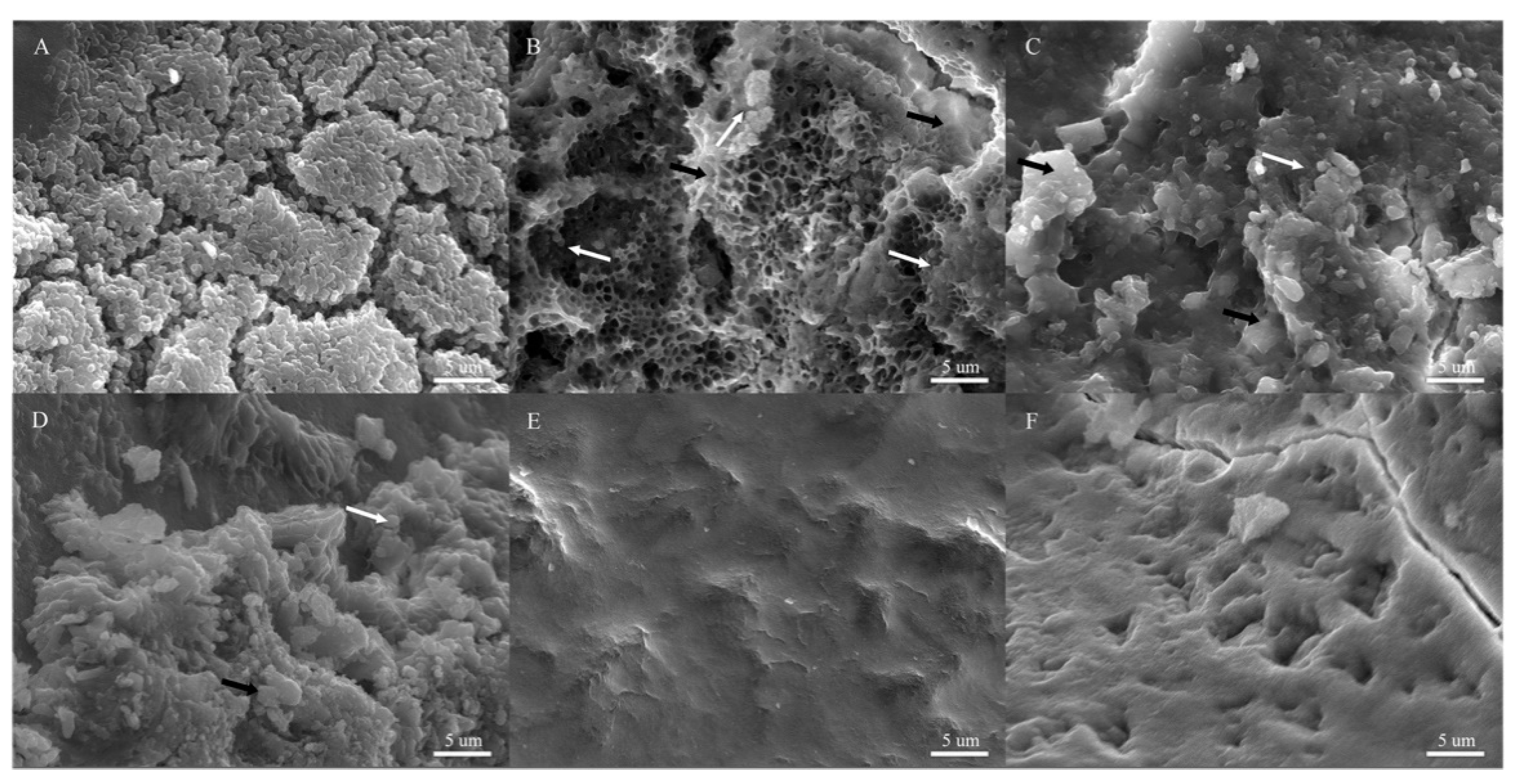
3.4. Anti-Biofilm Effect of Cu NPs against S. mutans on Tooth Crowns

To assess whether the observed anti-biofilm effect (Figure 3) also occurs under physiological conditions, biofilm formation on extracted human teeth was evaluated using scanning electron microscopy (SEM), focusing on the principal cariogenic bacterium, S. mutans (Figure 4). This assay utilized healthy extracted human teeth and pasteurized saliva to simulate real-life conditions for potential application. Due to the in vitro results obtained from the 96-well plate assay indicating no significant effect on S. salivarius and S. sanguinis (Figure 3) and the challenges associated with obtaining healthy teeth during the global COVID-19 pandemic, the analysis was not conducted for commensal bacteria.
In the absence of Cu NPs, we observed aggregated cocci over an extracellular polymeric substance (EPS) structure on all teeth (Figure 4A), indicating the formation of a regular biofilm. However, in the presence of Cu0 NPs, honeycomb-like structures representing EPS indicating natural cellular detachment from the biofilm at this time [38] were observed (Figure 4B, black arrow), with fewer bacterial aggregates (Figure 4B, white arrow). With oxide Cu NPs (CuxO NPs), a reduced biofilm was observed (Figure 4C,D), characterized by only a few amorphous bacteria present on the tooth surface (white arrow, Figure 4C,D). Consequently, Cu0 NPs decreased biofilm formation, while CuxO NPs showed an unclear biofilm structure formation at the concentrations tested compared with the control in Figure 4A.
Additionally, we evaluated the effect of NPs on biofilms already formed on tooth crowns (Figure 5), as described in the materials and methods section. After 48 h of incubation, S. mutans formed a mature biofilm on the tooth surface (Figure 5A, white arrow). By 72 h, the presence of EPS (Figure 5B, black arrow) and aggregated bacteria (Figure 5B, white arrow) was easily detected, along with a few small honeycomb-like structures (Figure 5B, black arrow). Upon the addition of Cu0 NPs (Figure 5C), fewer aggregated bacteria were observed (Figure 5C, white arrow), along with reduced EPS and honeycomb-like structures (Figure 5C, white arrow). After 72 h of mature biofilm formation, the addition of Cu2O NPs (Figure 5D) showed few bacterial cells (white arrow), alongside increased EPS and honeycomb-like structures (Figure 5D, black arrow). Notably, these structures were not observed in the “biofilm formation assay” previously shown with Cu2O NPs (Figure 4C), suggesting that these NPs may disrupt the biofilm and induce the detachment of cells without affecting biofilm formation at this concentration. Finally, the detachment effect was more pronounced in biofilms exposed to CuO NPs, where practically no EPS structures or bacteria were observed (Figure 5E), indicating that the exposure of biofilms formed on tooth crowns to CuO NPs led to the complete disruption of the biofilms. Overall, our results indicate that CuxO NPs strongly decrease biofilm formation and promote biofilm detachment from tooth crowns, with the total absence of biofilm and related structures observed upon CuO NP exposure.
This study presents the first comprehensive investigation of the effect of various Cu NPs on the growth and biofilm formation of four significant oral bacteria, revealing the distinctive impact of Cu NPs on each bacterial strain. CuO NPs emerged as the most potent in inhibiting both the growth and biofilm formation of pathogenic oral bacteria. While further competition assays involving all strains and viability/biofilm formation assays using oral samples such as saliva are essential, our findings provide a valuable foundation for the potential utilization of CuO NPs as anti-cavity agents, particularly for their pronounced effect on pathogenic bacteria.

4. Discussion

Worldwide, untreated cavities represent the most prevalent oral health concern. This disease poses a significant global health challenge, emphasizing the urgent need for its control [1]. Copper (Cu) and copper oxide nanoparticles (CuxO NPs) have emerged as focal points of research in biomedical applications, drawing attention because of their multifaceted advantages, which include enhancing drug stability, facilitating precise biodistribution, elevating therapeutic efficacy, and facilitating targeted delivery to specific sites through active or passive targeting mechanisms [39]. Added to medical applications, their remarkable antimicrobial properties make them a cornerstone of various industries worldwide. Harnessing the potential of Cu-based nanomaterials extends their applications across diverse sectors, such as agriculture, livestock management, water treatment, wood preservation, and textile manufacturing [40]. Moreover, the remarkable conductivity and cost-effectiveness of Cu nanomaterials position them as compelling alternatives to noble metal counterparts in pivotal fields like solar energy conversion, battery technology, and electrochemical sensing [40,41].
In this way, Cu NPs have been utilized to inhibit the growth of oral pathobionts in various dental materials, including metals and alloys; polymers and resins; cements; and other miscellaneous materials [15]. Because cavities result from oral dysbiosis characterized by an increase in acidogenic and aciduric bacteria [42], AOAs used to control the growth of these species should also promote the maintenance of a balanced microbial oral community, or eubiosis [43]. Currently, several commonly used antimicrobial agents against pathogenic oral bacteria include sodium fluoride, chlorhexidine, penicillin, chitosan, and daptomycin. Interestingly, S. sanguinis exhibits greater sensitivity to most standard antimicrobial oral agents compared with S. mutans, contrasting with the situation observed with Cu NPs (Table S1). Only chlorhexidine (CHX) and penicillin appear to be more toxic for S. mutans. However, it has been documented that CHX-containing mouthwash can alter the salivary microbiome, leading to a more acidic environment and reducing nitrite availability in healthy individuals [12]. Regarding antibiotics such as penicillin, studies have reported alterations in the oral microbiota, which may subsequently affect the concentration of salivary antibodies [13].
With the aim of evaluating the effects of Cu NPs on some important representative bacteria from the oral microbiome, we conducted in vitro analyses to assess the impact of three types of Cu NPs on S. mutans, L. rhamnosus, S. sanguinis, and S. salivarius.
In planktonic lifestyle, we observed that CuxO NPs exhibited a stronger antibacterial effect compared with Cu0, with MIC values ranging between 400 and 600 µg/mL in S. mutans and S. salivarius, while surprisingly showing high tolerance in S. sanguinis (MIC > 800 µg/mL). Interestingly, CuO NPs exerted a pronounced effect on cariogenic bacteria compared with commensal ones. This observation aligns with the viability assay, where S. sanguinis exhibited higher survival rates (11.4%) compared with the minimal cell survival observed in S. mutans and L. rhamnosus (less than 0.2%).
Previous studies have reported that CuO NPs induce higher levels of reactive oxygen species (ROS) than Cu2O and Cu0 NPs. CuO NPs generate ROS through Haber–Weiss and Fenton-type reactions, while Cu2O NPs only generate ROS through Fenton-type reactions [44]. Moreover, it has been reported that CuO NPs demonstrate a higher degree of internalization and better activity at lower concentrations [25]. This phenomenon could explain the antibacterial effect observed with CuO NPs. Additionally, considering that CuO NPs do not exhibit toxic effects on human cells even at high concentrations (up to 5000 µg/mL) [45], these NPs could be suitable candidates for AOA applications.
In Cu0 NPs, all Streptococcus genus bacteria exhibited high tolerance (MIC > 1000 µg/mL), with L. rhamnosus showing slight sensitivity (MIC 800 µg/mL). Previous studies have reported the greater antibacterial activity of Cu0 NPs compared with other Cu NPs in aerobic conditions, attributable to their stronger ability to accept electrons, leading to bacterial membrane rupture [46], and their superior capacity to release Cu+ ions, facilitating contact killing activity against bacteria [47]. However, our studies were conducted under microaerophilic conditions, where there is likely less oxidation, potentially reducing the release of Cu+ ions and altering the lethal contact effect.
The most tolerant strain to all Cu NPs was S. sanguinis. Although there are no reports of this bacterium being exposed to Cu NPs, it has shown resistance to high concentrations of copper salts (MIC, 1000 µg/mL) [48]. Previously, a decrease in Cu+ released in the presence of H2O2 has been reported [49]. The decrease in Cu released by NPs may occur because the OH radicals generated are adsorbed on the nanocrystal surface [49]. In this way, the production of H2O2 by S. sanguinis [9] could potentially protect this bacterium from the effects of Cu NPs.
In contrast, Cu NPs strongly affected L. rhamnosus. The sensitivity of L. rhamnosus to Cu may be attributable to amine and carboxyl groups on the cell surface, which have a greater affinity for the metal [50]. Generally, Lactobacillus strains display an electronegative charge with a cell surface dominated by anionic compounds [51], which could enhance the binding of Cu NPs (and Cu ions), leading to damage to the cell membrane.
In the biofilm assay, we did not detect a significant effect of Cu NPs on biofilm formation in S. sanguinis, and surprisingly, an increase in biofilm formation in S. salivarius was observed. To date, no studies reporting a positive effect of Cu NPs on biofilm formation have been published. However, the exposure of E. gracilis to Cu2+ stimulated biofilm formation, suggesting that biofilm formation could be considered a protective mechanism [52]. Cu NPs significantly reduced biofilm formation in S. mutans in all assays. On tooth crowns, we observed a decrease in biofilm structures in the presence of all NPs, consistent with previous reports involving Cu-containing NPs [29]. Honeycomb-like structures were observed in biofilms exposed to Cu0 NPs, indicating cell detachment from the biofilm [38]. Thus, it can be speculated that Cu0 NPs did not block attachment to the tooth but instead caused bacterial release [38].
In the case of CuO NPs, no EPS structures were observed, indicating that the oxides affected the formation of glucan matrix even in the presence of sucrose. A previous study showed that copper ions suppressed the expression of certain glucosyltransferase, gtf genes, which code to the enzymes responsible for synthesizing glucans from sucrose. These genes are crucial for the formation of glucan matrix in dental plaque, contributing to biofilm formation and dental caries development, thereby negatively affecting biofilm formation in S. mutans [53]. Additionally, it has been reported that biofilm formation is regulated by autoinducers (AIs) mediated by quorum sensing (QS) [54]. In 2021, it was demonstrated that copper inhibited the QS of S. agalactiae [55]. In S. mutans, Cu NPs could negatively affect biofilm formation through both of these mechanisms.

5. Conclusions

The impact of Cu NPs on key species within representative oral microbiome bacteria was thoroughly investigated, with a focus on fostering oral health (eubiosis). Our research uncovered the distinct effect of CuO NPs, showing the significant inhibition of pathogenic bacterial growth while demonstrating minimal influence on representative beneficial commensal species. Particularly noteworthy was the ability of CuO NPs to effectively deter biofilm formation, with limited adverse effects on the commensal bacteria tested. This was evidenced in biofilm analyses conducted on tooth surfaces, where both the prevention of biofilm formation and the disruption of already established mature biofilms were observed.
Moving forward, our research aims to expand into in vivo studies to analyze the in vivo effect of these Cu NPs. Overall, our findings underscore the potential of CuO NPs for targeted applications against oral pathogens, emphasizing their promise as a therapeutic strategy for enhancing oral health.

Supplementary Materials

The following supporting information can be downloaded at https://www.mdpi.com/article/10.3390/microorganisms12030624/s1. Table S1. Comparative Table of Antimicrobial agents used commonly over oral pathogens. References [56,57,58,59,60] are cited in the Supplementary Materials.

Author Contributions

Methodology, N.O., D.S., J.L.-M., R.O., D.B. and J.M.P.-D.; Validation, D.B. and J.M.P.-D.; Formal analysis, N.O., J.L.-M., D.B. and J.M.P.-D.; Investigation, N.O., D.S. and J.M.P.-D.; Resources, N.O.; Writing—original draft, N.O., J.L.-M., M.Q.-M., D.B. and J.M.P.-D.; Writing—review & editing, N.O., D.S., J.L.-M., M.Q.-M., D.B. and J.M.P.-D.; Project administration, N.O.; Funding acquisition, N.O., D.B. and J.M.P.-D. All authors have read and agreed to the published version of the manuscript.

Funding

This research was supported by the Agencia Nacional de Investigación y Desarrollo (ANID) Grant No. 3190555; Fondo Nacional de Desarrollo Científico y Tecnológico (FONDECYT) Grant No. 1200870; and Fondo Nacional de Desarrollo Científico y Tecnológico (FONDECYT) Grant No. 1200877.

Institutional Review Board Statement

All experiments complied with the ethical standards of the Bioethical Committee, Universidad Andrés Bello (Approval nª 017/2019). 14 May 2019.

Informed Consent Statement

José Manuel Pérez Donoso and all authors provided consent for the publication of all results and images of microorganisms.

Data Availability Statement

The original contributions presented in this study are included in the article and the associated Supplementary Materials.

Acknowledgments

We thank all participants for their financial and technical support for this study. N.O. created the experimental design, wrote the main manuscript text, and prepared all figures. D.S. performed experimental technician support. J.L.-M. is a dentistry professional who obtained all dental pieces and prepared them. R.O. performed microscopy technician support. M.Q.-M. wrote the main manuscript text. D.B. created the experimental design and wrote the main manuscript text. J.M.P.-D. created the experimental design and wrote the main manuscript text. All authors gave their final approval and agreed to be accountable for all aspects of the work.

Conflicts of Interest

The authors declare no competing interests.

Abbreviations

NPsNanoparticles
AOAAnticavity oral agents
Cu NPsCopper nanoparticles
CuxOCopper oxide NPs
OD600Optical Density at 600 nm
OD570Optical Density at 570 nm
EPSExtracellular polymeric substance
ROSReactive oxygen species
BHIBrain Heart Infusion
CFUColony-Forming Unit
BFIBiofilm Formation Index
SEMScanning electron microscopy
MICMinimal inhibitory concentration
MBCMinimum bactericidal concentration
AIsAutoinducers
QSQuorum sensing
H2O2Hydrogen peroxide
CHXChlorhexidine

References

  1. WHO. Global Oral Health Status Report; WHO: Geneva, Switzerland, 2022; Volume 57, ISBN 9789240061484. [Google Scholar]
  2. Deo, P.N.; Deshmukh, R. Oral Microbiome: Unveiling the Fundamentals. J. Oral Maxillofac. Pathol. 2019, 23, 122–128. [Google Scholar] [CrossRef] [PubMed]
  3. Belstrøm, D.; Constancias, F.; Liu, Y.; Yang, L.; Drautz-Moses, D.I.; Schuster, S.C.; Kohli, G.S.; Jakobsen, T.H.; Holmstrup, P.; Givskov, M. Metagenomic and Metatranscriptomic Analysis of Saliva Reveals Disease-Associated Microbiota in Patients with Periodontitis and Dental Caries. NPJ Biofilms Microbiomes 2017, 3, 23. [Google Scholar] [CrossRef]
  4. Bowen, W.H.; Burne, R.A.; Wu, H.; Koo, H. Oral Biofilms: Pathogens, Matrix, and Polymicrobial Interactions in Microenvironments. Trends Microbiol. 2018, 26, 229–242. [Google Scholar] [CrossRef]
  5. Radaic, A.; Kapila, Y.L. The Oralome and Its Dysbiosis: New Insights into Oral Microbiome-Host Interactions. Comput. Struct. Biotechnol. J. 2021, 19, 1335–1360. [Google Scholar] [CrossRef]
  6. Marsh, P.D. Are Dental Diseases Examples of Ecological Catastrophes? Microbiology 2003, 149, 279–294. [Google Scholar] [CrossRef] [PubMed]
  7. Takahashi, N.; Nyvad, B. Ecological Hypothesis of Dentin and Root Caries. Caries Res. 2016, 50, 422–431. [Google Scholar] [CrossRef]
  8. Horiuchi, M.; Washio, J.; Mayanagi, H.; Takahashi, N. Transient Acid-Impairment of Growth Ability of Oral Streptococcus, Actinomyces, and Lactobacillus: A Possible Ecological Determinant in Dental Plaque. Oral Microbiol. Immunol. 2009, 24, 319–324. [Google Scholar] [CrossRef]
  9. Valdebenito, B.; Tullume-Vergara, P.O.; González, W.; Kreth, J.; Giacaman, R.A. In Silico Analysis of the Competition between Streptococcus Sanguinis and Streptococcus Mutans in the Dental Biofilm. Mol. Oral Microbiol. 2018, 33, 168–180. [Google Scholar] [CrossRef]
  10. Burton, J.P.; Wescombe, P.A.; Cadieux, P.A.; Tagg, J.R. Beneficial Microbes for the Oral Cavity: Time to Harness the Oral Streptococci? Benef Microbes 2011, 2, 93–101. [Google Scholar] [CrossRef]
  11. Burton, J.P.; Wescombe, P.A.; Macklaim, J.M.; Chai, M.H.C.; Macdonald, K.; Hale, J.D.F.; Tagg, J.; Reid, G.; Gloor, G.B.; Cadieux, P.A. Persistence of the Oral Probiotic Streptococcus Salivarius M18 Is Dose Dependent and Megaplasmid Transfer Can Augment Their Bacteriocin Production and Adhesion Characteristics. PLoS ONE 2013, 8, e65991. [Google Scholar] [CrossRef]
  12. Bescos, R.; Ashworth, A.; Cutler, C.; Brookes, Z.L.; Belfield, L.; Rodiles, A.; Casas-Agustench, P.; Farnham, G.; Liddle, L.; Burleigh, M.; et al. Effects of Chlorhexidine Mouthwash on the Oral Microbiome. Sci. Rep. 2020, 10, 5254. [Google Scholar] [CrossRef]
  13. Cheng, X.; He, F.; Si, M.; Sun, P.; Chen, Q. Effects of Antibiotic Use on Saliva Antibody Content and Oral Microbiota in Sprague Dawley Rats. Front. Cell. Infect. Microbiol. 2022, 12, 721691. [Google Scholar] [CrossRef] [PubMed]
  14. Tartaglia, G.M.; Tadakamadla, S.K.; Connelly, S.T.; Sforza, C.; Martín, C. Adverse Events Associated with Home Use of Mouthrinses: A Systematic Review. Ther. Adv. Drug Saf. 2019, 10, 2042098619854881. [Google Scholar] [CrossRef] [PubMed]
  15. Yun, Z.; Qin, D.; Wei, F.; Xiaobing, L. Application of Antibacterial Nanoparticles in Orthodontic Materials. Nanotechnol. Rev. 2022, 11, 2433–2450. [Google Scholar] [CrossRef]
  16. Priyadarsini, S.; Mukherjee, S.; Mishra, M. Nanoparticles Used in Dentistry: A Review. J. Oral Biol. Craniofac. Res. 2018, 8, 58–67. [Google Scholar] [CrossRef]
  17. Haleem, A.; Javaid, M.; Singh, R.P.; Rab, S.; Suman, R. Applications of Nanotechnology in Medical Field: A Brief Review. Glob. Health J. 2023, 7, 70–77. [Google Scholar] [CrossRef]
  18. Pathakoti, K.; Manubolu, M.; Hwang, H.M. Nanotechnology Applications for Environmental Industry. In Handbook of Nanomaterials for Industrial Applications; Elsevier: Amsterdam, The Netherlands, 2018; pp. 894–907. [Google Scholar] [CrossRef]
  19. Xu, V.W.; Nizami, M.Z.I.; Yin, I.X.; Yu, O.Y.; Lung, C.Y.K.; Chu, C.H. Application of Copper Nanoparticles in Dentistry. Nanomaterials 2022, 12, 805. [Google Scholar] [CrossRef] [PubMed]
  20. Lövestam, G.; Rauscher, H.; Roebben, G.; Klüttgen, B.S.; Gibson, N.; Putaud, J.-P.; Stamm, H. Considerations on a Definition of Nanomaterial for Regulatory Purposes. JCR Ref. Rep. 2010, 80, 41. [Google Scholar]
  21. Gawande, M.B.; Goswami, A.; Felpin, F.X.; Asefa, T.; Huang, X.; Silva, R.; Zou, X.; Zboril, R.; Varma, R.S. Cu and Cu-Based Nanoparticles: Synthesis and Applications in Catalysis. Chem. Rev. 2016, 116, 3722–3811. [Google Scholar] [CrossRef]
  22. Santhoshkumar, J.; Agarwal, H.; Menon, S.; Rajeshkumar, S.; Venkat Kumar, S. A Biological Synthesis of Copper Nanoparticles and Its Potential Applications. In Green Synthesis, Characterization and Applications of Nanoparticles; Elsevier: Amsterdam, The Netherlands, 2019; pp. 199–221. [Google Scholar] [CrossRef]
  23. Ramos-Zúñiga, J.; Bruna, N.; Pérez-Donoso, J.M. Toxicity Mechanisms of Copper Nanoparticles and Copper Surfaces on Bacterial Cells and Viruses. Int. J. Mol. Sci. 2023, 24, 10503. [Google Scholar] [CrossRef]
  24. Begum, M.S.; Devi, R.K.; Professor, A. Wet Biochemical Synthesis of Copper Oxide Nanoparticles Coated on Titanium Dental Implants. Int. J. Adv. Res. Sci. Eng. Technol. 2016, 3, 1191–1194. [Google Scholar]
  25. Ma, X.; Zhou, S.; Xu, X.; Du, Q. Copper-Containing Nanoparticles: Mechanism of Antimicrobial Effect and Application in Dentistry—A Narrative Review. Front. Surg. 2022, 9, 905892. [Google Scholar] [CrossRef] [PubMed]
  26. Chatterjee, A.K.; Chakraborty, R.; Basu, T. Mechanism of Antibacterial Activity of Copper Nanoparticles. Nanotechnology 2014, 25, 135101. [Google Scholar] [CrossRef] [PubMed]
  27. Raffi, M.; Mehrwan, S.; Bhatti, T.M.; Akhter, J.I.; Hameed, A.; Yawar, W.; Ul Hasan, M.M. Investigations into the Antibacterial Behavior of Copper Nanoparticles against Escherichia coli. Ann. Microbiol. 2010, 60, 75–80. [Google Scholar] [CrossRef]
  28. Castro, M.; Fernandez, E.; Fluxá, P.P.; Gil, A.M.C.; Grez, P.V. Antimicrobial Effect against Streptococcus Mutans of an Adhesive System with Copper and Zinc Oxide Nanoparticles. Rev. Cuba. Investig. Biomed. 2020, 39, 1–14. [Google Scholar]
  29. Covarrubias, C.; Trepiana, D.; Corral, C. Synthesis of Hybrid Copper-Chitosan Nanoparticles with Antibacterial Activity against Cariogenic Streptococcus Mutans. Dent. Mater. J. 2018, 37, 379–384. [Google Scholar] [CrossRef] [PubMed]
  30. Modaresi, F. The Use of Synergistically Antiplaque Nanoparticles in Treating Dental Caries. J. Dent. Health Oral Disord. Ther. 2017, 6, 144–149. [Google Scholar] [CrossRef]
  31. Jarpa, P. Footwear Molded in Plastic Material with Ventilation Holes of the Crocs Type Where Said Footwear Contains Copper-Based Bactericide, CL2013002853A1. 4 October 2013. Available online: https://patents.google.com/patent/CL2013002853A1/en?oq=CL2013002853A1 (accessed on 17 March 2024).
  32. Jarpa, P. Quick-Drying Disinfectant Composition of the Gel Type for Hands and Skin, That Does Not Contain Alcohol. WO2015035530A8. 11 September 2014. Available online: https://patents.google.com/patent/WO2015035530A8/en?oq=WO2015035530A8 (accessed on 17 March 2024).
  33. Lucero-Mejía, J.E.; Romero-Gómez, S.d.J.; Hernández-Iturriaga, M. A New Classification Criterion for the Biofilm Formation Index: A Study of the Biofilm Dynamics of Pathogenic Vibrio Species Isolated from Seafood and Food Contact Surfaces. J. Food Sci. 2020, 85, 2491–2497. [Google Scholar] [CrossRef]
  34. Hu, C.; Zhang, F.; Kong, Q.; Lu, Y.; Zhang, B.; Wu, C.; Luo, R.; Wang, Y. Synergistic Chemical and Photodynamic Antimicrobial Therapy for Enhanced Wound Healing Mediated by Multifunctional Light-Responsive Nanoparticles. Biomacromolecules 2019, 20, 4581–4592. [Google Scholar] [CrossRef]
  35. Recalde, A.; van Wolferen, M.; Sivabalasarma, S.; Albers, S.V.; Navarro, C.A.; Jerez, C.A. The Role of Polyphosphate in Motility, Adhesion, and Biofilm Formation in Sulfolobales. Microorganisms 2021, 9, 193. [Google Scholar] [CrossRef]
  36. Ayoub, H.M.; Gregory, R.L.; Tang, Q.; Lippert, F. Influence of Salivary Conditioning and Sucrose Concentration on Biofilm-Mediated Enamel Demineralization. J. Appl. Oral Sci. 2020, 28, e20190501. [Google Scholar] [CrossRef]
  37. Thurnheer, T.; Bostanci, N.; Belibasakis, G.N. Microbial Dynamics during Conversion from Supragingival to Subgingival Biofilms in an in Vitro Model. Mol. Oral Microbiol. 2016, 31, 125–135. [Google Scholar] [CrossRef]
  38. Asahi, Y.; Miura, J.; Tsuda, T.; Kuwabata, S.; Tsunashima, K.; Noiri, Y.; Sakata, T.; Ebisu, S.; Hayashi, M. Simple Observation of Streptococcus Mutans Biofilm by Scanning Electron Microscopy Using Ionic Liquids. AMB Express 2015, 5, 6. [Google Scholar] [CrossRef]
  39. Woźniak-Budych, M.J.; Staszak, K.; Staszak, M. Copper and Copper-Based Nanoparticles in Medicine—Perspectives and Challenges. Molecules 2023, 28, 6687. [Google Scholar] [CrossRef]
  40. Harishchandra, B.D.; Pappuswamy, M.; PU, A.; Shama, G.; Pragatheesh, A.; Arumugam, V.A.; Periyaswamy, T.; Sundaram, R. Copper Nanoparticles: A Review on Synthesis, Characterization and Applications. Asian Pac. J. Cancer Biol. 2020, 5, 201–210. [Google Scholar] [CrossRef]
  41. Bakshi, M.; Kumar, A. Applications of Copper Nanoparticles in Plant Protection and Pollution Sensing: Toward Promoting Sustainable Agriculture. In Copper Nanostructures: Next-Generation of Agrochemicals for Sustainable Agroecosystems; Elsevier: Amsterdam, The Netherlands, 2022; pp. 393–413. [Google Scholar] [CrossRef]
  42. Thomas, C.; Minty, M.; Vinel, A.; Canceill, T.; Loubières, P.; Burcelin, R.; Kaddech, M.; Blasco-Baque, V.; Laurencin-Dalicieux, S. Oral Microbiota: A Major Player in the Diagnosis of Systemic Diseases. Diagnostics 2021, 11, 1376. [Google Scholar] [CrossRef]
  43. Baker, J.L.; Edlund, A. Exploiting the Oral Microbiome to Prevent Tooth Decay: Has Evolution Already Provided the Best Tools? Front. Microbiol. 2019, 10, 437902. [Google Scholar] [CrossRef] [PubMed]
  44. Giannousi, K.; Sarafidis, G.; Mourdikoudis, S.; Pantazaki, A.; Dendrinou-Samara, C. Selective Synthesis of Cu2O and Cu/Cu2O NPs: Antifungal Activity to Yeast Saccharomyces Cerevisiae and DNA Interaction. Inorg. Chem. 2014, 53, 9657–9666. [Google Scholar] [CrossRef]
  45. Allaker, R.P. Critical Review in Oral Biology & Medicine: The Use of Nanoparticles to Control Oral Biofilm Formation. J. Dent. Res. 2010, 89, 1175–1186. [Google Scholar] [CrossRef]
  46. Akhavan, O.; Ghaderi, E. Cu and CuO Nanoparticles Immobilized by Silica Thin Films as Antibacterial Materials and Photocatalysts. Surf. Coat. Technol. 2010, 205, 219–223. [Google Scholar] [CrossRef]
  47. Hans, M.; Erbe, A.; Mathews, S.; Chen, Y.; Solioz, M.; Mücklich, F. Role of Copper Oxides in Contact Killing of Bacteria. Langmuir 2013, 29, 16160–16166. [Google Scholar] [CrossRef]
  48. Grytten, J.; Scheie, A.A.; Giertsen, E. Synergistic Antibacterial Effects of Copper and Hexetidine against Streptococcus Sobrinus and Streptococcus Sanguis. Acta Odontol. Scand. 1988, 46, 181–183. [Google Scholar] [CrossRef]
  49. Du, T.; Vijayakumar, A.; Desai, V. Effect of Hydrogen Peroxide on Oxidation of Copper in CMP Slurries Containing Glycine and Cu Ions. Electrochim. Acta 2004, 49, 4505–4512. [Google Scholar] [CrossRef]
  50. Beveridge, T.J.; Murray, R.G.E. Sites of Metal Deposition in the Cell Wall of Bacillus Subtilis. J. Bacteriol. 1980, 141, 876–887. [Google Scholar] [CrossRef] [PubMed]
  51. Kirillova, A.V.; Danilushkina, A.A.; Irisov, D.S.; Bruslik, N.L.; Fakhrullin, R.F.; Zakharov, Y.A.; Bukhmin, V.S.; Yarullina, D.R. Assessment of Resistance and Bioremediation Ability of Lactobacillus Strains to Lead and Cadmium. Int. J. Microbiol. 2017, 2017, 9869145. [Google Scholar] [CrossRef] [PubMed]
  52. Morales-Calderón, L.S.; Armenta-Ortiz, N.; Méndez-Trujillo, V.; Ruiz-Sanchez, E.; González-Mendoza, D.; Grimaldo-Juarez, O.; Cervantes-Diaz, L.; Aviles-Marin, M. Laura Selene Morales-Calderón Copper Induced Biofilm Formation and Changes on Photosynthetic Pigment in Euglena Gracilis. Afr. J. Microbiol. Res. 2012, 6, 1833–1836. [Google Scholar] [CrossRef]
  53. Singh, K. Characterization of a Copper Resistance and Transport System in Streptococcus mutans. ProQuest Dissertations and Thesis, University of Toronto, Toronto, ON, Canada, 2015; p. 148. [Google Scholar]
  54. Zhou, L.; Zhang, Y.; Ge, Y.; Zhu, X.; Pan, J. Regulatory Mechanisms and Promising Applications of Quorum Sensing-Inhibiting Agents in Control of Bacterial Biofilm Formation. Front. Microbiol. 2020, 11, 589640. [Google Scholar] [CrossRef] [PubMed]
  55. Abdul-Hamza, H.K.; Mohammed, G.J. Anti-Quorum Sensing Effect of Streptococcus agalatiaceae by Zinc Oxide, Copper Oxide, and Titanium Oxide Nanoparticles. J. Phys. Conf. Ser. 2021, 1999, 012031. [Google Scholar] [CrossRef]
  56. Dong, L.; Tong, Z.; Linghu, D.; Lin, Y.; Tao, R.; Liu, J.; Tian, Y.; Ni, L. Effects of Sub-Minimum Inhibitory Concentrations of Antimicrobial Agents on Streptococcus Mutans Biofilm Formation. Int. J. Antimicrob. Agents 2012, 39, 390–395. [Google Scholar] [CrossRef]
  57. Qian, W.; Zhang, J.; Xiao, X. Research on Inhibition of Sodium Fluoride on Five Subgingival Bacteria in Vitro. Hua Xi Kou Qiang Yi Xue Za Zhi 1998.
  58. Li, X.; Wang, Y.; Jiang, X.; Zeng, Y.; Zhao, X.; Washio, J.; Takahashi, N.; Zhang, L. Investigation of Drug Resistance of Caries-Related Streptococci to Antimicrobial Peptide GH12. Front. Cell Infect. Microbiol. 2022, 12. [Google Scholar] [CrossRef]
  59. Doern, G.V.; Ferraro, M.J.; Brueggemann, A.B.; Ruoff, K.L. Emergence of High Rates of Antimicrobial Resistance among Viridans Group Streptococci in the United States. Antimicrob. Agents Chemother. 1996, 40, 891–894. [Google Scholar] [CrossRef] [PubMed]
  60. Aliasghari, A.; Khorasgani, M.R.; Vaezifar, S.; Rahimi, F.; Younesi, H.; Khoroushi, M. Evaluation of Antibacterial Efficiency of Chitosan and Chitosan Nanoparticles on Cariogenic Streptococci: An in Vitro Study. Iran. J. Microbiol. 2016, 8, 93. [Google Scholar]
Figure 1. Oral bacteria growth analysis (OD600) in the presence of different concentrations of Cu NPs. (A) S. mutans, (B) L. rhamnosus, (C) S. sanguinis, and (D) S. salivarius. The experiment was developed in an adequate medium for each strain. Bacteria were grown for 48 h at 37 °C and 90 rpm in the presence of Cu0, Cu2O, or CuO NPs. We used the absence of NPs as a positive control of growth. Data represent the mean ± SEM of DO600 values obtained in three independent experiments performed in triplicate. Asterisks represent statistically significant values (ns: no significative; * p < 0.05; ** p < 0.01; *** p < 0.001; **** p < 0.0001) compared with the related controls without NPs.
Microorganisms 12 00624 g001
Figure 2. Assessment of viable cells after exposure to 100, 300, and 500 µg/mL of CuO NPs. A viability assay was developed in an adequate liquid medium for 48 h at 37 °C and 90 rpm in the presence of CuO NPs. Then, bacteria were seeded in adequate agar plaque for (A) Colony-Forming Unit (CFU) determination. (B) Viability percentage with respect to growth in the absence of CuO NPs in each strain. We used the absence of NPs as a positive control of growth. Data represent the mean ± SEM of CFU/mL values obtained in three independent experiments performed in duplicate. Asterisks represent statistically significant values (ns: no significative; *** p < 0.001; **** p < 0.0001) compared with the controls.
Microorganisms 12 00624 g002
Figure 3. In vitro biofilm formation assay. (A) S. mutans, (B) S. sanguinis, and (C) S. salivarius were grown independently in the presence of different Cu NPs. Biofilm formation was analyzed with a crystal violet assay after 48 h of growth, and variation in biofilm formation was determined through the Biofilm Formation Index (DO570/DO600). Data represent the mean ± SEM of BFI values obtained in three independent experiments performed in triplicate. Asterisks represent statistically significant values (ns: no significative; * p < 0.05; ** p < 0.01; *** p < 0.001; **** p < 0.0001) compared with the related controls. The table at the bottom shows the percentage increase or decrease in biofilm formation.
Microorganisms 12 00624 g003
Figure 4. Effect of Cu NPs on S. mutans biofilm formation on tooth crown. Scanning electron micrography images of S. mutans biofilms on the crown of tooth surface for (A) bacteria grown on a tooth immersed in medium without Cu NPs; (B) bacteria grown on a tooth immersed in medium exposed to 200 µg/mL of Cu0 NPs, (C) 200 µg/mL of Cu2O NPs, and (D) 200 µg/mL of CuO NPs; and (E,F) controls without bacteria. White arrow: S. mutans; black arrow: EPS.
Microorganisms 12 00624 g004
Figure 5. Effect of Cu NPs on S. mutans biofilms formed on tooth crown. Scanning electron micrography images of tooth crowns after (A) 48 h of biofilm formation without Cu NPs (mature biofilm); (B) 72 h of biofilm formation without Cu NPs (positive control); and mature biofilm exposed 24 h to (C) 200 µg/mL of Cu0 NPs, (D) 200 µg/mL of Cu2O NPs, and (E) 200 µg/mL of CuO NP. (F) Tooth crown without bacteria: negative control. White arrow: S. mutans; black arrow: EPS.
Microorganisms 12 00624 g005
Table 1. Characteristics of the Cu NPs used in this work.
Cu0 NPsCu2O NPsCuO NPs
CAS number7440-50-81317-38-01317-38-0
Molecular weight63.5 g/mol143.9 g/mol79.6 g/mol
ColorBrown-redgreenblack
Size~40–70 nm~40–60 nm~40–60 nm
Batch180314-RN230315-SP190726-MA
Table 2. Minimal inhibitory concentration (MIC) values (µg/mL) of Cu0, Cu2O, and CuO NPs to S. mutans, L. rhamnosus, S. sanguinis, and S. salivarius.
MIC Cu0 NPs
(μg/mL)
MIC Cu2O NPs
(μg/mL)
MIC CuO NPs
(μg/mL)
S. mutans>1000500400
L. rhamnosus800300200
S. sanguinis>10001000800
S. salivarius>1000400600
Disclaimer/Publisher’s Note: The statements, opinions and data contained in all publications are solely those of the individual author(s) and contributor(s) and not of MDPI and/or the editor(s). MDPI and/or the editor(s) disclaim responsibility for any injury to people or property resulting from any ideas, methods, instructions or products referred to in the content.

