Article

The Uropathogenic Specific Protein Gene usp from Escherichia coli and Salmonella bongori is a Novel Member of the TyrR and H-NS Regulons

1
Biotechnical Faculty, University of Ljubljana, 1000 Ljubljana, Slovenia
2
National Institute of Chemistry, 1000 Ljubljana, Slovenia
*
Author to whom correspondence should be addressed.
Microorganisms 2020, 8(3), 330; https://doi.org/10.3390/microorganisms8030330
Submission received: 20 February 2020 / Accepted: 25 February 2020 / Published: 26 February 2020
(This article belongs to the Section Molecular Microbiology and Immunology)

Abstract

:
The Escherichia coli PAIusp is a small pathogenicity island encoding usp, for the uropathogenic specific protein (Usp), a genotoxin and three associated downstream imu1-3 genes that protect the producer against its own toxin. Bioinformatic analysis revealed the presence of the PAIusp also in publically available Salmonella bongori and Salmonella enterica subps. salamae genome sequences. PAIusp is in all examined sequences integrated within the aroP-pdhR chromosomal intergenic region. The focus of this work was identification of the usp promoter and regulatory elements controlling its activity. We show that, in both E. coli and S. bongori, the divergent TyrR regulated P3 promoter of the aroP gene, encoding an aromatic amino acid membrane transporter, drives usp transcription while H-NS acts antagonistically repressing expression. Our results show that the horizontally acquired PAIusp has integrated into the TyrR regulatory network and that environmental factors such as aromatic amino acids, temperature and urea induce usp expression.

1. Introduction

Escherichia coli and Salmonella bongori are both members of the Enterobacteriacae, (class gammaproteobacteria) and are presumed to have diverged from a common ancestor 120–150 million years ago [1,2]. The genus Salmonella consists of two species, S. enterica and S. bongori, that have diverged between 40–63.4 Myrs ago [3]. S. enterica is divided into six subspecies, enterica, salamae, arizonae, diarizonae, houtenae and indica [1]. While Salmonella serovars belonging to S. enterica, subspecies enterica, provoke disease in humans and other warm-blooded animals, S. bongori and the remaining five S. enterica subspecies are mostly considered nonpathogenic and commensals of cold-blooded animals. Therefore, only limited research into the remaining five S. enterica subspecies and S. bongori has been conducted. Nevertheless, S. bongori serovar 48:z35 strain RKS3044 is considered endemic in Sicily, Italy and has been reported to infect humans, mainly children aged 1 month to 3 years [4,5]. Furthermore, S. bongori strain N19-781 was recently isolated and a draft genome sequence published [6]. Nonetheless, the non-enterica subspecies do cause sporadic disease in mammals, with children and immuno-compromised individuals at highest risk [7,8]. On the other hand, Escherichia coli is a common commensal inhabitant of the human gastrointestinal tract however, strains that produce virulence factors provoke intestinal as well as extraintestinal infections.
Bacterial virulence factor genes are frequently encoded by pathogenicity islands (PAIs), inserted DNA segments that can be mobile and affect host pathobiology as well as bacterial fitness [9,10]. Gram-negative enteric pathogens have acquired a number of virulence factor genes central for infection via horizontal gene transfer (HGT). During evolution of bacterial pathogenesis, HGT of virulence genes has been a driving force for the emergence of new species and the adaptation of pathogens to new hosts as well as specific niches within their hosts [11].
The E. coli uropathogenic specific protein (Usp) is a genotoxin active against mammalian cells and is associated with strains that provoke prostatitis, pyelonephritis and bacteraemia [12,13,14]. Genotoxins have been shown to provoke carcenogenesis by promoting DNA damage in host cells [15,16,17,18]. Studies have found an increased presence of usp+ E. coli strains in colorectal cancer patients [19]. The small pathogenicity island PAIusp encodes usp and three associated downstream imu1-3 genes required for protection of the producer against its own toxin.
Among uropathogenic E. coli (UPEC), mostly belonging to the B2 phylogroup, PAIusp is located within the aroP-pdhR chromosomal intergenic region (Figure 1A). The aroP gene encodes an integral membrane protein that transports three aromatic amino acids, phenylalanine, tyrosine and tryptophan into the bacterial cell, while PdhR, the pyruvate dehydrogenase complex regulator controls the E. coli respiratory electron transport system. Three aroP promoters have been experimentally identified [20,21], P1 the major promoter, P2 located 21 bp downstream of P1, and the divergent P3 which overlaps with P1 (Figure 1A and Figure 2). The P1 promoter is repressed by the transcription regulator TyrR, with phenylalanine, tyrosine or tryptophan, acting as cofactors. The aroP gene is thus a member of the TyrR regulon that is involved in aromatic amino acids biosynthesis, catabolism, or transport. Furthermore, TyrR mediated repression of P1 was shown to recruit RNA polymerase to the divergent transcriptionally nonproductive P3 promoter [20,21,22]. The presence of the P3 promoter was proposed to fine tune the required expression levels of aroP with a mechanism whereby the substrates of the transporter regulate its synthesis [20,21,22,23].
The primary mechanism of TyrR-mediated gene regulation is repression, nevertheless activation by TyrR was reported for three genes, namely mtr, tyrP and the aroP promoter P3 [23]. A prerequisite for activation activity is a strong, appropriately located TyrR binding box DNA sequence [24], located upstream of the promoter -35 hexamer [23,25]. Members of the TyrR regulon may have two or more TyrR boxes that exhibit different affinities for the TyrR protein and are thus either strong or weak. To the former, TyrR in vitro binds alone, in the absence of aromatic amino acids while binding to the latter occurs only in the presence of tyrosine or phenylalanine and only in the vicinity of a strong box that is on the same face of the DNA helix [23].
H-NS (histone-like nucleoid structuring protein) is a nucleoid associated protein playing a key role in nucleoid organization in Gram negative bacteria. In addition, H-NS is a global regulator of gene expression involved in regulating global transcriptional responses to environmental stress as well as a silencer of xenogenic DNA acquired by horizontal gene transfer [26]. H-NS is a small 15,5 Kd protein that in solution exists as a dimer and binds DNA rich in A T sequences. Following DNA binding, H-NS subsequently oligomerizes and spreads along A T rich DNA mostly repressing gene expression.
In this study, we report the presence of the highly conserved PAIusp in S. bongori and S. enterica subsp. salamae strains. In all, PAIusp was found to be inserted at the same genomic location as in E. coli usp+ strains. Furthermore, we investigated and compared usp promoter regions and promoter activities from both S. bongori and E. coli, using reporter gene expression assays as well as Usp protein levels. In both species we identified the aroP P3, as the sole promoter of the usp gene. Our results also show that the horizontally acquired PAIusp has integrated into the TyrR regulatory network and that H-NS represses expression, while environmental factors such as aromatic amino acids, temperature and urea induce usp expression.

2. Materials and Methods

2.1. Bacterial Strains and Plasmids

The strains and plasmids used in this study are listed in Table 1. The usp promoter regions were PCR amplified from the uropathogenic E. coli strain TA211 which was isolated at the Institute of microbiology and immunology, Medical Faculty, University of Ljubljana, Ljubljana, Slovenia and from the S. bongori strain NCTC 12419. The E. coli Keio collection strain JW1889 and the tyrR defective JW1316 strain, were used throughout the study [27]. Bacteria were routinely grown at 37 °C in LB broth or M9 minimal medium. Media were supplemented with antibiotics when required at final concentrations of 100 µg/mL ampicillin, 50 µg/mL kanamycin or 12.5 µg/mL tetracycline (all from Sigma-Aldrich, St. Louis, MO, USA). To study promoter regulation, the L-aromatic amino acid phenylalanine was added to the M9 minimal medium at a final concentration of 1 mM. Freshly prepared urea stock was added to the M9 medium at a final concentration of 0.2, 0.4 or 0.6 M. To impose salt stress, 0.3 M NaCl was added to M9 minimal medium.

2.2. Computational Analysis and Genomic Island Sequence Comparisons

Genome mining for Usp homologs was carried out by PSI-BLAST and tBLASTn analysis using the E. coli UTI89 Usp protein (accession no. ABE05631.1) as queried against the NCBI non-redundant protein database. For phylogenetic analysis, multiple sequence alignments (restricted to complete genomes from NCBI Genome Information resource, July 2018) were based on a ClustalW program implemented in MEGA7 software [29], using the default settings. A neighbor-joining phylogenetic tree of Usp homologs from different bacterial species was generated using MEGA7.
The genomic regions of interest for comparison analysis were manually extracted from genomes of E. coli K-12 MG1655 (RefSeq NC_000913), E. coli UTI89 (RefSeq NC_007946.1), S. bongori NCTC 12419 (RefSeq NC_015761.1) and S. enterica subsp. salamae RKS2993 (RefSeq NZ_JXTT01000055). To compare genomic regions of the four strains, we performed nucleotide alignment using EasyFig version 2.1 [30], with the blastn algorithm with minimum length of hits set to 50 and maximum E- value set to 0. 001.
A basic comparative analysis of protein or nucleotide sequences was carried out using ClustalW version 2.0 [31]. Conserved protein domains encoded by the PAIusp were determined by cross reference of the Conserved Domain Database and Search Service version 3.16 [32].

2.3. Recombinant DNA Manipulations

All cloning was performed as described previously [33]. Plasmid DNA was isolated using the GeneJET plasmid miniprep kit (Thermo Fisher Scientific). Restriction enzymes and T4 DNA ligase (all Thermo Fisher Scientific) were used as directed by the manufacturers. PCR amplifications were carried out using Vent DNA polymerase (New England Biolabs). PCR and restriction fragments were purified using PCR purification kit (Thermo Fisher Scientific).
For usp-CAT reporter transcription fusions, the promoter region was PCR amplified from the S. bongori NCTC 12419 chromosomal DNA using the Bong_L_Pst_F and Bong_L_Bst_R primer pair (Table 2). The PCR product was cloned into the pJET1.2 cloning (high copy) plasmid (Thermo Fisher Scientific). The newly created PstI restriction site was then utilized to remove the upstream T7 promoter present in the vector. Digestion with PvuII and subsequent religation removed the downstream PlacUV5 promoter. This intermediate plasmid was designated pJBPLN3. The promoterless chloramphenicol resistance gene was amplified from the pLysS vector using primers Cm_Bst_F and Cm_Xba_R, with flanking BstEII and XbaI restriction sites. The digested PCR product was ligated into pJBPLN3, also digested with the same two enyzmes. The resulting plasmid, pJBLCm, was subsequently used in the construction of all usp promoter fragments from either S. bongori or E. coli, carrying different promoter variants by replacing the PstI and BstEII promoter fragment.
For usp-lacZ reporter fusions, PCR amplification was performed using primers listed in Table 2. Promoter fragments were then cloned into the low copy lacZ expression vector pRW50 [28]. Site-directed mutagenesis of the -10 or -35 promoter sequences was performed with synthetic primers as described by [34]. Mutations were confirmed by DNA sequence analysis.

2.4. Minimal Inhibitory Concentrations (MICs) for Chloramphenicol

All of the plasmids harboring promoter fragments (Table 1) were transformed into the E. coli K-12 MG1655 strain. The chloramphenicol MIC were determined using the broth microdilution method. Briefly, overnight cultures were diluted in fresh LB medium (1:4000) and subsequently 0.1 mL of the diluted culture was inoculated into a series of tubes with increasing concentrations of chloramphenicol and grown for 24 h. Background MIC values from cells carrying a promoterless pJCAT, were subtracted.

2.5. β-Galactosidase Assays

Plasmids harboring promoter fragments (Table 1) were transformed into the relevant bacterial strains. The overnight cultures were diluted in either fresh M9 minimal or LB medium (1:100) and grown at 37 °C for 24 h. Subsequently, cells were collected (1 mL) by centrifugation. β-galactosidase activity was assayed according to Miller, 1974 [35]. The presented data are the results of at least three independent experiments and are shown with standard deviations. Background β-galactosidase activity values, generated form cells carrying a promoterless pRW50, were subtracted.

2.6. Western Immunoblotting

E. coli K12 MG1655 strain, transformed with plasmids pUSP4L (containing UPEC usp gene under native P3 promoter) and pCUPBI (containing UPEC usp gene under native S. bongori P3 promoter), were grown at the designated temperatures with shaking in LB medium. After 24 h the cells, normalized to optical density, were collected by centrifugation. N-terminally His6-tagged Usp was affinity purified by Ni-NTA metal affinity chromatography (Qiagen, Germany) and eluted with 200 µl of buffer C (50 mM NaH2PO4 pH 8, 300 mM NaCl, 250 mM imidazole, 20 mM mercaptoethanol). Proteins in eluate were precipitated with TCA and washed with ice-cold acetone. The pellet was dried and resuspended in SDS loading buffer, separated on 12% SDS-PAGE gel and transferred at 200 mA for 90 min onto a PVDF membrane (Millipore, U.S.A.) for immunoblotting. The membrane was blocked in Tris-Buffered saline with 0.1% Tween 20 (TBS-T) containing 5% (w/v) bovine serum albumin (BSA). The membrane was subsequently incubated for 2 h with mouse anti-penta-histidine antibodies (Thermo Fisher Scientific) for anti-His staining, followed by 1 h incubation with horseradish peroxidase-conjugated rabbit anti-mouse IgG. The protein bands were visualized by an enhanced chemiluminescence kit (ECL; Amersham).

2.7. Protein Isolation and Electrophoretic Mobility Shift Assay (EMSA)

To isolate the purified TyrR and H-NS proteins, E. coli BL21 (DE3) cells containing the pET8HT or pET9HT expression plasmids were grown in LB medium and induced with 0.8 mM of isopropyl β-D-thiogalactopyranoside (IPTG) for 4 h at 37 °C. N-terminally His6-tagged TyrR protein was affinity purified by Ni-NTA metal affinity chromatography (Qiagen). The native purification protocol was followed according to the manufacturer’s instructions. Protein purity was assessed on a 10% SDS-PAGE gel, followed by Coomassie blue staining and the eluate was dialyzed overnight against a buffer (50 mM Tris-HCl pH 7.4, 150 mM NaCl, 5 mM dithiothreitol DTT, 10% glycerol) at 4 °C. The concentration of the purified TyrR protein was determined using NanoDrop 1000 (Thermo Fisher Scientific) with the extinction coefficients at 280 nm of 36,245 M−1 cm−1.
The reactions for EMSA assay were carried out as described previously with modifications [22]. Binding reactions were performed by mixing DNA fragments (~15 nM) with the purified His6-tagged TyrR protein (~15 nM), in a total volume of 10 µl of binding buffer (50 mM Tris-HCl pH 7.8, 50 mM NaCl, 3 mM magnesium acetate, 0.1 mM EDTA, 0.1 mM dithiothreitol (DTT), 25 µg/mL bovine serum albumin (BSA)) with added phenylalanine (0.1 mM) and ATP (0.1 mM). Protein-DNA binding reactions were incubated at 37 °C for 30 min and then analyzed on 2% agarose gel (2% agarose, 1% glycerol, 0.5x Tris - borate –EDTA, 1 mM MgCl2, 0.1 mM ATP, 0.1 mM phenylalanine). Protein-DNA complexes were resolved at 60 volts at 4 °C. The DNA fragments were stained with ethidium bromide and visualized. Binding reactions with the purified H-NS protein were performed as described previously [36].
Usp protein levels were quantified using GeneTools (Syngene International Ltd.).

2.8. Statistical Analysis

Data analyses were performed using GraphPad PRISM version 6.01 software. Statistically significant differences in gene expression from each promoter were determined by two-way analysis of variance (ANOVA) with post-hoc comparisons using the Tukey multiple comparison test.

3. Results

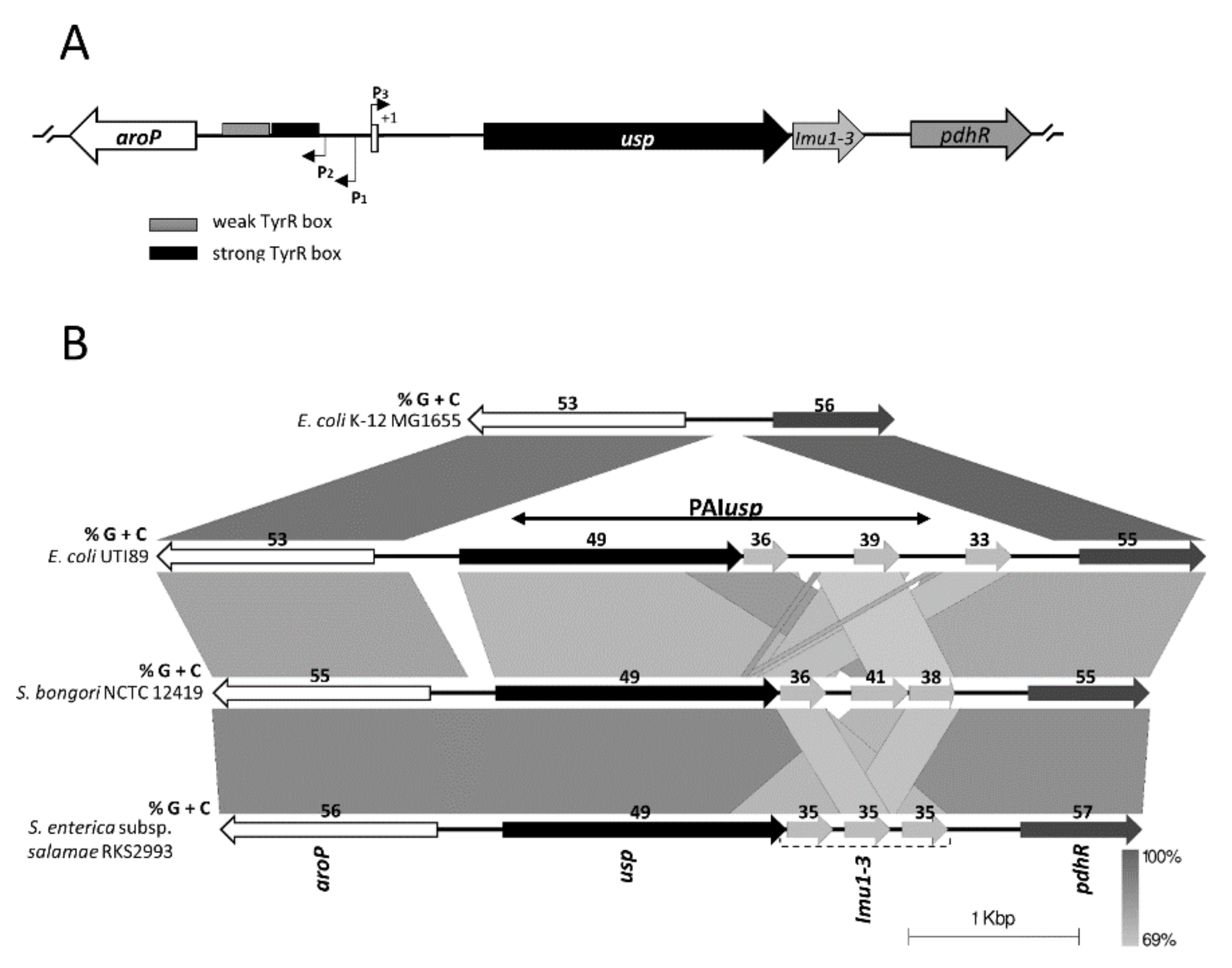
3.1. Bioinformatic Analysis of PAIusp prevalence

Whilst the PAIs of E. coli generally have a size of 30–100 kb and are associated with genes for tRNA, encode integrases and are flanked by direct repeats, usp is encoded on a small approximately 4 kb pathogenicity island, encoding only usp and the tightly associated imu genes. Our analysis showed that PAIusp is in all publicly available usp+ E. coli genome sequences, inserted into the intergenic region aroP-pdhR. The aroP gene encodes the aromatic amino acid permease and pdhR, the pyruvate dehydrogenase complex regulator. Interestingly, bioinformatic analysis revealed the presence of PAIusp homologs in all publicly available Salmonella bongori genomes, including the clinical isolate N268-08 [37] and in Salmonella enterica subsp. salamae genomes, inserted at exactly the same genome position as in all usp+E. coli genomes (Figure 1 and Table S1).
It has been reported that the Usp protein from E. coli strains harbors an N-terminal Hcp domain (PF05638), an extended C-terminal S-type pyocin domain (PF06958) and a colicin nuclease domain (PF12639) [38]. Pairwise alignment of the Usp proteins of E. coli and S. bongori strains, revealed ~76% sequence identity. Furthermore, the additional ORFs encoding Usp homologs in Salmonella enterica subsp. salamae strains show protein sequence identity of ~82% and ~77% compared to Usp from S. bongori strains and E. coli strains, respectively. All of the above-mentioned proteins from the various bacteria have the same domain structure as the Usp from E. coli (Figure S1A and Figure S1B). Phylogenetic analysis revealed that the Usp protein derived from E. coli clearly segregates as a separate lineage in the NJ tree from Salmonella which in turn splits into two clades, S. bongori and S. enterica subsp. salamae (Figure S1C).
Further analysis revealed that the E. coli usp flanking genes, aroP and pdhR, have a total G + C content (55%) similar to the average G + C content of an Escherichia genome (~51%) (indicating, that these are ancestral genes). On the other hand, all usp and associated immunity protein genes (imu1-3) have considerably lower G + C contents (49%, 36%, 38%, 38%, respectively) indicating acquisition via horizontal gene transfer (Figure 1B). G + C analysis showed similar results for Usp homologs in S. bongori and S. enterica subsp. salamae.
Herein we investigated activities of the E. coli and S. bongori usp promoters. Since data on usp expression were lacking, we initially set to in silico identify the usp promoter region. Sequence alignment of the aroP-usp intergenic regions of E. coli UTI89 and S. bongori NTCT 12419, revealed significant differences in the upstream proximal region (>200 bp relative to the usp start codon) (Figure 2). As in both genomes, we were unable to identify -35 (TTGACA) and -10 (TATAAT) consensus sequences [39], we speculated that the previously identified divergent aroP promoter P3 [20,21], located in the usp upstream distal region, could drive PAIusp transcription. In the aligned sequences of E. coli UTI89 and S. bongori NTCT 12419, the P3 promoter is located at exactly the same position as in E. coli K-12. The P3 -10 site is conserved in all available sequences, while the -35 sites from S. bongori and UTI89 differ compared to the commensal E. coli strain K-12 (without PAIusp), in three and two bases, respectively (Figure 2).

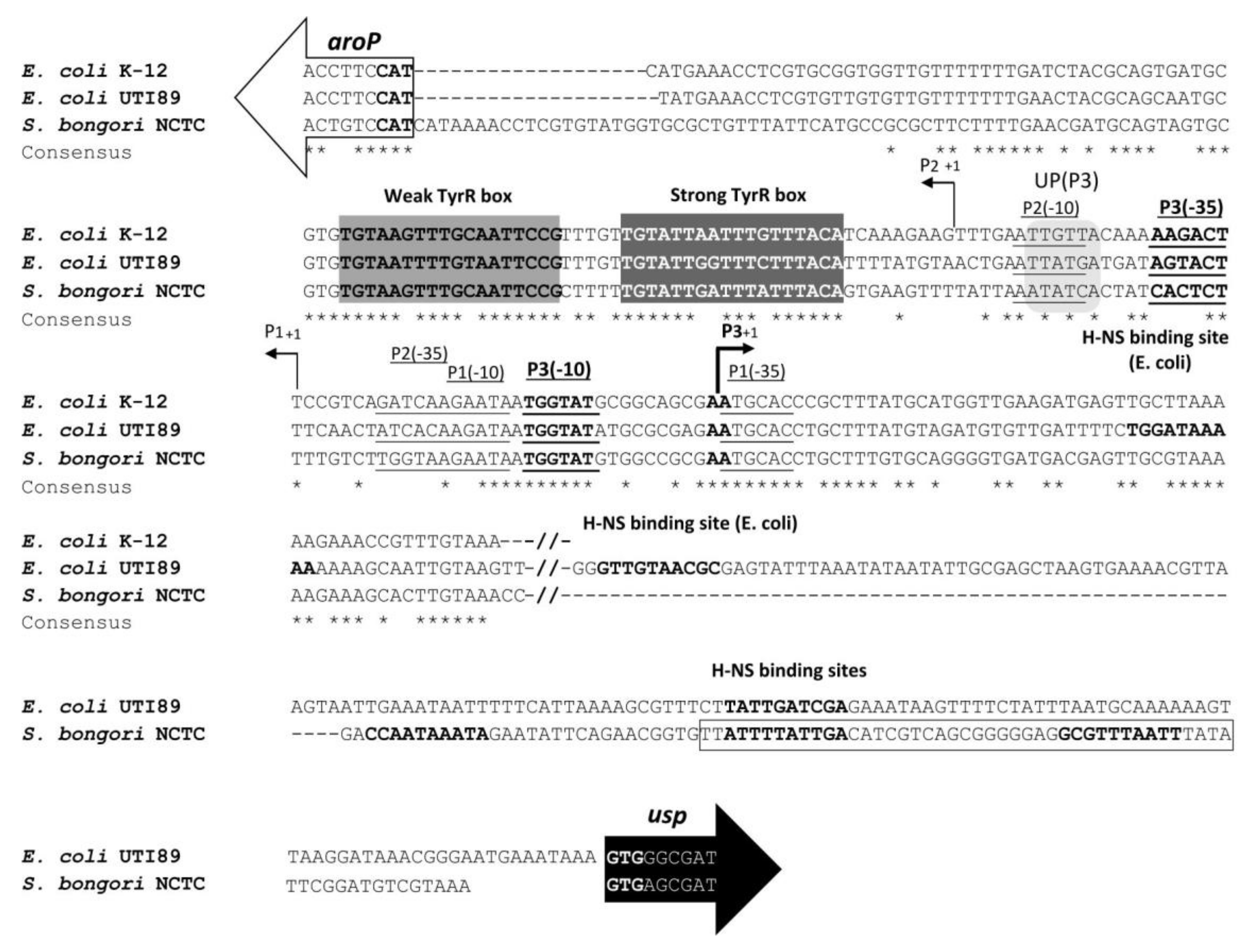
3.2. Expression of usp from the Divergent aroP P3 Promoter

Initially, promoter activity of DNA fragments from the S. bongori NCTC 12419 aroP-usp intergenic region, was investigated using the promoter-probe pJBLC plasmid with a chloramphenicol acetyl transferase reporter gene (CAT). Fragments with promoter activity drive CAT expression, resulting in resistance to chloramphenicol (Figure 3A). CAT assays were performed indirectly by measuring MIC of chloramphenicol. The DNA fragment encompassing the entire intergenic region (pJBLC, 403 bp) supported expression (MIC 70 µg/mL) in E. coli strain MG1655. On the other hand, MIC of chloramphenicol from a shorter DNA fragment (pJBSC, 274 bp), without the two (weak and strong) TyrR binding boxes, was approximately two fold lower, compared to that of the full length fragment indicating, that the TyrR protein acts as a transcription activator of the S. bongori P3 promoter. Furthermore, no significant effect on MIC of chloramphenicol was observed, if only the weak TyrR box was excluded (pJBTC, 315 bp). The smallest DNA fragment that could support significant expression was the 111 bp region harboring the strong TyrR box and the intact P3 promoter (not shown). Interestingly, when the 111 bp DNA fragment was extended for 64 bp (pJBPTD3), MIC of chloramphenicol was, in comparison to the full-length fragment (pJBLC), 4.4 fold higher. The same observation was made by Wang et al. (1997b) for the E. coli aroP promoter P3. They proposed that E. coli P3 and P1 have AT rich associated elements which could function as UP sequences and confer higher transcription levels (Figure 2). Bacterial UP elements are A T rich regions upstream of the -35 promoter element that enhance/activate transcription through contact with the alpha subunit carboxy-terminal domain of the RNA polymerase core enzyme [40,41].
Subsequently, we studied and compared aroP P3 promoter activity from E. coli and S. bongori strains on the basis of β-galactosidase activity from usp-lacZ transcription fusions (Figure 3B). Under the tested conditions we observed ~20 fold higher β-galactosidase activity from the S. bongori P3 compared to that from E. coli.
In an attempt to clarify the basis of the pronounced difference between S. bongori and E. coli P3 promoter activity, the effect of modifications in the -35 and -10 sites were investigated from the low copy number plasmid pRW50 (Table 1). The -10 site (wild type TGGTAT) was in both S. bongori and E. coli promoters altered to TTAATG, while the -35 site was in both promoters altered (wt. in S. bongori and E. coli, CACTCT and AGTACT, respectively) to CAAGCT (Figure 3B). The modifications introduced into the -10 region reduced P3 activity of both S. bongori and E. coli promoters, 3 fold (p-value < 0.0001) and 2 fold (p-value 0.008), respectively. On the other hand, modifications of the -35 regions exhibited no significant effect on promoter activity (Figure 3B). Thus, we conclude that the -10 site of promoter P3 plays a significant role in usp expression and furthermore, the observed difference in the level of transcription activity between the S. bongori and E. coli is not due to differences in the P3 -35 sites.

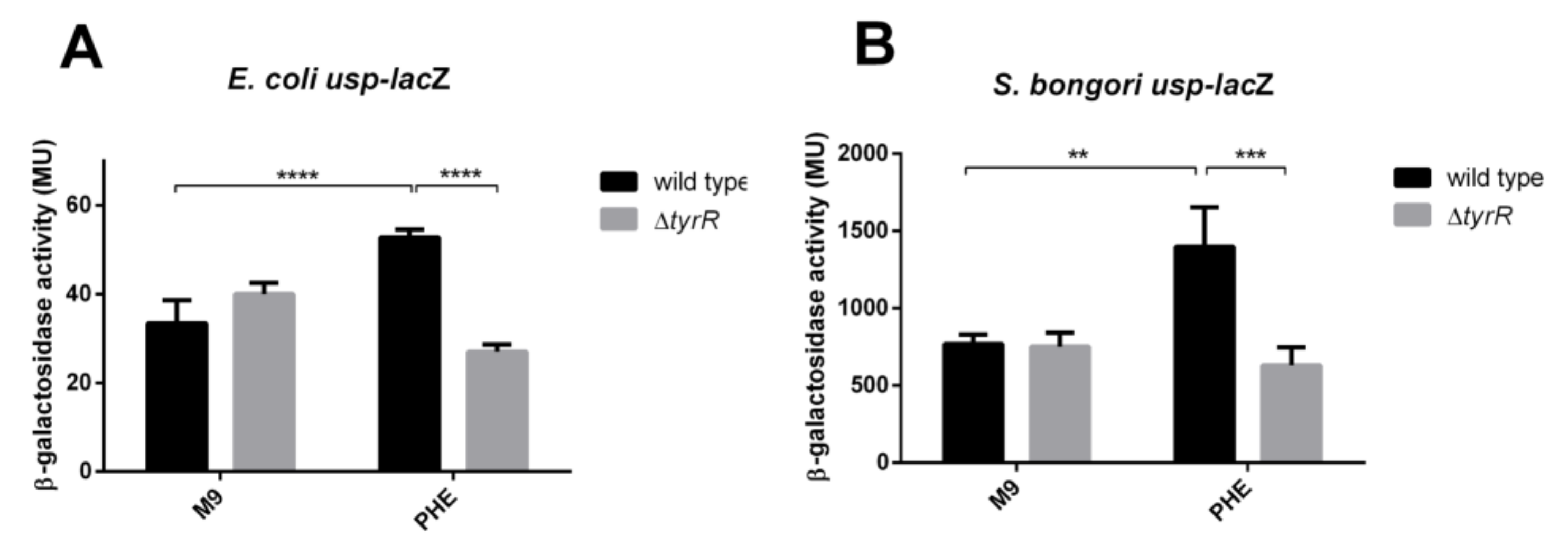
3.3. Positive Regulation of the usp P3 Promoter

In E. coli K-12, which does not harbor PAIusp, the P3 promoter was previously shown to exhibit very low-level activity [20]. To ascertain whether the aroP P3 promoter from E. coli and S. bongori, can be activated by aromatic amino acids via TyrR binding to the TyrR boxes, expression from the P3 usp-lacZ fusion was investigated in the tyrR+ (JW1889) and in the tyrR- defective strain (JW1316) in minimal medium with or without phenylalanine.
Our results showed that in the tyrR+ strain, the aromatic amino acid phenylalanine provoked a statistically significant increase in expression from both the S. bongori and E. coli P3 promoters 2-fold (p-value 0.0021) and 1.6 fold (p-value < 0.0001), respectively (Figure 4). On the other hand, in the tyrR- defective strain, no aromatic amino acid mediated activation of P3 promoter activity was detected (p-value 0.6641 in S. bongori and p-value 0.1097 in E. coli) indicating, that binding of the TyrR protein to the P3 upstream region, is critical for activation and regulation.

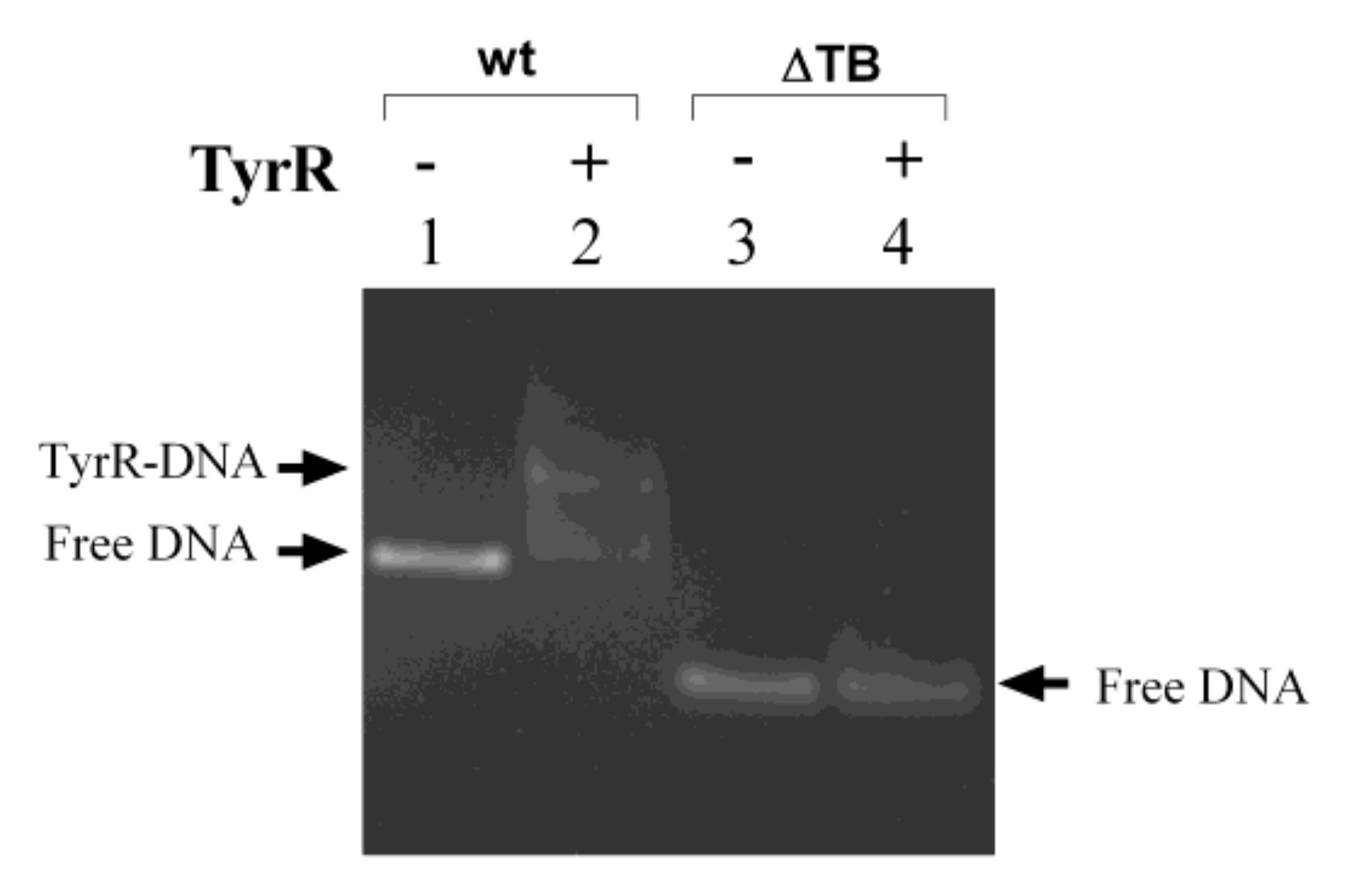
3.4. TyrR Binding to the S. bongori aroP-usp TyrR Boxes.

Previously, Wang et al., 1997a,b [20,21] demonstrated TyrR protein binding to the TyrR boxes within the intergenic region of aroP-pdhR in E. coli strain K-12. Here we demonstrated that the TyrR protein also specifically binds to the TyrR boxes within the S. bongori aroP-usp intergenic region. Incubation of the purified His6-tagged TyrR protein, with a DNA fragment encompassing the wild type aroP-usp intergenic region (403 bp) provoked, a mobility shift consistent with TyrR-DNA complex formation (Figure 5, Lanes 1 and 2). To prove unequivocally that binding of TyrR to the DNA fragment is specific, a control experiment was performed using the DNA fragment without TyrR binding boxes (274 bp) and no TyrR-DNA complex was observed (Figure 5, Lanes 3 and 4).

3.5. Repression of E. coli and S. bongori usp Expression by H-NS

To ascertain whether other transcription factors are involved in regulation of usp activity, an in silico search was performed using Virtual footprint [42]. Three putative H-NS binding sequences were found in the E. coli and S. bongori promoter regions. To corroborate the involvement of H-NS in regulation of usp expression, we subsequently investigated activity of P3 usp-lacZ fusions from E. coli and S. bongori as well as Usp protein levels in an hns+ (JW1889) and hns- defective E. coli strain (JW1225).
Our results showed higher activity of both usp-lacZ fusions as well as higher Usp protein levels in the hns- defective E. coli strain compared to the wild type indicating that H-NS acts as a repressor of usp expression (Figure 6). As temperature is an important signal regulating bacterial gene expression and as S. bongori is mostly considered restricted to cold-blooded animals we investigated usp promoter activity and Usp protein levels following growth at 25 °C, 30 °C, 37 °C, and 42 °C, in the wild type and in the hns mutant strain. Temperature dependent synthesis of the Usp protein was observed from the E. coli and S. bongori promoters. While under the tested conditions, usp-lacZ expression revealed higher S. bongori usp promoter activity compared to the E. coli promoter, protein levels from the latter were at all investigated temperatures in the w.t. and hns mutant strain significantly higher (Table S2).
From the E. coli promoter, the wild type strain produced approximately 5 fold higher levels of Usp at 37 °C and 42 °C than at 25 °C while in the hns mutant, Usp levels were higher and comparable at all three temperatures. From the S. bongori promoter, Usp levels were in the wild type strain grown at 37 °C and at 42 °C approximately eight fold higher than when grown at 25 °C while in the hns mutant strain, protein levels were at 37 °C and 42 °C higher, approximately 9 and 16 fold, respectively, than when cultivated at 25 °C. Our results indicate that in E. coli H-NS plays a key role in temperature dependent usp expression. Nevertheless, discrepancies between protein levels and promoter activities from the E. coli and S. bongori usp-lacZ fusions are evident. From the E. coli usp-lacZ fusion temperature dependent expression was observed in the hns defective strain indicating that other factors are also involved.

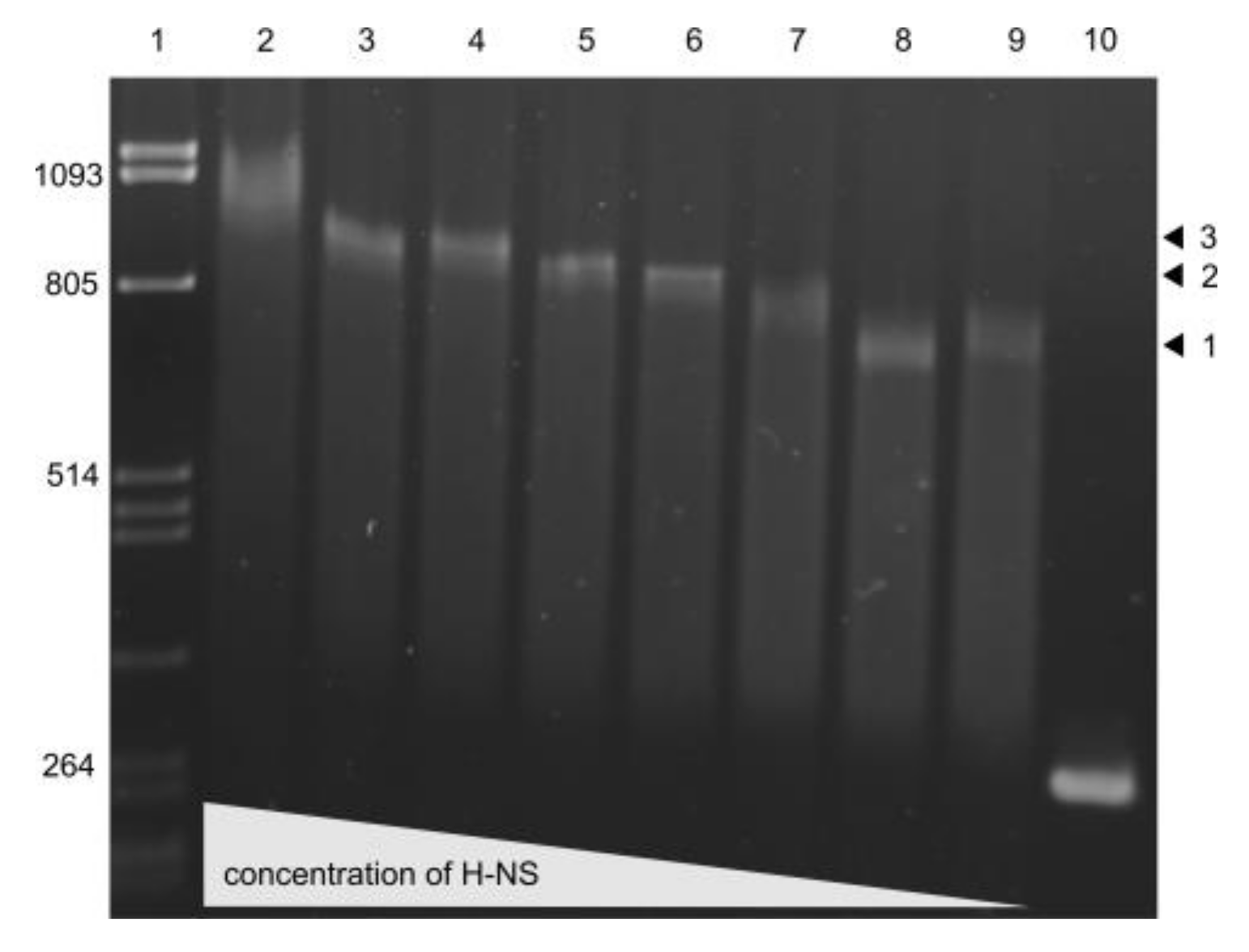
3.6. H-NS binding to usp Promoter Region

Our in silico analysis revealed three putative H-NS binding sites in the usp promoter regions (Figure 2). To assess H-NS binding, EMSA experiments were performed following incubation of the promoter fragment harboring the three binding sites with various concentrations of the purified H-NS protein (from 17.5 nM to 1.6 nM). As shown in Figure 7, increasing protein concentrations provoked a step wise increase in DNA shifts with a full shift observed at 17.5 nM H-NS due to binding to all three sites.

3.7. Urea Mediated Activation of Promoter P3

As PAIusp is prevalent among uropathogenic E. coli, that likely encounter osmotic stress in the urinary tract during infection, we postulated that urea and possibly another osmolyte NaCl, could affect usp expression from the aroP P3 promoter in E. coli and S. bongori. Expression from both, UPEC and S. bongori, usp-lacZ fusions was therefore investigated under urea induced stress in the tyrR+ and the tyrR- defective strain. All cultures were grown for 24 h in M9 minimal medium and in M9 minimal medium supplemented with 0.4M, 0.6M urea or 0.3 M NaCl, concentrations that are comparable to those in human urine [43]. To ascertain whether the TyrR transcription regulator is also involved in urea mediated regulation of aroP P3, β-galactosidase activity was also investigated in the tyrR-defective strain.
Our results showed statistically significant increases in β-galactosidase activity from E. coli and S. bongori promoter fusions in the presence of 0.4 M urea compared to a medium without urea, 3.5 (p-value < 0.0001) and 2.1 fold (p-value 0.0092), respectively (Figure 8). However, the effect of urea was independent of TyrR mediated regulation of promoter P3 (p-value > 0.5). Induction of S. bongori usp promoter activity was comparable in the presence of 0.6 M urea and 0.4 M urea. Surprisingly, we observed an ~1.8– fold (p-value 0.0001) reduction of E. coli P3 activity in the presence of 0.6 M urea compared to 0.4 M urea. On the other hand, 0.3 M NaCl provoked no significant increase in P3 promoter activity (data not shown).

4. Discussion

Here we present a study of the dissemination and regulation of the horizontally acquired PAIusp, usp promoter activity. Point mutations and DNA rearrangements are significant for evolution and speciation of bacterial species nevertheless HGT, the ability to acquire foreign genetic material, enables much more rapid adaptation and survival in novel ecological niches [9,44]. HGT occurs in all free-living bacteria and has played a major role in acquisition of virulence factor and antibiotic resistance genes [45,46]. A paradigm of HGT in bacterial speciation is the divergence of E. coli and Salmonella spp., that presumably occurred approximately 140 million years ago [2].
Our analysis of publicly available genome sequences, indicated limited dissemination of the PAIusp. Interestingly, S. bongori was previously shown to have effectors with virulence associated homologues in enteropathogenic E. coli and enterohemorrhagic E. coli [47]. The presence and identical genomic location in E. coli, S. bongori and S. enterica subsp. salamae strains indicates a common yet unknown ancient source and that acquisition via HGT occurred only once, or alternatively, that the aroP-pdhR intergenic region represents a specific integration site. Regulatory systems control gene expression and genes acquired via HGT must be in the new host appropriately expressed. While exogenous sequences can be eventually incorporated into preexisting regulatory networks, expression may initially be downregulated by specific “xenogenic silencing proteins”, such as the H-NS family of proteins and others, that bind to AT-rich DNA [41]. Further, while expression of horizontally acquired genes is in a number of pathogens essential during infection, outside the host expression could be detrimental. Additionally, mutations in regulatory regions affect expression of virulence factor genes and thus pathogen evolution. Similarly, in E. coli and S. bongori the horizontally acquired PAIusp gene cluster appears to have evolved new regulation after transfer and integration into the aroP-pdhR intergenic region, enabling expression of an otherwise silent aroP promoter P3. Thus, while usp-lacZ fusions showed significantly higher levels of expression from the S. bongori P3 promoter compared to that of E. coli P3, higher Usp protein levels from the latter could indicate more efficient translation possibly, to fine tune usp expression in particular species and subspecies inhabiting distinct ecological niches. Furthermore, a number of differences in the E. coli and S. bongori regulatory regions, such as position of and subtle differences in H-NS binding sequences as well as possibly in DNA topology, could be responsible for the observed higher expression of the S. bongori usp-lacZ fusion.
Integration of virulence factor genes acquired by HGT often does not suffice to promote pathogenicity or to exhibit a newly acquired phenotype [48,49,50] possibly due to the lack of appropriate gene regulation or even the absence of a promoter region. Thus, evolution of regulation of acquired genes has broader implications for our understanding of HGT and evolution of virulence. Here we demonstrated that PAIusp usurped the divergent TyrR regulated aroP P3 promoter resulting in adjustment of PAIusp gene expression with amino acid availability in the environment. This process required specific promoter mutations which enabled activation of the otherwise silent promoter P3.
In our study TyrR in conjunction with aromatic amino acids, induced a significant increase in usp expression in both bacteria, making usp a new member of the TyrR regulon. This is the first report of TyrR mediated expression of a virulence gene in E. coli and S. bongori, extending the significance of the TyrR regulon. Nonetheless, evidence indicates that TyrR [23] is directly or indirectly involved in virulence of other pathogens namely, it has been shown to be required for Yersinia pestis pathogenesis and extracellular survival/proliferation [51]. Additionally, two aromatic pathway genes (aroE and aroA) play a crucial role in Y. pestis fitness in deep tissues during infection [52]. Aromatic amino acid biosynthesis has also been shown to be required for the intracellular replication of Listeria monocytogenes and Shigella flexneri [53,54].
H-NS is a repressor of a number of virulence factor genes frequently encoded on horizontally acquired pathogenicity islands, preventing their expression under non-permissive conditions, i.e., outside the host. The protein consists of an N terminal dimerization domain, a second, central dimerization domain and a C-terminal domain for DNA binding. The N terminal domain harbors four α-helices that allow self-association by contacts “head-to-head” and “tail-to-tail”. These interactions may occur simultaneously allowing H-NS oligomerization to form nucleoprotein filaments that are either bridged or linear. H-NS compacts DNA, repressing expression particularly at the level of transcription [55,56,57]. Our results demonstrated that H-NS is a repressor of Usp synthesis as protein levels were higher from the E. coli and S. bongori native promoters in a hns defective strain compared to the wild type. In accordance, usp-lacZ expression from the E. coli promoter was also higher in the hns mutant but, in contrast, expression from the S. bongori promoter was lower. These results indicate that in S. bongori additional, possibly indirect posttranscriptional mechanisms involving H-NS could regulate Usp synthesis. Furthermore, deletion of the promoter region harboring the putative H-NS binding site, significantly increased usp promoter activity as demonstrated by an increase in MIC for chloramphenicol from plasmid pJBPTD3 (Figure 3A). Temperature is an important signal regulating bacterial gene expression and H-NS has been previously shown to be a key regulator in responses to environmental stimuli including temperature, enabling bacteria to differentiate within host vs. ambient environment [58,59]. Therefore, usp promoter activity and Usp protein levels were investigated and compared following growth at a number of temperatures. Indeed, while from the E. coli promoter the wild type strain produced approximately 5 fold higher levels of Usp at 37 °C and 42 °C than at 25 °C, in the hns mutant, Usp levels were comparable at all three temperatures indicating, that in E. coli H-NS plays a significant role in temperature dependent usp regulation. At the molecular level, temperature provokes alterations in local DNA structure [60] as well as alterations in the H-NS protein [61]. Nevertheless, discrepancies are evident, as from the E. coli usp-lacZ fusion temperature dependent expression was observed in the hns defective strain indicating that, at the level of promoter activity, other as of yet unresolved factors, are also involved. Additional studies will be performed to further unravel the complex molecular mechanisms regulating usp expression in E. coli and S. bongori in response to environmental cues.
Urea freely penetrates cell membranes and destabilizes protein conformation [62], however no E. coli receptor or transcription regulator that responds to urea has been identified. As UPEC colonize the urinary tract they are exposed to high osmolality, due to significant amounts of urea and inorganic ions in urine. Previous studies have shown that H-NS plays a significant role in NaCl induced changes in osmolarity. However, our results demonstrate that urea, but not NaCl, mediate activation of P3 independently of the transcriptional regulator TyrR. Indeed, a previous gene array study also revealed that usp was upregulated in the presence of urea [63]. We therefore speculate on the possible existence of another, TyrR and H-NS independent mechanism, regulating expression from P3, both in E. coli and S. bongori. In UPEC strains, the presence of urea in urine could trigger upregulation of usp expression in the urinary tract.
In conclusion, our results highlight the complex regulation of the usp gene encoded by PAIusp. We further demonstrate that usp is a novel member of the TyrR and H-NS regulons due to its integration into the aroP pdhR intergenic region where the aroP P3 promoter drives its expression. TyrR and H-NS play antagonistic roles in regulating usp expression, with usp P3 promoter activity stimulated by TyrR and repressed by H-NS. In the host, various environmental factors such as aromatic amino acids, temperature as well as urea could induce significant increases in usp expression. Following acquisition of PAIusp by a common ancestor, species divergence and subsequent mutations in the usp P3 promoter region fine-tuned expression in the bacterial hosts.

Supplementary Materials

The following are available online at https://www.mdpi.com/2076-2607/8/3/330/s1, Figure S1: Usp homologs encoded by PAIusp, Figure S2: Isolation of E. coli strain conveying high P3 promoter activity on MacConkey agar plates, Table S1: PAIusp containing strains identified by PSI-BLAST and tBLASTn analysis, excluding E. coli strains; Table 2: Usp levels from E. coli and S. bongori usp promoters in w.t. E. coli and isogenic hns defective strain.

Author Contributions

E.R.: conception of the study, acquisition, analysis and interpretation of data, writing of the manuscript; D.Ž.B., conception of the study, analysis and interpretation of data, writing of the manuscript; Z.P., conception of the study, acquisition, analysis and interpretation of data, writing of the manuscript. All authors reviewed the paper. All authors have read and agreed to the published version of the manuscript.

Funding

This work was supported by grant P1-0198 from the Slovene Research Agency.

Conflicts of Interest

The authors declare no conflict of interest.

References

  1. Crosa, J.H.; Brenner, D.J.; Ewing, W.H.; Falkow, S. Molecular relationships among the Salmonelleae. J. Bacteriol. 1973, 115, 307–315. [Google Scholar] [CrossRef] [PubMed] [Green Version]
  2. Doolittle, R.F.; Feng, D.F.; Tsang, S.; Cho, G.; Little, E. Determining divergence times of the major kingdoms of living organisms with a protein clock. Science 1996, 271, 470–477. [Google Scholar] [CrossRef] [PubMed] [Green Version]
  3. McQuiston, J.R.; Fields, P.I.; Tauxe, R.V.; Logsdon, J.M.J. Do Salmonella carry spare tyres? Trends Microbiol. 2008, 16, 142–148. [Google Scholar] [CrossRef] [PubMed]
  4. Nastasi, A.; Mammina, C.; Villafrate, M.R.; Massenti, M.F.; Scarlata, G.; Diquattro, M. Multiple typing of strains of Salmonella enterica subsp. bongori ser. 48:Z35: Isolated in southern Italy. Ann. Inst. Pasteur Microbiol. 1988, 139, 605–612. [Google Scholar] [CrossRef]
  5. Giammanco, G.M.; Pignato, S.; Mammina, C.; Grimont, F.; Grimont, P.A.; Nastasi, A. Persistent endemicity of Salmonella bongori 48: Z (35): In Southern Italy: Molecular characterization of human, animal, and environmental isolates. J. Clin. Microbiol. 2002, 40, 3502–3505. [Google Scholar] [CrossRef] [Green Version]
  6. Stevens, M.J.A.; Cernela, N.; Muller, A.; Stephan, R.; Bloemberg, G. Draft Genome Sequence of Salmonella bongori N19-781, a Clinical Strain from a Patient with Diarrhea. Microbiol. Resour. Announc. 2019, 8. [Google Scholar] [CrossRef] [Green Version]
  7. Abbott, S.L.; Ni, F.C.Y.; Janda, J.M. Increase in extraintestinal infections caused by Salmonella enterica subspecies II–IV. Emerg. Infect. Dis. 2012, 18, 637–639. [Google Scholar] [CrossRef]
  8. Nair, S.; Wain, J.; Connell, S.; de Pinna, E.; Peters, T. Salmonella enterica subspecies II infections in England and Wales—The use of multilocus sequence typing to assist serovar identification. J. Med. Microbiol. 2014, 63, 831–834. [Google Scholar] [CrossRef]
  9. Blum, G.; Ott, M.; Lischewski, A.; Ritter, A.; Imrich, H.; Tschäpe, H.; Hacker, J. Excision of large DNA regions termed pathogenicity islands from tRNA-specific loci in the chromosome of an Escherichia coli wild-type pathogen. Infect. Immun. 1994, 62, 606–614. [Google Scholar] [CrossRef] [Green Version]
  10. Seth-Smith, H.M.B.; Fookes, M.C.; Okoro, C.K.; Baker, S.; Harris, S.R.; Scott, P.; Pickard, D.; Quail, M.A.; Churcher, C.; Sanders, M.; et al. Structure, diversity, and mobility of the Salmonella pathogenicity island 7 family of integrative and conjugative elements within Enterobacteriaceae. J. Bacteriol. 2012, 194, 1494–1504. [Google Scholar] [CrossRef] [Green Version]
  11. Ochman, H.; Lawrence, J.G.; Groisman, E.A. Lateral gene transfer and the nature of bacterial innovation. Nature 2000, 405, 299–304. [Google Scholar] [CrossRef] [PubMed]
  12. Yamamoto, S.; Nakano, M.; Terai, A.; Yuri, K.; Nakata, K.; Nair, G.B.; Kurazono, H.; Ogawa, O. The presence of the virulence island containing the USP gene in uropathogenic Escherichia coli is associated with urinary tract infection in an experimental mouse model. J. Urol. 2001, 165, 1347–1351. [Google Scholar] [CrossRef]
  13. Lloyd, A.L.; Rasko, D.A.; Mobley, H.L. Defining genomic islands and uropathogen-specific genes in uropathogenic Escherichia coli. J. Bacteriol. 2007, 189, 3532–3546. [Google Scholar] [CrossRef] [PubMed] [Green Version]
  14. Nipič, D.; Podlesek, Z.; Budič, M.; Črnigoj, M.; Žgur-Bertok, D. Escherichia coli uropathogenic-specific protein, Usp, is a bacteriocin-like genotoxin. J. Infect. Dis. 2013, 208, 1545–1552. [Google Scholar] [CrossRef] [Green Version]
  15. Grasso, F.; Frisan, T. Bacterial Genotoxins: Merging the DNA Damage Response into Infection Biology. Biomolecules 2015, 5, 1762–1782. [Google Scholar] [CrossRef] [Green Version]
  16. Nougayrede, J.P.; Homburg, S.; Taieb, F.; Boury, M.; Brzuszkiewicz, E.; Gottschalk, G.; Buchrieser, C.; Hacker, J.; Dobrindt, U.; Oswald, E. Escherichia coli induces DNA double-strand breaks in eukaryotic cells. Science 2006, 313, 848–851. [Google Scholar] [CrossRef]
  17. Cuevas-Ramos, G.; Petit, C.R.; Marcq, I.; Boury, M.; Oswald, E.; Nougayrède, J. Escherichia coli induces DNA damage in vivo and triggers genomic instability in mammalian cells. Proc. Nat. Acad. Sci. USA 2010, 107, 11537–11542. [Google Scholar] [CrossRef] [Green Version]
  18. Buc, E.; Dubois, D.; Sauvanet, P.; Raisch, J.; Delmas, J.; Darfeuille-Michaud, A.; Pezet, D.; Bonnet, R. High prevalence of mucosa-associated Escherichia coli producing cyclomodulin and genotoxin in colon cancer. PLoS ONE 2013, 8, e56964. [Google Scholar] [CrossRef] [Green Version]
  19. Roche-Lima, A.; Carrasquillo-Carrión, K.; Gómez-Moreno, R.; Cruz, J.M.; Velázquez-Morales, D.M.; Rogozin, I.B.; Baerga-Ortiz, A. The presence of genotoxic and/or pro-inflammatory bacterial genes in gut metagenomic databases and their possible link with inflammatory bowel diseases. Front. Genet. 2018, 9, 116. [Google Scholar] [CrossRef] [Green Version]
  20. Wang, P.; Yang, J.; Lawley, B.; Pittard, A.J. Repression of the aroP gene of Escherichia coli involves activation of a divergent promoter. J. Bacteriol. 1997, 179, 4213–4218. [Google Scholar] [CrossRef] [Green Version]
  21. Wang, P.; Yang, J.; Pittard, A.J. Promoters and transcripts associated with the aroP gene of Escherichia coli. J. Bacteriol. 1997, 179, 4206–4212. [Google Scholar] [CrossRef] [PubMed] [Green Version]
  22. Wang, P.; Yang, J.; Pittard, A.J. Demonstration that the TyrR protein and RNA polymerase complex formed at the divergent P3 promoter inhibits binding of RNA polymerase to the major promoter, P1, of the aroP gene of Escherichia coli. J. Bacteriol. 1998, 180, 5466–5472. [Google Scholar] [CrossRef] [PubMed] [Green Version]
  23. Pittard, J.; Camakaris, H.; Yang, J. The TyrR regulon. Mol. Microbiol. 2005, 55, 16–26. [Google Scholar] [CrossRef] [PubMed]
  24. Sarsero, J.P.; Pittard, A.J. Molecular analysis of the TyrR protein-mediated activation of mtr gene expression in Escherichia coli K-12. J. Bacteriol. 1991, 173, 7701–7704. [Google Scholar] [CrossRef] [PubMed] [Green Version]
  25. Lawley, B.; Fujita, N.; Ishihama, A.; Pittard, A.J. The TyrR protein of Escherichia coli is a class I transcription activator. J. Bacteriol. 1995, 177, 238–241. [Google Scholar] [CrossRef] [PubMed] [Green Version]
  26. Grainger, D.C. Structure and function of bacterial H-NS protein. Biochem. Soc. Trans. 2016, 44, 1561–1569. [Google Scholar] [CrossRef] [PubMed]
  27. Baba, T.; Ara, T.; Hasegawa, M.; Takai, Y.; Okumura, Y.; Baba, M.; Datsenko, K.A.; Tomita, M.; Wanner, B.L.; Mori, H. Construction of Escherichia coli K-12 in-frame, single-gene knockout mutants: The Keio collection. Mol. Syst. Biol. 2006, 2, 2006.0008. [Google Scholar] [CrossRef] [Green Version]
  28. Lodge, J.; Fear, J.; Busby, S.; Gunasekaran, P.; Kamini, N.R. Broad host range plasmids carrying the Escherichia coli lactose and galactose operons. FEMS Microbiol. Lett. 1992, 74, 271–276. [Google Scholar] [CrossRef]
  29. Sullivan, M.J.; Petty, N.K.; Beatson, S.A. Easyfig: A Genome comparison visualizer. Bioinformatics 2011, 27, 1009–1010. [Google Scholar] [CrossRef]
  30. Thompson, J.D.; Higgins, D.G.; Gibson, T.J. CLUSTAL W: Improving the sensitivity of progressive multiple sequence alignment through sequence weighting, position-specific gap penalties and weight matrix choice. Nucleic Acids Res. 1994, 22, 4673–4680. [Google Scholar] [CrossRef] [Green Version]
  31. Marchler-Bauer, A.; Bo, Y.; Han, L.; He, J.; Lanczycki, C.J.; Lu, S.; Chitsaz, F.; Derbyshire, M.K.; Geer, R.C.; Gonzales, N.R.; et al. CDD/SPARCLE: Functional classification of proteins via subfamily domain architectures. Nucleic Acids Res. 2017, 45, D200–D203. [Google Scholar] [CrossRef] [PubMed]
  32. Sambrook, J.; Maccallum, P.; Russel, D. Molecular Cloning: A Laboratory Manual, 3rd ed.; Cold Spring Harbour Laboratory Press: New York, NY, USA, 2001. [Google Scholar]
  33. Kumar, S.; Stecher, G.; Tamura, K. MEGA7: Molecular Evolutionary Genetics Analysis version 7.0 for bigger datasets. Mol. Biol. Evol. 2016, 33, 1870–1874. [Google Scholar] [CrossRef] [PubMed] [Green Version]
  34. Michael, S.F. Mutagenesis by incorporation of a phosphorylated oligo during PCR amplification. Biotechniques 1994, 16, 410–412. [Google Scholar] [PubMed]
  35. Miller, J.H. Experiments in Molecular Genetics; Cold Spring Harbor Laboratory Press: New York, NY, USA, 1974. [Google Scholar]
  36. Molan, K.; Podlesek, Z.; Hodnik, V.; Butala, M.; Oswald, E.; Žgur Bertok, D. The Escherichia coli colibactin resistance protein ClbS is a novel DNA binding protein that protects DNA from nucleolytic degradation. DNA Repair. 2019, 79, 50–54. [Google Scholar] [CrossRef] [PubMed]
  37. Marti, R.; Hagens, S.; Loessner, M.J.; Klumpp, J. Genome Sequence of Salmonella bongori Strain N268-08. Genome Announc. 2013, 1. [Google Scholar] [CrossRef] [Green Version]
  38. Parret, A.H.; De Mot, R. Escherichia coli’s uropathogenic-specific protein: A bacteriocin promoting infectivity? Microbiology 2002, 148, 1604–1606. [Google Scholar] [CrossRef]
  39. Harley, C.B.; Reynolds, R.P. Analysis of Escherichia coli promoter sequences. Nucleic Acids Res. 1987, 15, 2343–2361. [Google Scholar] [CrossRef]
  40. Chen, H.; Tang, H.; Ebright, R.H. Functional interaction between RNA polymerase α subunit C-terminal domain and σ70 in UP-element and activator-dependent transcription. Mol. Cell 2003, 11, 1621–1633. [Google Scholar] [CrossRef]
  41. Estrem, S.T.; Ross, W.; Gaal, T.; Chen, Z.W.S.; Niu, W.; Ebright, R.H.; Gourse, R.L. Bacterial promoter architecture: Subsite structure of UP elements and interactions with the carboxyterminal domain of the RNA polymerase α subunit. Genes Dev. 1999, 13, 2134–2147. [Google Scholar] [CrossRef]
  42. Münch, R.; Hiller, K.; Grote, A.; Scheer, M.; Klein, J.; Schobert, M.; Jahn, D. VirtualFootprint and PRODORIC: An integrative framework for regulon prediction in prokaryotes. Bioinformatics 2005, 21, 4187–4189. [Google Scholar] [CrossRef] [Green Version]
  43. Culham, D.E.; Lu, A.; Jishage, M.; Krogfelt, K.A.; Ishihama, A.; Wood, J.M. The osmotic stress response and virulence in pyelonephritis isolates of Escherichia coli: Contributions of RpoS, ProP, ProU and other systems. Microbiology 2001, 147, 1657–1670. [Google Scholar] [CrossRef] [PubMed] [Green Version]
  44. Navarre, W.W. The impact of gene silencing on horizontal gene transfer and bacterial evolution. Adv. Microb. Physiol. 2016, 69, 157–186. [Google Scholar] [PubMed]
  45. Hayek, N. Lateral transfer and GC content of bacterial resistance genes. Front. Microbiol. 2013, 4, 41. [Google Scholar] [CrossRef] [Green Version]
  46. Ravenhall, M.; Škunca, N.; Lassalle, F.; Dessimoz, C. Inferring Horizontal Gene Transfer. PLoS Comput. Biol. 2015, 11, e1004095. [Google Scholar] [CrossRef] [PubMed] [Green Version]
  47. Fookes, M.; Schroeder, G.N.; Langridge, G.C.; Blondel, C.J.; Mammina, C.; Connor, T.R.; Seth-Smith, H.; Vernikos, G.S.; Robinson, K.S.; Sanders, M.; et al. Salmonella bongori provides insights into the evolution of the Salmonellae. PLoS Pathog. 2011, 7, e1002191. [Google Scholar] [CrossRef] [PubMed] [Green Version]
  48. O’Gorman, L.E.; Krejany, E.O.; Bennett-Wood, V.R.; Robins-Browne, R.M. Transfer of attaching and effacing capacity from a strain of enteropathogenic Escherichia coli to E. coli K-12. Microbiol. Res. 1996, 151, 379–385. [Google Scholar] [CrossRef]
  49. Elliott, S.J.; Yu, J.; Kaper, J.B. The cloned locus of enterocyte effacement from enterohemorrhagic Escherichia coli O157:H7 is unable to confer the attaching and effacing phenotype upon E. coli K-12. Infect. Immun. 1999, 67, 4260–4263. [Google Scholar] [CrossRef] [Green Version]
  50. Tan, A.; Petty, N.K.; Hocking, D.; Bennett-Wood, V.; Wakefield, M.; Praszkier, J.; Tauschek, M.; Yang, J.; Robins-Browne, R. Evolutionary adaptation of an AraC-like regulatory protein in Citrobacter rodentium and Escherichia species. Infect. Immun. 2015, 83, 1384–1395. [Google Scholar] [CrossRef] [Green Version]
  51. Deng, Z.; Liu, Z.; He, J.; Wang, J.; Yan, Y.; Wang, X.; Cui, Y.; Yujing, B.; Zongmin, D.; Yajun, S.; et al. TyrR, the regulator of aromatic amino acid metabolism, is required for mice infection of Yersinia pestis. Front. Microbiol. 2015, 6, 110. [Google Scholar] [CrossRef] [Green Version]
  52. Palace, S.G.; Proulx, M.K.; Lu, S.; Baker, R.E.; Goguen, J.D. Genome-wide mutant fitness profiling identifies nutritional requirements for optimal growth of Yersinia pestis in deep tissue. MBio 2014, 5. [Google Scholar] [CrossRef] [Green Version]
  53. Cersini, A.; Salvia, A.M.; Bernardini, M.L. Intracellular multiplication and virulence of Shigella flexneri auxotrophic mutants. Infect. Immun. 1998, 66, 549–557. [Google Scholar] [CrossRef] [PubMed] [Green Version]
  54. Stritzker, J.; Janda, J.; Schoen, C.; Taupp, M.; Pilgrim, S.; Gentschev, I.; Schreier, P.; Geginat, G.; Goebel, W. Growth, virulence, and immunogenicity of Listeria monocytogenesaro mutants. Infect. Immun. 2004, 72, 5622–5629. [Google Scholar] [CrossRef] [PubMed] [Green Version]
  55. Shindo, H.; Ohnuki, A.; Ginba, H.; Katoh, E.; Ueguchi, C.; Mizuno, T.; Yamazaki, T. Identification of the DNA binding surface of H-NS protein from Escherichia coli by heteronuclear NMR spectroscopy. FEBS Lett. 1999, 455, 63–69. [Google Scholar] [CrossRef] [Green Version]
  56. Gordon, B.R.; Li, Y.; Cote, A.; Weirauch, M.T.; Ding, P.; Hughes, T.R.; Navarre, W.W.; Xia, B.; Liu, J. Structural basis forrecognition of AT-rich DNA by unrelated xenogeneic silencing proteins. Proc. Natl. Acad. Sci. USA 2011, 108, 10690–10695. [Google Scholar] [CrossRef] [Green Version]
  57. Gao, Y.; Foo, Y.H.; Winardhi, R.S.; Tang, Q.; Yan, J.; Kenney, L.J. Charged residues in the H-NS linker drive DNA binding and gene silencing in single cells. Proc. Natl. Acad. Sci. USA 2017, 114, 12560–12565. [Google Scholar] [CrossRef] [Green Version]
  58. Ono, S.; Goldberg, M.D.; Olsson, T.; Esposito, D.; Hinton, J.C.; Ladbury, J.E. H-NS is a part of a thermally controlled mechanism for bacterial gene regulation. Biochem. J. 2005, 391, 203–213. [Google Scholar] [CrossRef] [Green Version]
  59. Stella, S.; Falconi, M.; Lammi, M.; Gualerzi, C.O.; Pon, C.L. Environmental control of the in vivo oligomerization of nucleoid protein H-NS. J. Mol. Biol. 2006, 355, 169–174. [Google Scholar] [CrossRef]
  60. Prosseda, G.; Falconi, M.; Giangrossi, M.; Gualerzi, C.O.; Micheli, G.; Colonna, B. The virF promoter in Shigella: More than just a curved DNA stretch. Mol. Microbiol. 2004, 51, 523–537. [Google Scholar] [CrossRef]
  61. Shahul Hameed, U.F.; Liao, C.; Radhakrishnan, A.K.; Huser, F.; Aljedani, S.S.; Zhao, X.; Momin, A.A.; Melo, F.A.; Guo, X.; Brooks, C.; et al. H-NS uses an autoinhibitory conformational switch for environment-controlled gene silencing. Nucleic Acids Res. 2019, 47, 2666–2680. [Google Scholar] [CrossRef] [Green Version]
  62. Zeidel, M.L.; Ambudkar, S.V.; Smith, B.L.; Agre, P. Reconstitution of functional water channels in liposomes containing purified red cell CHIP28 protein. Biochemistry 1992, 31, 7436–7440. [Google Scholar] [CrossRef]
  63. Withman, B.; Gunasekera, T.S.; Beesetty, P.; Agans, R.; Paliy, O. Transcriptional responses of uropathogenic Escherichia coli to increased environmental osmolarity caused by salt or urea. Infect. Immun. 2013, 81, 80–89. [Google Scholar] [CrossRef] [PubMed] [Green Version]
Figure 1. Schematic representation of the aroP-usp intergenic region. (A), Schematic representation of the aroP-usp intergenic region, indicating the position of the TyrR binding boxes and aroP promoter sites (not drawn to scale). (B) Schematic comparison of the aroP and pdhR regions in E. coli K-12 MG1655, E. coli UTI88, S. bongori NCTC 12419 and S. enterica subsp. salamae RKS2993. The graphic was built with Easyfig 2.1 using the blastn algorithm. Similar genes are connected by lines indicative of segments that match in a blastn comparison. Vertical bar at the right bottom indicates the threshold of blastn nucleotide identity values in the alignments. Arrows represent ORFs and are drawn to scale and accurately positioned based on the genome sequences they depict. White, black, light gray and dark gray arrows represent aroP, usp, imu1-3 and pdhR, respectively. E. coli K-12 MG1655 does not harbor PAIusp. The G + C content for each of the genes is shown above the arrows.
Microorganisms 08 00330 g001
Figure 2. Sequence comparison of the distal aroP-usp intergenic regulatory region of E. coli K-12 MG1655, E. coli UTI89 and S. bongori NCTC 12419. Distal regulatory regions from different bacteria were aligned using ClustalW algorithm. Identical nucleotides are indicated below the alignment with an asterisk. Dashed lines represent introduced gaps to maximize the alignment. The distal regulatory region of the usp gene, represented by two slashes, differs greatly in UTI89 and S. bongori, and thus was not included in this alignment. White and black arrows represent aroP and usp, respectively. The strong and weak TyrR boxes are shown in grey boxes. Sites similar to the -35 and -10 sites to the E. coli K-12 promoters P1 and P2 are underlined, while P3 is underlined and in boldface. UP sequences of promoter P3 (overlapping with P2 -10) are indicated in light grey. The transcription starting point for P1, P2 and P3 is indicated with an arrow and +1. Putative H-NS binding sites, downstream of P3+1, in E. coli UTI189 and S. bongori NCTC are also in bold. The S. bongori region with the putative H-NS binding sites deleted in plasmid pJBTD3 (shown in Figure 3A) is boxed.
Microorganisms 08 00330 g002
Figure 3. P3 promoter activity from both E. coli UPEC and S. bongori. (A) Analysis of P3 promoter activity from S. bongori using promoter probe plasmid pJCAT. The tested DNA fragments and their locations relative to the transcription start site +1 of P3 are shown. The numbers refer to nucleotides. Boxes indicate strong ( Microorganisms 08 00330 i001) and weak ( Microorganisms 08 00330 i002
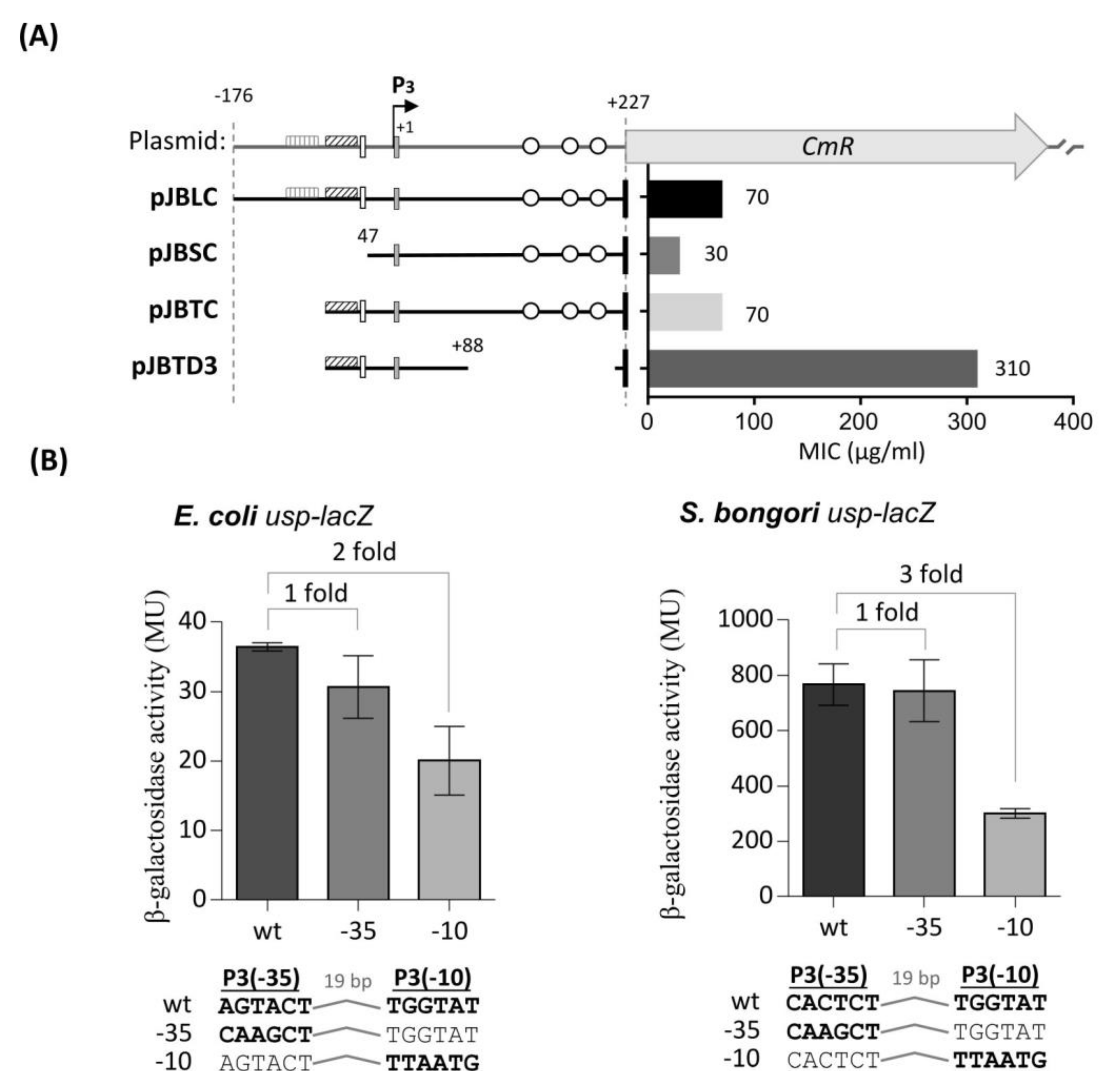
) TyrR boxes. The UP element is designated as an open square and H-NS binding sites as open circles. The ability of the fragments to drive CAT expression in usp-CAT transcriptional fusions was determined indirectly by measuring MIC of chloramphenicol. (B) Effects of site-specific mutations in either the -35 or -10 site of P3. Units for β-galactosidase assay are those defined by Miller, 1974 [35]. The values of β-galactosidase activities are averages of three independent experiments, with standard deviations below 15%. Values above columns are fold repression: specific activity of β-galactosidase of the wild-type promoters divided by that of site-specific mutated promoters.
Microorganisms 08 00330 g003
Figure 4. Expression of usp from P3 is modulated by the TyrR protein. Transcription from S. bongori (A) and E. coli (B) P3 in the presence or absence of aromatic amino acid phenylalanine in wild-type tyrR+ (black bars) and tyrR null phenotype strain (grey bars). Error bars represent standard deviation of the means of three independent experiments. M9, minimal medium; PHE, M9 supplemented with 1mM phenylalanine. Significant differences between or among groups are indicated, ** for p < 0.01, *** for p < 0.001 and **** for p < 0.0001 respectively.
Microorganisms 08 00330 g004
Figure 5. Electrophoretic mobility assay (EMSA) showing binding of the TyrR protein to the TyrR boxes within the S. bongori aroP-usp intergenic region. The DNA fragments corresponding to the wild type (wt, 403 bp) aroP-usp intergenic sequence (Lanes 1-2) and shorter DNA fragment without TyrR binding boxes (ΔTB, 274 bp) (Lanes 3-4). The DNA fragments, wt and ΔTB were each incubated in the presence or absence of TyrR protein (14 nM). The positions of free DNA and TyrR-DNA complexes are marked.
Microorganisms 08 00330 g005
Figure 6. Expression of E. coli and S. bongori usp genes in wild type (○), tyrR (□) and hns (Δ) defective strains at different temperatures. (A) Expression driven by the E. coli usp promoter. Expression from the usp-lacZ fusion of β-galactosidase and the inset shows the Western blot analysis of Usp protein levels in wild type and hns defective strains; (B) Expression driven by the S. bongori usp promoter, expression from usp-lacZ and inset, Usp protein levels; ○ wild type strain; □ tyrR mutant strain and Δ hns defective strain. Error bars represent standard deviations of the means of three independent experiments. Usp protein levels were determined at least three times and representative results are shown.
Microorganisms 08 00330 g006
Figure 7. Electrophoretic mobility assay (EMSA) showing binding of the H-NS protein to the H-NS binding sites within the S. bongori usp promoter region. Lane 1, PstI digested λ DNA; Lanes 1-9 usp promoter fragment with H-NS binding sites and increasing concentrations of purified H-NS protein (17.5 nM–1.6 nM); Lane 10, free DNA. Arrows 1, 2, and 3 designate protein concentration dependent increase in DNA shifts due to H-NS binding to 1, 2 or 3 binding sites.
Microorganisms 08 00330 g007
Figure 8. Expression of usp from P3 promoter is mediated by urea. Transcription from (A) S. bongori and (B) E. coli P3 in the presence or absence of urea in wild type tyrR+ (black bars) and in tyrR defective strain (grey bars). Units for β-galactosidase assay are those defined by Miller, 1974. Error bars represent standard deviations of the means of three independent experiments. M9, minimal medium; M9 supplemented with 0.4 M urea; M9 supplemented with 0.6 M urea. Significant differences between or among groups are indicated, ns for > 0.05, ** for p < 0.01 and **** for p < 0.0001.
Microorganisms 08 00330 g008
Table 1. The bacterial strains and plasmids used in this study.
Strains or PlasmidsRelevant Charasteristic(s) #Reference
Strains
E. coli DH5αCloning host; F– Φ80lacZΔM15 Δ(lacZYA-argF) U169 recA1 endA1 hsdR17 (rK−, mK+) phoA supE44 λ– thi-1 gyrA96 relA1Life Technologies
E. coli BL21 (DE3)FompT hsd SB(rBmB) dcmgalλ (DE3)Invitrogen
E. coli JW1889F-Δ(araD-araB)567, ΔlacZ4787(::rrnB-3), λ, ΔaraF751::kan, rph-1, Δ(rhaD-rhaB)568, hsdR514[27]
E. coli JW1316
E. coli JW1225
F-Δ(araD-araB)567, ΔlacZ4787(::rrnB-3), λ, ΔtyrR760::kan, rph-1, Δ(rhaD-rhaB)568, hsdR514
F-Δ(araD-araB)567, ΔlacZ4787(::rrnB-3), λ, Δhns746:: kan, rph-1, Δ(rhaD-rhaB)568, hsdR514
[27]
E. coli TA211wild-type strain (UPEC isolate)IMI
E. coli MG1655wild-type strainThis laboratory
S.bongori NCTC 12419wild-type strainATTC
Plasmids
pRW50TcR; Low-copy-number promoterless lacZ trancriptional fusion vector[28]
pRWBPTcR; 403 bp EcoRI-HindIII fragment from S. bongori NCTC 12419 aroP-usp intergenic region: usp-lacZ transcription fusionThis study
pRWBPM1TcR; pRWBP derivate; P3 (−35) mutation: CACTCT-->CAAGCTThis study
pRWBPM3TcR; pRWBP derivate; P3 (−10) mutation: TGGTAT-->TTAATGThis study
pRCPLTcR; 527 bp EcoRI-HindIII fragment from E. coli TA211 aroP-usp intergenic region: usp-lacZ trancription fusionThis study
pRWC10TcR; pRCPL derivate; P3 (−10) mutation: TGGTAT-->TTAATGThis study
pRWC35TcR; pRCPL derivate; P3 (−10) mutation: TGGTAT-->TTAATGThis study
pRWMGA-TcR; 215 bp EcoRI-HindIII fragment from E. coli K-12 MG1655aroP-pdhR intergenic region: aroP-lacZ transcription fusion
pJCATApR; High-copy-number promoterless cat trancription fusion vector (pJET1.2 cloning vector (Thermo Fisher Scientific) derivate)This study
pJBLCApR; 403 bp PstI-BstEII fragment from S. bongori aroP-usp intergenic region: usp-cat transcription fusion This study
pJBSCApR; pJBLC derivate; 247 bp fragment without both TyrR binging boxesThis study
pJBTCApR; pJBLC derivate; 315 bp fragment without weak TyrR binding boxThis study
pJBTD2ApR; 111 bp PstI-BstEII fragment from S. bongori aroP-usp intergenic region only with strong TyrR box and P3 promoter This study
pJBTD3ApR; pJBTD2 derivate: 111 bp fragment extended for 64 bpThis study
pET8HTApR; pET8c -derived expression vector with PT7 promoter: expression and production of recombinant N-terminally His6-tagged TyrRThis study
pET9HTApR; pET8c -derived expression vector with PT7 promoter: expression and production of recombinant N-terminally His6-tagged H-NSThis study
pUSP4R-LApR; T7 promoter of pUSP4 replaced with native E. coli TA211 usp promoter.[14]
pCUPBIApR; T7 promoter of pUSP4 replaced with native S. bongori NCTC 12419 promoterThis study
#—abbreviations: Tc, tetracycline; Ap, ampicillin; R, resistance; ATCC, American Type Culture Collection; IMI, Institute of microbiology and immunology, Medical Faculty, University of Ljubljana, Ljubljana, Slovenia.
Table 2. Primer sequences used in this study.
PrimerSequence (5′→3′)Purpose
Cm_Bst_FTTGGTAACCAAGAGAAAAAAATCACTGGATATACPCR amplification of cat gene from pLysS plasmid for cloning in pJBPLN3 (intermediate plasmid)
Cm_Xba_RTTTCTAGATTACGCCCCGCCCTGCCACTCAT
Bong_L_Pst_FCTGCAGCATAAAACCTCGTGTATGGTGCGCPCR amplification of S. bongori aroP-usp promoter region for cloning in pJBLC
Bong_S_Pst_FCTGCAGCACTATCACTCTTTTGTCTTGGTAAGPCR amplification of S. bongori aroP-usp promoter region for cloning in pJBSC and also used in EMSA
Bong_L_Bst_RGGTTACCCATTTTACGACATCCGAATATAAATTAAACGCCPCR amplification of S. bongori aroP-usp promoter region for cloning in pJBLC, pJBSCm, pJBTCm
Bong_promS_tyr_FTTCTGCAGCTTTTGTATTGATTTATTTACAGTGAPCR amplification of S. bongori aroP-usp promoter region for cloning in pJBTC, pJBTD2 and pJBTD3
Bong_prom_del2_RTTGGTTACCCATTTTACGACATCCGAACTCGTCATCACCCCTGCACAAAGCAGGTGCATPCR amplification of S. bongori aroP-usp promoter region for cloning in pJBTD2
Bong_prom_del3_RTTGGTTACCCATTTTACGACATCCGAATGCAAAATAAAGTGAGTAATGCTAAAAGGGPCR amplification of S. bongori aroP-usp promoter region for cloning in pJBTD3
Bong_L_Eco_FGAATTCATAAAACCTCGTGTATGGTGCGCPCR amplification of S. bongori aroP-usp promoter region for cloning in pRWBP, pRWBPM1, pRWBPM2 and also used in EMSA
Mut10_VspTGTCTTGGTAAGAATAATTAATTGTGACTGCGAATGCACPCR amplification of S. bongori aroP-usp promoter region for cloning in pRWBP, pRWBPM1, pRWBPM2
Mut35_HindATTAAATATCACTATCAAGCTTTTGTCTTGGTAAGAAPCR amplification of S. bongori aroP-usp promoter region for cloning in pRWBP, pRWBPM1, pRWBPM2
Bong_prom_Hind_RTTAAGCTTTACGACATCCGAATATAAATTAAACGCCPCR amplification of S. bongori aroP-usp promoter region for cloning in pRWBP, pRWBPM1, pRWBPM2 and also used in EMSA
Coli_L_Eco_FTTGAATTCTATGAAACCTCGTGTTGTGTTGTTPCR amplification of E. coli 211 aroP-usp promoter region for cloning in pRCPL, pRWC10 and pRWC35
Mut_coli_10_VspACTATCACAAGATAAATTAATATGCGCGAGAATGCACC
Mut_coli_35_HindACTGAATTATGATGATAAAGCTTTCAACTATCACAAG
Coli_L _R_HindTTAAGCTTTATTTCATTCCCGTTTATCCTTAAC
MG_prom_F_EcoTTGAATTCGAAACCTCGTGCGGTGGTTGTTTTTTTGPCR amplification of E. coli K-12 MG1655 aroP promoter region for cloning in pRWMGA-
MG_prom_R_HindTTAAGCTTTACAAACGGTTTCTTTTTAAGC
TyrR_Bam_FTTGGATCCCGTCTGGAAGTCTTTTGTGAAGACCPCR amplification of tyrR gene from E. coli DH5α for cloning in pET8HT
PCR amplification of hns gene from E. coli DH5α for cloning in pET9HT
TyrR_Mlu_R
H-NS Bam F
H-NS Mlu R
TTACGCGTTACTCTTCGTTCTTCTTCTGACTCAGAC
TTGGATCCAGCGAAGCACTTAAAATTCTGAAC
TTACGCGTTTATTGCTTGATCAGGAAATCGTCG

