Three-Dimensional Study of F. graminearum Colonisation of Stored Wheat: Post-Harvest Growth Patterns, Dry Matter Losses and Mycotoxin Contamination

 , , , , and
Abstract
: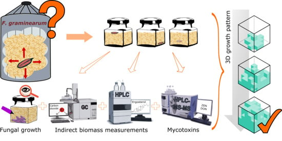
1. Introduction
2. Materials and Methods
2.1. Fungal Isolate and Spores Preparation
2.2. Grain Preparation, Inoculation and Incubation
2.3. Colonisation Pattern Assessment
2.4. Respiration Determination and Dry Matter Loss Estimation
2.5. Mycotoxins Extraction and Analysis
2.6. Ergosterol Analysis
2.7. Data Analysis
3. Results
3.1. Fungal Colonisation
3.2. Indirect Indicators of Fungal Growth
3.2.1. Fungal Respiration Dynamics
3.2.2. Dry Matter Loss Dynamics
3.2.3. Ergosterol Production Dynamics
3.3. Mycotoxin Production Dynamics
3.4. Correlation between Experimental Measures
3.4.1. Correlation between Fungal Respiration and Ergosterol Content
3.4.2. Correlation between Mycotoxin Contamination Levels and Indicators of Colonisation
4. Discussion
5. Conclusions
Supplementary Materials
Author Contributions
Funding
Acknowledgments
Conflicts of Interest
Appendix A. Computation of the Grain Colonised Volume

Appendix A.1. Top-Centre and Bottom-Centre Positions
- Mycelial expansion just observable on the top (bottom) side. Volumetric colonisation at the time t () was computed assuming a half ellipsoidal shape (Equation (A1))where a, b and c are elliptic radii. Radii b and c where computed as the maximum minus the minimum coordinates showing colonisation along X and Y axis divided by two, and a was assumed to be the . This assumes an upwards (downwards) radial reduction due to the fact that the mycelial expansion proceeds through a more tortuous path that the surface as observed experimentally [39].
- Mycelial expansion was observable on the vertical sides of the grain volume. Colonised volume was approximated as cubic shape and a half ellipsoid (Equation (A2)) as follows:where , and are mean distances obtained along axes X, Y and Z, respectively, showing colonisation. Radii a (in cm) was computed as 3.5 − if the face opposite to the inoculation point (bottom and top, respectively, for top-centre and bottom-centre inoculations, respectively) showed any sign of colonisation, or as the minimum value between and 3.5 − if not.
Appendix A.2. Bottom-Side Position
- First mycelial expansion not observable on either the East or West faces. The colonisation was assumed to follow a quarter of ellipsoid (Equation (A3)):where a, b and c are elliptic radii. Radius a was obtained on the North face as the mean value of the colonisation shown along the Z-axis. Radius c was obtained on the North face as the maximum minus the minimum coordinates showing colonisation along the X-axis divided by two. Radius b was obtained on the bottom face as the mean value of the colonisation shown along the Y-axis, assuming a minimum value of 1 square.
- Mycelial expansion observable on either the East or West faces. The growth was assumed to follow a cylindrical shape (Vc) and an ellipsoid shape (Ve) as expressed in Equation (A4):where is the wide of the jar (5.5 cm).
- Mycelial expansion observable on the top or South faces. Colonisation front was assumed to follow a cylindrical shape until the end of the colonisation (Equation (A5)) according the expression:where LT, and LN are the colonisation length attained by the fungi on the top and South faces, respectively. These were computed as the mean value of the colonisation shown along the Y-axis of the top face (LT) and the Z-axis of the South face (LS).
References
- European Commisison. Commission Regulation (EC) No 1881/2006 of 19 December 2006 setting levels for certain contaminants in foodstuffs. Off. J. Eur. Union 2009, 2006, 11–21. [Google Scholar]
- European Commisison. Commission Recommendation No 2006/576 of 17 August 2006 on the presence of deoxynivalenol, zearalenone, ochratoxin A, T-2 and HT-2 and fumonisins in products intended for animal feeding. Off. J. Eur. Union 2006, 229, 7–9. [Google Scholar]
- Mylona, K.; Magan, N. Fusarium langsethiae: Storage environment influences dry matter losses and T2 and HT-2 toxin contamination of oats. J. Stored Prod. Res. 2011, 47, 321–327. [Google Scholar] [CrossRef]
- Mylona, K.; Sulyok, M.; Magan, N. Relationship between environmental factors, dry matter loss and mycotoxin levels in stored wheat and maize infected with Fusarium species. Food Addit. Contam. Part A Chem. Anal. Control Expo. Risk Assess. 2012, 29, 1118–1128. [Google Scholar] [CrossRef] [Green Version]
- Garcia-Cela, E.; Magan, N.; Elsa, K.; Sulyok, M.; Medina, A. Fusarium graminearum in Stored Wheat: Use of CO2 Production to Quantify Dry Matter Losses and Relate This to Relative Risks of Zearalenone Contamination under Interacting Environmental Conditions. Toxins 2018, 10, 86. [Google Scholar] [CrossRef] [PubMed] [Green Version]
- Magan, N.; Aldred, D. Post-harvest control strategies: Minimizing mycotoxins in the food chain. Int. J. Food Microbiol. 2007, 119, 131–139. [Google Scholar] [CrossRef] [PubMed] [Green Version]
- Limay-Rios, V.; Miller, J.D.; Schaafsma, A.W. Occurrence of Penicillium verrucosum, ochratoxin A, ochratoxin B and citrinin in on-farm stored winter wheat from the Canadian Great Lakes Region. PLoS ONE 2017, 12, 1–22. [Google Scholar] [CrossRef] [PubMed]
- Ramirez, M.L.; Chulze, S.; Magan, N. Temperature and water activity effects on growth and temporal deoxynivalenol production by two Argentinean strains of Fusarium graminearum on irradiated wheat grain. Int. J. Food Microbiol. 2006, 106, 291–296. [Google Scholar] [CrossRef] [PubMed]
- Hope, R.; Aldred, D.; Magan, N. Comparison of environmental profiles for growth and deoxynivalenol production by Fusarium culmorum and F. graminearum on wheat grain. Lett. Appl. Microbiol. 2005, 40, 295–300. [Google Scholar] [CrossRef]
- Du, H.; Perré, P. A novel lattice-based model for investigating three-dimensional fungal growth on solid media. Phys. A Stat. Mech. Appl. 2020, 541, 123536. [Google Scholar] [CrossRef]
- de Ulzurrun, G.V.-D.; Baetens, J.M.; den Bulcke, J.V.; De Baets, B. Modelling three-dimensional fungal growth in response to environmental stimuli. J. Theor. Biol. 2017, 414, 35–49. [Google Scholar] [CrossRef] [PubMed]
- Marín, S.; Cuevas, D.; Ramos, A.J.; Sanchis, V. Fitting of colony diameter and ergosterol as indicators of food borne mould growth to known growth models in solid medium. Int. J. Food Microbiol. 2008, 121, 139–149. [Google Scholar] [CrossRef] [PubMed]
- Garcia, D.; Ramos, A.J.; Sanchis, V.; Marín, S. Modeling kinetics of aflatoxin production by Aspergillus flavus in maize-based medium and maize grain. Int. J. Food Microbiol. 2013, 162, 182–189. [Google Scholar] [CrossRef] [PubMed]
- Marín, S.; Ramos, A.J.; Sanchis, V. Comparison of methods for the assessment of growth of food spoilage moulds in solid substrates. Int. J. Food Microbiol. 2005, 99, 329–341. [Google Scholar] [CrossRef]
- Gourama, H.; Bullerman, L.B. Relationship between aflatoxin production and mold growth as measured by ergosterol and plate count. LWT-Food Sci. Technol. 1995, 28, 185–189. [Google Scholar] [CrossRef]
- Tothill, I.E.; Harris, D.; Magan, N. The relationship between fungal growth and ergosterol content of wheat grain. Mycol. Res. 1992, 96, 965–970. [Google Scholar] [CrossRef]
- Rao, B.S.; Rao, V.S.; Ramakrishna, Y.; Bhat, R.V. Rapid and specific method for screening ergosterol as an index of fungal contamination in cereal grains. Food Chem. 1989, 31, 51–56. [Google Scholar] [CrossRef]
- Lamper, C.; Teren, J.; Bartok, T.; Komoroczy, R.; Mesterházy, Á.; Sagi, F. Predicting DON contamination in Fusarium-infected wheat grains via determination of the ergosterol content. Cereal Res. Commun. 2000, 28, 337–344. [Google Scholar] [CrossRef]
- Booth, C. Chapter II: Fungal culture media. In Methods in Microbiology; Academic Press: London, UK, 1971; Volume 4, pp. 49–94. [Google Scholar]
- Garcia-Cela, E.; Kiaitsi, E.; Medina, A.; Magan, N. Interacting environmental stress factors affects targeted metabolomic profiles in stored wheat and that inoculated with F graminearum. Toxins (Basel) 2018, 10, 56. [Google Scholar] [CrossRef] [Green Version]
- Riquelme, M.; Reynaga-Peña, C.G.; Gierz, G.; Bartnicki-García, S. What determines growth direction in fungal hyphae? Fungal Genet. Biol. 1998, 24, 101–109. [Google Scholar] [CrossRef]
- Malachová, A.; Sulyok, M.; Beltrán, E.; Berthiller, F.; Krska, R. Optimization and validation of a quantitative liquid chromatography–tandem mass spectrometric method covering 295 bacterial and fungal metabolites including all regulated mycotoxins in four model food matrices. J. Chromatogr. A 2014, 1362, 145–156. [Google Scholar] [CrossRef] [PubMed] [Green Version]
- Ruzicka, S.; Norman, M.D.P.; Harris, J.A. Rapid ultrasonic method to determine ergosterol concentration in soil. Soil Biol. Biochem. 1995, 27, 1215–1217. [Google Scholar] [CrossRef]
- Petzoldt, T. Growthrates: Estimate Growth Rates from Experimental Data, R Package Version 0.8.1. 2019. Available online: https://cran.r-project.org/package=growthrates (accessed on 6 July 2020).
- Juyal, A.; Otten, W.; Falconer, R.; Hapca, S.; Schmidt, H.; Baveye, P.C.; Eickhorst, T. Combination of techniques to quantify the distribution of bacteria in their soil microhabitats at different spatial scales. Geoderma 2019, 334, 165–174. [Google Scholar] [CrossRef] [Green Version]
- Van den Bulcke, J.; Boone, M.; Van Acker, J.; Van Hoorebeke, L. Three-dimensional x-ray imaging and analysis of fungi on and in wood. Microsc. Microanal. 2009, 15, 395–402. [Google Scholar] [CrossRef] [PubMed] [Green Version]
- Maier, D.E.; Channaiah, L.H.; Martínez-Kawas, A.; Lawrence, J.; Chaves, E.; Coradi, P.; Fromme, G. Monitoring carbon dioxide concentration for early detection of spoilage in stored grain. In Proceedings of the 10th International Working Conference on Stored Product Protection, Estoril, Portugal, 27 June–2 July 2010; pp. 505–509, Julius Kühn Institut, Bundesforschungsinstitut für Kulturpflanzen. [Google Scholar] [CrossRef]
- Garcia-Cela, E.; Kiaitsi, E.; Sulyok, M.; Krska, R.; Medina, A.; Petit Damico, I.; Magan, N. Influence of storage environment on maize grain: CO2 production, dry matter losses and aflatoxins contamination. Food Addit. Contam. Part A 2019, 36, 175–185. [Google Scholar] [CrossRef] [PubMed] [Green Version]
- Borreani, G.; Tabacco, E.; Schmidt, R.J.; Holmes, B.J.; Muck, R.E. Silage review: Factors affecting dry matter and quality losses in silages. J. Dairy Sci. 2018, 101, 3952–3979. [Google Scholar] [CrossRef] [Green Version]
- Lacey, J.; Hamer, A.; Magan, N. Respiration and dry matter losses in wheat grain under different environmental factors. In Stored Product Protection 1994; Highley, E., Wright, E.J., Banks, H.J., Champ, B.R., Eds.; CAB International: Wallingford, UK, 1994; pp. 1007–1013. ISBN 0851989322. [Google Scholar]
- Stuper-Szablewska, K.; Kurasiak-Popowska, D.; Nawracała, J.; Perkowski, J. Study of metabolite profiles in winter wheat cultivars induced by Fusarium infection. Cereal Res. Commun. 2016, 44, 572–584. [Google Scholar] [CrossRef] [Green Version]
- Yong, R.K.; Cousin, M.A. Detection of moulds producing aflatoxins in maize and peanuts by an immunoassay. Int. J. Food Microbiol. 2001, 65, 27–38. [Google Scholar] [CrossRef]
- Ng, H.-E.; Raj, S.S.A.; Wong, S.H.; Tey, D.; Tan, H.-M. Estimation of fungal growth using the ergosterol assay: A rapid tool in assessing the microbiological status of grains and feeds. Lett. Appl. Microbiol. 2008, 46, 113–118. [Google Scholar] [CrossRef]
- Varga, M.; Bartók, T.; Mesterházy, Á. Determination of ergosterol in Fusarium-infected wheat by liquid chromatography-atmospheric pressure photoionization mass spectrometry. J. Chromatogr. A 2006, 1103, 278–283. [Google Scholar] [CrossRef]
- Zhang, H.; Wolf-Hall, C.; Hall, C. Modified microwave-assisted extraction of ergosterol for measuring fungal biomass in grain cultures. J. Agric. Food Chem. 2008, 56, 11077–11080. [Google Scholar] [CrossRef] [PubMed]
- Heidtmann-Bemvenuti, R.; Tralamazza, S.M.; Jorge Ferreira, C.F.; Corrêa, B.; Badiale-Furlong, E. Effect of natural compounds on Fusarium graminearum complex. J. Sci. Food Agric. 2016, 96, 3998–4008. [Google Scholar] [CrossRef] [PubMed]
- Ezekiel, C.N.; Odebode, A.C.; Fapohunda, S.O. Zearalenone production by naturally occurring Fusarium species on maize, wheat and soybeans from Nigeria. J. Biol. Environ. Sci. 2008, 2, 77–82. [Google Scholar]
- Kerry, R.; Ingram, B.; Garcia-Cela, E.; Magan, N. Spatial analysis of mycotoxins in stored grain to develop more precise management strategies. In Precision Agriculture; Wageningen Academic Publishers: Wageningen, The Netherlands, 2019; p. 330. [Google Scholar]
- Otten, W.; Gilligan, C.A. Effect of physical conditions on the spatial and temporal dynamics of the soil-borne fungal pathogen Rhizoctonia solani. New Phytol. 1998, 138, 629–637. [Google Scholar] [CrossRef]
 ), day 3 (
  ), day 4 (
  ), day 5 (
  ) and day 6 (
  ).
), day 3 ( ), day 4 ( ), day 5 ( ) and day 6 ( ).
).
), day 3 ( ), day 4 ( ), day 5 ( ) and day 6 ( ).


| Analyte | Retention Time (min) | Q1 (m/z) | DP (V) | Q3 (m/z) | CE (V) | CXP (V) |
|---|---|---|---|---|---|---|
| 3-AcetylDeoxynivalenol | 3.86 | 397.3 | −70 | −59.2/−307.1 | −38/−20 | −8/−7 |
| 15-AcetylDeoxynivalenol | 3.84 | 339.1 | 91 | 137.2/321.2 | 17/13 | 8/18 |
| Deoxynivalenol | 2.60 | 355.1 | −70 | −59.2/−265.2 | −40/−22 | −13/−10 |
| Zearalenone | 7.33 | 317.1 | −110 | −175/−121.1 | −34/−42 | −13/−8 |
| Water Activity (aw) | Inoculation | Replicate | Superficial Colonisation | Volumetric Colonisation | ||
|---|---|---|---|---|---|---|
| Lag Time (Days) | Rate (cm2·Day−1) | Lag Time (Days) | Rate (cm3·Day−1) | |||
| 0.95 | Top-centre | 1 | 2.71 | 59.50 | 2.66 | 48.93 |
| 0.95 | Top-centre | 2 | 3.46 | 45.30 | 2.75 | 48.76 |
| 0.95 | Top-centre | 3 | 2.90 | 64.25 | 3.25 | 60.67 |
| 0.95 | Bottom-side | 1 | 3.35 | 51.25 | 3.18 | 39.08 |
| 0.95 | Bottom-side | 2 | 3.04 | 47.50 | 3.40 | 59.93 |
| 0.95 | Bottom-side | 3 | 2.79 | 43.67 | 3.33 | 59.80 |
| 0.95 | Bottom-centre | 1 | 2.69 | 63.63 | 2.54 | 46.22 |
| 0.95 | Bottom-centre | 2 | 2.88 | 63.50 | 2.80 | 48.73 |
| 0.95 | Bottom-centre | 3 | 3.51 | 92.50 | 2.65 | 47.78 |
| 0.97 | Top-centre | 1 | 2.82 | 63.75 | 2.94 | 94.35 |
| 0.97 | Top-centre | 2 | 2.61 | 60.87 | 2.83 | 50.91 |
| 0.97 | Top-centre | 3 | 2.83 | 64.50 | 2.93 | 78.37 |
| 0.97 | Bottom-side | 1 | 2.85 | 43.93 | 3.27 | 54.07 |
| 0.97 | Bottom-side | 2 | 2.75 | 44.15 | 3.01 | 51.18 |
| 0.97 | Bottom-side | 3 | 3.04 | 47.88 | 3.46 | 64.12 |
| 0.97 | Bottom-centre | 1 | 2.65 | 65.50 | 2.87 | 75.23 |
| 0.97 | Bottom-centre | 2 | 2.92 | 66.38 | 2.84 | 81.10 |
| 0.97 | Bottom-centre | 3 | 2.79 | 65.62 | 2.89 | 76.33 |
| Water Activity Levels | 0.95 aw | 0.97 aw | 0.95 + 0.97 aw |
|---|---|---|---|
| Days 2, 4 and 6 | 0.9193 | 0.9329 | 0.7796 |
| Days 2, 4, 6, 8 and 10 | 0.8785 | 0.9404 | 0.8858 |
| Mycotoxins | Fungal Growth Indicator | 0.95 aw | 0.97 aw | ||
|---|---|---|---|---|---|
| Correlation | p-Value | Correlation | p-Value | ||
| DON (ng/g) | Volume (cm3) | 0.8149 | <0.0001 | 0.8528 | <0.0001 |
| Ergosterol (mg·g−1) | 0.9528 | <0.0001 | 0.8972 | <0.0001 | |
| Cumulative DML | 0.9220 | <0.0001 | 0.8660 | <0.0001 | |
| ZEN (ng/g) | Volume (cm3) | 0.4150 | 0.0313 | 0.7490 | <0.0001 |
| Ergosterol (mg·g−1) | 0.6686 | <0.0001 | 0.7856 | <0.0001 | |
| Cumulative DML | 0.5971 | <0.0001 | 0.8193 | <0.0001 | |
| DON (ng/g) | ZEN (ng·g−1) | 0.6417 | <0.0001 | 0.7604 | <0.0001 |
© 2020 by the authors. Licensee MDPI, Basel, Switzerland. This article is an open access article distributed under the terms and conditions of the Creative Commons Attribution (CC BY) license (http://creativecommons.org/licenses/by/4.0/).
Share and Cite
Portell, X.; Verheecke-Vaessen, C.; Torrelles-Ràfales, R.; Medina, A.; Otten, W.; Magan, N.; García-Cela, E. Three-Dimensional Study of F. graminearum Colonisation of Stored Wheat: Post-Harvest Growth Patterns, Dry Matter Losses and Mycotoxin Contamination. Microorganisms 2020, 8, 1170. https://doi.org/10.3390/microorganisms8081170
Portell X, Verheecke-Vaessen C, Torrelles-Ràfales R, Medina A, Otten W, Magan N, García-Cela E. Three-Dimensional Study of F. graminearum Colonisation of Stored Wheat: Post-Harvest Growth Patterns, Dry Matter Losses and Mycotoxin Contamination. Microorganisms. 2020; 8(8):1170. https://doi.org/10.3390/microorganisms8081170
Chicago/Turabian StylePortell, Xavier, Carol Verheecke-Vaessen, Rosa Torrelles-Ràfales, Angel Medina, Wilfred Otten, Naresh Magan, and Esther García-Cela. 2020. "Three-Dimensional Study of F. graminearum Colonisation of Stored Wheat: Post-Harvest Growth Patterns, Dry Matter Losses and Mycotoxin Contamination" Microorganisms 8, no. 8: 1170. https://doi.org/10.3390/microorganisms8081170