Article

Prevalence Rate and Molecular Characteristics of Oestrus ovis L. (Diptera, Oestridae) in Sheep and Goats from Riyadh, Saudi Arabia

by
Dina M. Metwally
1,2,*,
Shurug A. Albasyouni
1,
Ibrahim A.H. Barakat
1,3,
Isra M. Al-Turaiki
4,
Amal M. Almuhanna
1,
Muhammad A. Bashir
5,
Hanadi B. Baghdadi
6,7,
Manal F. El-Khadragy
8 and
Reem A. Alajmi
1
1
Zoology Department, College of Science, King Saud University, P.O. Box 2455, Riyadh 11451, Saudi Arabia
2
Parasitology Department, College of Veterinary Medicine, Zagazig University, Zagazig 44519, Egypt
3
Cell Biology Department, National Research Center, 33 Bohouth St., Dokki, Giza 12622, Egypt
4
Department of Information Technology, College of Computer and Information Sciences, King Saud University, Riyadh 11451, Saudi Arabia
5
Department of Plant Protection, Faculty of Agricultural Science Ghazi University, Dera Ghazi Khan 32200, Pakistan
6
Biology Department, College of Science, Imam Abdulrahman Bin Faisal University, Dammam City 31441, Saudi Arabia
7
Basic and Applied Scientific Research Center, Imam Abdulrahman Bin Faisal University, Dammam City 31441, Saudi Arabia
8
Department of Biology, Faculty of Science, Princess Nourah Bint Abdelrahman University, Riyadh 11451, Saudi Arabia
*
Author to whom correspondence should be addressed.
Animals 2021, 11(3), 689; https://doi.org/10.3390/ani11030689
Submission received: 28 January 2021 / Revised: 25 February 2021 / Accepted: 26 February 2021 / Published: 4 March 2021
(This article belongs to the Special Issue Veterinary Microbiology & Parasitology)

Abstract

:

Simple Summary

In the current study, we investigated the prevalence rate of Oestrus ovis (O. ovis) larvae in heads of sheep and goats obtained from abattoirs in Riyadh, Saudi Arabia. It was found that season did not have any significant effect on the characteristics of the larvae that affect sheep, except for the length of larva, for which those collected in spring differed from those collected in winter and autumn. In goats, the L3 instar number was significantly higher during summer. Oestrus ovis larvae were detected in sheep and goats by molecular analysis and they were most closely related to O. ovis larvae recovered from GenBank.

Abstract

Heads of sheep (n = 600) and goats (n = 800) slaughtered at Al-Aziziah Abattoir in Riyadh, Saudi Arabia, were inspected for the presence of O. ovis larvae (L). Heads were split along the longitudinal axes, and larvae (L1, L2, and L3) were gathered. The infestation rate was significantly higher in goats (44.5%; 356/800) than that in sheep (22.3%; 134/600). Out of the 151 collected larvae from sheep, 0% were L1, 1.3% were L2, and 98.7% were L3. Out of the total of 468 larvae from goats, 0% were L1, 1.2% were L2, and 98.8% were L3. The infestation rate was significantly higher in males than that in females. Myiasis-causing larvae collected from Riyadh, Saudi Arabia, were authenticated as O. ovis, according to morphological characteristics. Polymerase chain reaction (PCR) amplification of a partial fragment (600 bp) of the mitochondrial cytochrome c oxidase subunit I (mtCOI) gene further confirmed the species. Phylogenetic analysis based on the partial mtCOI gene sequence demonstrated that 23 unique sequences showed high similarity based on nucleotide pairs of O. ovis accessions retrieved from GenBank.

1. Introduction

The sheep gadfly O. ovis Linnaeus 1761 (Diptera, Oestridae) is one cause of myiasis in sheep and goats. Sneezing and nasal discharge are the most prominent clinical symptoms of infested animals [1]. Myiasis lowers the health of animals and causes major economic losses to the livestock industry due to abortion, reduced milk production, loss of weight gain, and fertility [2]. Oestrus ovis larvae persist in cranial cavities of sheep [3]. The incidence of O. ovis-related myiasis in the Jazan area was previously reported to be as high as 53.54% [1]. Clinical appearance, morphological characterizations of the larvae (e.g., slits of the posterior spiracles found on the posterior spiracle plates), and occasional identification of the adult fly are among the many approaches leading to the diagnosis of myiasis [4]. Molecular methods facilitate in the diagnosis and detection of a wide variety of species, including flies that cause myiasis [5,6]. Molecular approaches can be efficiently used to identify all larval stages [7]. Multiple genetic markers of ribosomal DNA (16S rRNA, 28S rRNA) and mitochondrial DNA (cytochrome oxidase genes (COI, COII, and 12S mtDNA)) have been used [8]. A widely used method for the molecular identification of myiasis-causing larvae belonging to the Oestridae family is the sequencing of the mitochondrial cytochrome c oxidase subunit I (mtCOI) gene, which contributes to an essential database for molecular identification of organisms [9,10]. Phylogenetic relationships can be assessed by polymerase chain reaction (PCR) and PCR restriction fragment length polymorphism (PCR-RFLP) assays [7]. For some species belonging to Calliphoridae, Sarcophagidae, and Oestridae, the mitochondrial gene subunit has been isolated, proving effective for molecular phylogenetic studies [4,11,12,13,14,15,16]. Oestrus spp. larvae identification can be complicated, as there are considerable morphological similarities within the species. The objectives of the present study were to investigate the seasonal prevalence of O. ovis among slaughtered sheep and goats in Riyadh, Saudi Arabia, and to use the sequencing of the mtCOI gene to identify the phylogenies of O. ovis larvae. One genetic marker, COI, was used to identify larvae collected from domestic sheep and goats to accurately identify Oestrus parasites [17].

2. Materials and Methods

2.1. Larvae Collection and Morphological Identification

This study was conducted between March 2019 and January 2020 in the city of Riyadh, Saudi Arabia. Riyadh City is in the center of the Kingdom of Saudi Arabia. It extends between latitude: 24°38′ North and longitude:46°43′ East. It has a very dry and arid climate; summer temperatures rise very dramatically during the day but fall at night, ranging between 35 °C and 43 °C. In the winter, the temperature drops dramatically and may reach 0 °C. Riyadh City is located 600 m above sea level, with several valleys and rich sand dunes. A total of 600 sheep and 800 goat heads from the Al-Aziziah Abattoir were analyzed for O. ovis larvae. The sex and age (<1 year is young and >1 year old is adult) of slaughtered animals were registered. At the investigation site, the nasal and frontal sinuses were longitudinally divided into the dorsoventral planes using a sharp handsaw. Oestrus ovis present in the mucous membrane of the nasal septa and nasal segments were collected with forceps and preserved in 70% ethanol for molecular studies and morphometric identification. Larvae were identified as the first, second, and third larval stages [18,19]. The percentage of each larval instar was calculated according to the equation (Larval stage percent = number of each larval stage/Total number of larvae × 100). In each infested head, the larvae were counted with the aid of identification keys [20]. The severity of the infestation was calculated as the number of larvae/number of animals infested.

2.2. DNA Extraction and Polymerase Chain Reaction (PCR)

DNA from each individual larva (about 0.025 gm from anterior section of the larva) was extracted using a DNeasy Blood & Tissue Kit (250) (Cat No./ID: 69506, Qiagen, Hilden, Germany) following the manufacturers’ protocol. mtCOI gene was amplified using the primer pairs described in Table 1 [21] and a thermal cycler (Veriti® 96-Well Thermal Cycler, Model 9902, Biosystem). This procedure was performed in a mixture (20 μL) consisting of master mix (5×, 4 μL), RNase-free water (12 μL), and DNA template (2 μL). The PCR program consisted of a denaturation step of 94 °C for 2 min, followed by 40 cycles of denaturation for 30 s at 94 °C. PCR products were analyzed by 1.5% agarose gel electrophoresis.

2.3. Nucleotide Sequences of the mtCOI Gene

A total of 40 sequences (Table S1), which were coded according to properties of their host, were analyzed in this study. The sequences were processed using Geneious Prime Build 2020-04-07 08:42 [22]. Prior to processing, all sequences were truncated using the error probability method with a limit of 0.05 on both sides. Related sequences were retrieved from GenBank after performing a BLAST [23] search. Multiple sequence alignments were then generated using CLUSTAL Omega [24] implemented in Geneious software. A phylogenetic tree was created using the neighbor-joining method [25] with the Tamura_Nei model and using 10,000 replicates. All samples were deposited to GenBank, and accession numbers have been provided.

2.4. Statistical Analysis

All data concerning the effect of different seasons were analyzed using one-way analysis of variance (ANOVA), followed by Duncan’s test to compare means. Statistical analysis was conducted using SPSS software program version 24, and all results were expressed as means ± SEM. Differences were considered significant at p ≤ 0.05.

3. Results

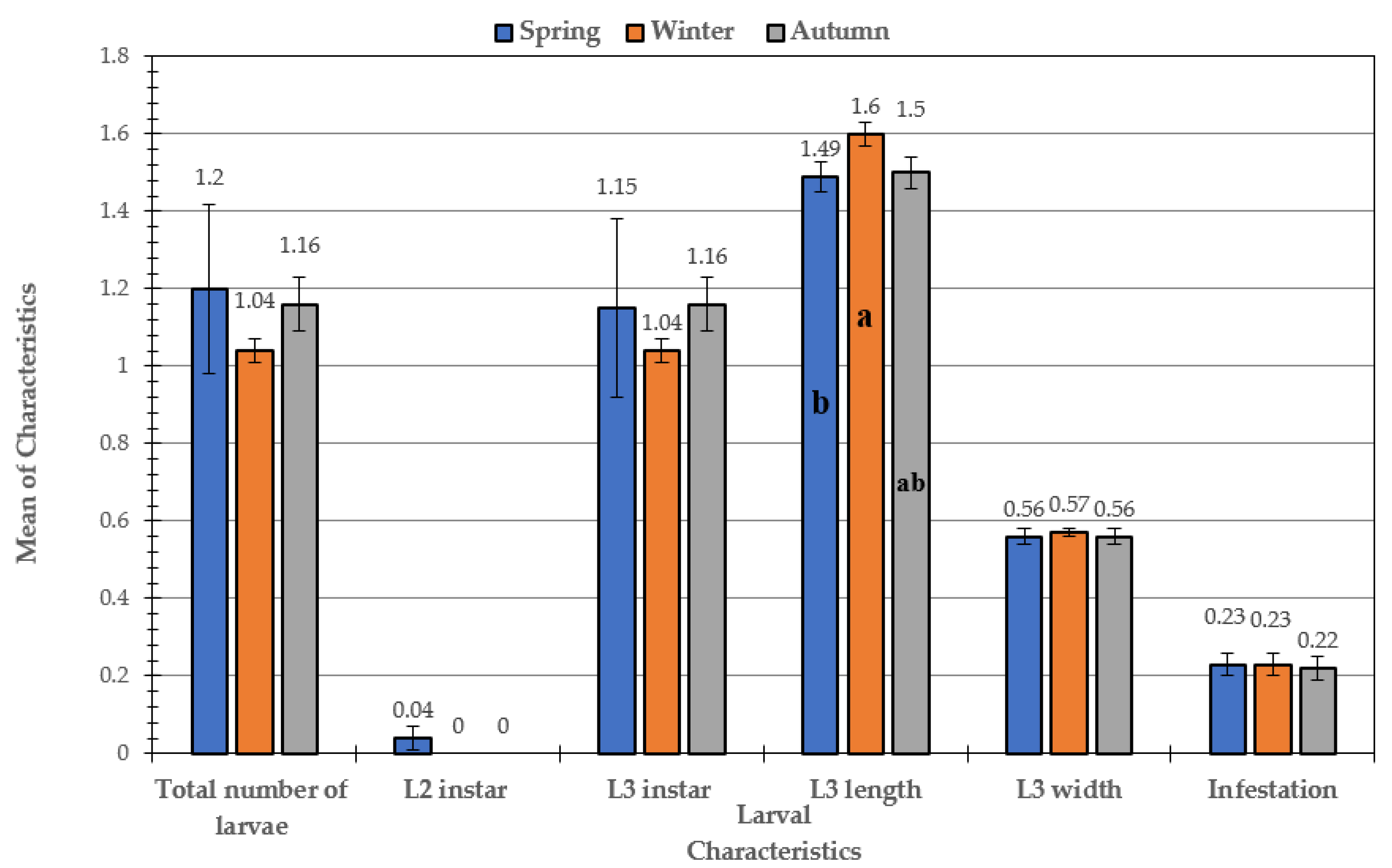
3.1. Prevalence of Infestation over the Course of the Study in Sheep and Goats

This study, which was conducted on 600 sheep and 800 goats (Table 2), showed that infestation rate with larvae was much higher in goats (44.5%; 356/800) than that in sheep (22.3%; 134/600). This also applied to the total number of larvae, where the total number of larvae in 356 head of goats was 486, while it was 151 in 134 head of sheep.
Results of statistical analysis concerning infestation rate showed that the ovine species is significantly more susceptible to infection with larvae than caprine species: the infestation mean in sheep was 1.78, and in goats was 1.56 at p ≤ 0.05, as a significant level.
The findings in Table 3 show that, in both species (goats and sheep), there was a disparity in the percentage of infestation between males and females, where the rate of infestation in males was higher than that in females (32.66% (98/300) and 65.50% (262/400) in sheep and goats, respectively). Additionally, the incidence of infestation was higher in female goats (32.50 percent; 94/400) than in female sheep (12.0 percent; 36/300). The seasonal effect on the characteristics of larvae specifically infesting goats (Figure 1) showed that there was a significant difference in the total number of larvae and L3 instar. The mean values of total larvae and L3 instar were significantly higher in the summer (1.94 ± 0.12 and 1.91 ± 0.12, respectively) than in other seasons.
In addition, it was found that there was no significant difference between the means of total number of larvae and L3 in the spring, winter, and autumn. Concerning the larvae width and infestation, the trend was the same in the two characteristics. No significant difference between the winter and autumn was observed, while the significant differences in width and infestation appeared between spring and summer. As for the larvae length characteristic, it was found that only the winter and summer seasons varied significantly, while there were no significant variations between the spring and autumn seasons. The results of the impact of season on the studied characteristics differed only in goats. It was found that seasons of the year did not have any significant effect on the characteristics of the larvae that affect sheep, in general, except for the length of larva, in which those collected in spring differed from those collected in winter and autumn (Figure 2).

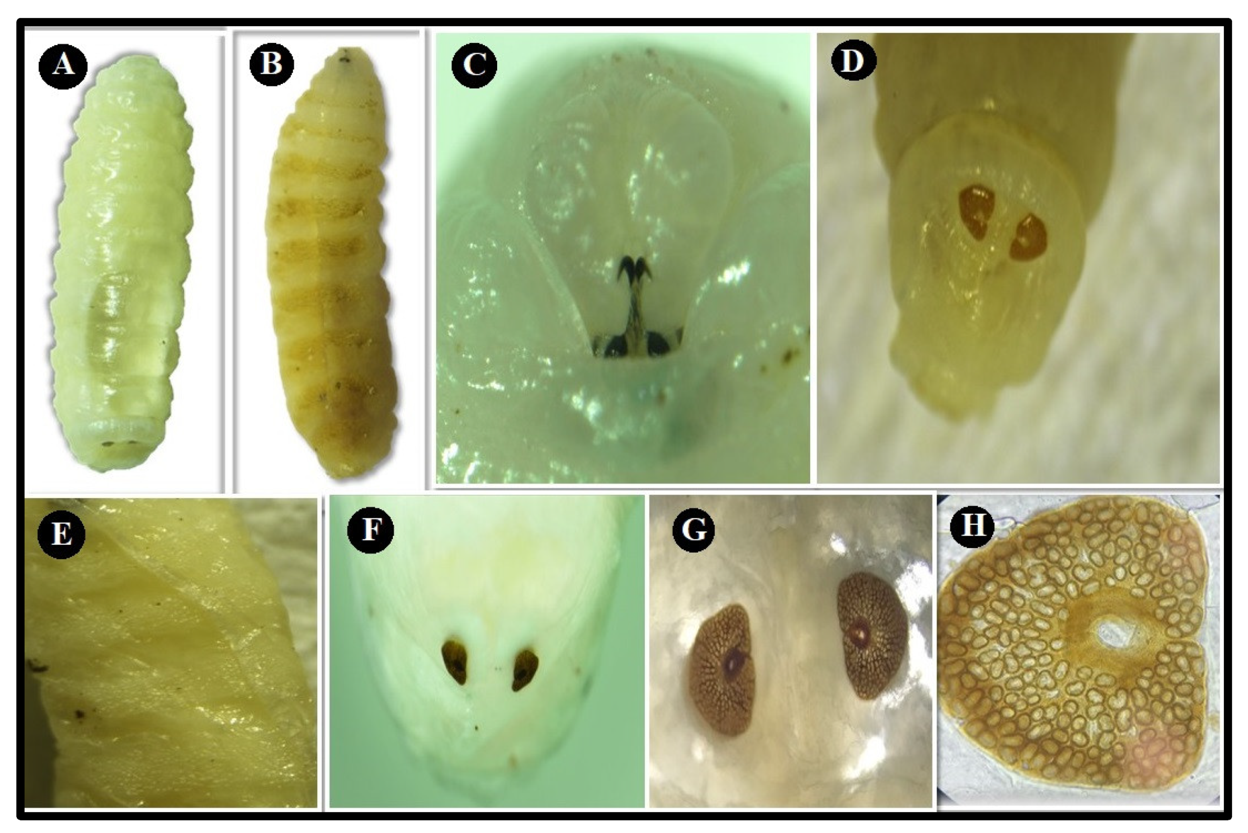
3.2. Morphological Examination

The identification of mature O. ovis larvae was based on morphological characteristic features of the larval stages. Second instar larvae (L2) were found only in young male goats in spring and summer; the average length and width were 0.84 ± 0.08 cm and 0.26 ± 0.05 cm, respectively, in spring, while the average length and width were 0.90 ± 0.10 cm and 0.30 ± 0.00 cm, respectively, in the summer. These L2 larvae are white in color, and the dorsal side shows only a few weak denticles on the second segment (Figure 3A). On the ventral side, the segments have rows of peculiarly shaped currycomb-like spines (Figure 3B). Antennary lobes are less separated giving to pseudocephalon triangular shape. The buccal funnel is well structured. The hooks of the first segment are less robust and more curved (Figure 3C). Furthermore, the median part of the postanal bulge is spinulose (Figure 3D). Ventrally the segments are provided with rows of peculiarly shaped currycomb-like spines, the thoracic and abdominal segments on the ventral side, show single-ended, caudally projected spines (Figure 3E). The posterior peritremes are more or less circular (Figure 3F), with the channel indicated by a distinct suture (Figure 3G,H).
Third instar larvae (L3) are yellow in color when young (Figure 4A), changing to a light brown later in the mature stage, and show broad transverse blackish bands on the dorsal side (Figure 4B). The second segment on the dorsal side presents with a variable number of small denticles; the following segments are bare but have a rough, leatherlike skin pattern, which is distinct only on the darkened parts. On the ventral side, the segments bear rows of strong spines (Figure 4C), which are irregularly placed on the third segment but are fairly regular on the following ones. The number of rows varies from two to five (Figure 4D). The postanal bulge shows fewer spines, while the preanal bulge is bare (Figure 4E). The posterior peritremes are circular, with a central button and lacking a distinct suture and the channel appears in the posterior peritremes (Figure 4F).

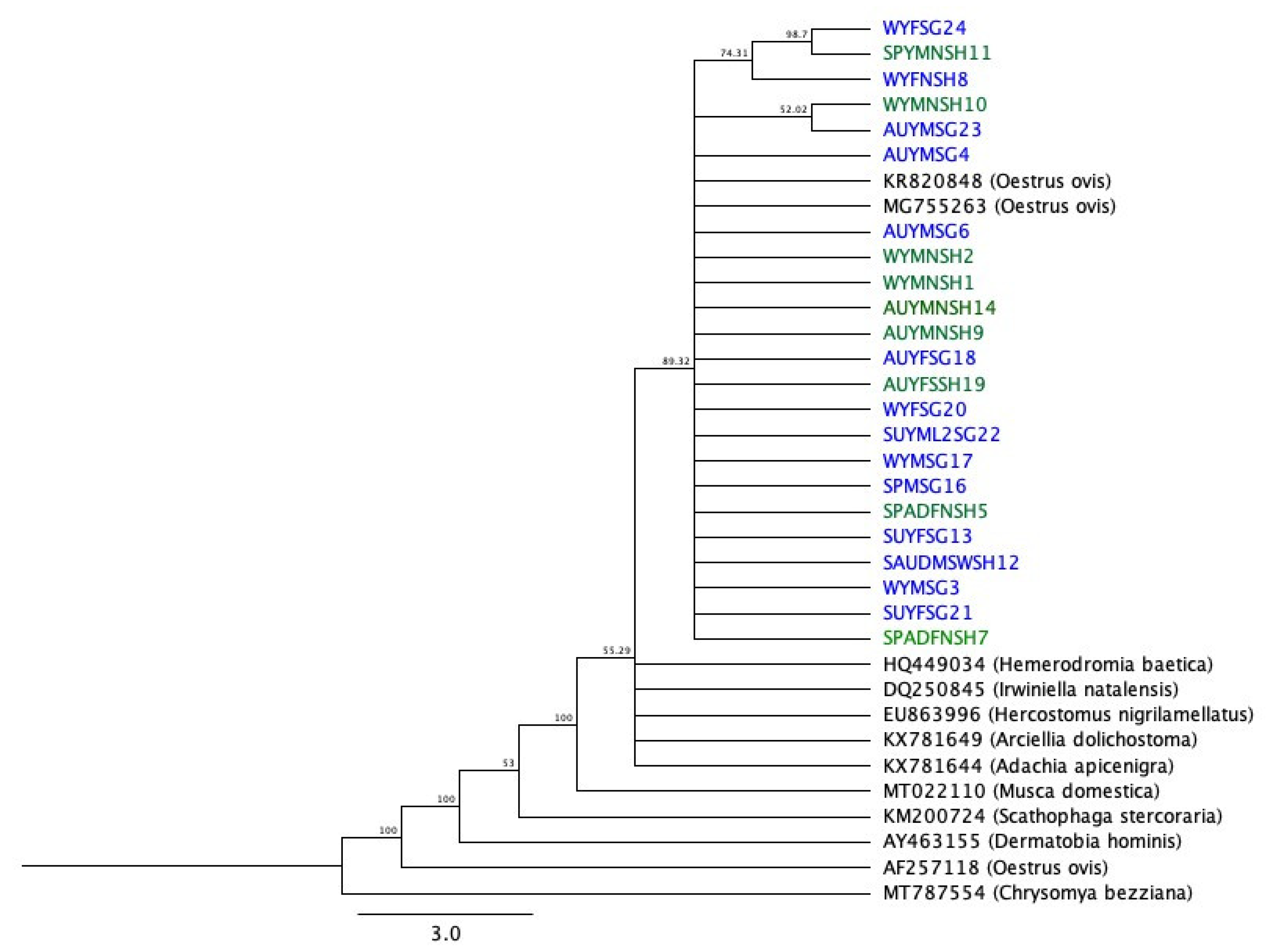
3.3. PCR Amplification and Nucleotide Sequence Analysis of the Partial mtCOI Gene

PCR amplification was performed using a set of primers (FFCOI and RHCO) to amplify a 600-bp fragment of the mtCOI gene from O. ovis larvae. A total of 23/40 sequences were analyzed in this study (sequences with HQ% (percent high quality) score less than 40 were discarded). The pairwise identity of sheep sequences was 97.9%, and that of goat sequences was 99.2%. Overall, the 23 sequences shared 98.1% pairwise identity and had 35.6% GC content. The results of BLAST search showed that all the sequences had high similarity to the O. ovis “sheep botfly” in GenBank. Figure 5 shows the phylogenetic tree indicating respective evolutionary relationships. MT787554 (Chrysomya bezziana) is used as an out-group.

4. Discussion

Out of the examined 600 sheep and 800 goat heads, 22.3% and 44.5%, respectively, were infested with O. ovis larvae. These percentages were high compared with previous studies in Saudi Arabia [26], which recorded 5.9% in Riyadh Region and 0.299% from adult O. ovis spp. in Jeddah [27], but similar to the 53.5% (257/480) reported in the Jazan Region [1]. Our results were also similar to those reported in Greece (43.2%) [28], Turkey (22.52%) [29], and Libya (42.33%) [30]. Our results were higher than those reported by other researchers around the world, such as by [31] in Egypt (8.67%), but less than those in Spain (71.1%) [32] and Italy (91.0%) [33]. These differences may be a result of differences in geographical area, animal breed, and climate. The infestation rate was higher in male sheep and goats than females (16.35% and 32.75%, respectively, compared to 6% and 11.75%; p ≤ 0.05). This was consistent with a report investigating sex differences in infestation in Libya [30].
In the central region of Saudi Arabia, the spring months are the best time of the year, with low temperature and humidity [26]. The disappearance of fly activity in most of the year is probably due to the hot, dry, and windy weather. The 27 °C was optimum temperature for adults emerge from pupa, while constant temperatures below 16 °C and above 32 °C were fatal [34].
Our results did not demonstrate any significant seasonal prevalence of infestation in sheep, suggesting that time of the year does not have any significant effect. This is in disagreement with a previous study [35], which mentioned that the peaks of infestation in Turkey were in the summer and spring, while the lowest infestation rates were in autumn. Another study in Egypt [36] concluded that heavier infestations were recorded in autumn (17.91%), while the lowest seasonal peak was in winter (7.85%). In Iraq, the highest recorded range was in the autumn, which was 76.92%, and the lowest range was in winter (30.76%) [37]. The reason for these differences may be changes in environmental conditions.
The present study recorded that 0% were L1, 1.5% were L2, and 111.2% were L3 of the total of 151 larvae collected from sheep, and 0% were L1, 1.7% were L2, and 135.4% were L3 of the of total 468 larvae collected from goats. The L1 result was lower than that recorded in previous studies [30,35] that reported that L1 larvae prevailed at 90.5% of the overall weight, and another study [37] that reported 13.14% L1 larvae. Our results were in agreement with one study [38] that reported a higher level of larvae in stage L3 (63.4%) than that in L2 (26.6%) and L1 (10%). It is possible that L1 larvae might have passed unseen due to their small size, that they were present in hidden places such as turbinates and ethmoid bones, or that numerous L1 were demolished in the nasal holes during the hypo-biotic period [39]. The morphological characters of L2 and L3 were in accordance with those stated in the identification key [20].
PCR analysis was performed using set of primers (FFCOI and RHCO) and successfully amplified 606bp fragment of mtCOI gene from O.ovis larvae. The sequence of amplified fragment showed genome arrangement typical of O. ovis sequence in Genbank. PCR amplification of mtCOI gene from eighteen species of Oestridae causing myiasis resulted in 686 bp [12]. Rooted phylogenetic trees elucidated phylogenetic relationships between O.ovis larvae amplicons and other members of Family: Oestridae published in Genbank.
The results of BLAST search showed that all the sequences had high similarity to the O. ovis “sheep botfly” identified in Brazil (accession number, KR820703) [40], with pairwise identity between 94.8% and 99.5% and coverage more than 86%. The sequences are also similar to the O. ovis sheep botfly identified in Turkey (accession number, MT124626) [41], with pairwise identity between 94.2% and 98.70% and coverage greater than 86.18%.

5. Conclusions

In this study, the results indicated that the infestation rate was significantly higher in sheep than goat, male than female, and spring season than other seasons of the year.

Supplementary Materials

The following are available online at https://www.mdpi.com/2076-2615/11/3/689/s1, Table S1: Accession numbers of samples deposited to GenBank.

Author Contributions

All the authors contributed equally to this work. All authors have read and agreed to the published version of the manuscript.

Funding

This research was funded by Deanship of Scientific Research at Princess Nourah Bint Abdulrahman University through the Fast-Track research funding program.

Institutional Review Board Statement

Ethical review and approval were waived for this study, due to collection of samples from slaughtered animals. The study did not involve alive animals.

Data Availability Statement

All relevant data are within the paper.

Conflicts of Interest

The authors declare no conflict of interest. The funders had no role in the design of the study; in the collection, analyses, or interpretation of data; in the writing of the manuscript, or in the decision to publish the results.

References

  1. Hanan, B.A. Seasonal prevalence of Oestrus ovis L. (Diptera: Oestridae) larvae in infested sheep in Jazan Region. Saudi Arabia. J. Parasitol. Vector Biol. 2013, 5, 66–71. [Google Scholar]
  2. Traversa, D.; Otranto, D. A new approach for the diagnosis of myiasis of animals: The example of horse nasal myiasis. Vet. Parasitol. 2006, 141, 186–190. [Google Scholar] [CrossRef]
  3. Hidalgo, A.; Palma, H.; Oberg, C.; Fonseca-Salamanca, F. Oestrus ovis infestation of grazing sheep during summer in southern Chile. Pesqui. Veterinária Bras. 2015, 35, 497–500. [Google Scholar] [CrossRef] [Green Version]
  4. Ogo, N.I.; Onovoh, E.; Okubanjo, O.O.; Galindo, R.C.; De La Lastra, J.-M.P.; De La Fuente, J.; De La Lastra, J.M.P. Molecular identification of Cordylobia anthropophaga Blanchard (Diptera: Calliphoridae) larvae collected from dogs (Canis familiaris) in Jos South, Plateau State, Nigeria. Onderstepoort J. Vet. Res. 2012, 79, 4. [Google Scholar] [CrossRef] [Green Version]
  5. Otranto, D.; Traversa, D. Molecular evidence indicating that Przhevalskiana silenus, P. aegagri and P. crossii (Diptera, Oestridae) are one species. Acta Parasitol. 2004, 49, 173–176. [Google Scholar]
  6. Hall, M.; Adams, Z.; Wyatt, N.; Testa, J.; Edge, W.; Nikolausz, M.; Farkas, R.; Ready, P. Morphological and mitochondrial DNA characters for identification and phylogenetic analysis of the myiasis causing flesh fly Wohlfahrtia magnifica and its relatives, with a description of Wohlfahrtia monegrosensis sp. n. Wyatt & Hall. Med. Vet. Entomol. 2009, 23, 59–71. [Google Scholar]
  7. Ames, C.; Turner, B.; Daniel, B. The use of mitochondrial cytochrome oxidase I gene (COI) to differentiate two UK blowfly species—Calliphora vicina and Calliphora vomitoria. Forensic Sci. Int. 2006, 164, 179–182. [Google Scholar] [CrossRef]
  8. Otranto, D.; Stevens, J.R. Molecular approaches to the study of myiasis-causing larvae. Int. J. Parasitol. 2002, 32, 1345–1360. [Google Scholar] [CrossRef]
  9. Otranto, D.; Puccini, V. Cytochrome oxidase I (COI) gene of some obligate myiasis causing larvae (Diptera: Oestridae): Which perspective for phylogenetic studies? Preliminary data. Proc. Cost Action 2000, 833, 195–201. [Google Scholar]
  10. Otranto, D.; Colwell, D.D.; Milillo, P.; Di Marco, V.; Paradies, P.; Napoli, C.; Giannetto, S. Report in Europe of nasal myiasis by Rhinoestrus spp. in horses and donkeys: Seasonal patterns and taxonomical considerations. Vet. Parasitol. 2004, 122, 79–88. [Google Scholar] [CrossRef] [PubMed]
  11. Otranto, D.; Colwell, D.D.; Traversa, D.; Stevens, J.R. Species identification of Hypoderma affecting domestic and wild ruminants by morphological and molecular characterization. Med. Vet. Entomol. 2003, 17, 316–325. [Google Scholar] [CrossRef]
  12. Otranto, D.; Traversa, D.; Guida, B.; Tarsitano, E.; Fiorente, P.; Stevens, J.R. Molecular characterization of the mitochondrial cytochrome oxidase I gene of Oestridae species causing obligate myiasis. Med. Vet. Entomol. 2003, 17, 307–315. [Google Scholar] [CrossRef]
  13. Otranto, D.; Milillo, P.; Traversa, D.; Colwell, D.D. Morphological variability and genetic identity in Rhinoestrus spp. causing horse nasal myiasis. Med. Vet. Entomol. 2005, 19, 96–100. [Google Scholar] [CrossRef]
  14. Li, X.; Cai, J.F.; Guo, Y.D.; Wu, K.L.; Wang, J.F.; Liu, Q.L.; Wang, X.H.; Chang, Y.F.; Yang, L.; Lan, L.M.; et al. The availability of 16S rRNA for the identification of forensically important flies (Diptera: Muscidae) in China. Trop. Biomed. 2010, 27, 155–166. [Google Scholar] [PubMed]
  15. Xinghua, W.; Jufeng, C.; Yadong, G.; Yufeng, C.; Kunlu, W.; Qinlai, L.; Jifeng, C.; Yunfeng, C.; Jiangfeng, W.; Yang, L.; et al. The availability of 16SrDNA gene for identifying forensically important blowflies in China. Rom. J. Leg. Med. 2010, 18, 43–50. [Google Scholar] [CrossRef] [Green Version]
  16. Hendawy, S.H.; Allam, N.A.; Kandil, O.M.; Zayed, A.A.; Desouky AR, A.; Mahmoud, A. Partial COI and 16S rRNA genes sequences of Cephalopina titillator mitochondrial dna: Evidence for variation in evolutionary rates within myiasis causing species. Indian J. Anim. Res. 2012, 52, 105–110. [Google Scholar]
  17. Moreno, V.; Romero-Fernández, I.; Marchal, J.A.; Beltran, M.; Granados, J.E.; Habela, M.A.; Tamadon, A.; Rakhshandehroo, E.; Sarasa, M.; Pérez, J.M.; et al. Molecular characterization of bot flies, Oestrus spp., (Diptera, Oestridae), from domestic and wild Bovidae hosts. Vet. Parasitol. 2015, 212, 473–477. [Google Scholar] [CrossRef]
  18. Ferrar, P. A Guide to the Breeding Habits and Immature Stages of Diptera Cyclorrhapha; EJ Brill/Scandinavian Science Press: Copenhagen, Denmark, 1987; Volume 8, pp. 1–2. [Google Scholar]
  19. Smith, K.G. An Introduction to the Immature Stages of British Flies: Diptera Larvae, with Notes on Eggs, Puparia and Pupae; Royal Entomological Society: London, UK, 1989; Volume 10, p. 14. [Google Scholar]
  20. Zumpt, F. Myiasis in man and animals in the Old World. A textbook for physicians, veterinarians and zoologists. In Myiasis in Man and Animals in the Old World: A Textbook for Physicians, Veterinarians and Zoologists; Butterworth & Co. (Publishers) Ltd.: London, UK, 1965. [Google Scholar]
  21. Bosly, A.H. Molecular identification studies on Oestrus ovis L. (Diptera: Oestridae) larvae infested sheep in jazan region, Saudi Arabia. Indian J. Anim. Res. 2018, 52, 105–110. [Google Scholar]
  22. Drummond, A.J.; Ashton, B.; Buxton, S.; Cheung, M.; Cooper, A.; Duran, C.; Field, M.; Heled, J.; Kearse, M.; Markowitz, S.; et al. Geneious v5.3. Biomatters Ltd: Auckland, New Zealand. 2010. Available online: http://www.geneious.com (accessed on 28 January 2021).
  23. Altschul, S.F.; Gish, W.; Miller, W.; Myers, E.W.; Lipman, D.J. Basic local alignment search tool. J. Mol. Biol. 1990, 215, 403–410. [Google Scholar] [CrossRef]
  24. Sievers, F.; Wilm, A.; Dineen, D.; Gibson, T.J.; Karplus, K.; Li, W.; Lopez, R.; McWilliam, H.; Remmert, M.; Söding, J.; et al. Fast, scalable generation of high-quality protein multiple sequence alignments using Clustal Omega. Mol. Syst. Biol. 2011, 7, 539. [Google Scholar] [CrossRef] [PubMed]
  25. Saitou, N.; Nei, M. The neighbor-joining method: A new method for reconstructing phylogenetic trees. Mol. Biol. Evol. 1987, 4, 406–425. [Google Scholar] [CrossRef]
  26. Alahmed, A.M. Seasonal infestation of Oestrus ovis larvae in sheep heads in central region of Saudi Arabia. J. Egypt. Soc. Parasitol. 2000, 30, 895–901. [Google Scholar]
  27. Alikhan, M.; Al Ghamdi, K.; Al Zahrani, F.S.; Khater, E.I.; Allam, A.M. Prevalence and Salient Morphological Features of Myiasis Causing Dipteran Flies in Jeddah, Saudi Arabia. Biosci. Biotechnol. Res. Asia 2018, 15, 101–109. [Google Scholar] [CrossRef]
  28. Papadopoulos, E.; Chaligiannis, I.; Morgan, E.R. Epidemiology of Oestrus ovis L. (Diptera: Oestridae) larvae in sheep and goats in Greece. Small Rumin. Res. 2010, 89, 51–56. [Google Scholar] [CrossRef]
  29. Karatepe, B.; Karatepe, M.; Güler, S. Epidemiology of Oestrus ovis L. infestation in sheep in Nigde province, Turkey. Rev. Médecine Vétérinaire 2014, 165, 225–230. [Google Scholar]
  30. Mohsen, M.N.; Raham, S.E.; Gasim, M.H. Oestrus ovis larval infestation among sheep and goats of green moun-tain areas in Libya. J. Adv. Vet. Anim. Res. 2015, 2, 382–387. [Google Scholar]
  31. Amin, A.R.; Morsy, T.A.; Shoukry, A.; Mazyad, S.A. Oestrid head maggots in slaughtered sheep in Cairo abattoir. J. Egypt. Soc. Parasitol. 1997, 27, 855–861. [Google Scholar] [PubMed]
  32. Alcaide, M.; Reina, D.; Sánchez, J.; Frontera, E.; Navarrete, I. Seasonal variations in the larval burden distribution of Oestrus ovis in sheep in the southwest of Spain. Vet. Parasitol. 2003, 118, 235–241. [Google Scholar] [CrossRef] [PubMed]
  33. Caracappa, S.; Rilli, S.; Zanghi, P.; Di Marco, V.; Dorchies, P. Epidemiology of ovine oestrosis (Oestrus ovis Linné 1761, Diptera: Oestridæ) in Sicily. Vet. Parasitol. 2000, 92, 233–237. [Google Scholar] [CrossRef]
  34. Rogers, C.E.; Knapp, F.W. Bionomics of the Sheep Bot Fly, Oestrus Ovis. Environ. Entomol. 1973, 2, 11–23. [Google Scholar] [CrossRef]
  35. Arslan, M.O.; Kara, M.; Gicik, Y. Epidemiology of Oestrus ovis infestations in sheep in Kars province of north-eastern Turkey. Trop. Anim. Health Prod. 2009, 41, 299–305. [Google Scholar] [CrossRef] [PubMed]
  36. Ramadan, M.Y.; Khater, H.F.; Omer, S.F.; Rahman, A.A. Epidemiology of Oestrus ovis infesting Egyptian sheep. In Proceedings of the XX International Congress of Mediterranean Federation of Health and Production of Ruminants, Assiut, Egypt, 19–22 February 2013; pp. 19–22. [Google Scholar]
  37. Mohammed, R.G.; Josef, S.S.; Abed, K.J. Prevalence of Oestrus ovis Larvae in Slaughtered Sheep of Misan City, Iraq. Syst. Rev. Pharm. 2020, 11, 652–655. [Google Scholar]
  38. Yilma, J.; Dorchies, P. Epidemiology of Oestrus ovis in southwest France. Vet. Parasitol. 1991, 40, 315–323. [Google Scholar] [CrossRef]
  39. Bart, A.G.; Minár, J. Probability description of regulation on the level of population and individual in the host-parasite system using Oestrus ovis (Diptera: Oestridae) as an example. Folia Parasitol. 1992, 39, 75–83. [Google Scholar]
  40. Marinho, M.A.T.; Wolff, M.; Ramos-Pastrana, Y.; de Azeredo-Espin, A.M.L.; Amorim, D.D.S. The first phylo-genetic study of Mesembrinellidae (Diptera: Oestroidea) based on molecular data: Clades and congruence with morpholog-ical characters. Cladistics 2017, 33, 134–152. [Google Scholar] [CrossRef]
  41. Karademir, G.K.; Usluğ, S.; Okur, M.; Inci, A.; Yıldırım, A. Molecular Characterization and Phylogenetic Analyses of Oestrus ovis Larvae Causing Human Naso-pharyngeal Myiasis Based on CO1 Barcode Sequences. Turk. J. Parasitol. 2020, 44, 43–47. [Google Scholar] [CrossRef] [PubMed]
Figure 1. Effect of season on larval characteristics in infested goats. Mean values with different letters (a, b, c) are significantly different at p ≤ 0.05 between seasons.
Animals 11 00689 g001
Figure 2. Effect of seasons on larval characteristics in infested sheep. Mean values with different letters (a and b) are significantly different at p ≤ 0.05 between seasons.
Animals 11 00689 g002
Figure 3. Second instar larva (L2). (A) Dorsal view; (B) ventral view; (C) the front end containing the cephaloskeleton ; (D) the median part of the postanal bulge is spinulose; (E) ventral view shows spines on anterior segment ; (F) posterior spiracle; (G) stigmal plates and channel appear with clear distinct suture; (H) posterior spiracle (10×).
Animals 11 00689 g003
Figure 4. Third instar larva (L3). (A) Yellow color; (B) dorsal view shows broad transverse blackish bands; (C) ventral view shows spines on segments; (D) magnified view of the spines on the ventral segments; (E) postanal bulge shows fewer spines; (F) posterior spiracle is in a D-shape with central button but no distinct suture.
Animals 11 00689 g004
Figure 5. Genetic relationships of the mtCOI gene collected samples with other species retrieved from GenBank. The tree is generated using the neighbor-joining method, implemented in the Geneious software, with the Tamura_Nei model.
Animals 11 00689 g005
Table 1. Set of primers used for the amplification of mitochondrial cytochrome c oxidase subunit I (mtCOI) gene using extracted DNA of O. ovis larvae.
GenePrimersSequencesReferences
mtCOIFFCOI5′-GGAGCATTAATYGGRGAYG-3′[21]
RHCO5′-TAAACTTCAGGGTGACCAAAAATCA-3′
Table 2. Prevalence of Oestrus larvae in sheep and goats over the course of the study.
SpeciesNo. of Head ExaminedInfestation Rate (%)Mean of InfestationNo. of Larvae (%)Total Larvae (%)
L1L2L3
Sheep600134 (22.3)1.78 ± 0.02 a0.0 (0.0)2.0 (1.5)149.0 (111.2)151.0 (112.7)
Goat800356 (44.5)1.56 ± 0.02 b0.0 (0.0)6.0 (1.7)482.0 (135.4)486 (136.5)
Mean values with different superscript in column differed significantly at p ≤ 0.05.
Table 3. Sex differences on the prevalence of Oestrus larvae in sheep and goats.
SpeciesSexNo. of Examined AnimalsNo. of Infested (%)
SheepMale30098 (32.66)
Female30036 (12)
GoatMale400262 (65.5)
Female40094 (32.5)
Total1400490 (35.00)
Publisher’s Note: MDPI stays neutral with regard to jurisdictional claims in published maps and institutional affiliations.

