Article

A Comparison of Dobutamine, Norepinephrine, Vasopressin, and Hetastarch for the Treatment of Isoflurane-Induced Hypotension in Healthy, Normovolemic Dogs

by
Natalia Henao-Guerrero
1,
Carolina H. Ricco-Pereira
2 and
Vaidehi V. Paranjape
1,*
1
Department of Small Animal Clinical Sciences, Virginia-Maryland College of Veterinary Medicine, Blacksburg, VA 24061, USA
2
Department of Veterinary Clinical Sciences, The Ohio State University-College of Veterinary Medicine, Columbus, OH 43210, USA
*
Author to whom correspondence should be addressed.
Animals 2023, 13(16), 2674; https://doi.org/10.3390/ani13162674
Submission received: 26 July 2023 / Revised: 15 August 2023 / Accepted: 17 August 2023 / Published: 19 August 2023
(This article belongs to the Special Issue Anaesthesia and Analgesia in Companion Animals)

Abstract

:

Simple Summary

In anesthetized dogs, hypotension, defined as a mean arterial pressure (MAP) of <60–70 mmHg, has an incidence of up to 60%. It is therefore imperative to avoid the occurrence of hypotension and undertake immediate strategies to treat it. Treatment protocols for the correction of isoflurane-mediated hypotension include lowering or terminating isoflurane administration; lowering isoflurane requirement by using anesthetic adjuncts and analgesics; intravenous crystalloid and/or colloid bolus; and delivering anticholinergics, positive inotropes, and vasopressor drugs. The present study evaluated and compared the hemodynamic effects of dobutamine (DOB), norepinephrine (NEP), vasopressin (VAS), and hetastarch (HES) for the treatment of isoflurane-induced hypotension and established the most effective dosage regimen for the correction of hypotension. In healthy, anesthetized Beagle dogs undergoing acute, severe isoflurane-induced hypotension, NEP administered as an infusion was the most efficient therapy for correcting MAP. However, the DOB, VAS, and HES treatments were unsuccessful in stabilizing the MAP to >65 mmHg, even at maximum doses.

Abstract

Isoflurane is a commonly used inhalation anesthetic in species undergoing veterinary care that induces hypotension, impacting organ perfusion, making it imperative to minimize its occurrence or identify effective strategies for treating it. This study evaluated and compared the hemodynamic effects of DOB, NEP, VAS, and HES in twelve isoflurane-anesthetized Beagle dogs. The order of the first three treatments was randomized. HES was administered last. Data were collected before treatments (baseline) and after 10 min of a sustained MAP of <45 mmHg induced by a high end-tidal isoflurane concentration (T0). Once treatment was initiated and the target MAP was achieved (65 to 80 mmHg) or the maximum dose reached, data were collected after 15 min of stabilization (T1) and 15 min after (T2). A 15 min washout period with a MAP of ≥65 mmHg was allowed between treatments. The intravenous dosage regimens started and were increased by 50% every five minutes until the target MAP or maximum dose was reached. The dosages were as follows: DOB, 5–15 μg/kg/min; NEP, 0.1–2 μg/kg/min; VAS, 0.5–5 mU/kg/min; and HET, 6% 1–20 mL/kg/min. DOB improved CO, DO2, and VO2, but reduced SVR. VAS elevated SVR, but decreased CO, DO2, and VO2. HES minimally changed BP and mildly augmented CO, DO2, and VO2. These treatments failed to reach the target MAP. NEP increased the arterial BP, CO, MPAP, and PAWP, but reduced HR. Norepinephrine infusion at 0.44 ± 0.19 μg/kg/min was the most efficient therapy for correcting isoflurane-induced hypotension.

1. Introduction

Volatile anesthetics are an integral component of general anesthesia in research settings and clinical practice in veterinary and human medicine. Throughout the past several decades, a large number of studies that have defined the cardiac, vascular, and autonomic effects of volatile anesthetics have been published. These effects become evident immediately following anesthetic induction and during anesthetic maintenance. Some of the key elements of the cardiovascular system that are impacted include: (i) myocardial contractility, (ii) systemic vascular resistance (SVR), and (iii) cardiac electrophysiology [1].
The dose-dependent depression of myocardial contractility is primarily due to the dysregulation of the sarcolemmal L-type Ca2+ channels, sarcoplasmic reticulum, and contractile proteins [2,3]. Experimental canine studies have revealed that there are differences between inhalant anesthetics in this regard and that halothane and enflurane exert equal but more potent negative inotropic effects than isoflurane, desflurane, or sevoflurane [4,5]. Halothane exhibits the most pronounced reduction in blood pressure (BP) via myocardial depression and decrease in arterial tone, while isoflurane, sevoflurane, and desflurane primarily alter the left ventricular afterload and SVR [2,6,7,8]. Peripheral vasodilation and hypotension worsen with increasing concentrations of volatile anesthetics in a dose-dependent fashion. The association between intraoperative hypotension and its duration with postoperative mortality and organ dysfunction after general anesthesia is well documented [9]. All volatile agents attenuate the baroreceptor reflex, where every constituent of the reflex arc (i.e., afferent and efferent activity, central processing) can be inhibited [10,11]. Volatile anesthetics also slow down the rate of sinoatrial node discharge by affecting the sinoatrial node automaticity, along with having direct action on cardiac pacemaker cells and conduction pathways [2,12]. Canine studies have suggested that the myocardial sensitization to arrythmias is halothane > enflurane > sevoflurane = isoflurane [13,14].
Isoflurane is a commonly used inhalation anesthetic in animal species under veterinary care. Just like other volatile anesthetics, isoflurane anesthesia induces hypotension and consequently results in a decline in organ perfusion and poor patient outcomes [15]. In anesthetized dogs, hypotension, defined as a mean arterial pressure (MAP) of <60–70 mmHg [16,17], has an incidence of up to 60%, which even includes elective surgeries [18,19]. It is therefore imperative to avoid the occurrence of hypotension and undertake immediate strategies to treat it. Treatment protocols for the correction of isoflurane-mediated hypotension include lowering or terminating isoflurane administration; lowering isoflurane requirement by using anesthetic adjuncts and analgesics, intravenous crystalloid and/or colloid bolus; and delivering anticholinergics, positive inotropes, and vasopressor drugs [16,20,21]. Dobutamine exhibits a positive inotropy via β1 adrenergic agonist activity, thus increasing cardiac output (CO) in a dose-dependent fashion. Another routine treatment for hypotension is norepinephrine, which has potent agonistic actions on the α1 and α2 adrenergic receptors, resulting in peripheral vasoconstriction and venoconstriction, along with well-established β1 agonism-driven cardiac stimulation [16,20]. Vasopressin is also used for its vasoconstrictor effects, through its V1a receptor action. In the face of acidemia, it can enhance the SVR and can be useful in treating refractory hypotension, as seen in sepsis or hypovolemic shock [20,21]. These drugs are commonly selected for maintaining the BP in hypotensive and critically ill small animals. Synthetic colloidal solutions are popular in veterinary medicine and can be effective plasma volume expanders, administered alone or following a crystalloid or hypertonic saline bolus [22,23].
Even though these therapeutic options are well described in the literature [20,21,22,23], there are limited studies specifically focused on which of these therapies is most effective against severe isoflurane-induced hypotension in anesthetized dogs. Moreover, the dosages of dobutamine, norepinephrine, and vasopressin required to successfully combat and correct severe hypotension from isoflurane are relatively unknown. The objectives of the present study concerning healthy, normovolemic anesthetized dogs were: (i) to evaluate and compare the hemodynamic effects of dobutamine, norepinephrine, vasopressin, and hetastarch for the treatment of isoflurane-induced hypotension and (ii) to establish the most effective dosage regimen for dobutamine, norepinephrine, and vasopressin during the correction of hypotension. We hypothesized that isoflurane-induced hypotension would be similarly and successfully treated with dobutamine, norepinephrine, and vasopressin, but not with hetastarch.

2. Materials and Methods

2.1. Animals

Twelve adult, healthy, female, purpose-bred, and university-owned Beagles aged 12–24 months, with body weights of 8.5 ± 1.2 kg, participated in this prospective, crossover, randomized experimental study. The dogs were certified as ‘healthy’ based on a thorough physical examination, cardio-pulmonary auscultation, heart worm test, complete blood count, and serum chemistry. The experimental study and use of animals were approved by the Virginia Tech University Institutional Animal Care and Use Committee (protocol number 12-094-CVM). Six weeks after the end of the study, all the dogs underwent surgical sterilization, vaccination, and deworming, and were successfully adopted into single-family homes.
Based on previous canine studies evaluating the effect of an intervention on CO [24,25], a sample size of a minimum of ten animals was required to demonstrate a 25% significant difference in CO in response to the treatment administered, assuming a statistical power of 0.8 and an alpha level of 0.05 (http://estatistica.bauru.usp.br/calculoamostral/; accessed on 23 August 2012). An additional two dogs were added to the sample size to improve the statistical power of the study, as well as to account for unforeseen circumstances (e.g., missing data or challenging instrumentation).

2.2. General Anesthesia and Anesthetic Monitoring

After the completion of a two-week acclimatization period, each dog was randomly selected for the experiment, and food was withheld for 12 h with ad libitum access to water. On the day of the experiment, a cephalic catheter was aseptically placed, followed by anesthetic induction via isoflurane delivery in 100% oxygen (4 L/min) using a fitted facemask connected to a small animal anesthesia machine via a Bain non-rebreathing system. Once the appropriate anesthetic plane and adequate muscle relaxation for intubation were confirmed, the trachea was intubated, and a cuffed endotracheal tube was secured to the dog with a tie gauze and connected to the non-rebreathing circuit and anesthesia machine (Apollo; Draeger Medical Inc., Telford, PA, USA). The dogs were then placed in dorsal recumbency for the entire anesthetic period. Isoflurane in 100% oxygen (1–2 L/min) was used for anesthetic maintenance. The end-tidal concentration of isoflurane (ETISO), as continuously measured using an infrared gas analyzer, was included in a multiparameter monitor (Datex-Ohmeda S/5 Compact anesthesia monitor; GE Healthcare, Chicago, IL, USA) and was targeted at 1.4–1.6%, except during the experimental procedures, which required increments in ETISO. The other standard anesthetic monitoring included in the same monitor were a lead II electrocardiogram for heart rate (HR) and rhythm, an end-tidal concentration of carbon dioxide, esophageal temperature, a peripheral oxygen saturation of hemoglobin (SpO2), and spirometry. Normothermia (36.9 to 37.8 °C) was maintained in all dogs throughout the anesthetic period with a forced-air warming device and circulating water blanket in place. Standard monitoring variables were recorded every five minutes as per routine standards throughout the anesthetic period. No maintenance fluids were administered.
The dogs were mechanically ventilated using volume-controlled ventilation settings with a tidal volume of 10–12 mL/kg, and the respiratory rate was adjusted to maintain the end-tidal carbon dioxide concentration between 35 and 45 mmHg. Mechanical ventilation was initiated to maintain the consistent values in a specified range for the end-tidal carbon dioxide and isoflurane concentrations throughout the study. Aseptic arterial catheterization in the dorsal pedal artery was performed for recording invasive systolic (SAP), diastolic (DAP), and mean arterial blood pressure (MAP), as well as periodic arterial blood gas sampling. The same multi-parametric monitor was connected to a heparinized, saline-flushed (3 IU/mL) disposable pressure transducer system (Deltran II; Utah Medical Products Inc., Midvale, UT, USA), which was zeroed and leveled approximately at the location of the right atrium. Prior to the beginning of the experiment, the transducer and monitor were calibrated against a digital pressure manometer. With the help of a 2-point calibration technique (0 and 150 mmHg), the accuracy and linearity of the transducer were assessed via verification against the mercury manometer. The calibration was deemed to be acceptable when the difference between the manometer and displayed monitor pressures was <2 mmHg.

2.3. Instrumentation for Intermittent Pulmonary Artery Thermodilution to Measure CO and Other Hemodynamic Variables

By performing a modified Seldinger technique, a 6 Fr 8.5 cm hemostasis introducer (Fast-Cath; Abbott Cardiovascular, Plymouth, MN, USA) was aseptically inserted into the right jugular vein, through which a 5 Fr 75 cm Swan Ganz thermodilution catheter (132FS; Edwards Lifesciences Corp., Irvine, CA, USA) was advanced. The Swan Ganz catheter was maneuvered based on the pressure–waveform relationship displayed on the CO monitor (Carescape B850; GE Healthcare, Chicago, IL, USA). This was continued until the distal tip was located in the pulmonary artery. A 3 mL, 2 °C to 5 °C cold saline bolus was used as an indicator and injected into the proximal injectate port for the CO determination. The other hemodynamic variables measured using this catheter were the central venous pressure (CVP; via proximal port), mean pulmonary artery pressure (MPAP; via distal port), and pulmonary artery wedge pressure (PAWP; via distal port and balloon inflation). A detailed explanation of the thermodilution procedure in dogs was published previously and is available for review [26,27,28]. Each CO reading was timed at the end of the expiration, and the saline bolus was injected for over more than three seconds. At each datapoint, the CO reading recorded equaled the mean of 3 consecutive measurements within a 10% variation. This thermistor-tipped Swan Ganz catheter was also used to measure the core body temperature.

2.4. Calculations for Other Hemodynamic Variables Derived from the Thermodilution Catheter

Various other hemodynamic variables, such as the systemic vascular resistance (SVR), pulmonary vascular resistance (PVR), arterial oxygen content (CaO2), venous oxygen content (CvO2), oxygen delivery (DO2), oxygen consumption (VO2), and oxygen extraction ratio (OER), were calculated using the following formulae [19,27,29]:
SVR   ( dyn · sec / cm 5 ) =   ( Mean   Arterial   Pressure   Central   Venous   Pressure )   ×   80 Cardiac   output   by   Thermodilution
PVR   ( dyn · sec / cm 5 ) =   ( Mean   Pulmonary   Arterial   Pressure   Pulmonary   Artery   Wedge   Pressure )   ×   80 Cardiac   output   by   Thermodilution
CaO2 (mL/dL) = (1.34 × SaO2 × Hb) + (0.003 × PaO2)
CvO2 (mL/dL) = (1.34 × SvO2 × Hb) + (0.003 × PvO2)
DO2 (mL/min) = 10 × CaO2 × CO
VO2 (mL/min) = CO × (CaO2 − CvO2)
OER (%) = (CaO2 − CvO2)/CaO2 × 100
where SaO2 = arterial oxygen saturation; SvO2 = venous oxygen saturation; Hb = hemoglobin concentration; PaO2 = arterial oxygen tension; and PvO2 = venous oxygen tension.

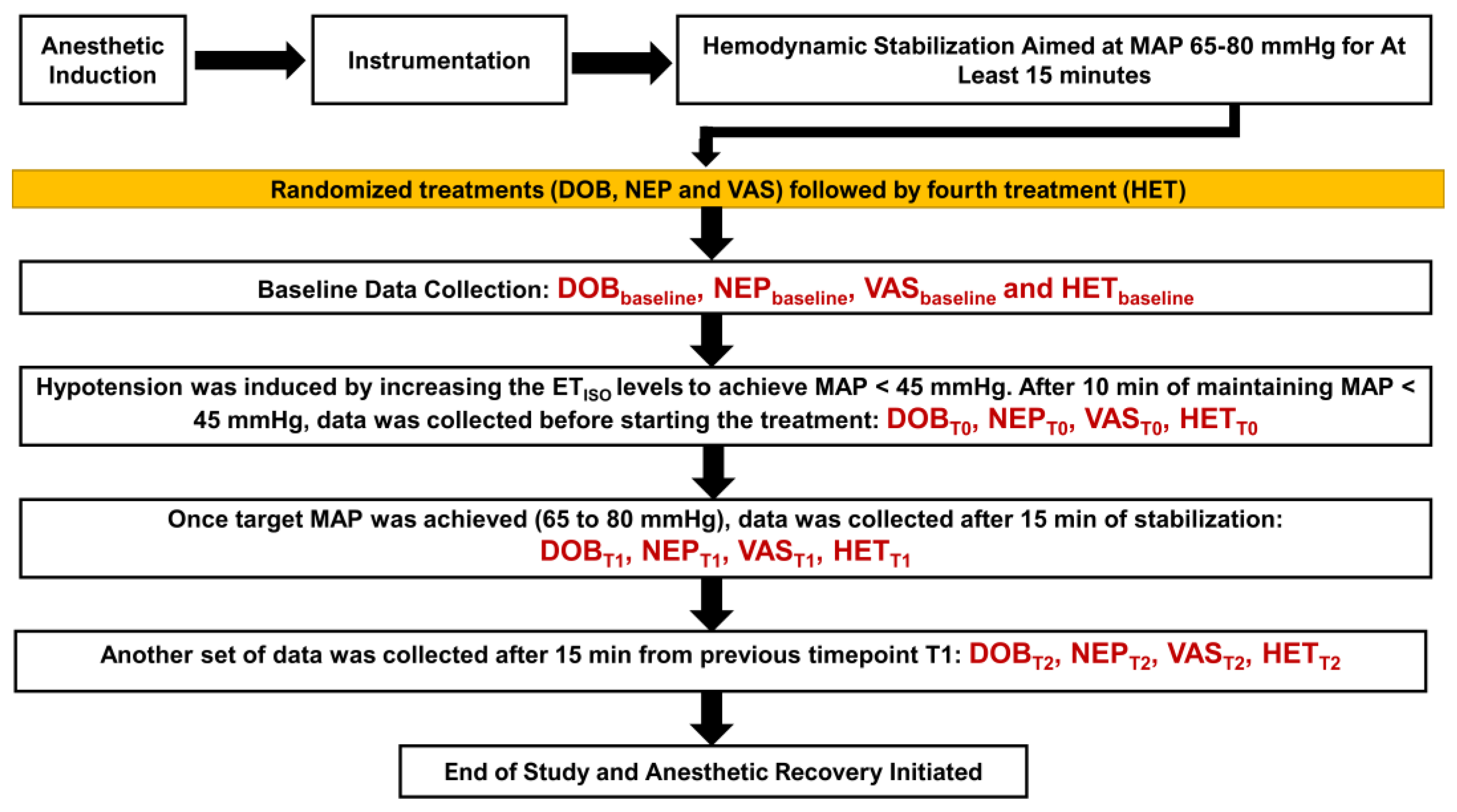
2.5. Administration for Treatments and Data Collection

Once the instrumentation was completed, hemodynamic stabilization was achieved by aiming for an MAP of 65 to 80 mmHg for at least 15 min. Then, each dog underwent four treatments (Figure 1). Three treatments, i.e., dobutamine (DOB), norepinephrine (NEP), and vasopressin (VAS), were always administered in a randomized order using an online website module (https://www.randomizer.org/; accessed on 25 August 2012). However, the colloid treatment (HET) always followed after the other three treatments were administered. Baseline data were collected before the DOB (DOBbaseline), NEP (NEPbaseline), VAS (VASbaseline), and 6% 670/0.75 hetastarch (HETbaseline) treatments. After collecting the baseline data for the selected treatment, hypotension was induced by increasing the ETISO levels to achieve an MAP of <45 mmHg. After 10 min of maintaining an MAP of <45 mmHg, data were collected before starting treatment (i.e., DOBT0, NEPT0, VAST0, and HETT0). The dosage regimens for the four treatments were: (i) DOB—intravenous dobutamine via an infusion pump (BD Alaris™ Infusion System; Becton, Dickinson and Company, Franklin Lakes, NJ, USA) was administered at a starting dose of 5 μg/kg/min, and the dose was increased by 50% every five minutes until the MAP was 65 to 80 mmHg; (ii) NEP—intravenous norepinephrine via an infusion pump (BD Alaris™ Infusion System) was administered at a starting dose of 0.1 μg/kg/min, and the dose was increased by 50% every five minutes until the MAP was 65 to 80 mmHg; (iii) VAS—intravenous vasopressin via an infusion pump (BD Alaris™ Infusion System) was administered at a starting dose of 0.5 mU/kg/min, and the dose was increased by 50% every five minutes until the MAP was 65 to 80 mmHg; and (iv) HET—intravenous 6% 670/0.75 hetastarch via an infusion pump (BD Alaris™ Infusion System) was administered at 1 mL/kg/min until the MAP was 65 to 80 mmHg. The maximum allowable limit for the dosages were 15 μg/kg/min (DOB), 2 μg/kg/min (NEP), 5 mU/kg/min (VAS), and 20 mL/kg (HET). Once the treatment was initiated and the target MAP was achieved (65 to 80 mmHg) or the maximum dosage was reached, data were collected after 15 min of stabilization (i.e., DOBT1, NEPT1, VAST1, and HETT1), followed by another data collection, which was 15 min after timepoint T1 (i.e., DOBT2, NEPT2, VAST2, and HETT2). At least a 15 min washout period was assigned between the treatments, and it was ensured that the MAP values were ≥65 mmHg during the rest/washout period. All the hemodynamic data were acquired during the expiratory phase of the respiratory cycle. An arterial and venous blood gas analysis was performed at baseline and timepoints T0 and T2.

2.6. Anesthetic Recovery

After the HET data collection, the jugular and arterial catheters were removed and external pressure was applied to confirm that bleeding and hematoma were absent. The delivery of isoflurane was terminated. After extubation, 0.2 mg/kg of IV butorphanol was administered to all the dogs and they were moved to individual kennels. A blood sample was obtained after 24 h to confirm that the hematocrit, total protein, and blood urea nitrogen levels were normal. The cardiorespiratory parameters, pain assessment and catheter sites were monitored periodically every 4–8 h for the next 48 h.

2.7. Statistical Analysis

The outcomes included ETISO, SpO2, HR, SAP, MAP, DAP, CO, CVP, MPAP, PAWP, SVR, PVR, DO2, VO2, and OER. Normal probability plots and a Shapiro–Wilk test showed that all the outcomes followed a normal distribution. Subsequently, the data were summarized as mean ± standard deviation. The effects of the treatment (DOB, NEP, VAS, and HET) and time period (baseline, T0, T1, and T2) on each outcome were assessed using a mixed-model analysis of variance. The generalized linear model specified treatment, time, and the interaction between treatment and time as fixed effects. Dog identification was the random effect, and a Kenward–Roger approximation was the degrees of freedom denominator. The interaction between treatment and time was further analyzed to compare the treatments at each time point and compare the time points within each treatment. The overall p-values for the main effects (treatment and time) across all the outcomes were adjusted for multiple testing using the Benjamini–Hochberg false discovery rate method. For each outcome with a significant overall p-value (after the Benjamini–Hochberg adjustment), p-values for 2-way comparisons were adjusted for multiple comparisons using Tukey’s procedure. Significance was set at p < 0.05. All the analyses were performed using SAS version 9.3 (Cary, NC, USA).

3. Results

All the dogs exhibited smooth and uneventful anesthetic induction, maintenance, and recovery. The successful placement of a Swan Ganz catheter was performed in all the dogs without any complications arising from the right heart catheterization. Even though all twelve dogs successfully underwent the experiment, significant missing data were reported in two dogs under the HET and DOB treatments. This made a data analysis challenging, and these two dogs were thus eliminated from the study and their data were not included. The dogs were normothermic (37.3 ± 0.5 °C) and normocapnic (40 ± 4 mmHg) throughout the anesthetic period. The jugular and arterial catheter sites did not present any evidence of hematoma formation or subcutaneous bruising. Four out of the twelve dogs required an additional dose of 0.3 mg/kg of IV butorphanol due to a display of mild pain at the 4 h timepoint post-extubation based on a pain assessment. The cardiorespiratory parameters, appetite, demeanor, and excretory functions were within the normal limits throughout the 48 h post-extubation period. The blood samples obtained at 24 h showed the hematocrit, total protein, and blood urea nitrogen levels to be within normal limits for all the dogs. Between the ten dogs, there were no differences in the: (i) total anesthesia time (p = 0.94); (ii) time spent between anesthetic induction to recording DOBbaseline (p = 0.43), NEPbaseline (p = 0.89), VASbaseline (p = 0.21), and HETbaseline (p = 0.55); (iii) time spent between the baseline and T2 data collection (p = 0.41); and (iv) time spent obtaining data for timepoints T1 (p = 0.67) and T2 (p = 0.14) during the treatments. The final doses required to achieve the target MAP values (65 to 80 mmHg) during isoflurane-induced hypotension were 9.64 ± 4.33 μg/kg/min for dobutamine infusion, 0.44 ± 0.19 μg/kg/min for norepinephrine infusion, 5 ± 0 mU/kg/min for vasopressin infusion, and 20 ± 0 mL/kg for hetastarch. Additionally, there was no change in the parameters recorded from the arterial and venous blood gas analysis performed at baseline or timepoints T0 and T2.
There were no differences between the baseline data for the measured parameters during the four treatments, as shown in Table 1. The ETISO values that caused an MAP of <45 mmHg in the four treatment groups were not different from one another. Once the hypotension set point from isoflurane was established, the dobutamine infusion caused mild increments in the heart rate, especially at high doses, with significant increases in the arterial BP, CO, MPAP, and PAWP values at T1 and T2 as compared to T0 (p < 0.001); however, the CVP values were significantly higher only at T2 compared to baseline (p < 0.001). Although the MAP values were increased as compared to hypotension, a consistent target MAP (65 to 80 mmHg) was not achieved with the doses used in the study. The norepinephrine infusion significantly decreased HR at T1 and T2 as compared to baseline and T0, with a subsequent increase in arterial BP, CO, MPAP, and PAWP as compared to T0 (p < 0.001). However, CO was significantly lower at T1 (p = 0.033) and T2 (p = 0.026) versus at baseline. Additionally, there was a small increase in CVP at T1 (p = 0.015) and T2 (p = 0.037) as compared to baseline. The administration of vasopressin also caused a significantly lower HR at T1 and T2 as compared to baseline and T0, with a concurrent increase in arterial BP, MPAP, and PAWP in comparison to T0 (p < 0.001). Even with the highest dose limit, a consistent target MAP (65 to 80 mmHg) was not achieved. The CO values were observed to deteriorate further with vasopressin, while no change in CVP was seen—as opposed to at T0. During the hetastarch treatment, HR was significantly elevated at T1 and T2 as compared to baseline and T0, but hypotension was not corrected. CO mildly increased with a significant rise in CVP, MPAP, and PAWP at T1 and T2 as compared to T0.
Similar to the measured parameters, there were no differences between the baseline data for the calculated parameters during the four treatments, as shown in Table 2. Dobutamine significantly decreased the SVR at T1 and T2 as compared to baseline and T0, while lowering the PVR at T1 and T2 as compared to T0 (p < 0.001). Meanwhile, norepinephrine and vasopressin caused significant increments in the SVR and PVR at T1 and T2 as compared to baseline (p < 0.001). The administration of hetastarch significantly lowered the SVR with concurrent elevations in the PVR at T2 as compared to baseline and T0 (p < 0.001). The dobutamine infusion led to significant increases in DO2 and VO2 at T1 and T2 as compared to T0 (p < 0.001); however, these values had no difference from baseline. During severe hypotension, norepinephrine significantly improved DO2 and VO2 at T1 and T2; however, these values were significantly lower than at baseline (p < 0.001). Vasopressin significantly diminished DO2 and VO2 at T1 and T2 as compared to baseline and T0 (p < 0.001). Hetastarch augmented DO2 and VO2 when the MAP was <45 mmHg. The OER did not show any significant changes throughout the experiment.

4. Discussion

The pathways that control arterial BP depend upon the length of period during which abnormal fluctuations in BP occur, i.e., short- and long-term effects. When acute variations in BP occur over minutes to hours, short-term control regulates BP through changes impacting CO, SVR, arterial compliance, heart rate, blood volume, and myocardial contractility [30]. Autoregulation ensures that the optimum blood flow and oxygen delivery are provided to the critical organs by keeping the blood pressure values within a physiologic range. The main elements of the control system that aims to stabilize adequate perfusion are the baroreceptors, chemoreceptors, central pressure centers in the medulla oblongata, and the renin–angiotensin–aldosterone pathway. Volatile anesthetics can alter autoregulatory mechanisms and, therefore, the vigilant monitoring and treatment of blood pressure can be a crucial step in maintaining a steady hemodynamic status under general anesthesia. Hypotension, i.e., an MAP of <60–70 mmHg under general anesthesia, can be triggered by decreases in HR, CO, stroke volume, and SVR [15,16]. Balanced anesthesia techniques incorporating analgesic infusions and regional anesthesia permit a decrease in the requirement for the inhalant anesthetic in order to diminish its vasodilatory impact, while other treatment strategies revolve around (i) the normalization of venous return and CO with intravenous fluid therapy using crystalloids, hypertonic saline, and colloids; (ii) the manipulation of vasomotor tone with sympathomimetic vasopressors such as norepinephrine and non-adrenergic vasopressors such as vasopressin; (iii) improving myocardial contractility with specific inotropes such as dobutamine; and (iv) the treatment of bradycardia with anticholinergics. Addressing other causative factors (i.e., arrhythmias, hypoproteinemia, hypoglycemia, and electrolyte imbalances) can also be beneficial in correcting BP values [16]. In the present study, we selected pharmacological interventions based on their mechanism of action, hemodynamic variable targeted (i.e., venous return, CO, SVR, and inotropy), and outcome on BP values. We found the norepinephrine infusion to be the most effective intervention during the treatment of isoflurane-induced hypotension (MAP < 45 mmHg) at 0.44 ± 0.19 μg/kg/min.
The detrimental effects of inhalant anesthetics can be centered on inotropy, vasomotor tone, and electrophysiologic function. The reduction in calcium ion flux through sarcolemmal L-type Ca2+ channels diminishes the availability of intracellular Ca2+ that can bind to the troponin–actin–myosin complex, which is responsible for myocardial contraction. Another mechanism directly activates the ryanodine-sensitive sarcoplasmic reticulum Ca2+ channels, thus negatively affecting Ca2+ storage and release during systole [2,3]. The severity of these changes may be greater in a preexistent depressed myocardium than in a normal myocardium. If contractility declines, it further lowers the ventricular emptying during systole, thus increasing the end-systolic volume and decreasing the stroke volume. The end result of this cascade is diminished CO and a subsequent reduction in blood pressure. A similar phenomenon occurs at the level of arterioles, where a decline in Ca2+ ion influx in the smooth muscles results in a diminished resting tone and, consequently, a reduced SVR and BP. Endothelial-dependent mechanisms include an effect on bradykinin levels, the activation of endothelial nitric oxide synthase, the release of nitric oxide, endothelin-1 production, angiotensin II-induced vascular smooth muscle contraction, Ca2+ ion sensitivity, and a calcium-induced calcium release process [2]. Halothane and enflurane are more potent at impairing the baroreceptor reflex arc than isoflurane, sevoflurane, and desflurane. Older inhalant anesthetics such as halothane may not appreciably change the heart rate in humans as opposed to isoflurane or desflurane, which increase the heart rate in response to a decrease in BP due to the preservation of the baroreceptor reflex [10,11]. Outcomes include the shortening of the cardiac action potential and effective refractory period duration in normal Purkinje fibers, prolonged ventricular conduction times, a delayed atrioventricular conduction time, and refractoriness [2,12]. Hence, these agents can cause bradycardia or bradyarrhythmias, which, if untreated, can contribute to hypotension. The molecular mechanisms underlying a reduction in the arrhythmogenic threshold for epinephrine exhibited by the inhalant anesthetics are poorly understood. The proposed theory includes the modulation of L-type Ca2+ ion channels that shorten the refractory period or an inhibitory action on voltage-gated K+ channels that may disturb repolarization and increase the risk of ventricular arrhythmias [13].
Isotonic crystalloids have been extensively evaluated for fluid resuscitation in hemodynamically unstable patients and are often the first choice during sepsis to normalize the MAP, lactate levels, and central venous oxygen saturation. Interestingly, it has been shown that the intravenous administration of isotonic fluid therapy at 15 to 80 mL/kg per hour during normotension and hypotension does not increase the BP in anesthetized normovolemic canine patients [31,32,33]. If hypotension in anesthetized canines was an outcome of a deep plane of anesthesia, administering isotonic fluids at 1 mL/kg/min did not increase CO or stroke volume. In recovery, vomiting, facial pitting edema, and nasal discharge were observed in some of the dogs due to the high-volume crystalloids administered [34]. Diarrhea, respiratory distress, and protrusion of the ocular globe have also been reported [31]. When an isotonic crystalloid or a colloid was infused intravenously during isoflurane-induced hypotension, lactated Ringer’s solution did not increase SAP, but decreased blood viscosity, packed cell volume, and total protein concentration, while hetastarch increased colloid osmotic pressure. Both types of fluids increased CO and blood volume, but decreased SVR. The volume of lactated Ringer’s solution to restore SAP was higher and this change in BP was achieved approximately three times slower in anesthetized dogs with blood loss-associated hypotension [25]. These canine studies caution against the use of crystalloid fluids to counteract hypotension under general anesthesia in normovolemic patients [31,32,33,34].
In numerous studies with anesthetized dogs, hetastarch has been proven to be beneficial over a crystalloid solution in normalizing hemodynamics during hypovolemic shock and hypotension [25,31,35,36]. In our study, we selected hetastarch for its superior effects on volume expansion, the longer duration of its effect, and the optimization of plasma colloid osmotic pressure. We observed increases in HR, CO, CVP, MPAP, PAWP, DO2, and VO2, with minimal to no change in arterial BP. It is possible that, in anesthetized, hypotensive, normovolemic patients, such as our study dogs, colloids have a lower impact on BP since the infused volume also lowers the SVR. In this scenario, Ohm’s law, i.e., BP = CO × SVR, explains why the BP remains stable as the colloid infusion causes an expansion of the blood and plasma volume. The effect of the volume expansion can also increase inotropy, which further augments the stroke volume and CO. The administration of colloids can lower the blood viscosity via the hemodilution of the packed cell volume and total protein [37], which, in correspondence with a reduced SVR, will ease cardiac workload and functioning [23,31,34]. We attribute the rise in HR in response to the hetastarch infusion to the Bainbridge or atrial reflex, where the acute increase in blood volume improved the venous return to the heart, thus accelerating the HR via a reduced parasympathetic tone via the efferent vagal pathway, triggered by the stimulation of atrial stretch receptors [38]. The current evidence demonstrates that there may be a lower risk for hetastarch-associated acute kidney injury in small animals when small bolus doses (<20 mL/kg) are used for a short duration. Having said that, its use in pre-existing azotemia is not recommended [23]. Colloid use in critically ill and septic patients might be risky due to the possible damage to the endothelial glycocalyx that can exacerbate the extravasation of fluids. Moreover, the negative effects of hetastarch in dogs, with or without preexisting coagulopathy, can be observed through coagulation tests even at doses of <20 mL/kg [39]. In a retrospective study on the intra-anesthetic predictors of prolonged hospitalization and survival for canine patients at a veterinary teaching hospital, colloid use had a significant impact [40]. Hence, clinicians must exercise caution and weigh the benefits against the risks associated with colloid therapy, along with a continuous assessment of the hemodynamic status in anesthetized canine patients.
Dobutamine, when used clinically, is a racemic mixture, where the pharmacodynamic activity depends on the interactions of the individual properties of the (+) and (−) stereoisomers. The (−)-dobutamine is a potent adrenergic α1 agonist with weak β1 and β2 activity effects, while the (+)-dobutamine predominantly stimulates the β1 and β2 adrenoreceptors and exhibits α1 antagonism [20,41]. In a dose-dependent manner, dobutamine boosts myocardial contractility and CO due to its effects on the β1 adrenergic receptors. The heart rate initially remains unchanged and then increases with higher doses, while the stroke volume only increases at lower doses [20,41,42,43]. It is not uncommon to observe positive chronotropy without an improvement in BP values at higher doses. Its influence on the vascular tone is via weak α1 activity and strong β2 activity—thus, peripheral vasodilation and a reduction in pulmonary vascular resistance can occur [20,42]. An older study on anesthetized dogs reported a dose-dependent rise in systolic aortic pressure during normovolemia, as well as increases in the systolic, diastolic, and mean aortic pressures during hypovolemia (a 10 mL/kg withdrawal of circulating blood volume). With dose increments from 1 to 10 μg/kg/min, the HR, CO, and pulmonary artery pressure were higher during both volume states [44]. Dobutamine at 10 μg/kg/min resulted in improved CO and myocardial performance with the greatest increase in inotropy seen at this dose in isoflurane-anesthetized normovolemic dogs [45]. In dogs, dobutamine induced minimal changes in BP, but increased the cardiac index, which coincided with significant increases in end-tidal carbon dioxide concentration. However, a decrease in the SVR and afterload was evident, which was deemed to be beneficial for the cardiac workload [46]. Similar findings were identified in another canine study, where dobutamine infusions of 1–8 μg/kg/min administered during isoflurane induced hypotension, which resulted in significant, dose-dependent increases in HR and stroke volume, subsequently improving the cardiac index. The concurrent, dose-dependent reduction in SVR due to β2 receptor agonism did not allow the MAP to improve [47]. In an endotoxic shock model in dogs, a dobutamine infusion of 5–10 μg/kg/min consistently increased tissue oxygen delivery and consumption; however, oxygen extraction deteriorated [48]. Our study exhibited mild increments in heart rate, especially at high dobutamine doses, with significant increases in arterial BP, CO, MPAP, PAWP, DO2, and VO2 and an unchanged OER. However, dobutamine failed to consistently maintain an MAP of >65 mmHg, even at the highest dose of 15 μg/kg/min, which is likely attributed to the simultaneous decrease in SVR.
According to the ‘Surviving Sepsis International Guidelines’, norepinephrine is strongly recommended as the first-line drug for treating vasodilatory shock through its α1 and β1 adrenergic actions that help to improve BP and CO [49]. In recent times, norepinephrine has been regarded as the most common first-line vasopressor for small animals to treat vasodilatory hypotension in emergency and critical care medicine [50]. Norepinephrine is a sympathomimetic amine derived from tyrosine and is structurally similar to epinephrine, although it lacks a methyl group on its nitrogen atom. This chemical difference is attributed to its primary α1, α2, and β1 adrenergic agonism. When compared to epinephrine, norepinephrine-associated peripheral vasoconstriction and venoconstriction in the venous beds (except the coronary vasculature) are more intense, while the β1 activity is milder [20,50]. Increases in heart rate and myocardial contractility may be observed with β1 agonism, but baroreceptor-reflex-mediated bradycardia is usually more common in response to α1- and α2-mediated vasoconstriction [20,43]. Potent vasoconstriction can be concerning, potentially leading to decreased organ blood flow [51]. At 0.1–1.5 μg/kg/min in endotoxemic, anesthetized dogs, norepinephrine increased the MAP, cardiac index, and stroke volume index in a dose-dependent manner, with a lack of effects observed on the SVR and HR [48,52]. Even though the tissue oxygen delivery increased, the oxygen extraction significantly diminished with 0.5–1 μg/kg/min [52]. An improvement in CO was only seen at 0.4 μg/kg/min, but no improvement in the mixed venous oxygen saturation was evident in another canine study [53]. Incremental doses of norepinephrine (0.05–2 μg/kg/min) in a recent canine study reported dose-dependent increases in MAP, CO, and oxygen delivery without insignificant changes in SVR. Stroke volume increased at lower doses, and HR increased at higher doses. No change in oxygen consumption and lactate concentrations was noted during infusion [19]. In our study on hypotensive dogs, we observed some contradictory findings, where norepinephrine did cause a baroreceptor-reflex-mediated decrease in HR, with subsequent improvements in arterial BP, CO, MPAP, PAWP, DO2, and VO2 and an unchanged OER. Additionally, the percentage increase in CO observed with norepinephrine was much lower than that with dobutamine at T1 and T2.
Arginine vasopressin, also referred to as the antidiuretic hormone, is a peptide hormone produced in the hypothalamus and stored or released from the posterior pituitary gland. The prime triggers for arginine vasopressin release are increased plasma osmolality, hypotension, and hypovolemia. A decrease in the circulating blood volume and blood pressure shifts the osmolality–vasopressin response curve in such a way that normal osmolality is only maintained in the presence of increased arginine vasopressin. A diminished arterial baroreceptor reflex will lead to the disinhibition of this hormone during hypotension, eventually leading to its increased secretion [20,21,54]. Similar to the α 1, β1, and β2 adrenergic receptors, vasopressin receptors are also excitatory G protein-coupled receptors. There are five vital vasopressin receptors in the body, i.e., V1R, V2R, V3R, oxytocin receptor, and the P2 class of purinoreceptors, which cause cellular effects that are specific to the receptor type. Direct systemic vasoconstriction is the end result of V1R stimulation. In vitro studies have suggested that arginine vasopressin may have higher vasoconstricting properties than norepinephrine, phenylephrine, and even angiotensin II. Exogenous or synthetic vasopressin is commonly recommended intravenously during cardiopulmonary resuscitation to aid in the return of spontaneous circulation or to treat vasodilatory and hemorrhagic shock [50,51,54]. Vasopressin is the most common second-choice vasopressor for cats and dogs in intensive care units for the treatment of vasodilatory shock [50]. During experimentally induced hemorrhagic shock in dogs, vasopressin significantly improved the arterial blood pressures and SVR index, with a simultaneous reduction in cardiac index. The pressor effect of vasopressin was steady and lasted longer than epinephrine [55]. In a similar canine model, three doses of vasopressin were compared (0.1 IU/kg, 0.4 IU/kg, and 1.6 IU/kg), and it was observed that the administration of high- and middle-dose vasopressin caused a superior increase in blood pressure and systemic vascular resistance when compared to low-dose. A total of 0.4 IU/kg of vasopressin was the most effective dose for improving hemodynamics during the decompensatory phase of hemorrhagic shock in dogs [56]. Additionally, vasopressin administration before crystalloid resuscitation was observed to be more beneficial in improving the hemodynamic and oxygen delivery functions in dogs with acute hemorrhage [57]. Our study indicated that vasopressin not only failed to stabilize the MAP values to >65 mmHg, but also had a significant negative impact on CO, DO2, and VO2. These findings warrant caution, as vasopressin infusion, when selected as a therapy for isoflurane hypotension, can compromise CO and thus organ perfusion, causing detrimental effects on patient health.
The need for fluid resuscitation in anesthetized small animals is routinely based on key cardiovascular variables, such as BP. Physiologically supported by the Frank–Starling cardiac curve, a crystalloid fluid bolus can serve as an intervention that is aimed at improving tissue perfusion by increasing a venous return and CO, with a subsequent improvement in BP. However, clinicians must keep in mind that blood pressure is influenced by not just the blood flow, but also the arterial vascular tone. In this regard, the concept of arterial load is significant to understanding the ventricular–arterial coupling [58]. The Windkessel model focuses on the net afterload and the inclusion of net arterial compliance, SVR, characteristic aortic impedance, total arterial inertance, and the effects of arterial wave reflections. Historically, the physiologic basis of arterial load evaluation was based on a two-element Windkessel model of arterial circulation comprising static and dynamic components. The static component describes the pressure–volume relationship and is supported by a resistive component, i.e., SVR, and a pulsatile component, i.e., net arterial compliance. The integrative parameter of arterial load is the effective arterial elastance, which amalgamates both of these components, but was not studied in the present study. Our study dogs displayed decreases in their SVR with the dobutamine and hetastarch treatments and improvements in their SVR values with norepinephrine and with vasopressin more strongly. On the other hand, dobutamine and norepinephrine caused decreases in the PVR, while vasopressin and hetastarch significantly increased this parameter.
Our experimental canine study had multiple limitations. The sample size was small, although adequate for maintaining the statistical power of the study, as per the power analysis. One of the influencing reasons behind this was ethical considerations and restricting unnecessary animal use. This model was based in a research environment; thus, the factors that could potentially influence the hemodynamics were strictly monitored and controlled, such as body temperature, end-tidal carbon dioxide concentration, sympathetic stimulation, and anesthetic depth. Moreover, after achieving a severe hypotension phase where the MAP was <45 mmHg, the vaporizer setting was not changed to lower the ETISO, which is a common clinical intervention and one of the first steps for treating hypotension in small animals. This was crucial for the testing of our study hypothesis and determining which of the four treatments was the most effective in treating isoflurane-induced hypotension, as well as investigating what the effective dosage regimen for each was. We periodically monitored the parameters that could indicate renal perfusion and function, not just during the phases of hypotension, but also while studying the impact of the vasopressor, inotrope, and hetastarch infusions. The small number of timepoints (baseline, T0, T1, and T2) for the data collection restricted the number of paired observations for robust comparisons of the hemodynamic variables between treatments. The treatments had a predetermined dose assigned to them, so it was difficult to predict what the clinical response would have been outside of the dose range, or if more time had been given for the clinical response to occur. Additionally, the researchers were not blinded to the treatments, which could contribute to a bias. However, the study goal was to describe the hemodynamic effects of the treatments during severe hypotension and describe a dosage regimen, which made this scenario inevitable. Lastly, this study utilized healthy Beagle dogs as its study population. Hence, evaluating these treatments and their effects on the hemodynamics in anesthetized canine patients with systemic diseases is warranted.

5. Conclusions

In healthy, anesthetized Beagle dogs undergoing acute, severe hypotension (MAP < 45 mmHg) induced by high doses of isoflurane, alongside pharmacologic interventions such as dobutamine, norepinephrine, vasopressin, and hetastarch, norepinephrine administered as an infusion was the most efficient therapy for correcting inhalant-anesthetic-induced hypotension at 0.44 ± 0.19 μg/kg/min. The dobutamine infusion aided in improving CO, DO2, and VO2, but was unsuccessful in stabilizing the MAP to >65 mmHg even at higher doses, which instead caused a simultaneous reduction in SVR. Vasopressin’s strong vasoconstriction was evident from the elevations observed in SVR; however, it negatively influenced CO, DO2, and VO2, which could be concerning for both critically ill and healthy canine patients. The hetastarch response to arterial BP was minimal, with a marginal augmentation of CO, DO2, and VO2, suggesting an inadequate hemodynamic response during the volatile anesthetic-induced vasodilatory state. Further research on canine patients suffering from systemic diseases anesthetized for surgical, medical, or diagnostic procedures is imperative to help to compare these data with the findings of our study. Volatile anesthetic-induced hypotension is one of the most commonly occurring cardiovascular complications in small animals during the intraoperative phase; thus, knowledge regarding the best therapeutic approach to correcting BP could be key for the optimization of patient monitoring and management in anesthetized animal patients.

Author Contributions

Conceptualization, N.H.-G. and C.H.R.-P.; methodology, N.H.-G. and C.H.R.-P.; software, N.H.-G. and V.V.P.; validation, N.H.-G. and V.V.P.; formal analysis, N.H.-G. and V.V.P.; investigation, N.H.-G. and C.H.R.-P.; resources, N.H.-G.; data curation, N.H.-G., C.H.R.-P. and V.V.P.; writing—original draft preparation, N.H.-G. and V.V.P.; writing—review and editing, N.H.-G., C.H.R.-P. and V.V.P.; visualization, N.H.-G.; supervision, N.H.-G.; project administration, N.H.-G.; funding acquisition, N.H.-G. All authors have read and agreed to the published version of the manuscript.

Funding

This research was funded by the Virginia-Maryland College of Veterinary Medicine—Veterinary Memorial Fund (grant no. 441777).

Institutional Review Board Statement

The study design, procedures, and animal use were approved by the Virginia Tech University—Institutional Animal Care and Use Committee (protocol number: 12-094-CVM; date of approval: 27 June 2012).

Data Availability Statement

Data supporting the central findings of this research study are contained within the article. Other data pertaining to studying animals may be available on request and are subjected to evaluation on a case-by-case basis respecting the Virginia Polytechnic Institute and State University regulations and policies on data handling.

Acknowledgments

The authors thank Noah Pavlisko for helping with data collection and Maria Killos, Andre C. Shih, Patricia Queiroz-Williams and Anderson Da Cunha for contributing to early study planning.

Conflicts of Interest

The authors declare no conflict of interest.

References

  1. Mitter, N.; Grogan, K.; Berkowitz, D.E.; Nyhan, D. Pharmacology of Anesthetic Drugs. In Kaplan’s Cardiac Anesthesia: For Cardiac and Noncardiac Surgery, 7th ed.; Kaplan, J.A., Ed.; Elsevier: Philadelphia, PA, USA, 2017; pp. 247–291. [Google Scholar]
  2. Meng, T.; Bu, W.; Ren, X.; Chen, X.; Yu, J.; Eckenhoff, R.G.; Gao, W.D. Molecular mechanism of anesthetic-induced depression of myocardial contraction. FASEB J. 2016, 30, 2915–2925. [Google Scholar] [CrossRef]
  3. Hüneke, R.; Fassl, J.; Rossaint, R.; Lückhoff, A. Effects of volatile anesthetics on cardiac ion channels. Acta Anaesthesiol. Scand. 2004, 48, 547–561. [Google Scholar] [CrossRef]
  4. Yamada, T.; Takeda, J.; Koyama, K.; Sekiguchi, H.; Fukushima, K.; Kawazoe, T. Effects of sevoflurane, isoflurane, enflurane, and halothane on left ventricular diastolic performance in dogs. J. Cardiothorac. Vasc. Anesth. 1994, 8, 618–624. [Google Scholar] [CrossRef]
  5. Conzen, P.F.; Hobbhahn, J.; Goetz, A.E.; Habazettl, H.; Granetzny, T.; Peter, K.; Brendel, W. Myocardial contractility, blood flow, and oxygen consumption in healthy dogs during anesthesia with isoflurane or enflurane. J. Cardiothorac. Anesth. 1989, 3, 70–77. [Google Scholar] [CrossRef]
  6. Torri, G. Inhalation anesthetics: A review. Minerva Anestesiol. 2010, 76, 215–228. [Google Scholar] [PubMed]
  7. Tanaka, S.; Tsuchida, H.; Nakabayashi, K.; Seki, S.; Namiki, A. The effects of sevoflurane, isoflurane, halothane, and enflurane on hemodynamic responses during an inhaled induction of anesthesia via a mask in humans. Anesth. Analg. 1996, 82, 821–826. [Google Scholar] [CrossRef] [PubMed]
  8. Brioni, J.D.; Varughese, S.; Ahmed, R.; Bein, B. A clinical review of inhalation anesthesia with sevoflurane: From early research to emerging topics. J. Anesth. 2017, 31, 764–778. [Google Scholar] [CrossRef] [PubMed]
  9. Wesselink, E.M.; Kappen, T.H.; Torn, H.M.; Slooter, A.J.C.; van Klei, W.A. Intraoperative hypotension and the risk of postoperative adverse outcomes: A systematic review. Br. J. Anaesth. 2018, 121, 706–721. [Google Scholar] [CrossRef] [PubMed]
  10. Muzi, M.; Ebert, T.J. Randomized, prospective comparison of halothane, isoflurane, and enflurane on baroreflex control of heart rate in humans. Adv. Pharmacol. 1994, 31, 379–387. [Google Scholar] [CrossRef]
  11. Picker, O.; Scheeren, T.W.; Arndt, J.O. Inhalation anaesthetics increase heart rate by decreasing cardiac vagal activity in dogs. Br. J. Anaesth. 2001, 87, 748–754. [Google Scholar] [CrossRef]
  12. Raatikainen, M.J.; Trankina, M.F.; Morey, T.E.; Dennis, D.M. Effects of volatile anesthetics on atrial and AV nodal electrophysiological properties in guinea pig isolated perfused heart. Anesthesiology 1995, 89, 434–442. [Google Scholar] [CrossRef] [PubMed]
  13. Hayashi, Y.; Sumikawa, K.; Tashiro, C.; Yamatodani, A.; Yoshiya, I. Arrhythmogenic threshold of epinephrine during sevoflurane, enflurane, and isoflurane anesthesia in dogs. Anesthesiology 1988, 69, 145–147. [Google Scholar] [CrossRef] [PubMed]
  14. Imamura, S.; Ikeda, K. Comparison of the epinephrine-induced arrhythmogenic effect of sevoflurane with isoflurane and halothane. J. Anesth. 1987, 1, 62–68. [Google Scholar] [CrossRef]
  15. Wijnberge, M.; Schenk, J.; Bulle, E.; Vlaar, A.P.; Maheshwari, K.; Hollmann, M.W.; Binnekade, J.M.; Geerts, B.F.; Veelo, D.P. Association of intraoperative hypotension with postoperative morbidity and mortality: Systematic review and meta-analysis. Br. J. Surg. Open 2021, 5, zraa018. [Google Scholar] [CrossRef]
  16. Grubb, T.; Sager, J.; Gaynor, J.S.; Montgomery, E.; Parker, J.A.; Shafford, H.; Tearney, C. 2020 AAHA Anesthesia and Monitoring Guidelines for Dogs and Cats. J. Am. Anim. Hosp. Assoc. 2020, 56, 59–82. [Google Scholar] [CrossRef]
  17. Skelding, A.; Valverde, A. Review of non-invasive blood pressure measurement in animals: Part 2—Evaluation of the performance of non-invasive devices. Can. Vet. J. 2020, 61, 481–498. [Google Scholar]
  18. Costa, R.S.; Raisis, A.L.; Hosgood, G.; Musk, G.C. Preoperative factors associated with hypotension in young anaesthetised dogs undergoing elective desexing. Aust. Vet. J. 2015, 93, 99–104. [Google Scholar] [CrossRef]
  19. Kojima, K.; Ishizuka, T.; Sasaki, N.; Nakamura, K.; Takiguchi, M. Cardiovascular effects of dose escalating of norepinephrine in healthy dogs anesthetized with isoflurane. Vet. Anaesth. Analg. 2021, 48, 654–662. [Google Scholar] [CrossRef] [PubMed]
  20. Skelding, A.M.; Valverde, A. Sympathomimetics in veterinary species under anesthesia. Vet. J. 2020, 258, 105455. [Google Scholar] [CrossRef]
  21. Valverde, A. Fluid Resuscitation for Refractory Hypotension. Front. Vet. Sci. 2021, 8, 621696. [Google Scholar] [CrossRef] [PubMed]
  22. Montealegre, F.; Lyons, B.M. Fluid Therapy in Dogs and Cats with Sepsis. Front. Vet. Sci. 2021, 8, 622127. [Google Scholar] [CrossRef] [PubMed]
  23. Adamik, K.N.; Yozova, I.D. Colloids Yes or No?—A “Gretchen Question” Answered. Front. Vet. Sci. 2021, 8, 624049. [Google Scholar] [CrossRef] [PubMed]
  24. Chen, H.C.; Sinclair, M.D.; Dyson, D.H. Use of ephedrine and dopamine in dogs for the management of hypotension in routine clinical cases under isoflurane anesthesia. Vet. Anaesth. Analg. 2007, 34, 301–311. [Google Scholar] [CrossRef]
  25. Muir, W.W.; Wiese, A.J. Comparison of lactated Ringer’s solution and a physiologically balanced 6% hetastarch plasma expander for the treatment of hypotension induced via blood withdrawal in isoflurane-anesthetized dogs. Am. J. Vet. Res. 2004, 65, 1189–1194. [Google Scholar] [CrossRef] [PubMed]
  26. Paranjape, V.V.; Henao-Guerrero, N.; Menciotti, G.; Saksena, S. Volumetric evaluation of fluid responsiveness using a modified passive leg raise maneuver during experimental induction and correction of hypovolemia in anesthetized dogs. Vet. Anaesth. Analg. 2023, 50, 211–219. [Google Scholar] [CrossRef]
  27. Paranjape, V.V.; Henao-Guerrero, N.; Menciotti, G.; Saksena, S. Esophageal Doppler-derived indices and arterial load variables provide useful hemodynamic information during assessment of fluid responsiveness in anesthetized dogs undergoing acute changes in blood volume. Am. J. Vet. Res. 2023, 84, 1–11. [Google Scholar] [CrossRef]
  28. Paranjape, V.V.; Henao-Guerrero, N.; Menciotti, G.; Saksena, S.; Agostinho, M. Agreement between Electrical Cardiometry and Pulmonary Artery Thermodilution for Measuring Cardiac Output in Isoflurane-Anesthetized Dogs. Animals 2023, 13, 1420. [Google Scholar] [CrossRef]
  29. Grasso, S.C.; Ko, J.C.; Weil, A.B.; Paranjape, V.; Constable, P.D. Hemodynamic influence of acepromazine or dexmedetomidine premedication in isoflurane-anesthetized dogs. J. Am. Vet. Med. Assoc. 2015, 246, 754–764. [Google Scholar] [CrossRef]
  30. Guyton, A.C. The relationship of cardiac output and arterial pressure control. Circulation 1981, 64, 1079–1088. [Google Scholar] [CrossRef]
  31. Aarnes, T.K.; Bednarski, R.M.; Lerche, P.; Hubbell, J.A.; Muir, W.W. Effect of intravenous administration of lactated Ringer’s solution or hetastarch for the treatment of isoflurane-induced hypotension in dogs. Am. J. Vet. Res. 2009, 70, 1345–1353. [Google Scholar] [CrossRef]
  32. Hauptman, J.G.; Richter, M.A.; Wood, S.L.; Nachreiner, R.F. Effects of anesthesia, surgery, and intravenous administration of fluids on plasma antidiuretic hormone concentrations in healthy dogs. Am. J. Vet. Res. 2000, 61, 1273–1276. [Google Scholar] [CrossRef] [PubMed]
  33. Gaynor, J.S.; Wertz, E.M.; Kesel, L.M.; Baker, G.E.; Cecchini, C.; Rice, K.; Mallinckrodt, C.M. Effect of intravenous administration of fluids on packed cell volume, blood pressure, and total protein and blood glucose concentrations in healthy halothane-anesthetized dogs. J. Am. Vet. Med. Assoc. 1996, 208, 2013–2015. [Google Scholar] [PubMed]
  34. Valverde, A.; Gianotti, G.; Rioja-Garcia, E.; Hathway, A. Effects of high-volume, rapid-fluid therapy on cardiovascular function and hematological values during isoflurane-induced hypotension in healthy dogs. Can. J. Vet. Res. 2012, 76, 99–108. [Google Scholar]
  35. McBride, D.; Raisis, A.L.; Hosgood, G.; Smart, L. Hydroxyethyl starch 130/0.4 compared with 0.9% NaCl administered to greyhounds with haemorrhagic shock. Vet. Anaesth. Analg. 2017, 44, 444–451. [Google Scholar] [CrossRef]
  36. Udelsmann, A.; Bonfim, M.R.; Silva, W.A.; Moraes, A.C. Hemodynamic effects of volume replacement with saline solution and hypertonic hydroxyethyl starch in dogs. Acta Cir. Bras. 2009, 24, 87–92. [Google Scholar] [CrossRef]
  37. Chohan, A.S.; Greene, S.A.; Grubb, T.L.; Keegan, R.D.; Wills, T.B.; Martinez, S.A. Effects of 6% hetastarch (600/0.75) or lactated Ringer’s solution on hemostatic variables and clinical bleeding in healthy dogs anesthetized for orthopedic surgery. Vet. Anaesth. Analg. 2011, 38, 94–105. [Google Scholar] [CrossRef] [PubMed]
  38. Crystal, G.J.; Salem, M.R. The Bainbridge and the “reverse” Bainbridge reflexes: History, physiology, and clinical relevance. Anesth. Analg. 2012, 114, 520–532. [Google Scholar] [CrossRef] [PubMed]
  39. Iannucci, C.; Dirkmann, D.; Howard, J.; Adamik, K.N. A prospective randomized open-label trial on the comparative effects of 6% hydroxyethyl starch 130/0.4 versus polyionic isotonic crystalloids on coagulation parameters in dogs with spontaneous hemoperitoneum. J. Vet. Emerg. Crit. Care 2021, 31, 32–42. [Google Scholar] [CrossRef]
  40. Smith, M.D.; Barletta, M.; Young, C.N.; Hofmeister, E.H. Retrospective study of intra-anesthetic predictors of prolonged hospitalization, increased cost of care and mortality for canine patients at a veterinary teaching hospital. Vet. Anaesth. Analg. 2017, 44, 1321–1331. [Google Scholar] [CrossRef]
  41. Ruffolo, R.R., Jr.; Messick, K. Effects of dopamine, (+/−)-dobutamine and the (+)- and (−)-enantiomers of dobutamine on cardiac function in pithed rats. J. Pharmacol. Exp. Ther. 1995, 235, 558–565. [Google Scholar]
  42. Robie, N.W.; Nutter, D.O.; Moody, C.; McNay, J.L. In vivo analysis of adrenergic receptor activity of dobutamine. Circ. Res. 1974, 34, 663–671. [Google Scholar] [CrossRef]
  43. Vainionpää, V.; Nuutinen, L.; Kairaluoma, M.; Mokka, R.; Tuononen, S. Haemodynamic comparison of dopamine and dobutamine in normovolaemic and hypovolaemic dogs. Acta Anaesthesiol. Scand. 1983, 27, 490–494. [Google Scholar] [CrossRef]
  44. Curtis, M.B.; Bednarski, R.M.; Majors, L. Cardiovascular effects of vasopressors in isoflurane-anesthetized dogs before and after hemorrhage. Am. J. Vet. Res. 1989, 50, 1866–1871. [Google Scholar]
  45. Dyson, D.H.; Sinclair, M.D. Impact of dopamine or dobutamine infusions on cardiovascular variables after rapid blood loss and volume replacement during isoflurane-induced anesthesia in dogs. Am. J. Vet. Res. 2006, 67, 1121–1130. [Google Scholar] [CrossRef] [PubMed]
  46. Paranjape, V.V.; Garcia-Pereira, F.L.; Menciotti, G.; Saksena, S.; Henao-Guerrero, N.; Ricco-Pereira, C.H. Agreement of cardiac output measurements by esophageal doppler and transesophageal echocardiography with intermittent pulmonary artery thermodilution during pharmacologic manipulation of hemodynamics in anesthetized dogs. Am. J. Vet. Res. 2023. advance online publication. [Google Scholar] [CrossRef]
  47. Rosati, M.; Dyson, D.H.; Sinclair, M.D.; Sears, W.C. Response of hypotensive dogs to dopamine hydrochloride and dobutamine hydrochloride during deep isoflurane anesthesia. Am. J. Vet. Res. 2007, 68, 483–494. [Google Scholar] [CrossRef] [PubMed]
  48. Bakker, J.; Vincent, J.L. Effects of norepinephrine and dobutamine on oxygen transport and consumption in a dog model of endotoxic shock. Crit. Care Med. 1993, 21, 425–432. [Google Scholar] [CrossRef] [PubMed]
  49. Evans, L.; Rhodes, A.; Alhazzani, W.; Antonelli, M.; Coopersmith, C.M.; French, C.; Machado, F.R.; Mcintyre, L.; Ostermann, M.; Prescott, H.C.; et al. Surviving sepsis campaign: International guidelines for management of sepsis and septic shock 2021. Intensive Care Med. 2021, 47, 1181–1247. [Google Scholar] [CrossRef]
  50. Shankar, A.; Gurumurthy, G.; Sridharan, L.; Gupta, D.; Nicholson, W.J.; Jaber, W.A.; Vallabhajosyula, S. A Clinical Update on Vasoactive Medication in the Management of Cardiogenic Shock. Clin. Med. Insights Cardiol. 2022, 16, 11795468221075064. [Google Scholar] [CrossRef]
  51. Silverstein, D.C.; Beer, K.A. Controversies regarding choice of vasopressor therapy for management of septic shock in animals. J. Vet. Emerg. Crit. Care 2015, 25, 48–54. [Google Scholar] [CrossRef]
  52. Melchior, J.C.; Pinaud, M.; Blanloeil, Y.; Bourreli, B.; Potel, G.; Souron, R. Hemodynamic effects of continuous norepinephrine infusion in dogs with and without hyperkinetic endotoxic shock. Crit. Care Med. 1987, 15, 687–691. [Google Scholar] [CrossRef]
  53. Luo, X.; Huang, Y.; Hayes, J.K.; Wong, K.C.; Yee, J.B.; McJames, S. Effects of dobutamine, epinephrine and norepinephrine on the hemodynamics of dogs during hemorrhagic shock. Acta Anaesthesiol. Sin. 1997, 35, 61–71. [Google Scholar] [PubMed]
  54. Holt, N.F.; Haspel, K.L. Vasopressin: A review of therapeutic applications. J. Cardiothorac. Vasc. Anesth. 2010, 24, 330–347. [Google Scholar] [CrossRef] [PubMed]
  55. Yoo, J.H.; Kim, M.S.; Park, H.M. Hemodynamic characteristics of vasopressin in dogs with severe hemorrhagic shock. J. Vet. Med. Sci. 2006, 68, 967–972. [Google Scholar] [CrossRef] [PubMed]
  56. Yoo, J.H.; Park, C.; Hahm, D.H.; Lee, H.J.; Park, H.M. Determination of optimal dose of arginine vasopressin in hemorrhagic shock in dogs. J. Vet. Med. Sci. 2007, 69, 755–758. [Google Scholar] [CrossRef]
  57. Yoo, J.H.; Kim, M.S.; Eom, K.D.; Park, J.I.; Park, C.; Park, H.M. Vasopressor therapy using vasopressin prior to crystalloid resuscitation in irreversible hemorrhagic shock under isoflurane anesthesia in dogs. J. Vet. Med. Sci. 2007, 69, 459–464. [Google Scholar] [CrossRef]
  58. Monge García, M.I.; Santos, A. Understanding ventriculo-arterial coupling. Ann. Transl. Med. 2020, 8, 795. [Google Scholar] [CrossRef]
Figure 1. Study timeline and data collection from twelve isoflurane-anesthetized Beagle dogs subjected to four treatments. Three treatments, i.e., dobutamine (DOB), norepinephrine (NEP), and vasopressin (VAS) were always administered in a randomized order. The colloid treatment (HET) was always administered last. Baseline data were collected before DOB (DOBbaseline), NEP (NEPbaseline), VAS (VASbaseline), and hetastarch (HETbaseline) treatments. Immediately after this, hypotension was induced by increasing the end-tidal isoflurane concentration to achieve mean arterial pressure (MAP) < 45 mmHg. After 10 min of maintaining MAP < 45 mmHg, data were collected before starting treatment (i.e., DOBT0, NEPT0, VAST0, and HETT0). The intravenous dosage regimens for the four treatments were: (i) DOB—starting dose of 5 μg/kg/min, which was increased by 50% every five minutes until MAP was 65 to 80 mmHg or maximum dose of 15 μg/kg/min; (ii) NEP—starting dose of 0.1 μg/kg/min, which was increased by 50% every five minutes until MAP was 65 to 80 mmHg or maximum dose of 2 μg/kg/min; (iii) VAS—starting dose of 0.5 mU/kg/min, which was increased by 50% every five minutes until MAP was 65 to 80 mmHg or maximum dose of 5 mU/kg/min; and (iv) HET—6% 670/0.75 hetastarch administered at 1 mL/kg/min until MAP was 65 to 80 mmHg or maximum dose of 20 mL/kg. Once the treatment was initiated and target MAP was achieved (65 to 80 mmHg) or maximum dose was reached, data were collected after 15 min of stabilization (i.e., DOBT1, NEPT1, VAST1, and HETT1) and 15 min after timepoint T1 (i.e., DOBT2, NEPT2, VAST2, and HETT2). There was a minimum 15 min washout period between treatments where MAP values were ≥65 mmHg.
Animals 13 02674 g001
Table 1. Mean ± standard deviation of the measured parameters, i.e., the end-tidal concentration of isoflurane (ETISO), the peripheral oxygen saturation of hemoglobin (SpO2), heart rate (HR), systolic arterial pressure (SAP), mean arterial pressure (MAP), diastolic arterial pressure (DAP), intermittent bolus pulmonary artery thermodilution cardiac output (CO), central venous pressure (CVP), mean pulmonary artery pressure (MPAP), and pulmonary artery wedge pressure (PAWP) recorded in ten healthy, mechanically ventilated, isoflurane-anesthetized Beagle dogs (missing data from the other two dogs were eliminated) subjected to four pharmacological interventions used to treat isoflurane-induced hypotension. Baseline data were collected before DOB (DOBbaseline), NEP (NEPbaseline), VAS (VASbaseline), and hetastarch (HETbaseline) treatments. Immediately after these treatments, hypotension was induced by increasing the end-tidal isoflurane concentration to achieve MAP < 45 mmHg. After 10 min of maintaining MAP < 45 mmHg, data were collected before starting treatment (i.e., DOBT0, NEPT0, VAST0, and HETT0). The intravenous dosage regimens for the four treatments were: (i) DOB—starting dose of 5 μg/kg/min, which was increased by 50% every five minutes until MAP was 65 to 80 mmHg or max dose of 15 μg/kg/min; (ii) NEP—starting dose of 0.1 μg/kg/min, which was increased by 50% every five minutes until MAP was 65 to 80 mmHg or max dose of 2 μg/kg/min; (iii) VAS—starting dose of 0.5 mU/kg/min, which was increased by 50% every five minutes until MAP was 65 to 80 mmHg or max dose of 5 mU/kg/min; and (iv) HET—6% 670/0.75 hetastarch administered at 1 mL/kg/min until MAP was 65 to 80 mmHg or max dose of 20 mL/kg. Once the treatment was initiated and target MAP was achieved (65 to 80 mmHg), data were collected after 15 min of stabilization (i.e., DOBT1, NEPT1, VAST1, and HETT1) and 15 min after timepoint T1 (i.e., DOBT2, NEPT2, VAST2, and HETT2). There was a minimum 15 min washout period between treatments, where MAP values were ≥65 mmHg.
Treatment TimepointETISO
(%)
SpO2
(%)
HR
(bpm)
SAP
(mmHg)
MAP
(mmHg)
DAP
(mmHg)
CO
(L/min)
CVP
(mmHg)
MPAP
(mmHg)
PAWP
(mmHg)
DOBbaseline1.4 ± 0.199 ± 1100 ± 15118 ± 872 ± 949 ± 71.75 ± 0.324.0 ± 0.311 ± 37 ± 2
NEPbaseline1.5 ± 0.199 ± 296 ± 11115 ± 769 ± 1046 ± 81.69 ± 0.443.6 ± 0.210 ± 26 ± 1
VASbaseline1.5 ± 0.098 ± 2102 ± 12120 ± 868 ± 942 ± 81.72 ± 0.253.8 ± 0.111 ± 27 ± 2
HETbaseline1.5 ± 0.198 ± 193 ± 10119 ± 771 ± 747 ± 61.64 ± 0.293.4 ± 0.111 ± 37 ± 1
DOBT03.0 ± 0.2 *99 ± 1107 ± 1289 ± 5 *42 ± 4 *19 ± 4 *0.88 ± 0.13 *4.6 ± 0.47 ± 1 *3 ± 1 *
NEPT02.9 ± 0.1 99 ± 1100 ± 1186 ± 3 41 ± 3 18 ± 5 0.95 ± 0.19 4.1 ± 0.36 ± 2 4 ± 1
VAST03.0 ± 0.1 98 ± 1108 ± 1090 ± 5 44 ± 2 21 ± 4 0.99 ± 0.11 4.4 ± 0.27 ± 2 3 ± 1
HETT03.0 ± 0.1 §98 ± 199 ± 1185 ± 5 §43 ± 2 §22 ± 4 §0.87 ± 0.15 §4.0 ± 0.26 ± 2 §4 ± 1 §
DOBT13.0 ± 0.1 *98 ± 2115 ± 10123 ± 663 ± 3 *33 ± 4 *2.10 ± 0.32 *4.9 ± 0.2 *11 ± 27 ± 1
NEPT12.9 ± 0.2 98 ± 276 ± 11 †,c130 ± 5 †,c72 ± 6 c43 ± 3 c1.26 ± 0.40 †,c4.6 ± 0.2 15 ± 3 †,c10 ± 2 †,c
VAST13.0 ± 0.1 99 ± 280 ± 9 ‡,c102 ± 5 ‡,c58 ± 4 36 ± 5 0.76 ± 0.16 ‡,c4.8 ± 0.3 14 ± 3 ‡,c8 ± 2
HETT13.0 ± 0.1 §98 ± 1134 ± 12 §,c83 ± 5 §,c47 ± 5 §,c29 ± 4 §1.06 ± 0.24 §,c5.9 ± 0.3 §,c19 ± 2 §,c10 ± 2 §,c
DOBT22.9 ± 0.1 *98 ± 1120 ± 11 *122 ± 7 *60 ± 6 *29 ± 5 *1.97 ± 0.22 *5.1 ± 0.1 *13 ± 27 ± 1
NEPT22.9 ± 0.1 98 ± 170 ± 10 †,d133 ± 8 †,d75 ± 8 d46 ± 6 d1.40 ± 0.31 †,d4.8 ± 0.2 17 ± 3 †,d13 ± 2 †,d
VAST23.0 ± 0.1 98 ± 277 ± 8 ‡,d109 ± 5 ‡,d61 ± 7 37 ± 4 0.68 ± 0.23 ‡,d4.7 ± 0.2 16 ± 2 ‡,d10 ± 2
HETT23.0 ± 0.1 §98 ± 1138 ± 13 §,d84 ± 6 §,d48 ± 5 §,d30 ± 5 §1.14 ± 0.17 §,d7.4 ± 0.2 §,d21 ± 3 §,d14 ± 1 §,d
* Significant difference (p < 0.05) between DOBbaseline and DOBT0, DOBT1, and DOBT2; significant difference (p < 0.05) between NEPbaseline and NEPT0, NEPT1, and NEPT2; significant difference (p < 0.05) between VASbaseline and VAST0, VAST1, and VAST2; § significant difference (p < 0.05) between HETbaseline and HETT0, HETT1, and HETT2. No significant difference (p < 0.05) noted between DOBbaseline and NEPbaseline, VASbaseline, and HETbaseline; no significant difference (p < 0.05) noted between DOBT0 and NEPT0, VAST0, and HETT0; c significant difference (p < 0.05) between DOBT1 and NEPT1, VAST1, and HETT1; and d significant difference (p < 0.05) between DOBT2 and NEPT2, VAST2, and HETT2.
Table 2. Mean ± standard deviation of the calculated parameters, i.e., systemic vascular resistance (SVR), pulmonary vascular resistance (PVR), arterial oxygen content (CaO2), venous oxygen content (CvO2), oxygen delivery (DO2), oxygen consumption (VO2), and oxygen extraction ratio (OER) recorded in ten healthy, mechanically ventilated, isoflurane-anesthetized Beagle dogs (missing data from the other two dogs were eliminated) subjected to four pharmacological interventions used to treat isoflurane-induced hypotension. Baseline data were collected before DOB (DOBbaseline), NEP (NEPbaseline), VAS (VASbaseline), and hetastarch (HETbaseline) treatments. Immediately after these treatments, hypotension was induced by increasing the end-tidal isoflurane concentration to achieve MAP < 45 mmHg. After 10 min of maintaining MAP < 45 mmHg, data were collected before starting treatment (i.e., DOBT0, NEPT0, VAST0, and HETT0). The intravenous dosage regimens for the four treatments were: (i) DOB—starting dose of 5 μg/kg/min, which was increased by 50% every five minutes until MAP was 65 to 80 mmHg or max dose of 15 μg/kg/min; (ii) NEP—starting dose of 0.1 μg/kg/min, which was increased by 50% every five minutes until MAP was 65 to 80 mmHg or max dose of 2 μg/kg/min; (iii) VAS—starting dose of 0.5 mU/kg/min, which was increased by 50% every five minutes until MAP was 65 to 80 mmHg or max dose of 5 mU/kg/min; and (iv) HET—6% 670/0.75 hetastarch administered at 1 mL/kg/min until MAP was 65 to 80 mmHg or max dose of 20 mL/kg. Once the treatment was initiated and target MAP was achieved (65 to 80 mmHg), data were collected after 15 min of stabilization (i.e., DOBT1, NEPT1, VAST1, and HETT1) and 15 min after timepoint T1 (i.e., DOBT2, NEPT2, VAST2, and HETT2). There was a minimum 15 min washout period between treatments, where MAP values were ≥65 mmHg.
Treatment TimepointSVR
(dyn · s/cm5)
PVR
(dyn · s/cm5)
DO2
(mL/min)
VO2
(mL/min)
OER
(%)
DOBbaseline3108 ± 387182 ± 14362 ± 1977 ± 821 ± 2
NEPbaseline3095 ± 293189 ± 15321 ± 1671 ± 722 ± 2
VASbaseline2986 ± 327186 ± 12326 ± 1479 ± 924 ± 1
HETbaseline3297 ± 304195 ± 13336 ± 1768 ± 620 ± 2
DOBT03400 ± 319363 ± 21 *168 ± 12 *39 ± 9 *23 ± 2
NEPT03107 ± 358336 ± 20 185 ± 15 43 ± 7 23 ± 3
VAST03200 ± 304323 ± 22 191 ± 13 41 ± 7 21 ± 1
HETT03586 ± 371367 ± 19 §170 ± 9 §36 ± 6 §21 ± 2
DOBT12213 ± 385 *153 ± 16 *413 ± 1190 ± 722 ± 1
NEPT14279 ± 406 †,c318 ± 14 †,c244 ± 10 †,c56 ± 7 †,c23 ± 1
VAST15600 ± 392 ‡,c631 ± 20 ‡,c146 ± 13 ‡,c31 ± 6 ‡,c21 ± 2
HETT13101 ± 333 c679 ± 23 §,c206 ± 9 §,c47 ± 6 §,c23 ± 2
DOBT22067 ± 402 *244 ± 21 *386 ± 1288 ± 622 ± 1
NEPT24011 ± 366 †,d228 ± 18 266 ± 9 †,d60 ± 5 †,d23 ± 2
VAST26343 ± 459 ‡,d705 ± 22 ‡,d129 ± 11 ‡,d28 ± 5 ‡,d22 ± 1
HETT22849 ± 427 §,d491 ± 24 §,d222 ± 10 §,d50 ± 7 §,d23 ± 1
* Significant difference (p < 0.05) between DOBbaseline and DOBT0, DOBT1, and DOBT2; significant difference (p < 0.05) between NEPbaseline and NEPT0, NEPT1, and NEPT2; significant difference (p < 0.05) between VASbaseline and VAST0, VAST1, and VAST2; § significant difference (p < 0.05) between HETbaseline and HETT0, HETT1, and HETT2. No significant difference (p < 0.05) noted between DOBbaseline and NEPbaseline, VASbaseline, and HETbaseline; No significant difference (p < 0.05) noted between DOBT0 and NEPT0, VAST0, and HETT0; c significant difference (p < 0.05) between DOBT1 and NEPT1, VAST1, and HETT1; and d significant difference (p < 0.05) between DOBT2 and NEPT2, VAST2, and HETT2.
Disclaimer/Publisher’s Note: The statements, opinions and data contained in all publications are solely those of the individual author(s) and contributor(s) and not of MDPI and/or the editor(s). MDPI and/or the editor(s) disclaim responsibility for any injury to people or property resulting from any ideas, methods, instructions or products referred to in the content.

