Article

Effects of Mining Activities on Gerbillus nanus in Saudi Arabia: A Biochemical and Histological Study

1
Zoology Department, College of Science, King Saud University, Riyadh 11451, Saudi Arabia
2
Department of Biology, King Khalid Military Academy, Riyadh 14625, Saudi Arabia
3
Zoology Department, Faculty of Science, Beni-Suef University, Beni-Suef 62514, Egypt
*
Authors to whom correspondence should be addressed.
Animals 2019, 9(9), 664; https://doi.org/10.3390/ani9090664
Submission received: 26 June 2019 / Revised: 29 August 2019 / Accepted: 3 September 2019 / Published: 7 September 2019
(This article belongs to the Section Ecology and Conservation)

Abstract

:

Simple Summary

Mining is the extraction of materials of economic importance from the earth. It is an important source of employment and economic development. However, mining activities can impact on biodiversity, the environment, and human health, mainly through the emission of large quantities of heavy metals. Our objective was to investigate the impacts of gold mining on the Balochistan gerbil, a rodent that inhabits the natural environments of Saudi Arabia. Our results demonstrate the accumulation of heavy metals in the soil, in Arabian boxthorn plants, and in the different tissues of the gerbils. In addition, the gerbils exhibited liver, kidney, and lung injury associated with decreased antioxidants. These data may be of public interest and may call attention to the evaluation of the impacts of gold mining on the environment and nearby communities.

Abstract

Mining can impact the environment, biodiversity, and human health through direct and indirect practices. This study investigated the effects of gold mining on Gerbillus nanus, in relation to organ dysfunction and redox imbalance. Soil samples, Lycium shawii, and G. nanus were collected from a site near a mining plant, and a control site. Soil and L. shawii samples from the mining site showed significantly higher cadmium (Cd), copper (Cu), mercury (Hg), arsenic (As), zinc (Zn), lead (Pb), and vanadium (V) levels. Hepatic, renal, and pulmonary Cd, Pb, Hg, Zn, Cu, Fe, As, and V concentrations were significantly higher in G. nanus from the mining site. Markers of liver and kidney function were elevated in serum, and several histological manifestations were observed in the liver, kidney, and lung of G. nanus from the mining site. Malondialdehyde and nitric oxide levels increased, and glutathione and antioxidant enzymes decreased in the liver and kidney of G. nanus. In conclusion, mining practices trigger tissue damage and oxidative stress in G. nanus that live close to the mining site. These findings can represent a scientific basis for evaluating the environmental and health impacts of mining on nearby communities.

1. Introduction

Mining refers to the extraction of geological materials such as minerals, coal, limestone, and other materials of economic interest from the earth. Mining activities include both small- and large-scale activities, and have a significant contribution to economic growth and development. However, mining activities can impact the environmental and social systems, living standards, health, and traditional practices of nearby communities [1,2,3]. Erosion, altered soil profile, contamination of soil and local streams, and emissions are among the environmental impacts that result from the construction, operation, and maintenance of mines [4,5]. Despite its importance as a source of employment, and the progress in occupational health and safety, the mining sector remains a high-risk environment where workers are highly susceptible to occupational injuries [6].
The widespread contamination of the environment with heavy metals (HMs) is one of the main consequences of mining [7]. Owing to their non-degradative nature, an increase in HM concentrations can exert long-term negative impacts on the ecosystem and human health [8]. HMs possess a high tendency for bioaccumulation and biomagnification; hence, they trigger serious health problems [9,10]. Additionally, several reports have investigated the toxic in vitro and in vivo effects of HMs [11,12]. Lead (Pb), cadmium (Cd), arsenic (As), mercury (Hg), zinc (Zn), and copper (Cu) have been identified in mining waste. Some of these HMs function as essential micronutrients and play central roles in redox processes, biochemical reactions, and the electron transport chain, whereas the nonessential HMs have no biological importance and are very toxic to living organisms [13]. Cd is a potent HM pollutant with a high tendency for bioaccumulation in living organisms [14]. Exposure to Cd is common in the mining industry, and hence poses a health threat to humans [15]. Pb is a well-documented HM pollutant with hazardous health effects and a high worldwide emission rate [9,14]. The long-term exposure to Pb and Cd is linked to the development of liver and neurological disorders, cancer, osteoporosis [11,16], and cardiovascular disease [17]. In rats chronically exposed to Pb and Cd, liver and heart tissue injuries and oxidative damage have been observed [12]. As is a very dangerous pollutant that attracts significant worldwide attention due to its potential toxic effects [18]. Lung, kidney, and skin cancers, cardiovascular and neural alterations, hepatomegaly, peripheral vascular disease, and other diseases may be triggered from exposure to As (reviewed in [19]). Hg is another HM that can induce cell death by binding the cysteine residues of proteins and depleting cellular antioxidants [20].
Given the hazardous impact of mining, and the resulting HM waste on the environment, society, and health, we investigated the effect of mining on the Balochistan gerbil (Gerbillus nanus) in Riyadh (Saudi Arabia). G. nanus belongs to the murid subfamily Gerbillinae [21] and inhabits natural environments in Saudi Arabia [22], and is therefore selected for investigation.

2. Materials and Methods

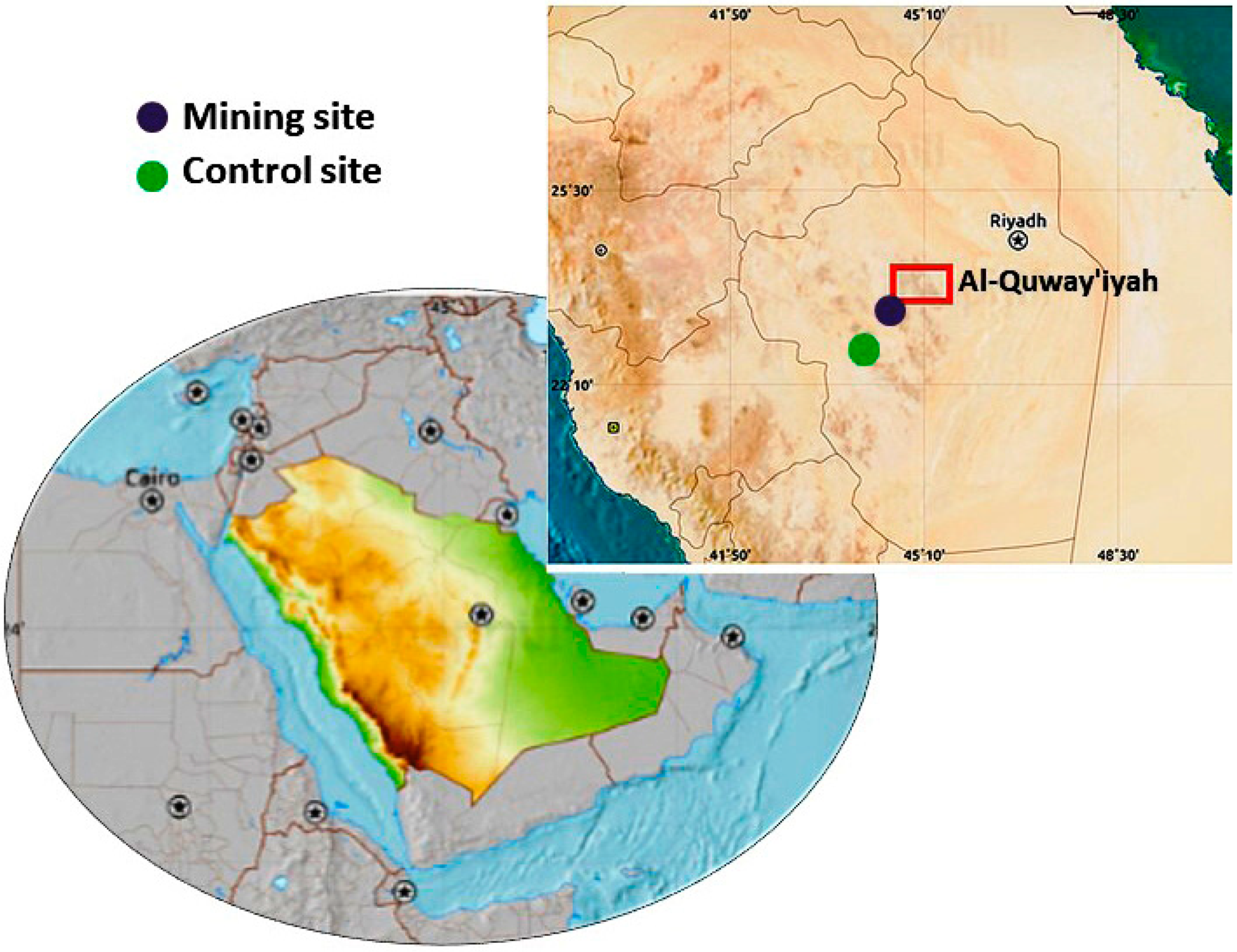
2.1. Study Site and Collection of Samples

Samples were collected from the area of mining activities in Al-Quway’iyah, a city located 165 km west of Riyadh Province (Saudi Arabia). This city is one of the large governorates in Saudi Arabia, and hence selected for the study.
The investigated gold mine site is located southwest of Al-Quway’iyah city. The samples were collected from two sites, as follows (Figure 1):
  • Site 1 (Mining): Located 500 m away from the gold mine, between E45° 05′ and N23° 47′.
  • Site 2 (Control): Located 20,000 m away from the gold mine, between E45° 06′ and N23° 36′.
Samples of the soil at 20 cm depth (n = 8) and Lycium shawii (Arabian boxthorn) (n = 8) were collected from both sites for the determination of HMs. Eight G. nanus were collected from each site with the help of specific rodent traps and transferred to the laboratory. The animals were sacrificed under anesthesia, and blood and tissue samples were collected for analysis. The protocol and procedures were approved by the institutional animal ethics committee of King Saud University (No. KSU-20165).

2.2. Determination of HMs

The concentrations of HMs were determined in the soil, L. shawii, and G. nanus kidneys, liver, and lungs using ELAN 9000 ICP/MS (Perkin Elmer Sciex Instruments, Concord, ON, Canada).

2.3. Assay of Liver and Kidney Function Markers

Serum alanine transaminase (ALT), aspartate transaminase (AST), alkaline phosphatase (ALP), creatinine, urea, and uric acid were measured using Biomerieux (Craponne, France) reagent kits, following the manufacturer’s instructions.

2.4. Assay of Oxidative Stress Markers and Antioxidants

Samples from the liver and kidney were homogenized in phosphate buffered saline (10% w/v) and centrifuged, and the supernatant was used for the assays. Malondialdehyde (MDA) [23], nitric oxide (NO) [24], reduced glutathione (GSH) [25], superoxide dismutase (SOD) [26], and catalase (CAT) [27] were assayed in the liver and kidney homogenate samples. Protein content was determined using Bradford assay.

2.5. Histological Study

Samples from the kidneys, liver, and lungs of G. nanus were fixed with 10% neutral buffered formalin and processed for paraffin embedding. 5 μm sections were cut, stained with hematoxylin and eosin (H&E), and examined using a light microscope.

2.6. Statistical Analysis

Data were analyzed using GraphPad Prism 7 (La Jolla, CA, USA) and expressed as means ± standard error of means (SEM). All statistical comparisons were performed using t-test, and a p value <0.05 was considered significant.

3. Results

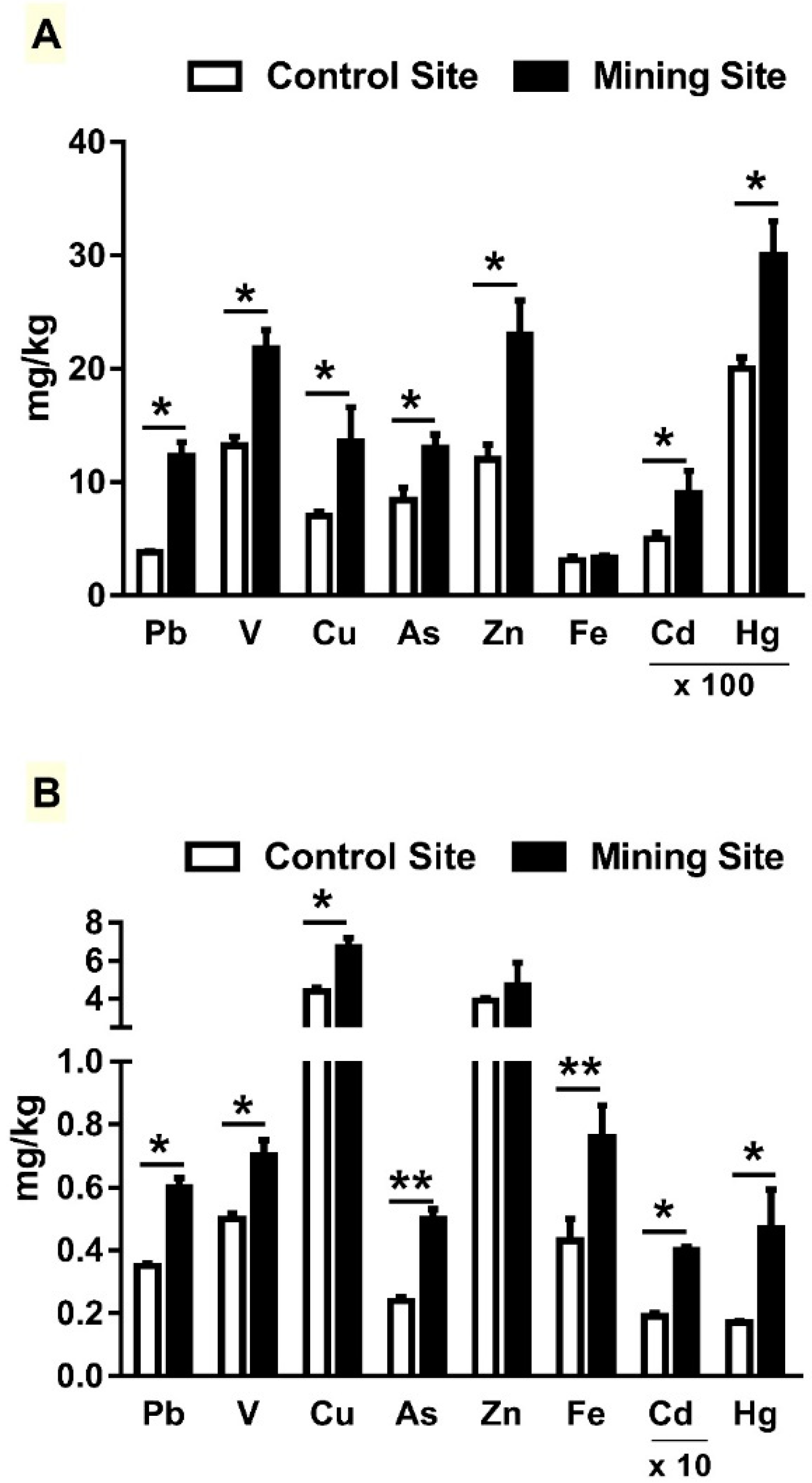
3.1. HM Concentration in Soil and L. shawii Samples

The concentration of HMs has been determined in soil samples collected from the control and mining sites. Pb, V, Cu, As, Zn, Cd, and Hg concentrations were significantly higher (p < 0.05) at the mining site, when compared with the control site (Figure 2A). The results revealed non-significant difference in Fe concentration between both sites. L. shawii samples at the mining site showed higher concentrations of Pb, V, Cu, As, Fe, Cd, and Hg, whereas Zn concentration was not significantly different when compared with the control site (Figure 2B).

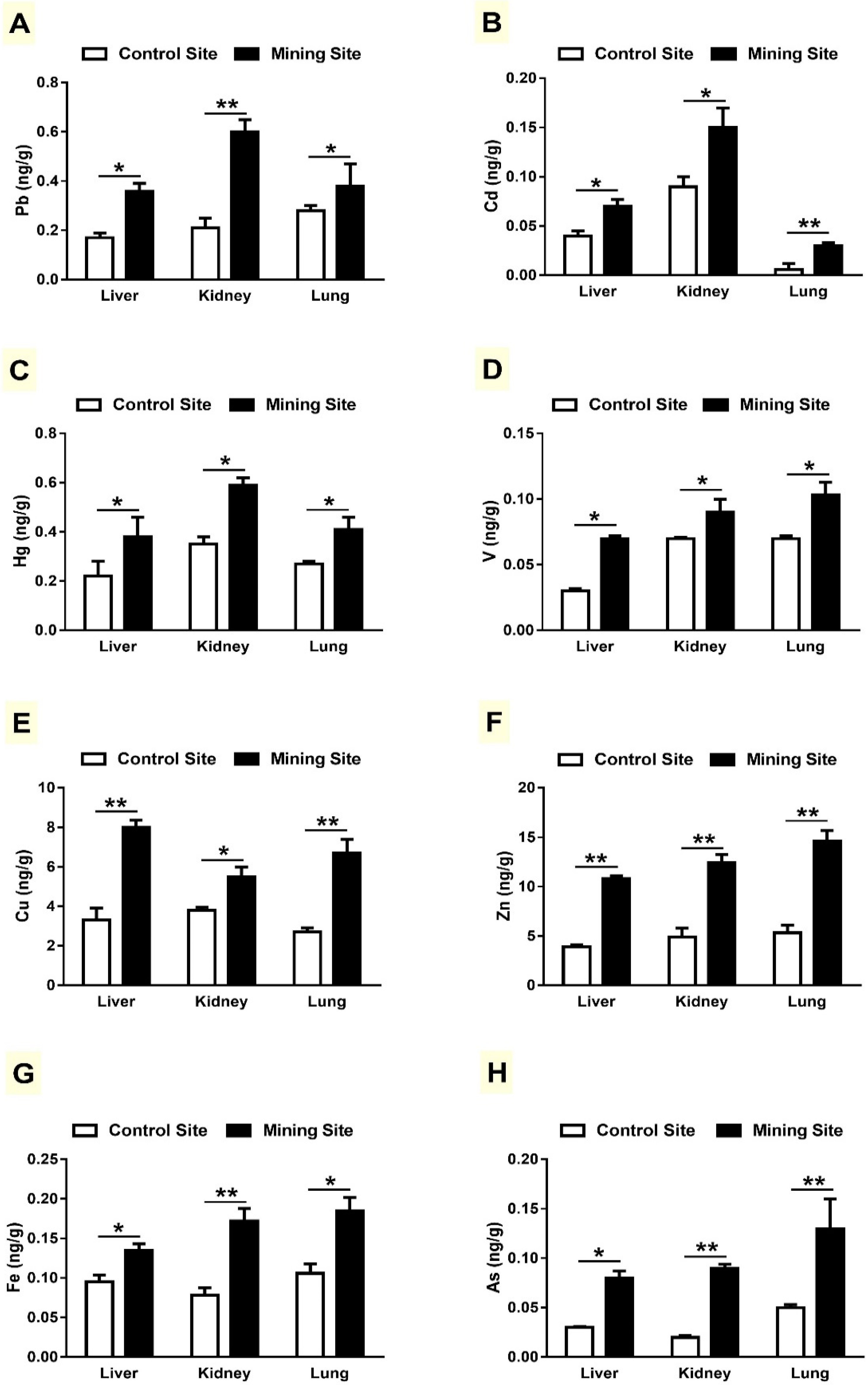
3.2. Concentration of HMs in Liver, Kidneys and Lungs of G. nanus

Pb, Cd, and Hg levels were significantly higher in the liver, kidneys, and lungs of G. nanus collected from the mining site, when compared with the control site animals, as depicted in Figure 3A–C. Similarly, concentrations of V (Figure 3D), Cu (Figure 3E), and Zn (Figure 3F) were significantly higher in the target tissues of G. nanus from the mining site. Concentrations of Fe (Figure 3G) and As (Figure 3H) were also remarkably higher in the liver, kidneys, and lungs of G. nanus from the mining site.

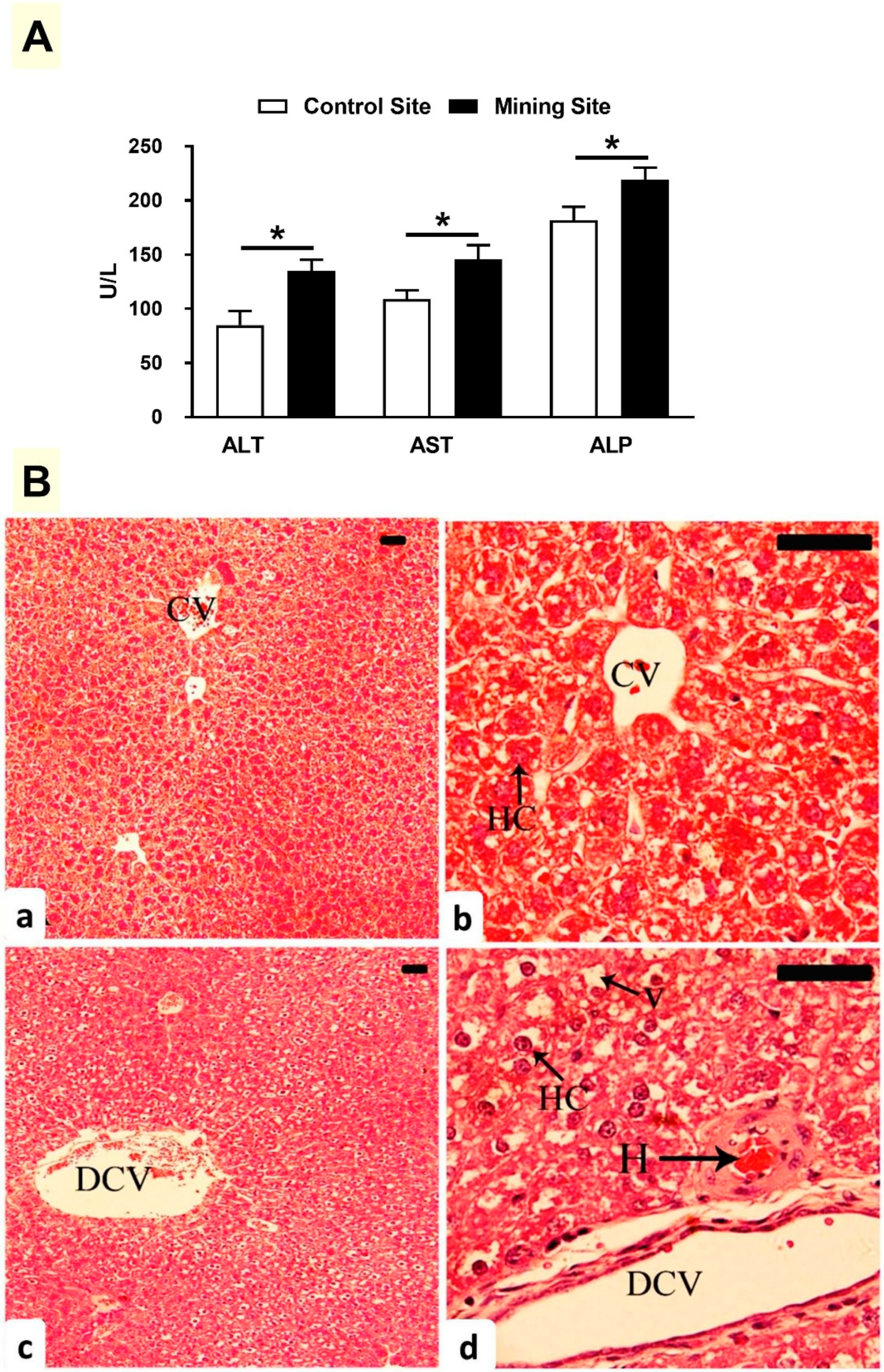
3.3. Effect of Mining on Liver Function and Histology in G. nanus

The liver function markers ALT, AST, and ALP were elevated in the serum of G. nanus from the mining site (p < 0.05) when compared with the corresponding control site animals (Figure 4A). Examination of the liver section showed normal structure of the hepatic lobules and hepatocytes in G. nauns from the control site (Figure 4B(a,b)). By contrast, G. nanus from the mining site showed hepatocyte vacuolations, hemorrhage, and dilated central veins (Figure 4B(c,d)).

3.4. Effect of Mining on Kidney Function and Histology in G. nanus

Serum creatinine, urea, and uric acid were elevated significantly (p < 0.05; p < 0.01; p < 0.05) in G. nanus from the mining site (Figure 5A). While G. nanus from the control site showed normal kidney (Figure 5B(a,b)), animals from the mining site exhibited glomerular degeneration (Figure 5B(c,d)).

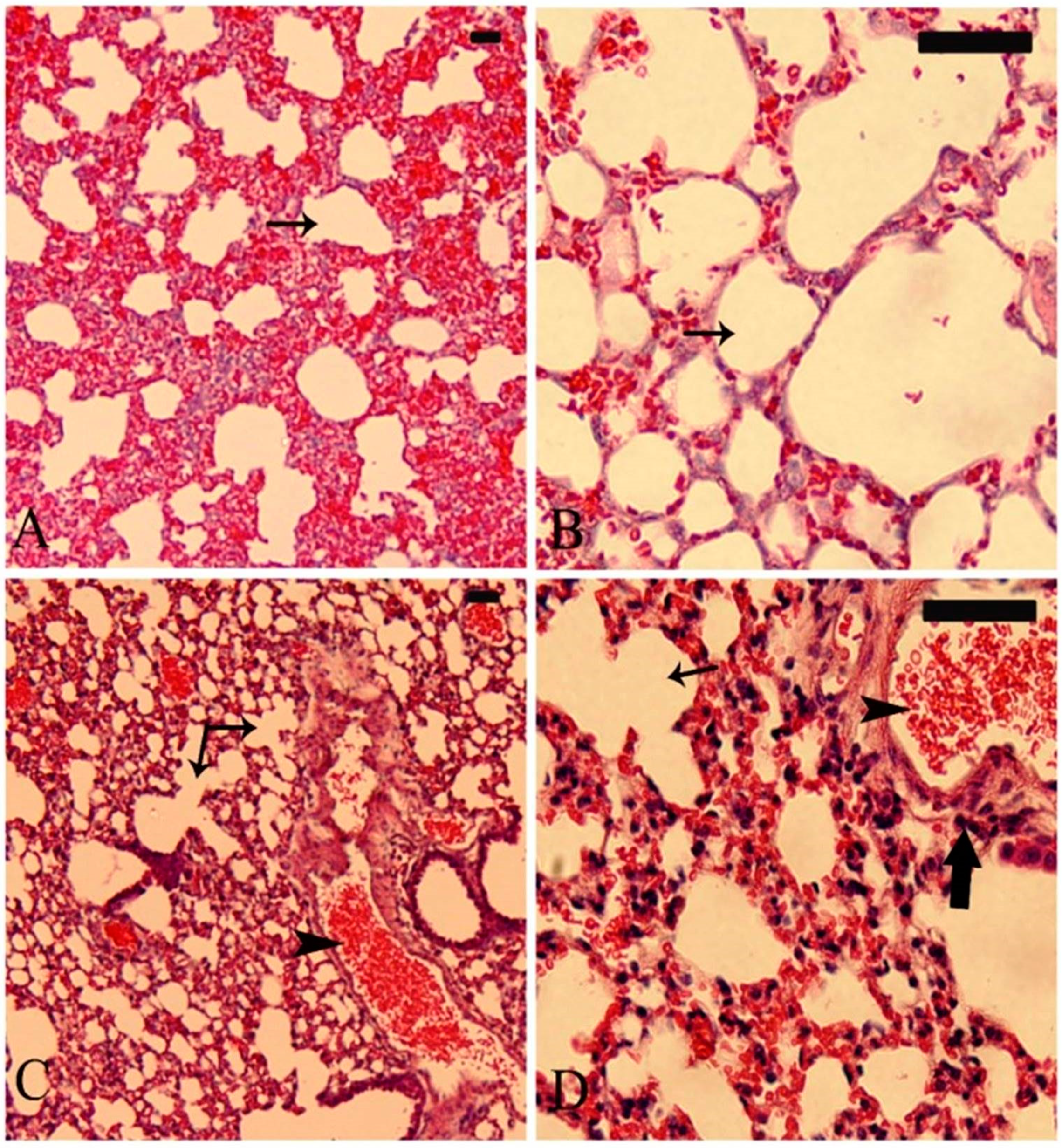
3.5. Effect of Mining on the Lung of G. nanus

As represented in Figure 6A,B, the lung of G. nanus from the control site showed normal structure of the bronchioles and alveoli. By contrast, the lungs of animals from the mining site showed dilated alveoli, inflammatory cell infiltration, and congested blood vessels (Figure 7C,D).

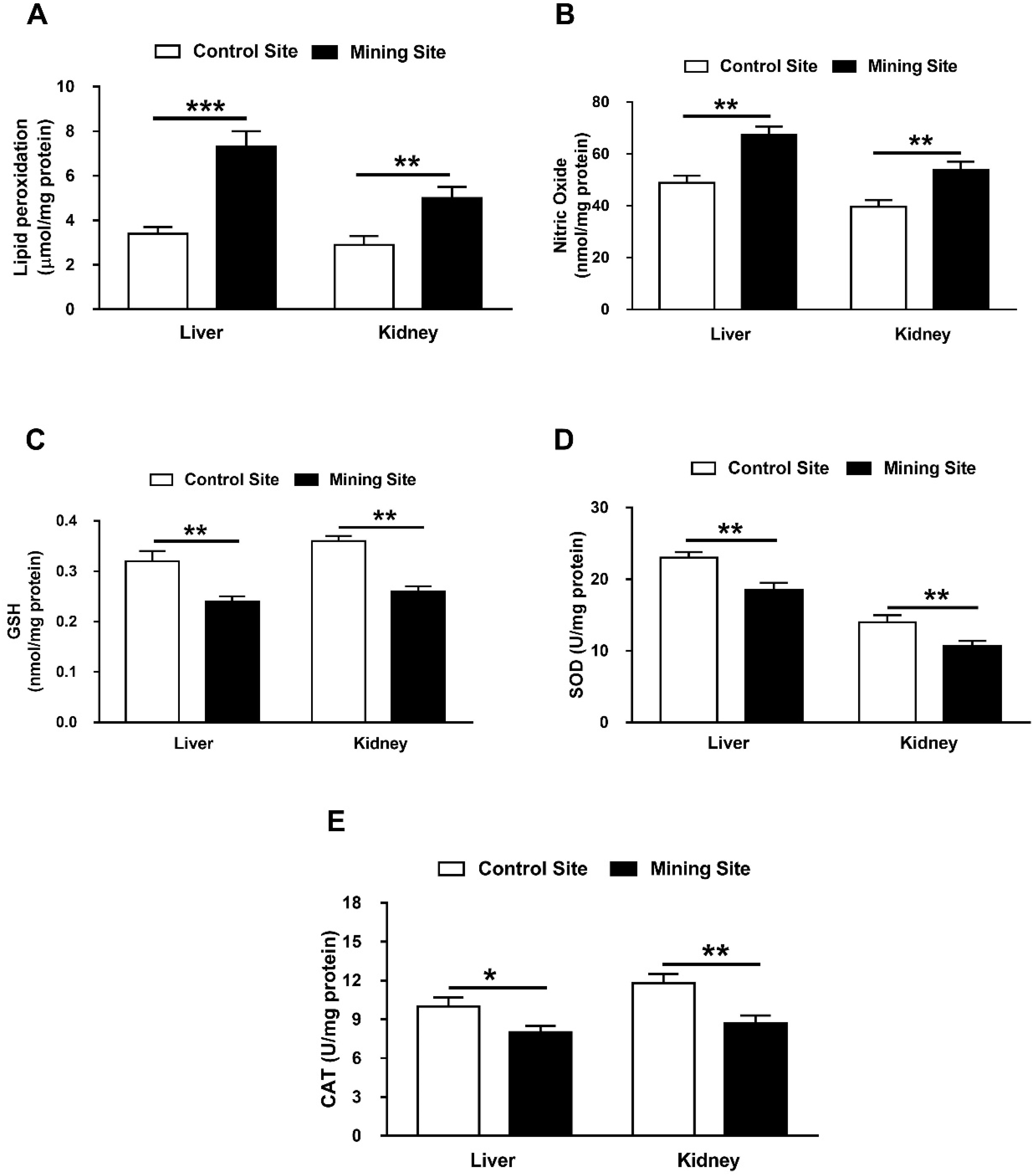
3.6. Effect of Mining on Hepatic and Renal Redox Balance in G. nanus

To evaluate the impact of mining on the redox balance in G. nanus, we determined lipid peroxidation (LPO), NO, and cellular antioxidants in the liver and kidney. G. nanus from the mining site showed a remarkable increase of hepatic (p < 0.001) and renal (p < 0.01) LPO (Figure 7A). Similarly, hepatic, and renal NO was increased significantly (p < 0.01) in G. nanus from the mining site (Figure 7B). When compared with the control site, animals from the mining site showed a decrease in hepatic and renal GSH content (p < 0.01; p < 0.01; Figure 7C), SOD (p < 0.01; p < 0.01; Figure 7D), and CAT (p < 0.05; p < 0.01; Figure 7E).

4. Discussion

This study evaluated the impact of mining activities in Al-Quway’iyah, Riyadh (Saudi Arabia) on G. nanus, with an emphasis on HM accumulation and redox imbalance. To evaluate the emission and distribution of HMs at the mining site, we determined the concentrations of Pb, V, Cu, As, Zn, Cd, Fe, and Hg in soil, L. shawii samples, and different organs of G. nanus. Pb, V, Cu, As, Cd, and Hg were increased in both the collected samples of soil and L. shawii. Fe and Zn concentrations were not significantly increased in soil and L. shawii, respectively. These findings show the effect of mining on the soil profile, as well as on plants growing in the areas nearby.
Despite their negative impact on the ecosystem and adverse health effects, exposure to HMs continues to increase in many regions [28]. Here, Pb, V, Cu, As, Zn, Cd, Fe, and Hg concentrations were increased in the liver, kidneys, and lungs of G. nanus. Besides their environmental impact, these HMs have proven to be a major threat to the body and its proper functioning [28]. Accordingly, liver and kidney dysfunction, along with histopathological manifestations in the livers, kidneys, and lungs of G. nanus were observed. These hazardous effects are directly connected to the accumulation of HMs in different tissues of G. nanus.
Pb, Cd, As, and Hg were demonstrated to be common HM pollutants produced through various human activities, including mining, and are all hazardous to human health. Pb is a highly toxic HM whose widespread use has resulted in health problems as well as environmental contamination. It has been estimated that 540,000 deaths per year, particularly in developing countries, occur due to Pb contamination [29] and that 26 million people are at risk [30]. The most common depositories of Pb within the body are the liver and kidneys [31], and this very likely explains the observed hepato- and nephrotoxicity in G. nanus from the mining site. The accumulation of Pb in the liver and kidney elevated transaminases, ALP, and renal function markers, along with the histopathological changes indicated by the injury of liver and kidney tissues. In this context, in vivo studies have demonstrated both hepato- and nephrotoxicity in rodents exposed to low concentrations of Pb [32,33,34]. The toxicity of Pb has been attributed mainly to its ionic properties and induction of redox imbalance [35]. It can disturb cellular signaling and metabolism, ionic transportation, and enzyme activities by replacing mono- and bivalent cations [36]. In addition to its ionic activity, Pb triggers the generation of reactive oxygen species (ROS), which promotes oxidative damage to lipids, DNA, and proteins [37]. Accordingly, elevated LPO and NO was accompanied by a depletion of cellular antioxidants within the liver and kidneys of G. nanus, demonstrating an oxidative stress status. Furthermore, Pb concentration was increased in the lungs of G. nanus, which showed dilation of alveoli, infiltration of inflammatory cells, and congestions. The relationship between lung injury and Pb accumulation was supported by the findings of Li et al., who demonstrated increased Pb concentration in the lungs of rats following exposure to atmospheric fine particulate matter [38].
Cd is a highly toxic, nonessential HM and a serious environmental pollutant. When released into the environment, Cd can remain for several decades in the soil and sediment because of its lack of degradability [39,40]. It has a high rate of soil-to-plant transfer and therefore accumulates in plants, predominantly in vegetables and fruits, and ultimately reaches the human body [41]. Our study shows the accumulation of Cd in the soil and L. shawii samples from the mining site. In addition, Cd concentrations were increased in different tissues of G. nanus, and its accumulation was associated with oxidative stress, hepato-/nephrotoxicity, and lung injury. Although the exact mechanisms of Cd toxicity have not been fully elucidated, the role of oxidative stress has been well-acknowledged. Cd produces hydrogen peroxide, which acts as a source of free radicals generated via Fenton reaction [42]. Its toxicity has been reported to cause damage to the liver and kidneys [43,44]. Upon exposure, Cd binds to albumin and is taken up and deposits primarily in the liver [45]. Within the liver, Cd promotes ROS generation, LPO, membrane damage, GSH depletion, and cell death [46]. Cd binds to metallothionine, and the produced complexes induce liver injury and circulate to the kidney, where they accumulate and cause renal tissue injury [47]. The activity of SOD and CAT was decreased in the liver and kidney of G. nanus from the mining site, and was a result of the binding of Cd with the thiol groups of these enzymes [48]. In addition, Cd can inhibit complex III of the mitochondrial electron transport chain [48]. The observed nephrotoxicity in this study was associated with glomerular degeneration, which has been shown in rats exposed to Cd [49]. Furthermore, the lungs represent one of the main routes through which Cd enters the body [40]. In this study, G. nanus showed lung injury associated with elevated Cd concentrations. The potential of Cd to provoke pulmonary toxicity has been supported by studies reporting bronchial and pulmonary irritation, transient bronchial inflammation [50], diminished pulmonary function [51], and primary lung cancers [52] following exposure to Cd.
Mining is one of the major sources of As and Hg pollution. Accordingly, As and Hg were found in high concentrations in the soil, L. shawii, and different tissues of G. nanus collected from the mining site. As is extensively available in different forms and possesses toxic and carcinogenic effects [53]. The toxicity of As forms inorganic arsenic species (iAs), monomethylarsonic acid, and dimethylarsinic acid, which produces methylated arsenicals via enzymatic conversion. Monomethylarsonic acid, an intermediate product produced during the transformation of iAs, is highly toxic and carcinogenic [53]. The toxicity of As has been associated with its ability to increase ROS levels, activate apoptosis signaling, alter metabolism and energy generation, and replace metal ions in cellular biomolecules [54]. Therefore, the accumulation of As in G. nanus has a role in the observed oxidative stress and hepato-/nephrotoxicity. Recent reports have shown As-induced hepatic, renal, and pulmonary manifestations in experimental animals [55,56,57].
Hg is a well-known HM pollutant, and its toxicity is commonly responsible for acute HM poisoning. Although neurotoxicity is the most common effect of Hg and methylmercury, which induces LPO and cell death [58], it can also cause nephrotoxicity [59] and hepatotoxicity [60]. Hence, Hg accumulation in G. nanus contributes, at least in part, to liver and kidney injury and redox imbalance.
V, Cu, Zn, and Fe were the other HMs shown to accumulate in the soil, L. shawii, and different tissues of G. nanus from the mining site. V is an environmental pollutant that possesses hazardous effects, and the inhalation of vanadium pentoxide dust has caused occupational toxicity [61]. Liver injury and other toxic effects have been demonstrated in rats exposed to V [62], and inhalation of V caused bronchopneumonia and chronic productive cough [63,64]. Cu exists in oxidized Cu(II) and reduced Cu(I) states, and is therefore considered potentially toxic. Generation of ROS during the conversion between the oxidized and reduced states represents the main reason of Cu toxicity [65]. Zn is an essential micronutrient needed for vital cell functions in microorganisms, plants, and humans; however, concentrations beyond the physiological value are toxic. At high concentrations, Zn interacts with sulfhydryl groups or replaces other essential metals in different cellular proteins [66]. Iron is a vital nutrient that acts as a cofactor for multiple enzymes within living cells. It mediates reactions that can catalyze the formation of free radicals, leading to cell damage. Hence, iron overload can induce toxicity particularly in children [67], as well as hepatotoxicity [68] and nephrotoxicity [69]. Therefore, excess V, Cu, Zn, and Fe are associated with ROS generation and contribute to liver, kidney, and lung injury in G. nanus that live close to the mining site.

5. Conclusions

These findings highlight the hazardous effects of mining activities on soil, plants, and animals. HM concentrations were higher in the soil and L. shawii from the mining site, which demonstrates the negative environmental impact of mining. G. nanus living at the mining site exhibit hepato- and nephrotoxicity, and lung manifestations. The deleterious effects of mining activities on G. nanus were associated with oxidative stress and the depletion of antioxidants. These results demonstrate the environmental and health impacts of mining and can provide a scientific basis for evaluating the effects of mining on nearby communities.

Author Contributions

Conceptualization, A.M.M., J.A., and A.A.A.; methodology, A.M.A., A.M.M., J.A., A.A.A., and S.N.M.; validation, J.A., A.A.A., and A.M.M.; formal analysis, A.M.M.; investigation, A.M.A., A.M.M., J.A., A.A.A., S.N.M., F.S.A.-O., and N.A.; resources, F.S.A.-O. and N.A.; data curation, A.M.A. and A.M.M.; writing—original draft preparation, A.M.M.; writing—review and editing, A.M.M.; visualization, J.A., A.A.A., A.M.A., and A.M.M.; supervision, A.M.M., J.A., and A.A.A.; project administration, J.A., A.M.M., and A.A.A.; funding acquisition, J.A.

Funding

This research was funded by the Dean of Scientific Research, King Saud University, grant number RGP-VPP-240.

Acknowledgments

We extend our appreciation to the Dean of Scientific Research, King Saud University, for funding the work through the research group project number RGP-VPP-240.

Conflicts of Interest

The authors declare no conflict of interest.

References

  1. Krook, J.; Svensson, N.; Eklund, M. Landfill mining: A critical review of two decades of research. Waste Manag. 2012, 32, 513–520. [Google Scholar] [CrossRef] [PubMed]
  2. Loayza, N.; Rigolini, J. The local impact of mining on poverty and inequality: Evidence from the commodity boom in Peru. World Dev. 2016, 84, 219–234. [Google Scholar] [CrossRef]
  3. Emmanuel, A.Y.; Jerry, C.S.; Dzigbodi, D.A. Review of environmental and health impacts of mining in Ghana. J. Health Pollut. 2018, 8, 43–52. [Google Scholar] [CrossRef] [PubMed]
  4. Dudka, S.; Adriano, D.C. Environmental impacts of metal ore mining and processing: A review. J. Environ. Qual. 1997, 26, 590–602. [Google Scholar] [CrossRef]
  5. Warhate, S.R.; Yenkie, M.K.; Chaudhari, M.D.; Pokale, W.K. Impacts of mining activities on water and soil. J. Environ. Sci. Eng. 2006, 48, 81–90. [Google Scholar]
  6. Nowrouzi-Kia, B.; Gohar, B.; Casole, J.; Chidu, C.; Dumond, J.; McDougall, A.; Nowrouzi-Kia, B. A systematic review of lost-time injuries in the global mining industry. Work (Read. Mass.) 2018, 60, 49–61. [Google Scholar] [CrossRef] [Green Version]
  7. Fashola, M.O.; Ngole-Jeme, V.M.; Babalola, O.O. Heavy metal pollution from gold mines: Environmental effects and bacterial strategies for resistance. Int. J. Environ. Res. Public Health 2016, 13, 1047. [Google Scholar] [CrossRef]
  8. Singh, R.; Gautam, N.; Mishra, A.; Gupta, R. Heavy metals and living systems: An overview. Indian J. Pharmacol. 2011, 43, 246–253. [Google Scholar] [CrossRef]
  9. Jarup, L. Hazards of heavy metal contamination. Br. Med Bull. 2003, 68, 167–182. [Google Scholar] [CrossRef] [Green Version]
  10. Rzymski, P.; Niedzielski, P.; Klimaszyk, P.; Poniedzialek, B. Bioaccumulation of selected metals in bivalves (Unionidae) and Phragmites australis inhabiting a municipal water reservoir. Environ. Monit. Assess. 2014, 186, 3199–3212. [Google Scholar] [CrossRef]
  11. de Kok, T.M.; Hogervorst, J.G.; Briede, J.J.; van Herwijnen, M.H.; Maas, L.M.; Moonen, E.J.; Driece, H.A.; Kleinjans, J.C. Genotoxicity and physicochemical characteristics of traffic-related ambient particulate matter. Environ. Mol. Mutagenesis 2005, 46, 71–80. [Google Scholar] [CrossRef] [PubMed]
  12. Markiewicz-Górka, I.; Januszewska, L.; Michalak, A.; Prokopowicz, A.; Januszewska, E.; Pawlas, N.; Pawlas, K. Effects of chronic exposure to lead, cadmium, and manganese mixtures on oxidative stress in rat liver and heart. Arh. Za Hig. Rada I Toksikol. 2015, 66, 51–62. [Google Scholar] [CrossRef] [PubMed]
  13. Rzymski, P.; Tomczyk, K.; Rzymski, P.; Poniedzialek, B.; Opala, T.; Wilczak, M. Impact of heavy metals on the female reproductive system. Ann. Agric. Environ. Med. AAEM 2015, 22, 259–264. [Google Scholar] [CrossRef] [PubMed]
  14. Lin, Y.F.; Aarts, M.G. The molecular mechanism of zinc and cadmium stress response in plants. Cell. Mol. Life Sci. CMLS 2012, 69, 3187–3206. [Google Scholar] [CrossRef] [PubMed]
  15. Nordberg, G.F.; Nogawa, K.; Nordberg, M.; Friberg, L.T. Chapter 23—Cadmium. In Handbook on the Toxicology of Metals, 3rd ed.; Academic Press: Burlington, NJ, USA, 2007; pp. 445–486. [Google Scholar]
  16. Brzoska, M.M.; Moniuszko-Jakoniuk, J. Low-level exposure to cadmium during the lifetime increases the risk of osteoporosis and fractures of the lumbar spine in the elderly: Studies on a rat model of human environmental exposure. Toxicol. Sci. Off. J. Soc. Toxicol. 2004, 82, 468–477. [Google Scholar] [CrossRef] [PubMed]
  17. Alissa, E.M.; Ferns, G.A. Heavy metal poisoning and cardiovascular disease. J. Toxicol. 2011, 2011, 870125. [Google Scholar] [CrossRef]
  18. Choudhury, B.; Chowdhury, S.; Biswas, A.K. Regulation of growth and metabolism in rice (oryza sativa L.) by arsenic and its possible reversal by phosphate. J. Plant Interact. 2011, 6, 15–24. [Google Scholar] [CrossRef]
  19. Rahman, Z.; Singh, V.P. The relative impact of toxic heavy metals (THMs) (arsenic (As), cadmium (Cd), chromium (Cr)(VI), mercury (Hg), and lead (Pb)) on the total environment: An overview. Environ. Monit. Assess. 2019, 191, 419. [Google Scholar] [CrossRef]
  20. Valko, M.; Rhodes, C.J.; Moncol, J.; Izakovic, M.; Mazur, M. Free radicals, metals and antioxidants in oxidative stress-induced cancer. Chem. Biol. Interact. 2006, 160, 1–40. [Google Scholar] [CrossRef]
  21. Harrison, D.L.; Bates, P.J.J. The Mammals of Saudi Arabia; Benn: London, UK, 1991; pp. 354–367. [Google Scholar]
  22. Azizi, K.; Moemenbellah-Fard, M.D.; Fakoorziba, M.R.; Fekri, S. Gerbillus nanus (rodentia: Muridae): A new reservoir host of leishmania major. Ann. Trop. Med. Parasitol. 2011, 105, 431–437. [Google Scholar] [CrossRef]
  23. Preuss, H.G.; Jarrell, S.T.; Scheckenbach, R.; Lieberman, S.; Anderson, R.A. Comparative effects of chromium, vanadium and gymnema sylvestre on sugar-induced blood pressure elevations in shr. J. Am. Coll. Nutr. 1998, 17, 116–123. [Google Scholar] [CrossRef] [PubMed]
  24. Grisham, M.B.; Johnson, G.G.; Lancaster, J.R., Jr. Quantitation of nitrate and nitrite in extracellular fluids. Methods Enzym. 1996, 268, 237–246. [Google Scholar]
  25. Beutler, E.; Duron, O.; Kelly, B.M. Improved method for the determination of blood glutathione. J. Lab. Clin. Med. 1963, 61, 882–888. [Google Scholar] [PubMed]
  26. Marklund, S.; Marklund, G. Involvement of the superoxide anion radical in the autoxidation of pyrogallol and a convenient assay for superoxide dismutase. FEBS Eur. J. Biochem. 1974, 47, 469–474. [Google Scholar] [CrossRef] [PubMed]
  27. Cohen, G.; Dembiec, D.; Marcus, J. Measurement of catalase activity in tissue extracts. Anal. Biochem. Anal. Biochem. 1970, 34, 30–38. [Google Scholar] [CrossRef]
  28. Jaishankar, M.; Tseten, T.; Anbalagan, N.; Mathew, B.B.; Beeregowda, K.N. Toxicity, mechanism and health effects of some heavy metals. Interdiscip. Toxicol. 2014, 7, 60–72. [Google Scholar] [CrossRef] [Green Version]
  29. World Health Organization. Lead Poisoning and Health. 2018. Available online: https://www.who.int/news-room/fact-sheets/detail/lead-poisoning-and-health (accessed on 1 May 2019).
  30. The New Top Six Toxic Threats: A Priority List for Remediation, World’s Worst Pollution Problems Report; Pure Earth: New York, NY, USA, 2015.
  31. Mudipalli, A. Lead hepatotoxicity & potential health effects. Indian J. Med Res. 2007, 126, 518–527. [Google Scholar]
  32. El-Nekeety, A.A.; El-Kady, A.A.; Soliman, M.S.; Hassan, N.S.; Abdel-Wahhab, M.A. Protective effect of Aquilegia vulgaris (L.) against lead acetate-induced oxidative stress in rats. Food Chem. Toxicol. Int. J. Publ. Br. Ind. Biol. Res. Assoc. 2009, 47, 2209–2215. [Google Scholar] [CrossRef]
  33. Jia, Q.; Ha, X.; Yang, Z.; Hui, L.; Yang, X. Oxidative stress: A possible mechanism for lead-induced apoptosis and nephrotoxicity. Toxicol. Mech. Methods 2012, 22, 705–710. [Google Scholar] [CrossRef]
  34. El-Tantawy, W.H. Antioxidant effects of spirulina supplement against lead acetate-induced hepatic injury in rats. J. Tradit. Complementary Med. 2016, 6, 327–331. [Google Scholar] [CrossRef]
  35. Lopes, A.C.; Peixe, T.S.; Mesas, A.E.; Paoliello, M.M. Lead Exposure and Oxidative Stress: A Systematic Review. Rev. Environ. Contam. Toxicol. 2016, 236, 193–238. [Google Scholar] [PubMed]
  36. Flora, S.J.S.; Flora, G.; Saxena, G. Environmental occurrence, health effects and management of lead poisoning. In Lead Chemistry, Analytical Aspects, Environmental Impacts and Health Effects; Cascas, S.B., Sordo, J., Eds.; Elsevier Publication: Amsterdam, The Netherlands, 2006; pp. 158–228. [Google Scholar]
  37. Adegbesan, B.O.; Adenuga, G.A. Effect of lead exposure on liver lipid peroxidative and antioxidant defense systems of protein-undernourished rats. Biol. Trace Elem. Res. 2007, 116, 219–225. [Google Scholar] [CrossRef] [PubMed]
  38. Li, Q.; Liu, H.; Alattar, M.; Jiang, S.; Han, J.; Ma, Y.; Jiang, C. The preferential accumulation of heavy metals in different tissues following frequent respiratory exposure to pm2.5 in rats. Sci. Rep. 2015, 5, 16936. [Google Scholar] [CrossRef] [PubMed]
  39. Jarup, L.; Akesson, A. Current status of cadmium as an environmental health problem. Toxicol. Appl. Pharmacol. 2009, 238, 201–208. [Google Scholar] [CrossRef] [PubMed]
  40. Thevenod, F.; Lee, W.K. Toxicology of cadmium and its damage to mammalian organs. Met. Ions Life Sci. 2013, 11, 415–490. [Google Scholar]
  41. Satarug, S.; Garrett, S.H.; Sens, M.A.; Sens, D.A. Cadmium, environmental exposure, and health outcomes. Cienc. Saude Coletiva 2011, 16, 2587–2602. [Google Scholar] [CrossRef]
  42. Casalino, E.; Sblano, C.; Landriscina, C. Enzyme activity alteration by cadmium administration to rats: The possibility of iron involvement in lipid peroxidation. Arch. Biochem. Biophys. 1997, 346, 171–179. [Google Scholar] [CrossRef]
  43. Jeyaprakash, K.; Chinnaswamy, P. Effect of spirulina and liv-52 on cadmium induced toxicity in albino rats. Indian J. Exp. Biol. 2005, 43, 773–781. [Google Scholar]
  44. Vicente-Sanchez, C.; Egido, J.; Sanchez-Gonzalez, P.D.; Perez-Barriocanal, F.; Lopez-Novoa, J.M.; Morales, A.I. Effect of the flavonoid quercetin on cadmium-induced hepatotoxicity. Food Chem. Toxicol. Int. J. Publ. Br. Ind. Biol. Res. Assoc. 2008, 46, 2279–2287. [Google Scholar] [CrossRef]
  45. Shimada, H.; Yasutake, A.; Hirashima, T.; Takamure, Y.; Kitano, T.; Waalkes, M.P.; Imamura, Y. Strain difference of cadmium accumulation by liver slices of inbred wistar-imamichi and fischer 344 rats. Toxicol. Vitro Int. J. Publ. Assoc. BIBRA 2008, 22, 338–343. [Google Scholar] [CrossRef]
  46. Wang, J.; Hao, M.; Liu, C.; Liu, R. Cadmium induced apoptosis in mouse primary hepatocytes: The role of oxidative stress-mediated erk pathway activation and the involvement of histone h3 phosphorylation. RSC Adv. 2015, 5, 31798–31806. [Google Scholar] [CrossRef]
  47. Castagnetto, J.M.; Hennessy, S.W.; Roberts, V.A.; Getzoff, E.D.; Tainer, J.A.; Pique, M.E. Mdb: The metalloprotein database and browser at the scripps research institute. Nucleic Acids Res. 2002, 30, 379–382. [Google Scholar] [CrossRef] [PubMed]
  48. Wang, Y.; Fang, J.; Leonard, S.S.; Rao, K.M. Cadmium inhibits the electron transfer chain and induces reactive oxygen species. Free Radic. Biol. Med. 2004, 36, 1434–1443. [Google Scholar] [CrossRef] [PubMed]
  49. Akomolafe, R.O.; Imafidon, C.E.; Olukiran, O.S.; Oladele, A.A.; Ajayi, A.O. Livolin forte ameliorates cadmium-induced kidney injury in wistar rats. Serb. J. Exp. Clin. Res. 2016, 17, 107–116. [Google Scholar] [CrossRef]
  50. Bolognin, M.; Kirschvink, N.; Leemans, J.; De Buscher, V.; Snaps, F.; Gustin, P.; Peeters, D.; Clercx, C. Characterisation of the acute and reversible airway inflammation induced by cadmium chloride inhalation in healthy dogs and evaluation of the effects of salbutamol and prednisolone. Vet. J. 2009, 179, 443–450. [Google Scholar] [CrossRef] [PubMed]
  51. Oberdorster, G. Airborne cadmium and carcinogenesis of the respiratory tract. Scand. J. Work Environ. Health 1986, 12, 523–537. [Google Scholar] [CrossRef]
  52. Takenaka, S.; Oldiges, H.; Konig, H.; Hochrainer, D.; Oberdorster, G. Carcinogenicity of cadmium chloride aerosols in w rats. J. Natl. Cancer Inst. 1983, 70, 367–373. [Google Scholar]
  53. Singh, N.; Kumar, D.; Sahu, A.P. Arsenic in the environment: Effects on human health and possible prevention. J. Environ. Biol. 2007, 28, 359–365. [Google Scholar]
  54. Hughes, M.F.; Beck, B.D.; Chen, Y.; Lewis, A.S.; Thomas, D.J. Arsenic exposure and toxicology: A historical perspective. Toxicol. Sci. 2011, 123, 305–332. [Google Scholar] [CrossRef]
  55. Thangapandiyan, S.; Ramesh, M.; Miltonprabu, S.; Hema, T.; Jothi, G.B.; Nandhini, V. Sulforaphane potentially attenuates arsenic-induced nephrotoxicity via the pi3k/akt/nrf2 pathway in albino wistar rats. Environ. Sci. Pollut. Res. Int. 2019, 26, 12247–12263. [Google Scholar] [CrossRef]
  56. Ling, S.; Shan, Q.; Liu, P.; Feng, T.; Zhang, X.; Xiang, P.; Chen, K.; Xie, H.; Song, P.; Zhou, L.; et al. Metformin ameliorates arsenic trioxide hepatotoxicity via inhibiting mitochondrial complex i. Cell Death Dis. 2017, 8, e3159. [Google Scholar] [CrossRef] [PubMed]
  57. Mariappan, N.; Zafar, I.; Husain, M.; Vaid, M.; Surolia, R.; Kashyap, M.P.; Srivastava, R.; Ahmad, S.; Agarwal, A.; Athar, M.; et al. Pulmonary manifestations of inhaled arsenic trioxide following an acute accidental exposure. Am. J. Respir. Crit. Care Med. 2018, 197, A2974. [Google Scholar]
  58. Patrick, L. Mercury toxicity and antioxidants: Part 1: Role of glutathione and alpha-lipoic acid in the treatment of mercury toxicity. Altern. Med. Rev. A J. Clin. Ther. 2002, 7, 456–471. [Google Scholar]
  59. Boroushaki, M.T.; Mollazadeh, H.; Rajabian, A.; Dolati, K.; Hoseini, A.; Paseban, M.; Farzadnia, M. Protective effect of pomegranate seed oil against mercuric chloride-induced nephrotoxicity in rat. Ren. Fail. 2014, 36, 1581–1586. [Google Scholar] [CrossRef] [PubMed]
  60. Brandao, F.; Cappello, T.; Raimundo, J.; Santos, M.A.; Maisano, M.; Mauceri, A.; Pacheco, M.; Pereira, P. Unravelling the mechanisms of mercury hepatotoxicity in wild fish (Liza aurata) through a triad approach: Bioaccumulation, metabolomic profiles and oxidative stress. Met. Integr. Biometal Sci. 2015, 7, 1352–1363. [Google Scholar] [CrossRef] [PubMed]
  61. Altamirano-Lozano, M.A.; Alvarez-Barrera, L.; Mateos-Nava, R.A.; Fortoul, T.I.; Rodriguez-Mercado, J.J. Potential for genotoxic and reprotoxic effects of vanadium compounds due to occupational and environmental exposures: An article based on a presentation at the 8th international symposium on vanadium chemistry, biological chemistry, and toxicology, Washington, DC, USA, 15–18 August 2012. J. Immunotoxicol. 2014, 11, 19–27. [Google Scholar]
  62. Castellini, C.; Mourvaki, E.; Sartini, B.; Cardinali, R.; Moretti, E.; Collodel, G.; Fortaner, S.; Sabbioni, E.; Renieri, T. In vitro toxic effects of metal compounds on kinetic traits and ultrastructure of rabbit spermatozoa. Reprod. Toxicol. 2009, 27, 46–54. [Google Scholar] [CrossRef]
  63. Roshchin, A.V.; Ordzhonikidze, E.K.; Shalganova, I.V. Vanadium--toxicity, metabolism, carrier state. J. Hyg. Epidemiol. Microbiol. Immunol. 1980, 24, 377–383. [Google Scholar]
  64. Assem, F.L.; Levy, L.S. Inhalation toxicity of vanadium. In Vanadium: Biochemical and Molecular Biological Approaches; Michibata, H., Ed.; Springer: Dordrecht, The Netherlands, 2012; pp. 209–224. [Google Scholar]
  65. Stern, B.R. Essentiality and toxicity in copper health risk assessment: Overview, update and regulatory considerations. J. Toxicol. Environ. Health. Part A 2010, 73, 114–127. [Google Scholar] [CrossRef]
  66. Kox, L.F.; Wosten, M.M.; Groisman, E.A. A small protein that mediates the activation of a two-component system by another two-component system. EMBO J. 2000, 19, 1861–1872. [Google Scholar] [CrossRef] [Green Version]
  67. Albretsen, J. The toxicity of iron, an essential element. Vet. Med. Bonn. Springs Edw. 2006, 101, 82–90. [Google Scholar]
  68. Ramm, G.A.; Ruddell, R.G. Hepatotoxicity of iron overload: Mechanisms of iron-induced hepatic fibrogenesis. Semin. Liver Dis. 2005, 25, 433–449. [Google Scholar] [CrossRef] [PubMed]
  69. Zager, R.A.; Johnson, A.C.M.; Hanson, S.Y. Parenteral iron nephrotoxicity: Potential mechanisms and consequences1. Kidney Int. 2004, 66, 144–156. [Google Scholar] [CrossRef] [PubMed] [Green Version]
Figure 1. Location map of the mining and control sites.
Animals 09 00664 g001
Figure 2. Concentration of heavy metals in (A) soil and (B) L. shawii from the mining and control sites. Data are means ± SEM, (n = 8). * p < 0.05 and ** p < 0.01.
Animals 09 00664 g002
Figure 3. Concentration of heavy metals in the liver, kidneys and lungs of G. nanus from the mining and control sites. Data are means ± SEM, (n = 8). * p < 0.05 and ** p < 0.01.
Animals 09 00664 g003
Figure 4. (A) Effect of mining activities on liver function markers of G. nanus. Data are means ± SEM, (n = 8). * p < 0.05. (B) Photomicrographs of H&E-stained sections in the liver of G. nanus from the control site (a,b) showing normal structure of hepatocytes (HC) and central vein (CV); and animals from the mining site (c,d) showing a dilated central vein (DCV), hemorrhage (H), and vacuolations (V). Scale bar = 100 µm.
Animals 09 00664 g004
Figure 5. (A) Effect of mining activities on renal function markers of G. nanus. Data are means ± SEM, (n = 8). * p < 0.05 and ** p < 0.01. (B) Photomicrographs of H&E-stained sections in the kidney of G. nanus from the control site (a,b) showing normal structure of the glomeruli (G) and renal tubules (T); and animals from the mining site (c,d) showing degenerated glomeruli (DG). Scale bar = 100 µm.
Animals 09 00664 g005
Figure 6. Photomicrographs of H&E-stained sections in lung sample of G. nanus from the control site (A,B) showing normal structure of the bronchioles and alveoli (arrow), and the lung of animals from the mining site (C,D) showing dilated alveoli (thin arrow), inflammatory cell infiltration (thick arrow), and congested blood vessels (arrow head). Scale bar = 100 µm.
Animals 09 00664 g006
Figure 7. Effect of mining activities on oxidative stress and antioxidants in liver and kidney of G. nanus. Animals from the mining site showed an increase in (A) lipid peroxidation and (B) nitric oxide, and decreased (C) GSH, (D) SOD, and (E) CAT in liver and kidney. Data are means ± SEM, (n = 8). * p < 0.05, ** p < 0.01 and *** p < 0.001.
Animals 09 00664 g007

