Bioactive Glasses and Glass/Polymer Composites for Neuroregeneration: Should We Be Hopeful?


1
Tissue Engineering Research Group (TERG), Department of Anatomy and Cell Biology, School of Medicine, Mashhad University of Medical Sciences, Mashhad 917794-8564, Iran
2
Department of Tissue Engineering & Regenerative Medicine, Faculty of Advanced Technologies in Medicine, Iran University of Medical Sciences (IUMS), Tehran 1449614535, Iran
3
Department of Anatomy and Cell Biology, School of Medicine, Mashhad University of Medical Sciences, Mashhad 917794-8564, Iran
4
Institute of Materials Physics and Engineering, Applied Science and Technology Department, Politecnico di Torino, Corso Duca degli Abruzzi 24, 10129 Torino, Italy
*
Authors to whom correspondence should be addressed.
Appl. Sci. 2020, 10(10), 3421; https://doi.org/10.3390/app10103421
Submission received: 14 April 2020
/
Revised: 2 May 2020
/
Accepted: 12 May 2020
/
Published: 15 May 2020
(This article belongs to the Special Issue Biopolymers, Natural Molecules and Nanomaterials for Tissue Regeneration)
Abstract
:Bioactive glasses (BGs) have been identified as highly versatile materials in tissue engineering applications; apart from being used for bone repair for many years, they have recently shown promise for the regeneration of peripheral nerves as well. They can be formulated in different shapes and forms (micro-/nanoparticles, micro-/nanofibers, and tubes), thus potentially meeting the diverse requirements for neuroregeneration. Mechanical and biological improvements in three-dimensional (3D) polymeric scaffolds could be easily provided by adding BGs to their composition. Various types of silicate, borate, and phosphate BGs have been examined for use in neuroregeneration. In general, BGs show good compatibility with the nervous system compartments both in vitro and in vivo. Functionalization and surface modification plus doping with therapeutic ions make BGs even more effective in peripheral nerve regeneration. Moreover, the combination of BGs with conductive polymers is suggested to improve neural cell functions at injured sites. Taking advantage of BGs combined with novel technologies in tissue engineering, like 3D printing, can open new horizons in reconstructive approaches for the nervous system. Although there are great potential opportunities in BG-based therapies for peripheral nerve regeneration, more research should still be performed to carefully assess the pros and cons of BGs in neuroregeneration strategies.
1. Introduction
The nervous system is vital in the human body and is made from two distinct components, the central nervous system (CNS) and the peripheral nervous system (PNS). Both parts are potentially subjected to various acute and chronic diseases and injuries, causing reduced quality of life [1,2,3]. In the case of peripheral nerve injuries, mechanical stress; exposure to heat, cold, and irradiation; tumors; and focal inflammation are the main reasons for partial or total loss of motor, sensory, and autonomic functions [4]. For example, cutting of the nerve as a result of traumatic injuries may result in complications like inability to move muscles, dysfunctional or no feeling of normal sensations, and eventually painful neuropathies [5]. To date, huge numbers of experimental studies have been conducted to provide appropriate substitutes for the reconstruction of lost/injured peripheral nerves. Autografts, mostly harvested from the sural nerve, are still identified as the gold standard for the regeneration of nerves as regards lack of rejection and providing a proper biological environment for axon reconstruction [6]. Another approach for restoring segmental nerve injuries involves the use of allografts, which depends on the viability of host and donor Schwann cells since they are responsible for the proper re-myelination process and serve as the main source for neurotrophic factors [7,8,9]. In the era of tissue engineering and regenerative medicine, the use of natural and synthetic biopolymers offers notable opportunities in neuroregeneration approaches [10,11,12]. Furthermore, scientists are steadily applying engineering approaches for the modification and improvement of polymeric tissue substitutes to accelerate neural tissue repair and regeneration. In this context, the use of bioactive glasses (BGs) for neural tissue engineering is emerging as an innovative strategy.
BGs have been conventionally applied for the repair and regeneration of osseous tissue in orthopedic and dental applications due to their capability of forming a chemical bond with living bone [13]. This unique property relies on an ion-exchange mechanism between glass and biological fluids, leading to the formation of an interfacial layer of nano-hydroxyapatite. The fundamentals of BG processing, properties, and applications in contact with bone have been comprehensively reviewed elsewhere [14,15,16].
Furthermore, it can be stated that there is a consensus among the experts in the field on the suitability of BGs for soft tissue engineering applications, too. Several studies have shown and summarized the use of different types of glasses in the management of soft-tissue-related damage and injuries [17,18]. Peripheral nerve and spinal cord regeneration have also been amongst the targets of BG-based therapies [19]. Many relevant studies have proposed the use of silicate-based BGs for nerve regeneration [20,21]; however, these materials often suffer from mechanical fragility and brittleness and, even more importantly, lack of degradability in biological environments, which hinders their application as effective tissue-engineered implantable temporary device [22]. Therefore, other types of BGs, i.e., phosphate- and borate-based glasses, were applied as alternatives in neuroregeneration strategies to overcome the limitations of silicate-based BGs [23,24]. In this regard, glasses in different shapes (particles and fibers) were added to polymeric matrixes to fabricate three-dimensional (3D) composite scaffolds which are flexible substitutes for neural tissue engineering [24,25]. Despite the promising early results, there are many substantial questions that should be addressed before the extensive usage of BGs, including the toxicity of BG by-products in the defect sites. Furthermore, tuning the degradation time of BGs should be carefully assessed as it may affect the final clinical outcome. Even more importantly, the electrical insulation feature of these glasses seems a big drawback as prior studies showed that electrical stimulation enhances functional nerve regeneration [26,27].
In this review article, we discuss the results of studies dealing with the application of BGs in nerve tissue engineering, emphasizing the latest advances, and try to draw an outline for future research in the field. As most of the studies are related to the regeneration of peripheral nerves by using BGs, this work mainly focuses on their potential in the management of relevant injuries and damage.
2. The Structure of Peripheral Nerves—Looking at What We Want to Regenerate
The nervous system in the human being is generally divided into the CNS and PNS, controlling voluntary and involuntary activities of the human body such as skeletal muscle contractions [28]. The CNS is composed of the brain and spinal cord containing interneurons, while the PNS includes peripheral cranial and spinal nerves with sensory and motor neurons [29]. The peripheral cranial and spinal nerves originate from the brainstem and the dorsal or ventral roots of the spinal cord, respectively (see Figure 1). Sensory axons are placed in dorsal roots and carry signals into the CNS, while motor axons are found in ventral roots and carry signals from CNS-originating neurons to muscles and glands [30].
Neurons are specialized cells of the nervous system serving as compartments for receiving and sending signals to other cells and tissues. Transfer of the electrical signals is conducted via neuron extensions, i.e., axons and dendrites. A peripheral nerve contains both dendrites and axons in its structure [32]. Histologically, peripheral nerves are complex tissues which are composed of parenchyma (Schwann cells and axons) and a stroma (a “natural scaffold” made of connective tissue elements) [33]. The nerve fibers are the smallest functional units of a peripheral nerve and can be categorized as myelinated or unmyelinated depending on the presence of Schwann cells. They are responsible for conducting information to (afferent) or from (efferent) the CNS. Due to their extension in the body, peripheral nerves are exposed to a broad range of diseases and injuries, such as neuropraxia, axonotmesis, neural mutilation, and nerve defects [34,35]. For instance, accidental trauma may result in physical and sensory malfunctions of the extremities and cause loss of movement and loss of skeletal muscle mass (muscle atrophy) [36]. Suturing nerve stumps together is commonly used for managing small defects, while the implantation of a substitute is necessary to make a bridge between the broken ends and support naturally regenerating axons to the distal segment. Most in vivo studies focused on peripheral nerve regeneration have been conducted in animal models of the sciatic nerve in the hindlimb; however, reconstruction of cranial nerves is of great importance, too, as their injuries might lead to permanent facial disfigurement [37].
3. Post-Injury Scenario—A Short Overview
Following the cutting of a nerve (axotomy), a series of molecular and cellular mechanisms, including the activation of cell survival pathways and up-regulation of regeneration-associated genes, happen in temporal and spatial coordination to encourage peripheral nerve regeneration in the injured site [38]. The main signaling pathways involved in peripheral nerve regeneration include phosphoinositide-3-kinase (PI3K)/protein kinase B (PKB or Akt) (PI3K/Akt) signaling (increasing survival, growth, and differentiation of cells), Ras–mitogen-activated protein kinase (Ras/MAPK) signaling (enhancing neurite outgrowth and Schwann cell differentiation and myelination), cyclic AMP (cAMP) signaling (improving guidance, differentiation, neurite outgrowth, and neuronal survival), and Rho/Rho-associated coiled-coil containing protein kinase (Rho/ROCK) signaling (modulating neurite outgrowth) [39,40]. Moreover, some studies emphasize the critical role of angiogenic signaling pathways in improving the quality of peripheral nerve repair [41].
Experimental evidence suggests that all these biomolecular events may result in axon regeneration at a rate of 1–3 mm/day, which significantly reduces with increasing time and distance from injury [42,43,44]. Therefore, peripheral nerves have no adequate regenerative capacity to rebuild their structure and function after severe injuries causing nerve gaps longer than 2 cm [45]. The use of autografts and allografts has a long history in the repair and regeneration of peripheral nerves; however, there are limitations in case of their extensive usage, including the shortage of donors, immune rejection, and the transfer of pathogens [46,47,48]. With the advent of the tissue engineering concept, polymers have achieved great success in peripheral nerve regeneration; however, other types of materials (ceramics and glasses) have also been examined for accelerating the healing process [23,49,50,51]. Specifically, bioactive materials could be applied alone or in combination with polymers; constructs containing cells (e.g., mesenchymal stem cells) and growth factors (e.g., vascular endothelial growth factor (VEGF)) facilitate the restoration of nerve structure and function via distinct approaches such as increased cell growth, improved angiogenesis, and providing tissue attachment possibilities in either bare or modified types [52,53,54,55]. Moreover, the use of materials capable of reducing inflammation and oxidative stress, such as nanoceria, gives further added value to peripheral nerve injury strategies [56,57].
The applicability of different types of BGs, including silicate-, phosphate-, and borosilicate-based glasses, has been assessed through a series of experimental studies [58,59,60,61]. To date, various forms and shapes of BGs have been produced and assessed in vitro and in vivo for nerve repair and regeneration, including glass tubes, glass powder/polymer tubes, and glass fiber/polymer warps. The concept behind the use of BGs in neuroregeneration relies on their ability to (1) improve cell growth and proliferation, (2) promote angiogenesis, (3) reduce inflammation, and (4) increase mechanical strength [23,62,63,64,65,66].
4. Silicate-Based BGs in Peripheral Nerve Regeneration
Looking at the literature, silicate glasses were apparently the first type of BGs to be proposed in the management of peripheral nerve injuries (Table 1). In 2005, Bunting et al. prepared Bioglass® 45S5 fibers (10 mm diameter) and entubulated them within silastic conduits to fabricate scaffolds with the ability to guide the re-growth of peripheral axons of adult rats [60]. The constructs were successfully implanted and allowed axonal regrowth in the animal model; specifically, the postoperative outcome was considered comparable with that obtained by using an autograft. Over time, ion-doped silicate glasses were also investigated as effective materials for accelerating peripheral nerve regeneration [67]. For instance, SiO2–Na2O–CaO–ZnO–CeO2 glasses have been suggested as suitable materials for the repair of peripheral nerve discontinuities due to the beneficial release of Ca2+ (19.26–3130 ppm) and Zn2+ (5.97–4904 ppm) ions stimulating cells towards regenerative pathways [59]. In 2014, the usability of a composite made of 45S5 glass nanoparticles and gelatin was evaluated for peripheral nerve regeneration [20]. The sol–gel glass particles were added to the gelatin matrix, and their interfacial bonding interaction was approved in the developed conduits. The in vitro data showed the positive effects of the composite conduits on cell viability, and in vivo implantation of the samples in the sciatic nerve of a male rat resulted in proper nerve regeneration (structurally and functionally) comparable to that of control groups three months after surgery (see Figure 2).
More recently, electrospun films made of polypyrrole/collagen polymers and strontium-substituted nano-sized BGs (PPY/Coll/n-Sr@BG) were evaluated for the promotion of sciatic nerve rejuvenation in vivo [68]. These constructs demonstrated a native-mimicking morphology, better porosity, and higher specific surface area than customary nerve channels, thus allowing appropriate transportation of nerve development factor and glucose, as well as hindering the section of lymphatic tissue and fibroblasts. Furthermore, PPY/Coll/n-Sr@BG provided a suitable substrate for the growth and expansion of neurilemma cells. PPY/Coll/n-Sr@BG resulted in viable recovery of sciatic nerve wounds at 24 weeks post-implantation in rats which was comparable to the results of autotransplants and superior to those of PPY/Coll groups.
Electrospun composites based on poly(glycolic acid) (PGA)/collagen/nano-sized BG (NBG) were fabricated as potential nerve conduits by Dehnavi et al. [69]. The obtained data proved the superior features of NBG-containing nanofibrous conduits in comparison to glass-free counterparts (PGA or PGA/collagen groups) in terms of mechanical/chemical properties, biocompatibility, and biodegradability.
Silicate-based BGs have also been applied in other shapes, including aligned microfibers. Souza et al. fabricated double-layered conduits by the incorporation of BG microfibers (SiO2–Na2O–K2O–MgO–CaO–P2O5 system) in nanofibrous poly(ε-caprolactone) (PCL) membranes [70]. The PCL polymer was electrospun upon BG microfibers (20 ± 2.3 µm) to make a two-layer bio-composite. The fabricated samples were permeable to water vapor, thus facilitating the exchange of cell metabolites between the inner portion of the nerve guide and the surrounding environment. In addition, a significant decrease in contact angle and an increase in mechanical properties were recorded as a result of the incorporation of BG fibers into the electrospun polymeric scaffolds.
5. Phosphate-Based BGs in Peripheral Nerve Regeneration
Although most studies involving phosphate-based BGs have addressed hard tissue engineering [71,72,73,74,75], there is recent evidence supporting the suitability of this kind of glass in peripheral nerve regeneration, too (Table 1). In fact, phosphate glass fibers were shown to play an effective role in promoting neurite outgrowth in vitro and peripheral nerve regeneration in vivo [24]. Kim et al. synthesized 50P2O5-40CaO-5Na2O-5Fe2O3 (mol.%) phosphate BG fibers (14.99 ± 2.77 μm in diameter) by using a melt-spinning method and successfully aligned the fibers on a compressed collagen matrix while rolling them into a nerve conduit. These composite tubular scaffolds showed positive effects on the outgrowth of adult dorsal root ganglion (DRG) neurons in vitro. Moreover, the authors observed the ability of the scaffolds to allow axon extension and the recovery of plantar muscle atrophy at 1 and 8 weeks post-implantation in transected sciatic nerves of rats. However, no significant differences were recorded between the BG-containing scaffolds and their glass-free counterparts regarding the number of axons, muscle atrophy, or motor and sensory functions 12 weeks after implantation (see Figure 3).
Full regeneration of a spinal cord injury is one of the ultimate targets in neuroregenerative medicine strategies. To this end, 3D composite hydrogel scaffolds made of phosphate BG and collagen were evaluated in a rodent model of transected rat spinal cord [76]. The glass formulation was 50P2O5–40CaO–5Na2O–5Fe2O3 (mol.%), and aligned fibers with a diameter of 18.49 ± 4.74 µm were produced to examine the healing properties. High levels of calcium and iron in the glass resulted in less soluble fibers to better support neuronal growth. The prepared constructs were implanted into completely transected rat spinal cords, and the outcomes were compared with those of glass-free collagen scaffolds. The obtained results revealed axon growth from the proximal and distal stumps to the glass-containing scaffolds after 12 postoperative weeks due to the directional guidance for outgrowing axons (Figure 4). Additionally, an improvement in locomotion ability was observed in the experimental groups receiving the glass-containing implant as compared to the controls after 12 weeks of implantation. At the same time, the mRNA levels of brain-derived neurotrophic factor (BDNF) in the bladder of rats were increased more in the glass-containing hydrogel scaffolds than in the control.
The use of glass fibers for the reconstruction of injured nerves has been a promising approach in tissue engineering strategies due to the relatively easy and precise tailoring of the fiber diameter and shape. As regards these critical issues, Vitale-Brovarone et al. synthesized titanium-containing phosphate glass fibers in a compositional range of 50P2O5–30CaO–9Na2O–3SiO2–3MgO–(5–x)K2O–xTiO2 mol.% (x = 0, 2.5, 5) to evaluate the materials’ efficacy in stimulating neuronal polarization and axonal growth along the fiber direction [77]. The degradation rate of the fibers in vitro was dependent on the glass composition since increasing TiO2 content decreased the glass solubility. In addition, the fiber diameter was reported to be another important parameter in the dissolution kinetics. The cellular assessments showed that aligned configuration of the glass fibers could provide a directional cue for axonal growth due to the formation of an anisotropic environment (see Figure 5).
Functionalization and surface modification are considered to be very valuable approaches in order to improve the inherent properties of BGs for tissue engineering strategies [78]. In this context, Ahn et al. prepared composites of carbon nanotubes (CNTs) interfaced with phosphate BG microfibers for the regeneration of transected sciatic nerve [79]. For this purpose, the authors chemically bonded aminated CNTs onto the surface of the aligned glasses and subsequently placed them into 3D poly(L/D-lactic acid) (PLDLA) tubes. The in vitro data demonstrated that these tubular scaffolds significantly improved the outgrowth of neurites of dorsal root ganglia and increased the maximal neurite length. In vivo results showed that the implantation of the scaffolds into a 10 mm transected sciatic nerve could successfully promote the tissue healing process at 16 postoperative weeks (Figure 6). The authors suggested that this improvement was due to the grafting of an electrically conductive nanomaterial (i.e., CNTs) to the BG-containing PLDLA scaffolds.
6. Borate-Based BGs in Peripheral Nerve Regeneration
The effectiveness of borate BGs for tissue-engineering-based therapies is completely accepted due to the capability of boron ions to improve cell proliferation, promote angiogenesis, and reduce inflammation [80,81,82]. The therapeutic effects of boric acid on peripheral nerve regeneration were previously studied [83]; the administration of 100 mg/kg of boric acid (four times per day) can reduce axonal and myelin damages in the sciatic nerve injury model in rats. Moreover, it has been reported that piezoelectric stimulation mediated by boron nitride nanotubes can be applied to improve the neurite length of PC12 cells in vitro [84]. Recent studies showed that boron can be easily loaded into chitosan/collagen hydrogels and promotes key biological functions such as angiogenesis [85].
Borate BGs have recently attracted much attention in the context of soft tissue healing applications [86]. In vitro experimental data confirmed that borate-based glasses have the ability to induce stem cells to secrete a series of proteins, including collagen, angiogenin, and VEGF [87]. There is a paucity of studies focused on nerve regeneration, but the early available results are promising (see Table 1) and justify further investigations. The cytocompatibility of 13-93B3 glass rods and microfibers (formulation: 53B2O3–20CaO–6Na2O–12K2O–5MgO–4P2O wt.%) with neuronal cells was documented by Marquardt et al. in 2013 [23]. Similar to silicate- and phosphate-based glasses, the borate glass scaffolds could increase the percentage of living neurons in vitro. Moreover, the aligned glass rods supported oriented neurite growth (see Figure 7).
Several experiments have documented that borate BGs show better mechanical, physicochemical, and biological properties after the incorporation of metallic cations (e.g., Ti4+, Cu2+, Zn2+, etc.) into their network [88,89]. These dopants typically increase borate glass durability upon contact with aqueous fluids. Ion-doped borate BGs have also been developed for potential use in peripheral nerve regeneration. Gupta et al. synthesized a series of borate-based BGs containing various therapeutic elements, including Ag, Ce, Cu, Fe, Ga, I, Y, and Zn [58]. The results showed that Fe-, Ga-, and Zn-doped BGs could successfully promote the survival and outgrowth of neurons, whereas I-doped BGs were the most detrimental to neurons.
Prior studies have shown that borate-based BGs can be mixed with polymers to fabricate electrospun composites as 3D biomimetic structures [90]. In addition, borate glasses have been successfully mixed with different polymers to prepare injectable hydrogels, which can be utilized in tissue engineering approaches [91,92,93]. With respect to neuroregeneration applications, the potential of composite sheets produced by adding borate-based BGs to PCL was examined in vitro [25]. Three different composites were prepared including 50 wt.% of PCL plus (I) 50 wt.% of 13–93 B3 borate BG, (II) 50 wt.% of 45S5 silicate BG, or (III) 50 wt.% of a blend of (I) and (II) in a 1:1 ratio. A higher degradation rate was recorded when 13–93B3 borate BG was added to PCL as compared to the composites containing only 45S5 silicate BGs; however, no adverse effects on neurite extension were observed after culturing of dorsal root ganglia (DRG) cells isolated from embryonic chicks on all the composites.
7. Conclusions and Future Perspectives
Although the research field of BGs for neuroregeneration is still in its beginning, these bioactive materials have shown great promise for the treatment of peripheral nerve and spinal cord injuries. For this purpose, silicate-, phosphate-, and borate-based BGs have been mainly proposed in the form of fibers and/or combined with soft polymers for the development of tubular devices to promote axonal directional growth. At present, BGs have proved suitable to repair small-to-mid peripheral nerve defects (typically < 2 cm) in animal models, but researchers have not experimented in human patients yet. Looking at the question posed in the manuscript title, the answer is “yes”—we should be hopeful about the use of BGs in this new and partially unexplored scenario.
The challenges to be tackled in the future will concern the improvement of tissue regeneration even for long nerve gaps, as well as investigation of the potential suitability of BG-based biomaterials in the repair of cranial nerves. The implementation of additive manufacturing technologies (AMTs), which have already been widely applied in bone tissue engineering [94], will indeed carry significant added value in this field, too. In fact, AMTs allow for (i) achieving accurate control of the geometry/porosity of the biomaterial/scaffold/fibrous construct and (ii) combining different biomaterials (e.g., BGs, polymers) and even cells, which can be printed simultaneously (biofabrication [95]).
Selection of the basic glass composition along with appropriate therapeutic dopants will be key to postoperative success. Metallic dopants can affect the chemical stability of the glass network and, hence, the BG reactivity in contact with biological fluids. In this regard, the resorption of the BG-based conduit should be comparable to that of nerve regeneration. Furthermore, the ion dissolution products released from BGs can have a direct effect on neural cell activity via pH variations and/or signaling pathway activation. At present, there is a paucity of studies on this topic; some ions, such as Ga3+ and Zn2+, seem to have stimulatory effects on neurons [58], but the biomolecular picture is still profoundly incomplete due to the complexity of interactions among the multiple ions which are typically released from BGs (not only the dopants but also silicate and phosphate species, Ca2+, and Na+).
The common components of BG structures are generally recognized to be safe for the human body. Although there have been no specific studies addressing the neuroregeneration scenario, the available results are quite promising. Lai et al. [96] described the removal pathways of silicon from silicate BG granules implanted in rabbits over a six-month follow-up period and analyzed the animal brain, heart, kidney, liver, lung, lymph nodes, spleen, and thymus by using biochemical and histopathological assays. It was shown that the average excretion rate of silicon was 2.4 mg/day, and all the implanted silicon was excreted by 19 weeks after implantation. No increased concentration of silicon ions was detected at the implant site (rabbit paraspinal muscle) or in the internal organs/structures mentioned above after 24 weeks, and no abnormal histological appearance was detected. It was also reported that both silicon derived from implanted silicate BG and boric acid derived from implanted borate BG are harmlessly excreted from the body through urine in metabolic processes [97,98].
Specific metallic cations with the ability to activate the signaling pathways involved in peripheral nerve regeneration deserve to be incorporated in BGs and investigated in the near future. Mg2+ ions were reported to promote the expression of nerve growth factor (NGF) and neurotrophic factor 3 (NTF3) in Schwann cells [99]. Strontium [100] and gadolinium [101] can activate the Ras/MAPK and PI3K/Akt signaling pathways, respectively.
It is also worth mentioning that BGs could have a pivotal role in nerve repair due to their proven pro-angiogenic properties [63], since some studies have recently highlighted the critical role of angiogenic signaling pathways in improving the quality of peripheral nerve repair [41].
The optimization of the glass-to-polymer ratio in composite biomaterials is also crucial for tailoring the physical, biological, and mechanical properties of the nerve guidance conduit. The use of electrically conductive biodegradable polymers [102] as matrixes for the production of BG/polymer composites also deserves to be investigated; in this way, the electrical insulation properties of BGs would be somehow compensated.
Another strategy to improve the outcome of nerve guidance conduits could be the incorporation of chemicals, drugs, or exogenous neurotrophic factors to be released once the biomaterial has been implanted [103,104]. Such drugs could be introduced into the lumen of hollow resorbable BG fibers undergoing progressive dissolution after implantation, thus allowing biomolecules to be released. Mesoporous bioactive glasses (MBGs) [105] could also be considered as versatile platforms for the controlled release of neural drugs and growth factor. For example, there is abundant literature showing that MBGs are excellent carriers for ibuprofen, which is commonly used due to its anti-inflammatory properties [106]; furthermore, this drug was reported to improve nerve functional recovery and increase the area and thickness of myelinated axons, too [107,108], and it could therefore be encapsulated in MBG-based drug delivery systems addressing neuroregeneration.
Author Contributions
Conceptualization: S.K.; literature search: S.K. and M.G.-K.; writing—original draft preparation: S.K.; writing—review and editing: F.B. and M.M.; funding acquisition: F.B. All authors have read and agreed to the published version of the manuscript.
Funding
This research received no external funding.
Conflicts of Interest
The authors declare no conflict of interest relevant to this article.
References
- Mietto, B.S.; Mostacada, K.; Martinez, A.M.B. Neurotrauma and inflammation: CNS and PNS responses. Mediat. Inflamm. 2015, 2015, 251204. [Google Scholar] [CrossRef]
- El Seblani, N.; Welleford, A.; Quintero, J.E.; van Horne, C.; Gerhardt, G.A. Invited Review: Utilizing Peripheral Nerve Regenerative Elements to Repair Damage in the CNS. J. Neurosci. Methods 2020, 335, 108623. [Google Scholar] [CrossRef] [PubMed]
- Naghavi Alhosseini, S.; Moztarzadeh, F.; Kargozar, S.; Dodel, M.; Tahriri, M. Development of polyvinyl alcohol fibrous biodegradable scaffolds for nerve tissue engineering applications: In vitro study. Int. J. Polym. Mater. Polym. Biomater. 2015, 64, 474–480. [Google Scholar] [CrossRef]
- Novajra, G.; Baino, F.; Raimondo, S.; Lousteau, J.; Milanese, D.; Vitale-Brovarone, C. Bioactive glasses for nerve regeneration. In Bioactive Glasses; RSC Publishing: London, UK, 2016; pp. 420–441. [Google Scholar]
- Dodla, M.C.; Alvarado-Velez, M.; Mukhatyar, V.J.; Bellamkonda, R.V. Peripheral nerve regeneration. In Principles of Regenerative Medicine; Elsevier: Amsterdam, The Netherlands, 2019; pp. 1223–1236. [Google Scholar]
- Matejčík, V. Peripheral nerve reconstruction by autograft. Injury 2002, 33, 627–631. [Google Scholar] [CrossRef]
- Cerqueira, S.R.; Lee, Y.-S.; Cornelison, R.C.; Mertz, M.W.; Wachs, R.A.; Schmidt, C.E.; Bunge, M.B. Decellularized peripheral nerve supports Schwann cell transplants and axon growth following spinal cord injury. Biomaterials 2018, 177, 176–185. [Google Scholar] [CrossRef]
- O’Rourke, C.; Day, A.; Murray-Dunning, C.; Thanabalasundaram, L.; Cowan, J.; Stevanato, L.; Grace, N.; Cameron, G.; Drake, R.; Sinden, J. An allogeneic ‘off the shelf’therapeutic strategy for peripheral nerve tissue engineering using clinical grade human neural stem cells. Sci. Rep. 2018, 8, 1–11. [Google Scholar] [CrossRef] [Green Version]
- Nishiura, Y.; Brandt, J.; Nilsson, A.; Kanje, M.; Dahlin, L. Addition of cultured Schwann cells to tendon autografts and freeze–thawed muscle grafts improves peripheral nerve regeneration. Tissue Eng. 2004, 10, 157–164. [Google Scholar] [CrossRef]
- Zha, F.; Chen, W.; Zhang, L.; Yu, D. Electrospun natural polymer and its composite nanofibrous scaffolds for nerve tissue engineering. J. Biom. Sci. Polym. Ed. 2020, 31, 519–548. [Google Scholar] [CrossRef]
- Amani, H.; Kazerooni, H.; Hassanpoor, H.; Akbarzadeh, A.; Pazoki-Toroudi, H. Tailoring synthetic polymeric biomaterials towards nerve tissue engineering: A review. Artif. Cells Nanomed. Biotechnol. 2019, 47, 3524–3539. [Google Scholar] [CrossRef] [Green Version]
- Liu, Y.; Hsu, S.-H. Biomaterials and neural regeneration. Neural Regen. Res. 2020, 15, 1243. [Google Scholar]
- Hench, L.L. Bioactive Ceramics. Ann. N. Y. Acad. Sci. 1988, 523, 54–71. [Google Scholar] [CrossRef] [PubMed]
- Jones, J.R. Review of bioactive glass: From Hench to hybrids. Acta Biomater. 2013, 9, 4457–4486. [Google Scholar] [CrossRef] [PubMed]
- Jones, J.R.; Brauer, D.S.; Hupa, L.; Greenspan, D.C. Bioglass and bioactive glasses and their impact on healthcare. Int. J. Appl. Glass Sci. 2016, 7, 423–434. [Google Scholar] [CrossRef]
- Baino, F.; Verné, E. Glass-based coatings on biomedical implants: A state-of-the-art review. Biomed. Glasses 2017, 3, 1–17. [Google Scholar] [CrossRef] [Green Version]
- Kargozar, S.; Mozafari, M.; Hamzehlou, S.; Baino, F. Using Bioactive Glasses for the Management of Burns: Hope or Hype? Front. Bioeng. Biotechnol. 2019, 7, 62. [Google Scholar] [CrossRef] [PubMed]
- Kargozar, S.; Hamzehlou, S.; Baino, F. Can bioactive glasses be useful to accelerate the healing of epithelial tissues? Mater. Sci. Eng. C 2019, 97, 1009–1020. [Google Scholar] [CrossRef] [PubMed]
- Zhang, X.; O’shea, H.; Kehoe, S.; Boyd, D. Time-dependent evaluation of mechanical properties and in vitro cytocompatibility of experimental composite-based nerve guidance conduits. J. Mech. Behav. Biomed. Mater. 2011, 4, 1266–1274. [Google Scholar] [CrossRef] [PubMed]
- Koudehi, M.F.; Fooladi, A.A.I.; Mansoori, K.; Jamalpoor, Z.; Amiri, A.; Nourani, M.R. Preparation and evaluation of novel nano-bioglass/gelatin conduit for peripheral nerve regeneration. J. Mater. Sci. 2014, 25, 363–373. [Google Scholar] [CrossRef] [PubMed]
- Lizarraga, V.L.; Nigmatullin, R.; Ladino, B.; Taylor, C.; Boccaccini, A.; Knowles, J.; Claeyssens, F.; Haycock, J.; Roy, I. Modulation of neuronal cell affinity of composites scaffolds based on polyhydroxyalkanoates and bioactive glasses. Biom. Mater. 2020. [Google Scholar] [CrossRef]
- Shan, D.; Ma, C.; Yang, J. Enabling biodegradable functional biomaterials for the management of neurological disorders. Adv. Drug Deliv. Rev. 2019, 148, 219–238. [Google Scholar] [CrossRef]
- Marquardt, L.M.; Day, D.; Sakiyama-Elbert, S.E.; Harkins, A.B. Effects of borate-based bioactive glass on neuron viability and neurite extension. J. Biomed. Mater. Res. Part A 2014, 102, 2767–2775. [Google Scholar] [CrossRef] [PubMed]
- Kim, Y.P.; Lee, G.S.; Kim, J.W.; Kim, M.S.; Ahn, H.S.; Lim, J.Y.; Kim, H.W.; Son, Y.J.; Knowles, J.C.; Hyun, J.K. Phosphate glass fibres promote neurite outgrowth and early regeneration in a peripheral nerve injury model. J. Tissue Eng. Regen. Med. 2015, 9, 236–246. [Google Scholar] [CrossRef] [PubMed]
- Mohammadkhah, A.; Marquardt, L.M.; Sakiyama-Elbert, S.E.; Day, D.E.; Harkins, A.B. Fabrication and characterization of poly-(ε)-caprolactone and bioactive glass composites for tissue engineering applications. Mater. Sci. Eng. C 2015, 49, 632–639. [Google Scholar] [CrossRef] [PubMed]
- Senger, J.-L.; Chan, K.M.; Macandili, H.; Chan, A.W.; Verge, V.M.; Jones, K.E.; Webber, C.A. Conditioning electrical stimulation promotes functional nerve regeneration. Exp. Neurol. 2019, 315, 60–71. [Google Scholar] [CrossRef] [PubMed]
- Senger, J.-L.B.; Chan, K.M.; Webber, C.A. Conditioning electrical stimulation is superior to postoperative electrical stimulation, resulting in enhanced nerve regeneration and functional recovery. Exp. Neurol. 2020, 325, 113147. [Google Scholar] [CrossRef] [PubMed]
- Del Vecchio, A.; Ubeda, A.; Sartori, M.; Azorin, J.M.; Felici, F.; Farina, D. Central nervous system modulates the neuromechanical delay in a broad range for the control of muscle force. J. Appl. Physiol. 2018, 125, 1404–1410. [Google Scholar] [CrossRef] [Green Version]
- Noback, C.R.; Ruggiero, D.A.; Demarest, R.J.; Strominger, N.L. The Human Nervous System: Structure and Function; Springer Science & Business Media: Berlin, Germany, 2005. [Google Scholar]
- Dalton, P.D.; Harvey, A.R.; Oudega, M.; Plant, G.W. Tissue engineering of the nervous system. In Tissue Engineering; Elsevier: Amsterdam, The Netherlands, 2014; pp. 583–625. [Google Scholar]
- Nicholls, K.; Furness, N.D. Peripheral nerve compression syndromes of the upper limb. Surgery 2019, 37, 288–293. [Google Scholar] [CrossRef]
- Johnson, E.O.; Zoubos, A.B.; Soucacos, P.N. Regeneration and repair of peripheral nerves. Injury 2005, 36, S24–S29. [Google Scholar] [CrossRef]
- Geuna, S.; Raimondo, S.; Ronchi, G.; Di Scipio, F.; Tos, P.; Czaja, K.; Fornaro, M. Histology of the peripheral nerve and changes occurring during nerve regeneration. Int. Rev. Neurobiol. 2009, 87, 27–46. [Google Scholar]
- Al-Majed, A.A.; Neumann, C.M.; Brushart, T.M.; Gordon, T. Brief electrical stimulation promotes the speed and accuracy of motor axonal regeneration. J. Neurosci. 2000, 20, 2602–2608. [Google Scholar] [CrossRef]
- Zhang, P.-X.; Han, N.; Kou, Y.-H.; Zhu, Q.-T.; Liu, X.-L.; Quan, D.-P.; Chen, J.-G.; Jiang, B.-G. Tissue engineering for the repair of peripheral nerve injury. Neural Regen. Res. 2019, 14, 51. [Google Scholar] [PubMed]
- Ciaramitaro, P.; Mondelli, M.; Logullo, F.; Grimaldi, S.; Battiston, B.; Sard, A.; Scarinzi, C.; Migliaretti, G.; Faccani, G.; Cocito, D. Traumatic peripheral nerve injuries: Epidemiological findings, neuropathic pain and quality of life in 158 patients. J. Peripher. Nerv. Syst. 2010, 15, 120–127. [Google Scholar] [CrossRef] [PubMed]
- Kornfeld, T.; Vogt, P.M.; Radtke, C. Nerve grafting for peripheral nerve injuries with extended defect sizes. Wiener Med. Wochenschr. 2019, 169, 240–251. [Google Scholar] [CrossRef] [PubMed] [Green Version]
- Van Niekerk, E.A.; Tuszynski, M.H.; Lu, P.; Dulin, J.N. Molecular and cellular mechanisms of axonal regeneration after spinal cord injury. Mol. Cell. Proteom. 2016, 15, 394–408. [Google Scholar] [CrossRef] [PubMed] [Green Version]
- Chan, K.M.; Gordon, T.; Zochodne, D.W.; Power, H.A. Improving peripheral nerve regeneration: From molecular mechanisms to potential therapeutic targets. Exp. Neurol. 2014, 261, 826–835. [Google Scholar] [CrossRef] [PubMed]
- Sun, Z.-G.; Ma, J.-T.; Liu, H.-W.; Hu, M.; Huang, H.-T. ERK/MAPK and PI3K/AKT signal channels simultaneously activated in nerve cell and axon after facial nerve injury. Saudi J. Biol. Sci. 2017, 24, 1853–1858. [Google Scholar] [CrossRef]
- Wang, H.; Zhu, H.; Guo, Q.; Qian, T.; Zhang, P.; Li, S.; Xue, C.; Gu, X. Overlapping mechanisms of peripheral nerve regeneration and angiogenesis following sciatic nerve transection. Front. Cell. Neurosci. 2017, 11, 323. [Google Scholar] [CrossRef]
- Gutmann, E.; Guttmann, L.; Medawar, P.; Young, J. The rate of regeneration of nerve. J. Exp. Biol. 1942, 19, 14–44. [Google Scholar] [CrossRef] [Green Version]
- Sunderland, S. Rate of regeneration in human peripheral nerves: Analysis of the interval between injury and onset of recovery. Arch. Neurol. Psychiatry 1947, 58, 251–295. [Google Scholar] [CrossRef]
- Fu, S.Y.; Gordon, T. Contributing factors to poor functional recovery after delayed nerve repair: Prolonged axotomy. J. Neurosci. 1995, 15, 3876–3885. [Google Scholar] [CrossRef] [Green Version]
- Bellamkonda, R.V. Peripheral nerve regeneration: An opinion on channels, scaffolds and anisotropy. Biomaterials 2006, 27, 3515–3518. [Google Scholar] [CrossRef] [PubMed]
- Tang, P.; Whiteman, D.R.; Voigt, C.; Miller, M.C.; Kim, H. No Difference in Outcomes Detected Between Decellular Nerve Allograft and Cable Autograft in Rat Sciatic Nerve Defects. JBJS 2019, 101, e42. [Google Scholar] [CrossRef] [PubMed]
- Roballo, K.C.S.; Bushman, J. Evaluation of the host immune response and functional recovery in peripheral nerve autografts and allografts. Transpl. Immunol. 2019, 53, 61–71. [Google Scholar] [CrossRef] [PubMed]
- Boriani, F.; Savarino, L.; Fazio, N.; Pedrini, F.A.; Fini, M.; Aldini, N.N.; Martini, L.; Zini, N.; Bernardini, M.; Bolognesi, F. Auto-Allo Graft Parallel Juxtaposition for Improved Neuroregeneration in Peripheral Nerve Reconstruction Based on Acellular Nerve Allografts. Ann. Plast. Surg. 2019, 83, 318–325. [Google Scholar] [CrossRef]
- Liu, M.; Huang, C.; Zhao, Z.; Wang, A.; Li, P.; Fan, Y.; Zhou, G. Nano-hydroxyapatite (n-HA) Involved in The Regeneration of Rat Nerve Injury Triggered by Overloading Stretch. Med. Novel Technol. Devices 2019, 4, 100022. [Google Scholar] [CrossRef]
- Itoh, S.; Yamaguchi, I.; Suzuki, M.; Ichinose, S.; Takakuda, K.; Kobayashi, H.; Shinomiya, K.; Tanaka, J. Hydroxyapatite-coated tendon chitosan tubes with adsorbed laminin peptides facilitate nerve regeneration in vivo. Brain Res. 2003, 993, 111–123. [Google Scholar] [CrossRef]
- Liu, M.; Zhou, G.; Hou, Y.; Kuang, G.; Jia, Z.; Li, P.; Fan, Y. Effect of nano-hydroxyapatite-coated magnetic nanoparticles on axonal guidance growth of rat dorsal root ganglion neurons. J. Biom. Mater. Res. Part A 2015, 103, 3066–3071. [Google Scholar] [CrossRef]
- Zhong, D.; Cao, Y.; Li, C.-J.; Li, M.; Rong, Z.-J.; Jiang, L.; Guo, Z.; Lu, H.-B.; Hu, J.-Z. Neural stem cell-derived exosomes facilitate spinal cord functional recovery after injury by promoting angiogenesis. Exp. Biol. Med. 2020, 1535370219895491. [Google Scholar] [CrossRef]
- Fang, Z.; Ge, X.; Chen, X.; Xu, Y.; Yuan, W.-E.; Ouyang, Y. Enhancement of sciatic nerve regeneration with dual delivery of vascular endothelial growth factor and nerve growth factor genes. J. Nanobiotechnology 2020, 18, 1–14. [Google Scholar] [CrossRef] [Green Version]
- Mozafari, M.; Banijamali, S.; Baino, F.; Kargozar, S.; Hill, R.G. Calcium carbonate: Adored and ignored in bioactivity assessment. Acta Biomater. 2019, 91, 35–47. [Google Scholar] [CrossRef]
- Kargozar, S.; Ramakrishna, S.; Mozafari, M. Chemistry of biomaterials: Future prospects. Curr. Opin. Biom. Eng. 2019, 10, 181–190. [Google Scholar] [CrossRef]
- Qian, Y.; Han, Q.; Zhao, X.; Li, H.; Yuan, W.-E.; Fan, C. Asymmetrical 3D nanoceria channel for severe neurological defect regeneration. iScience 2019, 12, 216–231. [Google Scholar] [CrossRef] [PubMed] [Green Version]
- Kargozar, S.; Baino, F.; Hoseini, S.J.; Hamzehlou, S.; Darroudi, M.; Verdi, J.; Hasanzadeh, L.; Kim, H.-W.; Mozafari, M. Biomedical applications of nanoceria: New roles for an old player. Nanomedicine 2018, 13, 3051–3069. [Google Scholar] [CrossRef] [PubMed]
- Gupta, B.; Papke, J.B.; Mohammadkhah, A.; Day, D.E.; Harkins, A.B. Effects of chemically doped bioactive borate glass on neuron regrowth and regeneration. Ann. Biomed. Eng. 2016, 44, 3468–3477. [Google Scholar] [CrossRef]
- Zhang, X.; Kehoe, S.; Adhi, S.; Ajithkumar, T.; Moane, S.; O’Shea, H.; Boyd, D. Composition–structure–property (Zn2+ and Ca2+ ion release) evaluation of Si–Na–Ca–Zn–Ce glasses: Potential components for nerve guidance conduits. Mater. Sci. Eng. C 2011, 31, 669–676. [Google Scholar] [CrossRef]
- Bunting, S.; Di Silvio, L.; Deb, S.; Hall, S. Bioresorbable Glass Fibres Facilitate Peripheral Nerve Regeneration. J. Hand Surg. 2005, 30, 242–247. [Google Scholar] [CrossRef]
- Parthasarathy, K.S.; Cheng, Y.-C.N.; McAllister II, J.P.; Shen, Y.; Li, J.; Deren, K.; Haacke, E.M.; Auner, G.W. Biocompatibilities of sapphire and borosilicate glass as cortical neuroprostheses. Magn. Reson. Imaging 2007, 25, 1333–1340. [Google Scholar] [CrossRef]
- Baino, F.; Hamzehlou, S.; Kargozar, S. Bioactive glasses: Where are we and where are we going? J. Funct. Biomater. 2018, 9, 25. [Google Scholar] [CrossRef] [Green Version]
- Kargozar, S.; Baino, F.; Hamzehlou, S.; Hill, R.G.; Mozafari, M. Bioactive glasses: Sprouting angiogenesis in tissue engineering. Trends Biotechnol. 2018, 36, 430–444. [Google Scholar] [CrossRef]
- Kargozar, S.; Lotfibakhshaiesh, N.; Ai, J.; Samadikuchaksaraie, A.; Hill, R.G.; Shah, P.A.; Milan, P.B.; Mozafari, M.; Fathi, M.; Joghataei, M.T. Synthesis, physico-chemical and biological characterization of strontium and cobalt substituted bioactive glasses for bone tissue engineering. J. Non-Cryst. Solids 2016, 449, 133–140. [Google Scholar] [CrossRef] [Green Version]
- Lee, S.; Meyers, J.L. Bioactive Glass Treatment of Inflammation in Skin Conditions. Google Patents US6423343B1, 23 July 2002. [Google Scholar]
- Mârza, S.M.; Magyari, K.; Bogdan, S.; Moldovan, M.; Peştean, C.; Nagy, A.; Tăbăran, F.; Licarete, E.; Suarasan, S.; Dreanca, A.; et al. Skin wound regeneration with bioactive glass-gold nanoparticles ointment. Biom. Mater. 2019, 14, 025011. [Google Scholar] [CrossRef] [PubMed]
- Sabbatini, M.; Boccafoschi, F.; Bosetti, M.; Cannas, M. Adhesion and differentiation of neuronal cells on Zn-doped bioactive glasses. J. Biomater. Appl. 2014, 28, 708–718. [Google Scholar] [CrossRef] [PubMed]
- Lin, B.; Dun, G.; Jin, D.; Du, Y. Development of polypyrrole/collagen/nano-strontium substituted bioactive glass composite for boost sciatic nerve rejuvenation in vivo. Artif. Cells Nanomed. Biotechnol. 2019, 47, 3423–3430. [Google Scholar] [CrossRef] [PubMed] [Green Version]
- Dehnavi, N.; Parivar, K.; Goodarzi, V.; Salimi, A.; Nourani, M.R. Systematically engineered electrospun conduit based on PGA/collagen/bioglass nanocomposites: The evaluation of morphological, mechanical, and bio-properties. Polym. Adv. Technol. 2019, 30, 2192–2206. [Google Scholar] [CrossRef]
- Souza, M.T.; Peitl, O.; Zanotto, E.D.; Boccaccini, A.R. Novel Double-Layered Conduit Containing Highly Bioactive Glass Fibers for Potential Nerve Guide Application. Int. J. Appl. Glass Sci. 2016, 7, 183–194. [Google Scholar] [CrossRef]
- Navarro, M.; del Valle, S.; Martínez, S.; Zeppetelli, S.; Ambrosio, L.; Planell, J.A.; Ginebra, M.P. New macroporous calcium phosphate glass ceramic for guided bone regeneration. Biomaterials 2004, 25, 4233–4241. [Google Scholar] [CrossRef]
- Neel, E.A.; Knowles, J. Physical and biocompatibility studies of novel titanium dioxide doped phosphate-based glasses for bone tissue engineering applications. J. Mater. Sci. 2008, 19, 377–386. [Google Scholar]
- Vitale-Brovarone, C.; Ciapetti, G.; Leonardi, E.; Baldini, N.; Bretcanu, O.; Verné, E.; Baino, F. Resorbable glass–ceramic phosphate-based scaffolds for bone tissue engineering: Synthesis, properties, and in vitro effects on human marrow stromal cells. J. Biomater. Appl. 2011, 26, 465–489. [Google Scholar] [CrossRef] [Green Version]
- Gentile, P.; Chiono, V.; Tonda-Turo, C.; Mattu, C.; Baino, F.; Vitale-Brovarone, C.; Ciardelli, G. Bioresorbable glass effect on the physico-chemical properties of bilayered scaffolds for osteochondral regeneration. Mater. Lett. 2012, 89, 74–76. [Google Scholar] [CrossRef]
- Bretcanu, O.; Baino, F.; Verné, E.; Vitale-Brovarone, C. Novel resorbable glass-ceramic scaffolds for hard tissue engineering: From the parent phosphate glass to its bone-like macroporous derivatives. J. Biomater. Appl. 2014, 28, 1287–1303. [Google Scholar] [CrossRef] [PubMed] [Green Version]
- Joo, N.-Y.; Knowles, J.C.; Lee, G.-S.; Kim, J.-W.; Kim, H.-W.; Son, Y.-J.; Hyun, J.K. Effects of phosphate glass fiber–collagen scaffolds on functional recovery of completely transected rat spinal cords. Acta Biomater. 2012, 8, 1802–1812. [Google Scholar] [CrossRef] [PubMed]
- Vitale-Brovarone, C.; Novajra, G.; Lousteau, J.; Milanese, D.; Raimondo, S.; Fornaro, M. Phosphate glass fibres and their role in neuronal polarization and axonal growth direction. Acta Biomater. 2012, 8, 1125–1136. [Google Scholar] [CrossRef] [PubMed]
- Kargozar, S.; Kermani, F.; Mollazadeh Beidokhti, S.; Hamzehlou, S.; Verné, E.; Ferraris, S.; Baino, F. Functionalization and Surface Modifications of Bioactive Glasses (BGs): Tailoring of the Biological Response Working on the Outermost Surface Layer. Materials 2019, 12, 3696. [Google Scholar] [CrossRef] [Green Version]
- Ahn, H.-S.; Hwang, J.-Y.; Kim, M.S.; Lee, J.-Y.; Kim, J.-W.; Kim, H.-S.; Shin, U.S.; Knowles, J.C.; Kim, H.-W.; Hyun, J.K. Carbon-nanotube-interfaced glass fiber scaffold for regeneration of transected sciatic nerve. Acta Biomater. 2015, 13, 324–334. [Google Scholar] [CrossRef] [Green Version]
- Dzondo-Gadet, M.; Mayap-Nzietchueng, R.; Hess, K.; Nabet, P.; Belleville, F.; Dousset, B. Action of boron at the molecular level. Biol. Trace Elem. Res. 2002, 85, 23–33. [Google Scholar] [CrossRef]
- Balasubramanian, P.; Hupa, L.; Jokic, B.; Detsch, R.; Grünewald, A.; Boccaccini, A.R. Angiogenic potential of boron-containing bioactive glasses: In vitro study. J. Mater. Sci. 2017, 52, 8785–8792. [Google Scholar] [CrossRef]
- Acaroz, U.; Ince, S.; Arslan-Acaroz, D.; Gurler, Z.; Kucukkurt, I.; Demirel, H.H.; Arslan, H.O.; Varol, N.; Zhu, K. The ameliorative effects of boron against acrylamide-induced oxidative stress, inflammatory response, and metabolic changes in rats. Food Chem. Toxicol. 2018, 118, 745–752. [Google Scholar] [CrossRef]
- Kızılay, Z.; Erken, H.A.; Çetin, N.K.; Aktaş, S.; Abas, B.İ.; Yılmaz, A. Boric acid reduces axonal and myelin damage in experimental sciatic nerve injury. Neural Regen. Res. 2016, 11, 1660–1665. [Google Scholar] [CrossRef]
- Ciofani, G.; Danti, S.; D’Alessandro, D.; Ricotti, L.; Moscato, S.; Bertoni, G.; Falqui, A.; Berrettini, S.; Petrini, M.; Mattoli, V.; et al. Enhancement of Neurite Outgrowth in Neuronal-Like Cells following Boron Nitride Nanotube-Mediated Stimulation. ACS Nano 2010, 4, 6267–6277. [Google Scholar] [CrossRef]
- Ahtzaz, S.; Sher Waris, T.; Shahzadi, L.; Anwar Chaudhry, A.; Ur Rehman, I.; Yar, M. Boron for tissue regeneration-it’s loading into chitosan/collagen hydrogels and testing on chorioallantoic membrane to study the effect on angiogenesis. Int. J. Polym. Mater. Polym. Biomater. 2020, 69, 525–534. [Google Scholar] [CrossRef]
- Balasubramanian, P.; Buettner, T.; Pacheco, V.M.; Boccaccini, A.R. Boron-containing bioactive glasses in bone and soft tissue engineering. J. Eur. Ceram. Soc. 2018, 38, 855–869. [Google Scholar] [CrossRef]
- Thyparambil, N.J.; Gutgesell, L.C.; Bromet, B.A.; Flowers, L.E.; Greaney, S.; Day, D.E.; Semon, J.A. Bioactive borate glass triggers phenotypic changes in adipose stem cells. J. Mater. Sci. Mater. Med. 2020, 31. [Google Scholar] [CrossRef] [PubMed]
- Schuhladen, K.; Wang, X.; Hupa, L.; Boccaccini, A.R. Dissolution of borate and borosilicate bioactive glasses and the influence of ion (Zn, Cu) doping in different solutions. J. Non-Cryst. Solids 2018, 502, 22–34. [Google Scholar] [CrossRef]
- Shafaghi, R.; Rodriguez, O.; Phull, S.; Schemitsch, E.H.; Zalzal, P.; Waldman, S.D.; Papini, M.; Towler, M.R. Effect of TiO2 doping on degradation rate, microstructure and strength of borate bioactive glass scaffolds. Mater. Sci. Eng. C 2020, 107, 110351. [Google Scholar] [CrossRef]
- Kolan, K.C.R.; Li, J.; Roberts, S.; Semon, J.A.; Park, J.; Day, D.E.; Leu, M.C. Near-field electrospinning of a polymer/bioactive glass composite to fabricate 3D biomimetic structures. Int. J. Bioprinting 2019, 5. [Google Scholar] [CrossRef]
- Tang, Y.; Pang, L.; Wang, D. Preparation and characterization of borate bioactive glass cross-linked PVA hydrogel. J. Non-Cryst. Solids 2017, 476, 25–29. [Google Scholar] [CrossRef]
- Kolan, K.C.R.; Semon, J.A.; Bindbeutel, A.T.; Day, D.E.; Leu, M.C. Bioprinting with bioactive glass loaded polylactic acid composite and human adipose stem cells. Bioprinting 2020, 18, e00075. [Google Scholar] [CrossRef]
- Kolan, K.; Liu, Y.; Baldridge, J.; Murphy, C.; Semon, J.; Day, D.; Leu, M. Solvent Based 3D Printing of Biopolymer/Bioactive Glass Composite and Hydrogel for Tissue Engineering Applications. Procedia CIRP 2017, 65, 38–43. [Google Scholar] [CrossRef]
- Baino, F.; Fiume, E.; Barberi, J.; Kargozar, S.; Marchi, J.; Massera, J.; Verné, E. Processing methods for making porous bioactive glass-based scaffolds—A state-of-the-art review. Int. J. Appl. Ceramic Technol. 2019, 16, 1762–1796. [Google Scholar] [CrossRef]
- Mironov, V.; Trusk, T.; Kasyanov, V.; Little, S.; Swaja, R.; Markwald, R. Biofabrication: A 21st century manufacturing paradigm. Biofabrication 2009, 1, 022001. [Google Scholar] [CrossRef] [PubMed] [Green Version]
- Lai, W.; Garino, J.; Flaitz, C.; Ducheyne, P. Excretion of resorption products from bioactive glass implanted in rabbit muscle. J. Biomed. Mater. Res. Part A 2005, 75, 398–407. [Google Scholar] [CrossRef] [PubMed]
- Lai, W.; Garino, J.; Ducheyne, P. Silicon excretion from bioactive glass implanted in rabbit bone. Biomaterials 2002, 23, 213–217. [Google Scholar] [CrossRef]
- Modglin, V.C.; Brown, R.F.; Jung, S.B.; Day, D.E. Cytotoxicity assessment of modified bioactive glasses with MLO-A5 osteogenic cells in vitro. J. Mater. Sci. 2013, 24, 1191–1199. [Google Scholar] [CrossRef] [PubMed]
- Sun, L.; Wang, M.; Chen, S.; Sun, B.; Guo, Y.; He, C.; Mo, X.; Zhu, B.; You, Z. Molecularly engineered metal-based bioactive soft materials–neuroactive magnesium ion/polymer hybrids. Acta Biomater. 2019, 85, 310–319. [Google Scholar] [CrossRef] [PubMed]
- Peng, S.; Zhou, G.; Luk, K.D.; Cheung, K.M.; Li, Z.; Lam, W.M.; Zhou, Z.; Lu, W.W. Strontium promotes osteogenic differentiation of mesenchymal stem cells through the Ras/MAPK signaling pathway. Cell. Phys. Biochem. 2009, 23, 165–174. [Google Scholar] [CrossRef] [PubMed]
- Shen, L.; Yang, A.; Yao, P.; Sun, X.; Chen, C.; Mo, C.; Shi, L.; Chen, Y.; Liu, Q. Gadolinium promoted proliferation in mouse embryo fibroblast NIH3T3 cells through Rac and PI3K/Akt signaling pathways. Biometals 2014, 27, 753–762. [Google Scholar] [CrossRef] [PubMed]
- Zhang, Z.; Rouabhia, M.; Wang, Z.; Roberge, C.; Shi, G.; Roche, P.; Li, J.; Dao, L.H. Electrically conductive biodegradable polymer composite for nerve regeneration: Electricity-stimulated neurite outgrowth and axon regeneration. Artif. Organs 2007, 31, 13–22. [Google Scholar] [CrossRef]
- Samadian, H.; Maleki, H.; Fathollahi, A.; Salehi, M.; Gholizadeh, S.; Derakhshankhah, H.; Allahyari, Z.; Jaymand, M. Naturally occurring biological macromolecules-based hydrogels: Potential biomaterials for peripheral nerve regeneration. Int. J. Biol. Macromol. 2020, 154, 795–817. [Google Scholar] [CrossRef]
- Ahangari, N.; Kargozar, S.; Ghayour-Mobarhan, M.; Baino, F.; Pasdar, A.; Sahebkar, A.; Ferns, G.A.; Kim, H.W.; Mozafari, M. Curcumin in tissue engineering: A traditional remedy for modern medicine. Biofactors 2019, 45, 135–151. [Google Scholar] [CrossRef]
- Kargozar, S.; Montazerian, M.; Hamzehlou, S.; Kim, H.-W.; Baino, F. Mesoporous bioactive glasses: Promising platforms for antibacterial strategies. Acta Biomater. 2018, 81, 1–19. [Google Scholar] [CrossRef] [PubMed]
- Wu, C.; Chang, J. Multifunctional mesoporous bioactive glasses for effective delivery of therapeutic ions and drug/growth factors. J. Control. Release 2014, 193, 282–295. [Google Scholar] [CrossRef] [PubMed]
- Wang, X.; Budel, S.; Baughman, K.; Gould, G.; Song, K.-H.; Strittmatter, S.M. Ibuprofen enhances recovery from spinal cord injury by limiting tissue loss and stimulating axonal growth. J. Neurotrauma 2009, 26, 81–95. [Google Scholar] [CrossRef] [PubMed]
- Madura, T.; Tomita, K.; Terenghi, G. Ibuprofen improves functional outcome after axotomy and immediate repair in the peripheral nervous system. J. Plast. Reconstr. Aesthetic Surg. 2011, 64, 1641–1646. [Google Scholar] [CrossRef] [PubMed]
Figure 1.
Schematic representation of a peripheral spinal nerve (cross-sectional view) showing its layers and components. Reproduced with permission from ref [31].
Figure 1.
Schematic representation of a peripheral spinal nerve (cross-sectional view) showing its layers and components. Reproduced with permission from ref [31].

Figure 2.
(A) SEM micrographs of the nano 45S5 Bioglass/gelatin conduits; cross-sectional view of the composites (200×) (a), outer layer of the conduit wall (500×) (b), and its inner layer (300×) (c). (B) The illustration (a1) shows the implantation process of nerve conduits made of 45S5 nano-Bioglass/gelatin into a 10 mm long gap defect in the sciatic nerve in rats and (a2) and a macroscopic image of the regenerated nerve at 3 months post-surgery. (b1) shows nano Bioglass/gelatin conduits prior to the implantation and (b2), (b3), and (b4) represent the conduits at 30, 60, and 90 days post-implantation. (C) Histological longitudinal section of the implanted conduits at 30 (a), 60 (b), and 90 (c) days post-surgery, confirming the nerve outgrowth from proximal (P, right) to distal (d, left) (×10). Reproduced with permission from ref [20].

Figure 3.
(A) Composite scaffolds of phosphate glass fibers (PGfs) and collagen (Col) designed for peripheral nerve regeneration: (a) and (b) demonstrate the manufacturing process of the scaffolds and their scanning microscopy (SEM) photographs (scale bar = 100 μm). (c), (d), and (e) show the surgery procedure related to complete transection of the sciatic nerve, the implantation of the composites (0.8 mm diameter, 3 mm long), and suturing of the neural sheath with 10-0 nylon suture, respectively. (B) Illustration of immunohistological staining of animal axons within the (a) cross-section of an intact sciatic nerve; (b) cross-section of the scaffold collagen group; and (c) PGf/collagen scaffolds at 7 days post-surgery. (d) and (e) exhibit magnified images of the yellow boxes in (c). (f) and (g) represent high-magnification images of the white boxes in (c) (white scale bar = 200 μm, yellow scale bar = 20 μm). Cross-sectional images 1 mm distal from (h) the collagen group and (i) the PGf/collagen group at 8 weeks post-implantation, and (j) the collagen group and (k) the PGf/collagen group after 12 weeks. (l) shows the number of axons at 1 mm distal to the scaffolds 8 and 12 weeks post-implantation. DIC: differential interference contrast microscope. DAPI: 4′,6-diamidino-2-phenylindole. Reproduced with permission from ref [24].
Figure 4.
(A) Representation of (a) SEM images of fabricated phosphate glass fibers (PGFs); (b) PGF–collagen gel complex post dehydration process; (c) and (d) SEM micrograph of the PGF–collagen scaffold and magnified image of the yellow box in (c), respectively. (B) Images illustrating the surgical procedure for implantation of the scaffolds into completely transected rat spinal cords (a–e). Reproduced with some modifications from ref [76].

Figure 5.
(a) Inverted microscope images presenting dorsal root ganglion (DRG) neurons on a glass coverslip at day 0, showing a rounded morphology; (b) SEM micrographs of DRG neurons at 3 days post-incubation on glass slices containing 2.5% Ti (mol.%); (c) SEM and (d) confocal microscope micrographs showing DRG neurons at 3 days post-incubation on glass coverslips. (e) Confocal microscope images exhibiting DRG neurons at 3 days post-incubation on glass coverslips, showing long neurites; (f) and (g) DRG neurons on 25 µm glass fibers containing 2.5% Ti (mol.%), presenting long neurites extended along the fiber axis direction. TiPS2.5: phosphate glasses with composition (mol.%) 50P2O5–30CaO–9Na2O–3SiO2–3MgO–(5 – x)K2O–xTiO2, with x equal to 2.5. Reproduced with permission from ref [77].

Figure 6.
(A) Schematic illustration of aminated phosphate glass fibers (PGFs) interfaced with carbon nanotubes (CNTs) through carboxyl groups to wrap around a poly(L/D-lactic acid) (PLDLA) electrospun nanofiber mat and fabricate a porous PLDLA cylindrical tube. (B) In vivo implantation of the composite scaffolds into the proximal and distal stumps of a completely transected sciatic nerve of rats (a) and histological and immunohistochemical images at 16 weeks post-implantation (b, c) clarifying the regenerative effect of the scaffolds. Yellow scale bar = 500 μm, black scale bar = 50 μm. Reproduced with some modifications from ref [79].

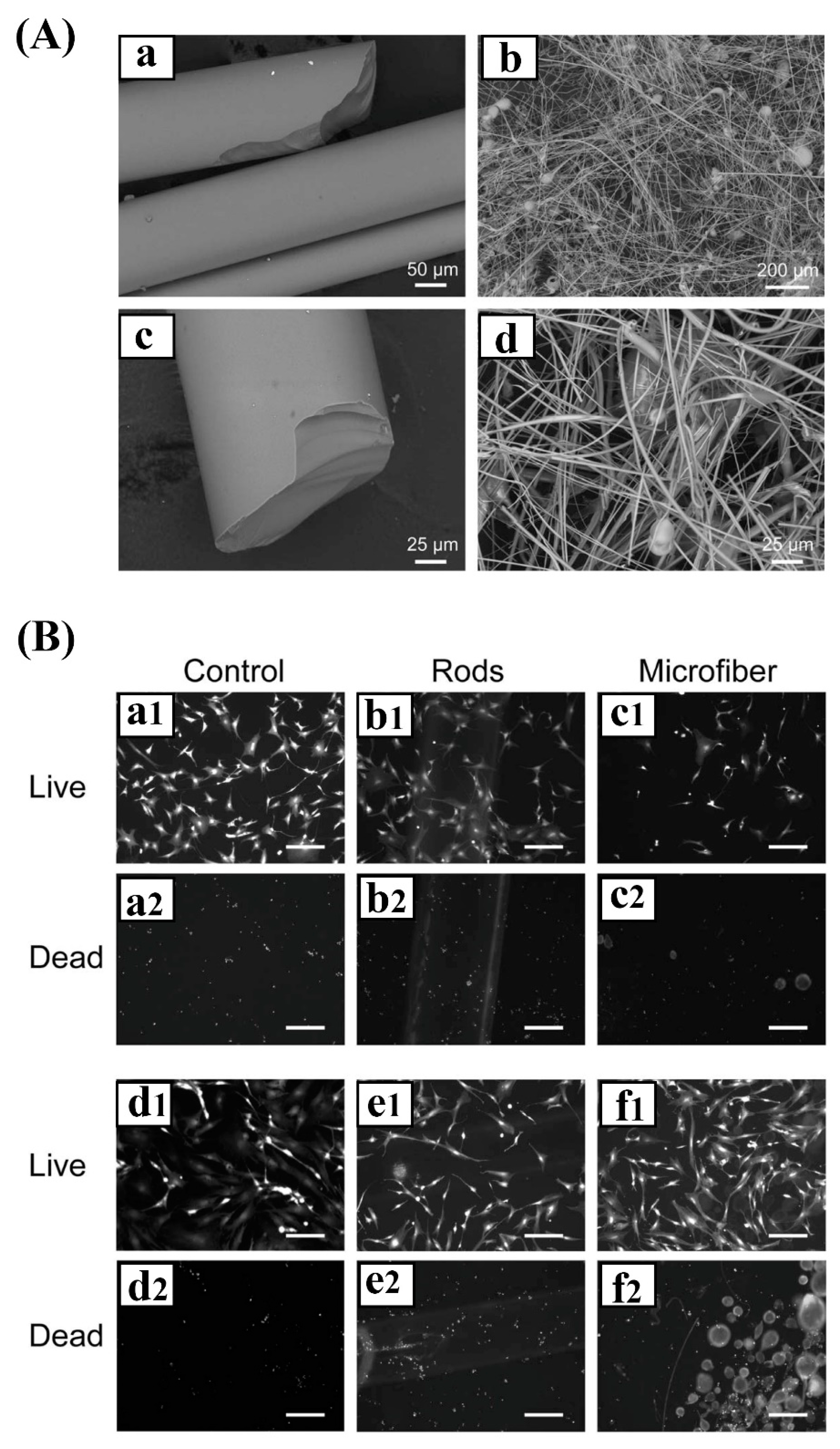
Figure 7.
(A) SEM micrographs of melt-derived 13-93 B3 borate BGs in rod (a, c) and microfiber (b, d) forms. (B) Microscopic images of embryonic chick dorsal root ganglia (DRG) cells alone (control groups a1/a2 and d1/d2) or treated with 13-93 B3 rods (b1/b2 and e1/e2) and microfibers (c1/c2 and f1/f2). Note that the images were captured from the cells after 6 days of culture. The culture medium was exchanged every other day (transient condition) in the two top rows, while no medium was exchanged throughout the experiment (static condition) in the two bottom rows. Scale bar is 100 μm. Reproduced with some modifications from ref [23].


Table 1.
A summary of currently available reports showing the suitability of different types of BGs and glass-based composites in peripheral nerve repair and regeneration.
Table 1.
A summary of currently available reports showing the suitability of different types of BGs and glass-based composites in peripheral nerve repair and regeneration.
| BG Composition/Synthesis Route | Applied Construct | Remarks | Ref (s) |
|---|---|---|---|
| 64 SiO2, 26 CaO, 5 MgO and 5 P2O5 (mol%), sol–gel method | Silicate glass nanoparticles/gelatin nanocomposites | - The composites showed no significant cytotoxicity - The composite showed great potential in the regeneration of myelinated axons in damaged sciatic nerves | [20] |
| 45S5 BG: 45.0 SiO2, 24.5 Na2O, 24.5 CaO, and 6.0 P2O5 (wt%) Borate glass: 53 SiO2, 20 CaO, 6 Na2O, 4 P2O5, 12 K2O, 5% MgO (wt%), melt-derived method | Silicate glass/polyhydroxyalkanoate (PHA) composites | - The composites exhibited good biocompatibility - The constructs improved the mechanical properties needed for the regeneration of peripheral nerves - The constructs supported growth and neuronal differentiation | [21] |
| Bioglass 45S5 (45% SiO2, 24% Na2O, 24.5% CaO and 6% P2O5) (wt%), melt-derived method | Silicate glass (45S5) fibers | - The glass fibers provided a suitable substrate for the adhesion and growth of rat Schwann cells - Axonal regeneration occurred through a Silastic conduit filled with Bioglass fibers - Axonal regrowth was observed in the Bioglass-fiber-treated rats comparable to an autograft 4 weeks after implantation in a 0.5 cm interstump gap in the sciatic nerves | [60] |
| 45S5 BG: 45% SiO2, 24% Na2O, 24.5% CaO and 6% P2O5 (wt%) substituted with Zn (5, 10, and 20 wt%) | Silicate glass (45S5) doped with Zn | - Bioglasses doped with low concentration of Zn supported cell adhesion and proliferation of undifferentiated SK-N-BE human neuroblastoma cells - Bioglasses doped with high Zn concentration lightly induced adhesion and phenotype characterization of the cells | [67] |
| Commercial nano-sized 45S5 BG | Silicate glass nanoparticles/polyglycolic acid (PGA), collagen electrospun composites | - The glass-containing nanofibers showed good compatibility with rats’ mesenchymal stem cells (MSCs) - The composite constructs provided a better substrate for cell adhesion and proliferation as compared to glass-free matrixes | [69] |
| SiO2–Na2O–K2O–MgO–CaO–P2O5 | Silicate glass microfibers incorporated in nanofibrous PCL | - Significant improvements in the mechanical properties and wettability were observed in the case of glass/polymer composites than the polymer matrix alone - The composites showed increased bioactivity | [70] |
| 50 P2O5, 40 CaO, 5 Na2O, 5 Fe2O3 (mol%), melt-derived method | Phosphate glass/collagen scaffolds | - Stimulated neurite outgrowth along the fibers in vitro - Enhanced axons extending along the scaffold at 7 days post-surgery - Caused recovery of plantar muscle atrophy at 8 weeks post-implantation - No significant differences in the case of functional capacity between the experimental groups and controls (collagen scaffolds lacking BGs) 12 weeks after implantation | [24] |
| 50 P2O5, 40 CaO, 5 Na2O, 5 Fe2O3 (mol%), melt-derived method | Phosphate glass fiber/collagen scaffolds | - The scaffolds induced axon growth from the proximal and distal stumps to the scaffold 12 weeks after implantation - The composites recovered locomotor and bladder functions at 8 weeks post-implantation - Endogenous BDNF levels were detected in the bladder at 12 weeks post-implantation | [76] |
| 50P2O5–30CaO–9Na2O–3SiO2–3MgO–(5– x)K2O–xTiO2 mol.% (x = 0, 2.5, 5) | Phosphate glass fibers with a diameter ranging between 25 and 82 µm | - The glass fibers provided a proper substrate for cell adhesion and proliferation - The aligned configuration of the fibers supported the growing axons of dorsal root ganglion (DRG) neurons along the fiber axis direction | [77] |
| 50P2O5–40CaO–5Na2O–5Fe2O3 (mol.%) | Phosphate glass microfibers–aminated carbon nanotubes (CNTs) incorporated into poly(L/D-lactic acid) (PLDLA) tubes | - Neurites of DRG outgrew along the aligned composite scaffolds - The constructs increased the number of regenerating axons and improved the electrophysiological functions | [79] |
| 13–93 B3 borate glass: 53B2O3, 20 CaO, 6Na2O, 12K2O, 5MgO, 4P2O (wt%) | Borate glass rods and microfiber/fibrin composites | - Borate glasses and the composite scaffolds improved neurite extension comparable to that of control fibrin scaffolds, clarifying the lack of significant effect of the glasses on neuronal health - Aligned glass scaffolds could guide neurite extension in an oriented manner | [23] |
| 13–93 B3 borate glass; 45S5 silicate glass; a blend of 13–93 B3 and 45S5 glasses | Borate and silicate glasses/PCL composites | - The composites containing 13–93 B3 borate glass exhibited a higher degradation rate than their counterparts containing only 45S5 silicate glass - None of the glasses caused adverse effects on neurite extension as compared to PCL alone - Neurite extension was increased in contact with PCL:45S5 PCL:13–93 B3 composites after 24 h of incubation | [25] |
| 13-93 B3 borate glass doped with Ag, Ce, Cu, Fe, Ga, iodine (I), Y, and Zn | Borate glass/PCL composite sheets | - Cu, Fe, Ga, Zn, and Sr-doped glasses promoted the survival and outgrowth of neurons as compared to undoped glasses - The Cu- and Ga-doped glasses showed the lowest average percent survival of support cells | [58] |
© 2020 by the authors. Licensee MDPI, Basel, Switzerland. This article is an open access article distributed under the terms and conditions of the Creative Commons Attribution (CC BY) license (http://creativecommons.org/licenses/by/4.0/).
Share and Cite
MDPI and ACS Style
Kargozar, S.; Mozafari, M.; Ghenaatgar-Kasbi, M.; Baino, F. Bioactive Glasses and Glass/Polymer Composites for Neuroregeneration: Should We Be Hopeful? Appl. Sci. 2020, 10, 3421. https://doi.org/10.3390/app10103421
AMA Style
Kargozar S, Mozafari M, Ghenaatgar-Kasbi M, Baino F. Bioactive Glasses and Glass/Polymer Composites for Neuroregeneration: Should We Be Hopeful? Applied Sciences. 2020; 10(10):3421. https://doi.org/10.3390/app10103421
Chicago/Turabian StyleKargozar, Saeid, Masoud Mozafari, Maryam Ghenaatgar-Kasbi, and Francesco Baino. 2020. "Bioactive Glasses and Glass/Polymer Composites for Neuroregeneration: Should We Be Hopeful?" Applied Sciences 10, no. 10: 3421. https://doi.org/10.3390/app10103421
Note that from the first issue of 2016, this journal uses article numbers instead of page numbers. See further details here.