
Evaluation of Adsorbent’s Efficiency by Using Biomarker Approaches in Farm Animals: A Systematic Review


Department of Food Engineering, School of Animal Science and Food Engineering, University of São Paulo, Av. Duque de Caxias Norte, 225, Pirassununga CEP 13635-900, SP, Brazil
*
Author to whom correspondence should be addressed.
Appl. Sci. 2022, 12(24), 13000; https://doi.org/10.3390/app122413000
Submission received: 1 December 2022
/
Revised: 14 December 2022
/
Accepted: 15 December 2022
/
Published: 18 December 2022
Abstract
:The secondary metabolism of toxigenic fungi can produce mycotoxins, substances that are toxic for both humans and animals. Mycotoxins and their by-products found in various biological tissues are considered biomarkers, and concentrations of these substances are directly proportional to the level of exposure. Mineral adsorbents are substances that may prevent mycotoxin absorption. The aim of this review is to study the feasibility of biomarkers as tools to assess the efficiency of mineral adsorbents against mycotoxin absorption in farm animals. In the systematic review, data from the scientific literature between the 2001 and 2022 were searched based on established criteria selection and eligibility. A total of 22 articles were included. The most used species as animal models were poultry and cattle, while the most common biological samples were milk, serum, and liver. Biomarkers most frequently analyzed were aflatoxin M1 (AFM1) and unmetabolized aflatoxin B1 (AFB1). The most used analytical method was liquid chromatography tandem mass spectrometry. Biomarkers are adequate tools to assess the efficiency of mineral adsorbents against the aflatoxins and deoxynivalenol in farm animals, but further studies are needed to provide reliable biomarkers for other mycotoxins.
1. Introduction
Mycotoxins are secondary metabolites of fungi produced mainly by genera Aspergillus, Fusarium, Penicillium, and Claviceps. The most common mycotoxins are aflatoxins (AFB1, AFB2, AFG1, AFG2), fumonisin B1 (FB1), ochratoxin A (OTA), deoxynivalenol (DON), zearalenone (ZEN), T-2 toxin (T-2), and HT-2 toxin (HT-2) [1,2,3]. The products that are most contaminated by mycotoxins are corn, wheat, sorghum, soy, coffee, wine and juice, fruits and vegetables, milk products, and animal feed, among others [3,4,5,6,7,8]. Animals that ingest contaminated feed may excrete mycotoxins in an unaltered form or as biotransformed products. Mycotoxins may also accumulate in meat, viscera, and eggs that may, later on, be ingested by humans [3].
Mycotoxin ingestion may lead to acute or chronic effects in humans and animals, depending on the amount ingested and the extent of exposure. Cases of acute intoxication may quickly lead to death, but they are less common, given the amount of mycotoxin that needs to be ingested [1,9,10]. Chronic cases occur when small amounts of mycotoxins—with their immunotoxic, neurotoxic, nephrotoxic, dermal toxicity, and teratogenic properties—are continuously ingested for long periods [1,10]. Mycotoxins also have mutagenic and carcinogenic potential, with AFB1, AFB2, AFG1, and AFG2 classified as class 1 human carcinogens (definitely carcinogenic to humans), and OTA, FB1, FB2, and AFM1 as class 2B (possibly carcinogenic to humans) [11].
Mycotoxins affect production animals. In pigs, FB1 causes swine pulmonary edema, and DON and FB cause immunosuppression and lesions in liver and lungs [12]. In dairy cattle, AFB1 ingestion leads to reduced milk yield, and in beef cattle, reduced feed intake and immunosuppression [1]. In broilers, feed contaminated with AFB1 and FB1 reduces weight gain and causes liver changes [13], and in laying hens, OTA reduces weight gain, feed intake, and egg yield, besides leading to liver and kidney changes. OTA residues may also be found in the eggs [14]. Horses that ingest feed contaminated with FB1 may die of equine leukoencephalomalacia, a highly lethal disease characterized by nervous symptoms [15].
In most countries, there are regulations that define maximum tolerated levels (MTL) of mycotoxins in human food. In Brazil, regulation RDC 07/2011 of the Brazilian Health Regulatory Agency (ANVISA) defines MTLs for AFB1, AFB2, AFG1, AFG2, AFM1, OTA, DON, ZEN, FB, and patulin (PAT) in different foods [16]. For example, in fluid milk, MTL for AFM1 is 0.5 µg/kg; in corn MTL for the sum of AFB1, AFB2, AFG1, and AFG2 is 20 µg/kg; in cocoa and chocolate-based products, the MTL of OTA is 5.0 µg/kg. For animal feed, recommendations are not determined for individual feeds and mycotoxins, but MLT for the sum of AFB1, AFB2, AFG1, and AFG2 in raw materials used for animal feed is 50 µg/kg [17]. The European Union, on the other hand, is stricter in terms of MTL in human foods; for instance, AFM1 limit in milk is 0.05 µg/kg, 10 times lower than the MTL for this product and mycotoxin in Brazil [18].
Different chemical, biological, and physical decontamination methods have been developed to reduce mycotoxin levels in food products [1,19]. Chemical methods aim at the structural degradation of mycotoxins, adding oxidizing or reducing agents, acids, and bases to the substrate [1,20]. Biological decontamination is based on the use of microorganisms, such as bacteria, fungi, and yeasts, which bind to mycotoxins and degrade or adsorb them [1,3,10,20,21]. Physical methods, on the other hand, may use irradiation, solvent extraction, or adsorption. Adsorption is a superficial phenomenon in which mycotoxins are attracted by electrostatic and polar interactions to an adsorbent substance [20].
The most used adsorbents are aluminosilicate clays, with silica, aluminum, oxygen, and hydroxyl groups in their basic composition [22]. These clays may be classified based on their chemical structure, such as phyllosilicates (montmorillonite, bentonite, smectite) and tectosilicates (zeolites) [23]. Examples of these clays are calcium aluminosilicate and hydrated sodium calcium aluminosilicate (HSCAS), bentonite, sepiolite, montmorillonite, diatomite, and zeolite. Activated charcoal, although not an aluminosilicate, is also used as an adsorbent [3,20]. However, adsorbents usually show wide variation in composition and physical-chemical properties. Thus, it is necessary to assess their efficacy using in vivo protocols, which are generally costly and labor-intensive, as they involve the administration of toxins at different levels, with and without the adsorbent, to assess the effect on animal productivity [16]. Besides, these assays generally require collection of histopathological and clinical data, among other issues that add to the cost and labor. In this context, the use of biomarker approaches to estimate the mycotoxin bioavailability of adsorbent efficiency in in vivo assays may reduce costs, besides being more practical, helping the standardization of the experimental trials, and making it possible to assess the effect of adsorbents in field conditions.
Biomarkers are substances that may be detected in biological samples (viscera, milk, eggs, feces, urine, blood), after exposure to a given agent, such as mycotoxins. In the case of exposure to mycotoxins, biomarkers may be the either the structurally unaltered toxin or a biotransformed product, depending on the toxicokinetic of the mycotoxin and the individual characteristics of the species [24,25,26,27]. The European Union has rules for the use of adsorbents in animal feed [28,29,30], and these products must be carefully tested to be approved [31]. Therefore, the objective of this systematic review was to evaluate the feasibility of biomarkers as tools to assess the efficiency of mineral adsorbents for mycotoxins in farm animals.
2. Methods
2.1. Search Strategy
This systematic review was based on the PRISMA methodology [32] and on the search in the Google Scholar, PubMed, and ScienceDirect databases. The following keywords were used: “farm animals” OR “production animals” AND “mycotoxins” AND “adsorbent” OR “clay” AND “biomarkers” OR “residue” OR “metabolites”. Articles published between 2001 and 2022 were screened and manually analyzed.
2.2. Inclusion and Exclusion Criteria and Data Collection
The following inclusion and eligibility criteria were used to select the articles: (1) keywords presented in the title and/or abstract, (2) research articles (not reviews), (3) published in English, (4) detailed experimental conditions, (5) articles described the use of mineral adsorbents (alone or as mixtures with yeasts, vitamins, mineral extracts, etc.) and biomarkers of mycotoxin in the assessment of adsorbent efficacy in production animals (cattle, buffaloes, pigs, broilers and laying hens, goats, and sheep).
The following exclusion criteria were used: (1) articles describing only in vitro studies and (2) incomplete description of the experimental methods.
All articles that remained after the initial screening were downloaded for further evaluation based on the inclusion and exclusion criteria. From the articles included in the review, the following data were collected: species used as experimental models, number of animals, concentration of the mycotoxins offered to the animals, adsorbent used and at what level, biomarkers analyzed, samples collected, and analytical method.
2.3. Calculation of Percent Reduction in Biomarker Concentration Caused by Treatments (RBTC)
Percent reduction in biomarker concentration caused by the treatment with the adsorbent was calculated in relation to the positive control (RBTC) in articles that did not have this result. RBTC is a quantitative measure of the efficiency of the adsorbent. RCTB calculation is shown in Equation (1), where “a” is the concentration of the biomarker in the treatment, and “b” is the concentration of the biomarker in the positive control.
y = (a ÷ b) × 100 RBTC = 100 − y (%)
3. Study Characteristics
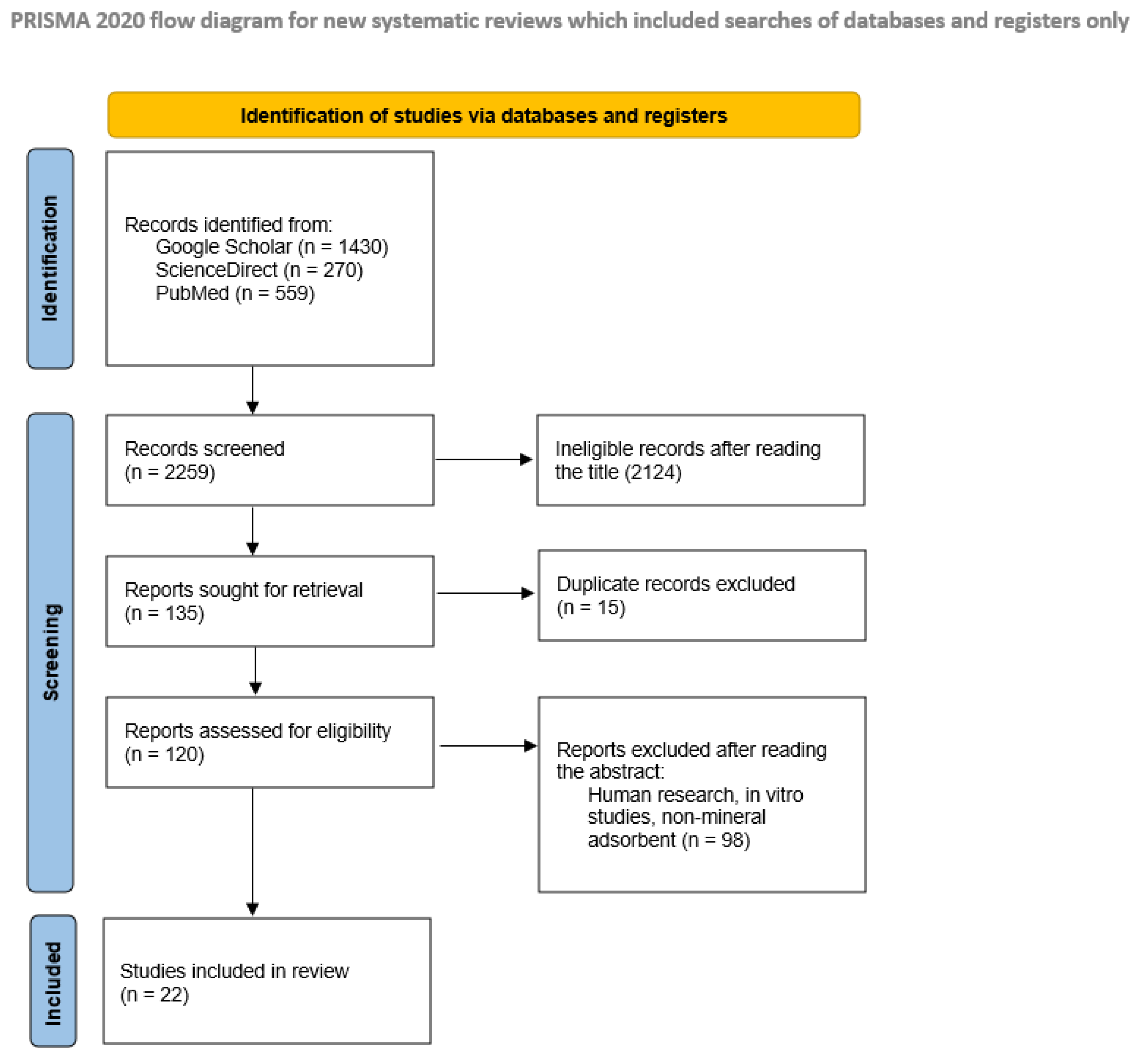
After the search using the keywords, 1430 records were found in Google Scholar, 270 in ScienceDirect, and 559 in PubMed, totaling 2259 articles. From these, 2124 were excluded because they did not meet the including criteria for the title. From the 135 remaining records, 15 were duplicated. The remainder of the reports were analyzed by their abstract and full text. A total of 98 articles did not meet the eligibility criteria and 22 articles were thus included in the systematic review. Figure 1 shows the flow diagram for identification, selection, inclusion, and exclusion of the articles.
4. Results
Five different species of production animal were used as experimental models: eight studies involved broilers or laying hens, eight used dairy cows, four used pigs, two used goats, and one used buffalo. Six different mycotoxins were used at different concentrations for the intoxication studies; as some of the articles reported the use of more than one mycotoxin, a total of 28 experimental intoxications were analyzed. AFB1 was the most used mycotoxin (18 times); DON was used three times; OTA, T-2, and ZEN were used twice; and HT-2 was used once.
Seventeen different adsorbents were used. Five of them were made up of a single material: bentonite (used nine times), HSCAS (five times), montmorillonite (once), and zeolite and aluminosilicate clay (once). An adsorbent with 95% activated sulfur bentonite and 5% phytogenic substances (organic material) was also included in this group, given it was mostly made up by a single mineral adsorbent.
Activated charcoal was also used as an adsorbent (two times), as well as mixtures of mineral adsorbents at different proportions. These mixtures were as follows: montmorillonite 50% + diatomite 50% (used once); sepiolite 44.44% + bentonite 55.56% (used once); and talc 69.4% + China clay 30% (used twice).
Eight different mixed adsorbents were also used. These were commercial products made up mineral adsorbents and biological decontaminants, such as yeasts, yeast cell walls, or mannan-oligosaccharides and additives (preservatives, antioxidants, etc.). Escent® S (Innovad SA, Essen, Belgium) and Solis Molis (Novus International Trading Co., Ltd., Shanghai, China) were used twice. All other mixed adsorbents were used only once: MMDA (Patent Co., Misicevo, Serbia), Toxy-Nil (Nutriad Animal Feed Additives, Dendermonde, Belgium), Unique Plus (Nutriad Animal Feed Additives, Dendermonde, Belgium), MTB-100® (Alltech Inc., Nicholasville, KY, USA), and other two mixed adsorbents that were not commercial products.
In total, 16 different biomarkers were used from the 44 analyzed in the studies. AFM1 was used 15 times, and was the most frequent biomarker in the studies, followed by AFB1, used 9 times. ZEN, T-2, HT-2, OTA, DON, and deoxynivalenol 3-glucuronide (DON-GlcA) were used twice. Deoxynivalenol-sulfate (DON-S), zearalenone- glucuronide (ZEN-GlcA), aflatoxin B1-lysin (AFB1-lysin), AFB2, AFG1, AFG2, and total aflatoxin and aflatoxicol (AFL), were each used in only one study.
For the detection of these biomarkers, 53 samples of 18 different types were used. Samples may be divided in four large groups: viscera, serum, animal products, and excreta. Among viscera, the liver was the most common sample in biomarker detection (used seven times), followed by kidneys (four times) and spleen (two times). Samples of the genital tract, heart, lungs, stomach, muscle, brain, bursa of Fabricius, thymus, and small intestine were used only once. Serum was used seven times. Among animal products, milk was used 11 times, and was the most common substrate in the analysis of biomarkers in this systematic review. Eggs were used once. Among excreta samples, urine was used five times, poultry excreta were used four times, and feces was used three times.
Seven different methods were used for the detection and quantification of biomarkers in the selected samples, divided in two main categories: chromatography (used three times) and immunoenzimatic methods (four times). Among chromatographic methods, liquid chromatography tandem mass spectrometry (LC-MS/MS) was the most common form of biomarker detection (used nine times); high-performance liquid chromatography coupled with fluorescence detector (HPLC-FLD) was used four times, and high-performance liquid chromatography was used twice. Among immunoenzymatic methods, the enzyme-linked immunosorbent assay (ELISA) was used four times; fluorescence polarization immunoassay, radioimmunoassay, and enzyme immunoassay were all used once. Table 1 compiles all data collected from the articles included in this review.
5. Discussion
The five species employed as experimental models are farm animals widely used for the production of meat, milk, and their products. The absence of horses as experimental models may be explained by the fact that these animals are generally not considered production animals, but as sport or companion animals [51]. A study carried out by Takagi et al. [52] quantified zearalenone biomarkers, α-zearalenol and β-zearalenol, in the urine of horses using LC-MS/MS and showed that urine is an adequate sample for the measurement of this mycotoxin and its metabolites.
The most common technique used in the detection of biomarkers was LC-MS/MS. Its popularity may be due to its high sensitivity and specificity, making it possible to analyze different mycotoxins as parts per billion (ppb) or even parts per trillion (ppt) [1,53,54]. As observed in the present review, liquid chromatography coupled with fluorescence detector, as well as ELISA, are also widely used for the detection of mycotoxins and their biomarkers in foods, and in human and animal samples [19,24,53,54,55,56,57].
In some studies, no biomarkers were detected in the samples [29]. For example, RBTC calculation could not be carried out in the study by Yang et al. [33] because levels of T-2 and HT-2 in the viscera analyzed were too low, either undetectable or below the limit of detection of the methods employed. It is important to emphasize that absence of detection of biomarkers may have different meanings. First, it may be related to the sensitivity of the analytical method. Therefore, the absence of detection of biomarkers does not mean absence of biomarkers, but lack of sensitivity of the method. Considering this, LC-MS/MS is, presently, the most sensitive technique for this quantification. Secondly, the amount of mycotoxin offered to the animals may not be enough for the detection of biomarkers. An example for this case is the study by Vasiljević [14]. Although they used LC-MS/MS, OTA was only detected in the eggs of hens that were treated with 1 mg/kg, and not in those that received 0.25 mg/kg of the toxin.
A total of 16 different biomarkers were searched for in the samples analyzed, either the unchanged mycotoxin—AFB1, for example—or its biotransformed form, an adduct or a conjugate, such as hydroxylated AFM1, AFB1-lysin, and ZEN-GlcA, respectively. Of all mycotoxins employed in the studies selected here, AFB1 was the most common one. This may be related to the high carcinogenic potential of AFB1, to the fact that AFB1 metabolites (which are also potentially carcinogenic) may be found in different and popular animal products [3,11], and the possible animal losses caused by acute and lethal intoxication by AFB1 [9].
AFM1 and AFB1 were also the most analyzed and quantified biomarkers, due to the same reasons [11]. Residues of these mycotoxins in animal products were found in many of the studies used in this review, mainly in poultry livers, a food that is widely consumed all over the world [46,50]. The large number of articles that aimed at testing the reduction in AFM1 concentration in the milk by mineral adsorbents is due to the great frequency of contamination of cattle feed with AFB1. Besides, regulations that determine MTLs for AFM1 in milk (in Brazil, 0.5 µg/kg, in the EU, 0.05 µg/kg) offer economic and commercial reasons for creating and testing products that may reduce the concentration of these mycotoxins in foods and animal feed [16,18].
In humans, many biomarkers are used in the assessment of mycotoxin exposure. However, there are few studies that used these biomarkers to test the efficacy of a mineral adsorbent. A review by Phillips et al. [58] cites the possible use of the adsorbent NovaSilTM and in the reduction of biomarkers in urine and blood of people exposed to high levels of mycotoxins. Among some of the most common biomarkers studied in animals and humans were the following: AFM1, AFB1-lysin, AFB1-N7-guanine, OTA, ochratoxin A glucuronide (OTA-GlcA), DON, deepoxy-deoxynivalenol (DOM-1), DON-GlcA, AFB1, AFB2, AFG1, AFG2, T-2, HT-2, ZEN, zearalenol (ZON), µ-ZON, β-ZON, FB1, FB2, and citrinin (CIT). [24,55,56,59] Many of these biomarkers were also used in the articles analyzed here. Another factor that explains the use of several biomarkers in animal studies is the EU regulation no. 386/2009 [30], which determines the use of biomarkers to assess the efficacy of additives that aim at reducing the concentration of mycotoxins in foods, or their bioavailability to animals [31].
Eighteen types of samples were used in the studies reported here. The most frequent one was milk, which corroborates the finding of AFM1 as the most detected biomarker. The high level of consumption of milk and the importance of this food in human nutrition all over the world, the carcinogenic risk posed by AFM1, and the MTLs determined all over the world for this mycotoxin explain the wide interest in the analysis of its residues [16,18]. Other popular biological samples used in biomarker quantification were liver, serum, urine, poultry excreta, and feces [3,19,24,59]. The choice of both the biomarker to be studied and the sample to be collected should be based on the knowledge about toxicokinetics, biotransformation, and accumulation trends of the mycotoxin. This is especially true when the aim of biomarker quantification is to evaluate the efficacy of a decontaminant, as the presence or absence of the biomarker may change as a function of toxicodynamic and toxicokinetic characteristics of the species.
The findings of this review corroborate those of a previous one by Escrivá et al. [55] on the use of biological samples in the determination of mycotoxins and their biomarkers, carried out in studies between 2005 and 2017. These authors listed samples of serum, urine, human and cow milk, liquor, feces, liver kidney, bile, muscle, spleen, heart, ovaries uterus, stomach, brain, small intestines, and lungs collected from humans, pigs, cattle, horses, fish, and rats to detect different toxins and their biomarkers. These biological materials and subjects were also found in the studies used in present review. LC-MS/MS was the most popular method, and HPLC-FLD and ELISA were frequently used [51], similar to the findings of the present review. However, in the review by Escrivá et al. [51], the most found mycotoxins were OTA, ZEN, and DON, and not AFM1 and AFB1, as observed in the present review.
The 17 mineral adsorbents reported in the present review showed differences in efficacy, given they were made up of a single mineral or a mixture of mineral and yeasts, or different minerals. Efficacy is linked to the affinity between the adsorbent material and the mycotoxin; affinity, in its turn, depends on polar and electrostatic interactions between the two compounds [20]. As it was not possible to standardize the different variables analyzed in this review, such as animal species, type of adsorbent, type of sample, biomarker, and analytical method, it is difficult to compare the efficacy of the different adsorbents. However, some patterns were visible and independent of the type of adsorbent.
In the studies by Cha et al. [36] and Kutz et al. [41] with the detection of AFM1 in milk, the same level of mycotoxin was offered to the groups treated with mineral adsorbents or mixed mineral-yeast adsorbent. In these studies, RBTC in the treatment with the mineral adsorbent was greater than with the mixed one, showing that the mineral adsorbent was more efficient in reducing AFM1 in milk. Another important finding is that there may be a direct proportion between time of adsorbent use and the RBTC. That is, the longer the treatment, the greater the reduction in biomarkers in the sample [25,49].
In the treatments that used only one concentration of mycotoxin and two different concentrations of the same adsorbent, greater ingestion of mineral adsorbent led to greater reduction in biomarker concentration, indicating a direct proportion between the amount of biomarker detected and the amount of adsorbent offered [26,37,39]. Besides, mycotoxin concentration showed an inverse proportion with RBTC in treatments that used a single concentration of mycotoxin. That is, the greater the mycotoxin concentration, the lower the RBTC and, consequently, the greater the biomarker concentration in the samples [14,35]. Thus, there is a limit for the efficacy of the adsorbent. When levels of the mycotoxin are too high, the limit of adsorption may be reached, and biomarker quantification in the samples may be higher. Because of this, the levels of exposure of the animals have to be determined before the implementation of strategies based on mineral adsorbents, so as to supply the correct concentration of adsorbent to decrease biomarkers to adequate levels.
6. Conclusions
Biomarkers are excellent tools to measure the efficacy of mineral adsorbents, especially the aflatoxins and DON in farm animals. Many factors impact the detection of these biomarkers, such as analytical methods, type of sample, and animal species, among others. The decision about the type of biomarker to analyze and the type of sample should be based on studies that describe the toxicokinetics of the mycotoxin, as well as its biotransformation, conjugation, and routes of elimination. Only then these biomarkers may be used to evaluate the efficacy of an adsorbent, and the results obtained may be reliable. More studies should be carried out with animals to find out other biomarkers, or for the simultaneous quantification of several biomarkers, to make them eligible for the evaluation of the efficacy of adsorbents against mycotoxins.
Author Contributions
Conceptualization, L.G.D.F. and C.A.F.d.O.; methodology, L.G.D.F.; validation, L.G.D.F., C.A.F.d.O. and S.A.; formal analysis, C.A.F.d.O.; investigation, L.G.D.F.; resources, C.A.F.d.O.; data curation, L.G.D.F.; writing—original draft preparation, L.G.D.F.; writing—review and editing, C.A.F.d.O. and S.A.; visualization, C.A.F.d.O.; supervision, C.A.F.d.O. and S.A.; project administration, C.A.F.d.O.; funding acquisition, C.A.F.d.O. All authors have read and agreed to the published version of the manuscript.
Funding
This research was funded by Fundação de Amparo à Pesquisa do Estado de São Paulo (FAPESP, grant numbers: 2022/05066-9; 2022/03952-1 and 2019/21603-1.
Institutional Review Board Statement
Not applicable.
Informed Consent Statement
Not applicable.
Data Availability Statement
Not applicable.
Acknowledgments
The authors would like to thank Fundação de Amparo à Pesquisa do Estado de São Paulo (FAPESP) for their financial support.
Conflicts of Interest
The authors declare no conflict of interest.
References
- Pleadin, J.; Frece, J.; Markov, K. Mycotoxins in Food and Feed. In Advances in Food and Nutrition Research; Academic Press Inc.: Cambridge, MA, USA, 2019; Volume 89, pp. 297–345. [Google Scholar]
- Berthiller, F.; Crews, C.; Dall’Asta, C.; de Saeger, S.; Haesaert, G.; Karlovsky, P.; Oswald, I.P.; Seefelder, W.; Speijers, G.; Stroka, J. Masked Mycotoxins: A Review. Mol. Nutr. Food Res. 2013, 57, 165–186. [Google Scholar] [CrossRef]
- Adegbeye, M.J.; Reddy, P.R.K.; Chilaka, C.A.; Balogun, O.B.; Elghandour, M.M.M.Y.; Rivas-Caceres, R.R.; Salem, A.Z.M. Mycotoxin Toxicity and Residue in Animal Products: Prevalence, Consumer Exposure and Reduction Strategies—A Review. Toxicon 2020, 177, 96–108. [Google Scholar] [CrossRef] [PubMed]
- Franco, L.T.; Petta, T.; Rottinghaus, G.E.; Bordin, K.; Gomes, G.A.; Oliveira, C.A.F. Co-Occurrence of Mycotoxins in Maize Food and Maize-Based Feed from Small-Scale Farms in Brazil: A Pilot Study. Mycotoxin Res. 2019, 35, 65–73. [Google Scholar] [CrossRef] [PubMed]
- Mokhtarian, M.; Tavakolipour, H.; Bagheri, F.; Fernandes Oliveira, C.A.; Corassin, C.H.; Khaneghah, A.M. Aflatoxin B1 in the Iranian Pistachio Nut and Decontamination Methods: A Systematic Review. Qual. Assur. Saf. Crop. Foods 2020, 12, 15–25. [Google Scholar] [CrossRef]
- Delage, N.; d’Harlingue, A.; Colonna Ceccaldi, B.; Bompeix, G. Occurrence of Mycotoxins in Fruit Juices and Wine. Food Control 2003, 14, 225–227. [Google Scholar] [CrossRef]
- Corassin, C.H.; Borowsky, A.; Ali, S.; Rosim, R.E.; de Oliveira, C.A.F. Occurrence of Aflatoxin M1 in Milk and Dairy Products Traded in São Paulo, Brazil: An Update. Dairy 2022, 3, 842–848. [Google Scholar] [CrossRef]
- Nan, M.; Xue, H.; Bi, Y. Contamination, Detection and Control of Mycotoxins in Fruits and Vegetables. Toxins 2022, 14, 309. [Google Scholar] [CrossRef] [PubMed]
- Batatinha, M.J.; Botura, M.B.; Górniak, S.L. Micotoxinas e Micotoxicoses. In Toxicologia Aplicada à Medicina Veterinária; Manole Santana de Parnaíba: São Paulo, Brazil, 2020; Volume 2, pp. 304–330. ISBN 9788520458976. [Google Scholar]
- Oliveira, C.; Augusto, F.; Bovo, F.; Corassin, C.H.; Jager, A.V.; Reddy, K.R. Recent Trends in Microbiological Decontamination of Aflatoxins in Foodstuffs. In Aflatoxins—Recent Advances and Future Prospects; IntechOpen: Rijeka, Croatia, 2013; pp. 59–92. ISBN 978-953-51-0904-4. [Google Scholar]
- International Agency for Research on Cancer IARC. Monographs on the Evaluation of Carcinogenic Risks to Humans Some Traditional Herbal Medicines, Some Mycotoxins, Naphthalene and Styrene; International Agency for Research on Cancer: Lyon, France, 2002; Volume 82, ISBN 9283212827. [Google Scholar]
- Grenier, B.; Loureiro-Bracarense, A.-P.; Lucioli, J.; Pacheco, G.D.; Cossalter, A.-M.; Moll, W.-D.; Schatzmayr, G.; Oswald, I.P. Individual and Combined Effects of Subclinical Doses of Deoxynivalenol and Fumonisins in Piglets. Mol. Nutr. Food Res. 2011, 55, 761–771. [Google Scholar] [CrossRef]
- Miazzo, R.; Peralta, M.F.; Magnoli, C.; Salvano, M.; Ferrero, S.; Chiacchiera, S.M.; Carvalho, E.C.Q.; Rosa, C.A.R.; Dalcero, A. Efficacy of Sodium Bentonite as a Detoxifier of Broiler Feed Contaminated with Aflatoxin and Fumonisin. Poult. Sci. 2005, 84, 1–8. [Google Scholar] [CrossRef]
- Vasiljević, M.; Marinković, D.; Milićević, D.; Pleadin, J.; Stefanović, S.; Trialović, S.; Raj, J.; Petrujkić, B.; Trialović, J.N. Efficacy of a Modified Clinoptilolite Based Adsorbent in Reducing Detrimental Effects of Ochratoxin A in Laying Hens. Toxins 2021, 13, 469. [Google Scholar] [CrossRef] [PubMed]
- Vendruscolo, C.P.; Frias, N.C.; Carvalho, C.B.; Sá, L.R.M.; Belli, C.B.; Baccarin, R.Y.A. Leukoencephalomalacia Outbreak in Horses Due to Consumption of Contaminated Hay. J. Vet. Intern. Med. 2016, 30, 1879–1881. [Google Scholar] [CrossRef]
- Agência Nacional de Vigilância Sanitária. Diário Oficial da União No 37; Sesão 1; Agência Nacional de Vigilância Sanitária: Brasília, Brazil, 2011.
- Ministério da Agricultura. Diário Oficial da União; Seção 1; Ministério da Agricultura: Brasília, Brazil, 1988; p. 21.968.
- European Union. Comission Regulation; Official Journal of the European Union; European Union: Brussels, Belgium, 2006. [Google Scholar]
- Gonçalves, B.L.; Gonçalves, J.L.; Rosim, R.E.; Cappato, L.P.; Cruz, A.G.; Oliveira, C.A.F.; Corassin, C.H. Effects of Different Sources of Saccharomyces Cerevisiae Biomass on Milk Production, Composition, and Aflatoxin M1 Excretion in Milk from Dairy Cows Fed Aflatoxin B1. J. Dairy Sci. 2017, 100, 5701–5708. [Google Scholar] [CrossRef] [Green Version]
- di Gregorio, M.C.; de Neeff, D.V.; Jager, A.V.; Corassin, C.H.; Carão, Á.C.D.P.; de Albuquerque, R.; de Azevedo, A.C.; Oliveira, C.A.F. Mineral Adsorbents for Prevention of Mycotoxins in Animal Feeds. Toxin. Rev. 2014, 33, 125–135. [Google Scholar] [CrossRef]
- Luo, Y.; Liu, X.; Li, J. Updating Techniques on Controlling Mycotoxins—A Review. Food Control 2018, 89, 123–132. [Google Scholar] [CrossRef]
- Daković, A.; Matijašević, S.; Rottinghaus, G.E.; Ledoux, D.R.; Butkeraitis, P.; Sekulić, Ž. Aflatoxin B1 Adsorption by Natural and Copper Modified Montmorillonite. Colloids Surf. B Biointerfaces 2008, 66, 20–25. [Google Scholar] [CrossRef] [PubMed]
- Rossetto, E.; Beraldin, R.; Penha, F.G.; Pergher, S.B.C. Caracterização de Argilas Bentonitas e Diatomitas e Sua Aplicação Como Adsorventes. Química Nova 2009, 32, 2064–2067. [Google Scholar] [CrossRef]
- Jager, A.V.; Tonin, F.G.; Baptista, G.Z.; Souto, P.C.M.C.; Oliveira, C.A.F. Assessment of Aflatoxin Exposure Using Serum and Urinary Biomarkers in São Paulo, Brazil: A Pilot Study. Int. J. Hyg. Environ. Health 2016, 219, 294–300. [Google Scholar] [CrossRef]
- di Gregorio, M.C.; Jager, A.V.; Souto, P.C.M.C.; Costa, A.A.; Rottinghaus, G.E.; Passarelli, D.; Budiño, F.E.L.; Corassin, C.H.; Oliveira, C.A.F. Determination of Serum Aflatoxin B1-Lysine to Evaluate the Efficacy of an Aflatoxin-Adsorbing Feed Additive in Pigs Fed an Aflatoxin B1-Contaminated Diet. Mycotoxin Res. 2017, 33, 93–102. [Google Scholar] [CrossRef]
- Raj, J.; Vasiljević, M.; Tassis, P.; Farkaš, H.; Männer, K. Efficacy of a Multicomponent Mycotoxin Detoxifying Agent on Concurrent Exposure to Zearalenone and T-2 Mycotoxin in Weaned Pigs. Livest. Sci. 2020, 242, 104295. [Google Scholar] [CrossRef]
- Rodrigues, R.O.; Rodrigues, R.O.; Ledoux, D.R.; McFadden, T.B.; Rottinghaus, G.E.; Borutova, R.; Averkieva, O. Feed Additives Containing Sequestrant Clay Minerals and Inactivated Yeast Reduce Aflatoxin Excretion in Milk of Dairy Cows. J. Dairy Sci. 2019, 102, 6614–6623. [Google Scholar] [CrossRef]
- European Union; European Parliament and of the Council; Official Journal of the European Union. Regulation (EC) No 1831/2003 of the European Parliament and of the Council; European Union: Brussels, Belgium, 2003; Volume 46, p. 29. [Google Scholar]
- European Union; European Parliament and of the Council; Official Journal of the European Union. Regulation (EC) No 1333/2008 Of The European Parliament And Of The Council; European Union: Brussels, Belgium, 2008; Volume 51. [Google Scholar]
- European Union; European Parliament and of the Council; Official Journal of the European Union. Commission Regulation (EC) No 386/2009 of May 2009 amending Regulation (EC) No 1831/2003 of the European Parliament and of the Council; European Union: Brussels, Belgium, 2009; Volume 37, p. 66. [Google Scholar]
- EFSA Panel on Additives or Substances used in Animal Feed (FEEDAP). Statement on the Establishment of Guidelines for the Assessment of Additives from the Functional Group ‘Substances for Reduction of the Contamination of Feed by Mycotoxins’. EFSA J. 2010, 8, 1693. [Google Scholar] [CrossRef]
- Page, M.J.; Moher, D.; Bossuyt, P.M.; Boutron, I.; Hoffmann, T.C.; Mulrow, C.D.; Shamseer, L.; Tetzlaff, J.M.; Akl, E.A.; Brennan, S.E.; et al. PRISMA 2020 Explanation and Elaboration: Updated Guidance and Exemplars for Reporting Systematic Reviews. BMJ 2021, 372, n160. [Google Scholar] [CrossRef]
- Yang, L.; Zhao, Z.; Deng, Y.; Zhou, Z.; Hou, J. Toxicity Induced by F. Poae-Contaminated Feed and the Protective Effect of Montmorillonite Supplementation in Broilers. Food Chem. Toxicol. 2014, 74, 120–130. [Google Scholar] [CrossRef] [PubMed]
- Xiong, J.L.; Wang, Y.M.; Zhou, H.L.; Liu, J.X. Effects of Dietary Adsorbent on Milk Aflatoxin M1 Content and the Health of Lactating Dairy Cows Exposed to Long-Term Aflatoxin B1 Challenge. J. Dairy Sci. 2018, 101, 8944–8953. [Google Scholar] [CrossRef] [Green Version]
- Xiong, J.L.; Wang, Y.M.; Nennich, T.D.; Li, Y.; Liu, J.X. Transfer of Dietary Aflatoxin B1 to Milk Aflatoxin M1 and Effect of Inclusion of Adsorbent in the Diet of Dairy Cows. J. Dairy Sci. 2015, 98, 2545–2554. [Google Scholar] [CrossRef] [PubMed] [Green Version]
- Cha, M.; Wang, E.; Hao, Y.; Ji, S.; Huang, S.; Zhao, L.; Wang, W.; Shao, W.; Wang, Y.; Li, S. Adsorbents Reduce Aflatoxin M1 Residue in Milk of Healthy Dairy Cow Exposed to Moderate Level Aflatoxin B1 in Diet and Its Exposure Risk for Humans. Toxins 2021, 13, 665. [Google Scholar] [CrossRef]
- Pate, R.T.; Paulus Compart, D.M.; Cardoso, F.C. Aluminosilicate Clay Improves Production Responses and Reduces Inflammation during an Aflatoxin Challenge in Lactating Holstein Cows. J. Dairy Sci. 2018, 101, 11421–11434. [Google Scholar] [CrossRef] [PubMed] [Green Version]
- Neeff, D.V.; Ledoux, D.R.; Rottinghaus, G.E.; Bermudez, A.J.; Dakovic, A.; Murarolli, R.A.; Oliveira, C.A.F. In Vitro and In Vivo Efficacy of a Hydrated Sodium Calcium Aluminosilicate to Bind and Reduce Aflatoxin Residues in Tissues of Broiler Chicks Fed Aflatoxin B1. Poult. Sci. 2013, 92, 131–137. [Google Scholar] [CrossRef] [PubMed]
- Queiroz, O.C.M.; Han, J.H.; Staples, C.R.; Adesogan, A.T. Effect of Adding a Mycotoxin-Sequestering Agent on Milk Aflatoxin M1 Concentration and the Performance and Immune Response of Dairy Cattle Fed an Aflatoxin B1-Contaminated Diet. J. Dairy Sci. 2012, 95, 5901–5908. [Google Scholar] [CrossRef] [PubMed]
- Magnoli, A.P.; Monge, M.P.; Miazzo, R.D.; Cavaglieri, L.R.; Magnoli, C.E.; Merkis, C.I.; Cristofolini, A.L.; Dalcero, A.M.; Chiacchiera, S.M. Effect of Low Levels of Aflatoxin B1 on Performance, Biochemical Parameters, and Aflatoxin B1 in Broiler Liver Tissues in the Presence of Monensin and Sodium Bentonite. Poult. Sci. 2011, 90, 48–58. [Google Scholar] [CrossRef] [PubMed]
- Kutz, R.E.; Sampson, J.D.; Pompeu, L.B.; Ledoux, D.R.; Spain, J.N.; Vázquez-Añón, M.; Rottinghaus, G.E. Efficacy of Solis, NovasilPlus, and MTB-100 to Reduce Aflatoxin M1 Levels in Milk of Early to Mid Lactation Dairy Cows Fed Aflatoxin B1. J. Dairy Sci. 2009, 92, 3959–3963. [Google Scholar] [CrossRef] [PubMed] [Green Version]
- Rizzi, L.; Simioli, M.; Roncada, P.; Zaghini, A. Aflatoxin B1 and Clinoptilolite in Feed for Laying Hens: Effects on Egg Quality, Mycotoxin Residues in Livers, and Hepatic Mixed-Function Oxygenase Activities. J. Food Prot. 2003, 66, 860–865. [Google Scholar] [CrossRef] [PubMed]
- Horky, P.; Gruberova, H.A.; Aulichova, T.; Malyugina, S.; Slama, P.; Pavlik, A.; Skladanka, J.; Skoric, M.; Skalickova, S. Protective Effect of a New Generation of Activated and Purified Bentonite in Combination with Yeast and Phytogenic Substances on Mycotoxin Challenge in Pigs. PLoS ONE 2021, 16, e0259132. [Google Scholar] [CrossRef] [PubMed]
- Wakade, B.A.; Ingole, S.D.; Bharucha, S.V.; Nagvekar, A.S. Effect of Toxin Binders on Immunity and Aflatoxin M1 Residues in Milk in Buffaloes. Indian J. Anim. Sci. 2019, 89, 944–950. [Google Scholar]
- Mugerwa, S.; Kabirizi, J.; Zziwa, E. Effect of Supplementing Lactating Goats Fed on Aflatoxin Contaminated Feed with Calcium Bentonite and Activated Charcoal on Aflatoxin M1 Concentration, Excretion and Carryover in Milk. Uganda J. Agric. Sci. 2016, 16, 83. [Google Scholar] [CrossRef]
- Liu, N.; Wang, J.; Deng, Q.; Gu, K.; Wang, J. Detoxification of Aflatoxin B1 by Lactic Acid Bacteria and Hydrated Sodium Calcium Aluminosilicate in Broiler Chickens. Livest. Sci. 2018, 208, 28–32. [Google Scholar] [CrossRef]
- Lauwers, M.; Croubels, S.; Letor, B.; Gougoulias, C.; Devreese, M. Biomarkers for Exposure as a Tool for Efficacy Testing of a Mycotoxin Detoxifier in Broiler Chickens and Pigs. Toxins 2019, 11, 187. [Google Scholar] [CrossRef] [Green Version]
- Soufiani, G.R.N.; Razmara, M.; Kermanshahi, H.; Barrientos Velázquez, A.L.; Daneshmand, A. Assessment of Aflatoxin B1 Adsorption Efficacy of Natural and Processed Bentonites: In Vitro and in Vivo Assays. Appl. Clay Sci. 2016, 123, 129–133. [Google Scholar] [CrossRef]
- Rao, S.B.N.; Chopra, R.C. Influence of Sodium Bentonite and Activated Charcoal on Aflatoxin M1 Excretion in Milk of Goats. Small Rumin. Res. 2001, 41, 203–213. [Google Scholar] [CrossRef]
- Fowler, J.; Li, W.; Bailey, C. Effects of a Calcium Bentonite Clay in Diets Containing Aflatoxin When Measuring Liver Residues of Aflatoxin B1 in Starter Broiler Chicks. Toxins 2015, 7, 3455–3464. [Google Scholar] [CrossRef] [Green Version]
- Travassos, G.F.; Coelho, A.B. Padrão de Substituição Entre Carnes No Consumo Domiciliar do Brasil. Rev. Econ. Sociol. Rural 2017, 55, 285–304. [Google Scholar] [CrossRef]
- Takagi, M.; Uno, S.; Kokushi, E.; Sato, F.; Wijayagunawardane, M.M.P.; Fink-Gremmels, J. Measurement of Urinary Concentrations of the Mycotoxins Zearalenone and Sterigmatocystin as Biomarkers of Exposure in Mares. Reprod. Domest. Anim. 2018, 53, 68–73. [Google Scholar] [CrossRef] [PubMed] [Green Version]
- Turner, N.W.; Subrahmanyam, S.; Piletsky, S.A. Analytical Methods for Determination of Mycotoxins: A Review. Anal. Chim. Acta 2009, 632, 168–180. [Google Scholar] [CrossRef] [PubMed]
- Turner, N.W.; Bramhmbhatt, H.; Szabo-Vezse, M.; Poma, A.; Coker, R.; Piletsky, S.A. Analytical Methods for Determination of Mycotoxins: An Update (2009–2014). Anal. Chim. Acta 2015, 901, 12–33. [Google Scholar] [CrossRef]
- Escrivá, L.; Font, G.; Manyes, L.; Berrada, H. Studies on the Presence of Mycotoxins in Biological Samples: An Overview. Toxins 2017, 9, 251. [Google Scholar] [CrossRef] [Green Version]
- Franco, L.T.; Ismail, A.; Amjad, A.; Oliveira, C.A.F. de Occurrence of Toxigenic Fungi and Mycotoxins in Workplaces and Human Biomonitoring of Mycotoxins in Exposed Workers: A Systematic Review. Toxin. Rev. 2021, 40, 576–591. [Google Scholar] [CrossRef]
- Zheng, M.Z.; Richard, J.L.; Binder, J. A Review of Rapid Methods for the Analysis of Mycotoxins. Mycopathologia 2006, 161, 261–273. [Google Scholar] [CrossRef]
- Phillips, T.D.; Afriyie-Gyawu, E.; Williams, J.; Huebner, H.; Ankrah, N.A.; Ofori-Adjei, D.; Jolly, P.; Johnson, N.; Taylor, J.; Marroquin-Cardona, A.; et al. Reducing Human Exposure to Aflatoxin through the Use of Clay: A Review. Food Addit. Contam. Part A Chem. Anal. Control Exp. Risk Assess. 2008, 25, 134–145. [Google Scholar] [CrossRef]
- Franco, L.T.; Petta, T.; Rottinghaus, G.E.; Bordin, K.; Gomes, G.A.; Alvito, P.; Assunção, R.; Oliveira, C.A.F. Assessment of Mycotoxin Exposure and Risk Characterization Using Occurrence Data in Foods and Urinary Biomarkers in Brazil. Food Chem. Toxicol. 2019, 128, 21–34. [Google Scholar] [CrossRef]
Figure 1.
Methodology of identification, selection, and inclusion of articles based on the PRISMA flow diagram.
Figure 1.
Methodology of identification, selection, and inclusion of articles based on the PRISMA flow diagram.

Table 1.
List of articles used as data sources.
| Species (N) | Mycotoxin Offered (µg/kg) | Adsorbent (Concentration) | Biomarkers | Collected Samples | Mean RBTC in Treatments (%) | Analytical Method | Reference |
|---|---|---|---|---|---|---|---|
| Pigs (24) | AFB1 (1100) | HSCAS a (0.5%) | AFB1-lisine | Serum | 63.64 | UPLC-MS/MS | [25] |
| Piglets (112) | T-2 (500) | T1—MMDA b (1 g/kg) T2—MMDA (3 g/kg) | T-2 HT-2 | Liver, kidney, spleen, and genital tract | T1—17.11 T2—20.05 | Fluorescence-Polarization-Immunoassay | [26] |
| ZEN (350) | T1—MMDA (1 g/kg) T2—MMDA (3 g/kg) | ZEN | Liver, kidney, spleen, and genital tract | T1—28.19 T2—49.11 | |||
| Dairy cows (32) | AFB1 (2800) | Toxy-Nil c (100 g/day) Unique Plus d (100 g/day) | AFM1 e AF | Milk, urine, and feces | TN—52 UP—42.03 | HPLC-FLD | [27] |
| Broilers (160) | T-2 (4000) HT-2 (667) | Montmorillonite e (5 g/kg) | T-2 HT-2 | Heart, liver, lung, kidney, muscular stomach, small intestine, muscle, and brain | T-2 liver—60.25 T-2 kidney—18.34 Other samples could not be calculated | LC-MS/MS | [33] |
| Dairy cows (40) | AFB1 (20) | Solis Molis f (0.25% DM) | AFM1 | Milk | 31.58 | LC-MS/MS | [34] |
| Dairy cows (24) | T1—AFB1 (20) T2—AFB1 (40) | Solis Molis f (0.25% DM) | AFM1 | Milk | T1—16 T2—1.92 | LC-MS/MS | [35] |
| Dairy cows (40) | AFB1 (8) | Adsorbent 1 g (0.07% DM) Adsorbent 2 h (0.07% DM) | AFM1 | Milk | Adsorbent 1—50.53 Adsorbent 2—45.16 | LC-MS/MS | [36] |
| Dairy cows (60) | AFB1 (100) | T1—Aluminosilicate clay i (113 g) T2—Aluminosilicate clay i (227 g) | AFM1 AFB1 and AFM1 AFB1 and AFM1 | Milk, urine, and feces | T1—12.17 T2—21.82 | LC-FLD | [37] |
| Broiler chicks (100) | AFB1 (2500) | HSCAS (0.5%) | AFB1, AFB2, AFM1, AFG1, AFG2, AFL | Liver and kidney | AF liver—82.78 AF kidney—47.62 | LC-FLD | [38] |
| Dairy cows (8) | AFB1 (75) | T1—Calibrin A j (0.2%) T2—Calibrin A j (1%) | AFM1 | Milk | T1—+ 12.28 * T2—19.28 | Radioimmunoassay | [39] |
| Broilers (160) | AFB1 (50) | Sodium bentonite k (0.3%) | AFB1 | Liver | 62.5 | LC-MS/MS | [40] |
| Dairy cows (12) | AFB1 (112) | Solis® l NovasilPlus® m MTB-100® n | AFM1 | Milk | SO—44.8 NOV—47.91 MTB—4.17 | LC-FLD | [41] |
| Laying hens (48) | T1—OTA (1000) T2—OTA (250) | Minazel Plus® o (0.2%) | OTA | Egg | T1—67 T2—ND * | LC-MS/MS | [14] |
| Laying hens (96) | AFB1 (2500) | Clinoptilolite p (2%) | AFB1 | Liver | < LoQ * | Enzyme immunoassay | [42] |
| Pigs (96) | DON (5000) | T1—activated bentonite q (1.5 kg/ton) T2 r—(2 kg/ton) T3 s—(3.5 kg/ton) | DON total/free DON-GlcA | Urine | T1—DON total and free—14.64 T2—DON total and free—26.12 T3—DON total and free—9.01 | ELISA | [43] |
| Buffaloes (48) | AFB1 (1771) | T1 t—(50 g/d) T2 u—(50 g/d) T3 u—(25 g/d) | AFM1 | Milk | T1—5.6 T2—2.12 T3—11.57 | ELISA | [44] |
| Goats (9) | AFB1 (100) | T2—calcium bentonite (1%) T3—activated charcoal (1%) | AFM1 | Milk | T2—6.7 T3—13.46 | HPLC | [45] |
| Broilers (480) | AFB1 (40) | HSCAS v (3 g/kg) | AFB1 | Serum Liver Kidney Spleen Bursa of Fabricius Thymus Excreta | Serum—56,31 Liver—64.12 Kidney—44.25 Spleen—47.95 Bursa of Fabricius—65.94 Thymus—45.84 Excreta—37.32 | ELISA | [46] |
| Broilers (160) | AFB1 (2000) DON (500) OTA (250) | Escent® S w (0.237 g/kg) | DON-S AFB1 OTA | Excreta Plasma | DON-S plasma—49 AFB1 plasma—40 OTA plasma—+13 Excreta—n.a. * | LC-MS/MS | [47] |
| Pigs (8) | DON (36) ZEN (3) | Escent® S w (0.1 g/kg) | DON ZEN DON-GlcA ZEN GlcA | Urine Feces Plasma Plasma and urine | DON urine—26 ZEN feces—+21 DON-GlcA plasma—13 ZEN-GlcA plasma—12 ZEN GlcA urine—4 | LC-MS/MS | |
| Dairy cows (12) | AFB1 (1,95) | T1—unprocessed bentonite (0.6%) T2—G.BindTM—processed bentonite (0.6%) T3—commercial bentonite (0.6%) | AFM1 | Milk | NC * | ELISA | [48] |
| Goats (9) | AFB1 (100) | T2—sodium bentonite (1%) T3—activated charcoal (1%) | AFM1 | Milk | T2—+12.17 T3—3.02 | HPLC | [49] |
| Broilers (366) | AFB1 (600) AFB1 (1200) AFB1 (1800) | Calcium bentonite TX4 (0.2%) | AFB1 | Liver | 35.03 | LC-MS | [50] |
* ND—not detected; LoQ—limit of quantification; (+X%)—treatment concentration higher than the positive control; n.a.—not applicable due to small number of samples; NC—not calculable due to absence of positive control; RBTC: Reduction in biomarker concentration in relation to positive control. a—HSCAS; Agfix®, Agroceres, Rio Claro, Brazil. b—MMDA; Patent Co., Misicevo, Serbia (modified zeolite, Bacillus spp., yeast cell wall, silymarin). c—Toxy-Nil; Nutriad Animal Feed Additives, Dendermonde, Belgium (clay minerals and inactivated S. cerevisiae). d—Unique Plus; Nutriad Animal Feed Additives, Dendermonde, Belgium (clay minerals, S. cerevisiae, botanical components, antioxidants, and preservatives). and—K-10 Montmorillonite; Sigma-Aldrich (St. Louis, MO, USA). f—Solis Molis; Novus International Trading Co., Ltd., Shanghai, China (Sodium montmorillonite with live yeast, yeast culture, mannan oligosaccharides, and vitamin E). g—Adsorbent 1 (montmorillonite and diatomite at a 50:50 ratio). h—Adsorbent 2 (montmorillonite, diatomite, yeast cell wall extracts, and sodium alginate). i—Aluminosilicate clay, adsorbent; FloMatrix, PMI Nutritional Additives, Arden Hills, MN, USA. j—Calibrin A; Amlan International, Chicago, IL, USA (HSCAS). k—Sodium bentonite; Mendoza Province, Argentina. l—Solis®, Novus International Inc., St. Charles, MO, USA. (HSCAS). m—NovasilPlus®, Engelhard Corp., Cleveland, OH, USA. (HSCAS). n—MTB-100® Alltech Inc., Nicholasville, KY, USA. (S. cerevisiae and HSCAS). o—Minazel Plus® (modified clinoptilolite). p—Clinotpilolita, Ecoclin S.r.l. ZolaPredosa, Bologna, Italy. q—Activated Bentonite, based on Fortisorb Premium, Addicoogroup, s.r.o., Czech Republic. r—T2, based in Fortisorb Phyto, Addicoo group, s.r.o., Czech Republic. (75% activated bentonite, 17.8% yeasts cell wall derivatives, 7.2% phytogenic substances). s—T3, Addicoo group, s.r.o., Czech Republic (95% activated, sulfur bentonite, 5% de phytogenic substances). t—T1 (44.44% exal (sepiolite) and 55.56% bentonite). u—T2 e T3 (69.4% talc and 30% China clay). v—HSCAS; Kemin Industries, Zhuhai, Chinaw—Escent®S, Innovad SA, Essen, Belgium (bentonite, sepiolite, yeasts cell wall components, preservatives and mold inhibitors, and a proprietary blend of plant extracts rich in natural polyphenols and antioxidants). w—Escent®S, Innovad SA, Essen, Belgium (bentonite, sepiolite, yeasts cell wall components, preservatives and mold inhibitors, and a proprietary blend of plant extracts rich in natural polyphenols and antioxidants).
Publisher’s Note: MDPI stays neutral with regard to jurisdictional claims in published maps and institutional affiliations. |
© 2022 by the authors. Licensee MDPI, Basel, Switzerland. This article is an open access article distributed under the terms and conditions of the Creative Commons Attribution (CC BY) license (https://creativecommons.org/licenses/by/4.0/).
Share and Cite
MDPI and ACS Style
Freire, L.G.D.; Ali, S.; de Oliveira, C.A.F. Evaluation of Adsorbent’s Efficiency by Using Biomarker Approaches in Farm Animals: A Systematic Review. Appl. Sci. 2022, 12, 13000. https://doi.org/10.3390/app122413000
AMA Style
Freire LGD, Ali S, de Oliveira CAF. Evaluation of Adsorbent’s Efficiency by Using Biomarker Approaches in Farm Animals: A Systematic Review. Applied Sciences. 2022; 12(24):13000. https://doi.org/10.3390/app122413000
Chicago/Turabian StyleFreire, Lucas Gabriel Dionisio, Sher Ali, and Carlos Augusto Fernandes de Oliveira. 2022. "Evaluation of Adsorbent’s Efficiency by Using Biomarker Approaches in Farm Animals: A Systematic Review" Applied Sciences 12, no. 24: 13000. https://doi.org/10.3390/app122413000
Note that from the first issue of 2016, this journal uses article numbers instead of page numbers. See further details here.