
DNA Damage of Iron-Gold Nanoparticle Heterojunction Irradiated by kV Photon Beams: A Monte Carlo Study


1
Radiation Medicine Program, Princess Margaret Cancer Centre, University Health Network, Toronto, ON M5G 1X6, Canada
2
Department of Radiation Oncology, University of Toronto, Toronto, ON M5T 1P5, Canada
3
Department of Physics, Toronto Metropolitan University, Toronto, ON M5B 2K3, Canada
*
Author to whom correspondence should be addressed.
Appl. Sci. 2023, 13(15), 8942; https://doi.org/10.3390/app13158942
Submission received: 5 July 2023
/
Revised: 31 July 2023
/
Accepted: 2 August 2023
/
Published: 3 August 2023
(This article belongs to the Special Issue Applications in Nanotechnology and Nanomedicine of Magnetic Nanomaterials)
Abstract
:Featured Application
This study aims to employ Monte Carlo simulation to explore nano-theranostic applications in radiotherapy, specifically focusing on the investigation of DNA damage in nanoparticle-enhanced radiotherapy. By harnessing the unique properties of iron-gold nanoparticles, the goal is to enhance the precision and effectiveness of cancer treatment through radiotherapy. Utilizing advanced Monte Carlo simulations, researchers can intricately delineate the interplay between these nanoparticles and ionizing radiation, unveiling the intricate mechanisms underlying nanoscale DNA damage. This pioneering approach holds significant promise for refining radiotherapy protocols, thereby leading to improved therapeutic outcomes and diminished patient side effects in the ongoing battle against cancer.
Abstract
This study aims to evaluate the dependence of DNA damage on the proportion of iron and gold in iron-gold nanoparticle heterojunctions using Monte Carlo simulations. The simulation setup included a spherical nanoparticle with varying percentages of iron and gold, irradiated by photon beams of different energies (50–150 keV). The Geant4-DNA Monte Carlo code was utilized for the accurate tracking of radiation transport. The results reveal that DNA damage increases with a higher percentage of gold volume in the heterojunction, primarily due to photoelectric enhancement. Furthermore, a lower photon beam energy of 50 keV induces greater DNA damage compared to energies of 100 keV and 150 keV. The findings suggest that for effective cancer cell eradication through DNA damage, the gold volume should be equal to or greater than 50% in the iron-gold nanoparticle heterojunction. In conclusion, the findings from this study will shed light on the potential of iron-gold nanoparticle heterojunctions in enhancing radiotherapy outcomes. The investigation of DNA damage resulting from the combination of contrast agents and radiosensitizers is crucial for advancing cancer research and treatment. The knowledge gained from this research will aid in the development of personalized and effective radiotherapy approaches, ultimately improving patient outcomes in cancer treatment.
1. Introduction
Cancer has had a significant impact on a large portion of the global population, remaining one of the leading causes of death worldwide and with more than 50 types identified. According to the latest data from worldwide cancer statistics in 2020, it was estimated that 0.225 percent of the population was affected by cancer [1]. A Canadian study released in 2021 predicted that one in four individuals will succumb to cancer. However, there is a glimmer of hope as the number of cancer survivors continues to rise. With advancements in healthcare and treatment, it is projected that the number of survivors will increase by 5.3 million by the year 2030 [2]. Despite this positive trend, there is an expected rise in the number of cancer diagnoses and deaths as well [3].
Radiotherapy remains a crucial component of treatment planning in hospitals, playing a role in the treatment of approximately 50 percent of cancer patients. Furthermore, radiotherapy has contributed to the cure of around 40 percent of these patients [4]. Given the significance of radiotherapy in treatment planning, it is imperative to enhance techniques and equipment. At its core, radiotherapy utilizes radiation to eradicate cancer cells. Ionizing radiation generates ions or charged particles that release energy as they pass through tissue cells. The amount of energy deposited per unit mass is referred to as the dose delivered to the cells. This dose prompts cellular apoptosis or genetic mutations that can result in cell death [5]. The administration of the dose is carried out using a medical linear accelerator (LINAC). These devices are employed to externally deliver photon and electron beams, a method known as external beam radiotherapy. LINACs are highly efficient as they feature a rotating head capable of delivering radiation from various angles depending on the patient’s case and the organs at risk. This approach minimizes unnecessary exposure to specific areas, granting medical physicists the flexibility to precisely target the tumor based on the treatment plan.
In conventional radiotherapy, photons are utilized as the primary form of ionizing radiation. Photons, including X-rays and gamma rays, have the ability to interact with electrons, whether they are bound to an atom or free electrons, as well as with the electric field of the nucleus. When photons interact with tightly bound electrons, it leads to the photoelectric effect or Rayleigh scattering. The photoelectric effect results in the release of a photoelectron, while Rayleigh scattering does not transfer energy and therefore does not contribute to the dose [6]. On the other hand, if a photon interacts with a free electron, it undergoes Compton scattering, resulting in the production of another photon and a recoil electron. Compton scattering involving X-rays is predominantly associated with free electrons due to their low binding energy compared to the incoming photon energy. However, in practice, Compton scattering can also occur for bound electrons, as long as the incoming photon possesses very high energies. Both the electrons generated by the photoelectric effect and Compton scattering contribute to the dose and overall DNA damage. Photons are produced in a LINAC through an X-ray target.
As technology advances in the clinical setting, the utilization of imaging for guidance in radiotherapy is becoming increasingly common. The enhanced speed of image acquisition methods enables real-time imaging guidance during radiotherapy. Imaging modalities such as computed tomography (CT) or magnetic resonance imaging (MRI) can be employed for this purpose. When conducting MRI scans, the use of contrast agents becomes crucial. These agents are substances that enhance the contrast and visibility of organs in an image [7]. One emerging area of research focuses on the use of iron oxide as a new contrast agent for MRI. While image-guided radiation therapy (IGRT) continues to evolve, the importance of contrast agents in medical imaging remains significant, making advancements in contrast agents relevant to ongoing imaging research [8,9].
Gold nanoparticles have gained significant attention in the field of radiotherapy due to their unique properties and potential benefits in enhancing radiotherapy effectiveness [10,11]. One key reason gold can be used in gold nanoparticle-enhanced radiotherapy is its high atomic number. Gold has a high atomic number (Z = 79), which means it is efficient at absorbing and scattering X-rays and photons, the types of radiation commonly used in radiotherapy. This property allows gold nanoparticles to act as potent radiosensitizers by increasing the dose delivered to cancer cells while minimizing the damage to surrounding healthy tissues. Furthermore, gold nanoparticles can be precisely targeted to tumor sites. Their small size and surface modifications enable specific attachment to cancer cells or tumor vasculature, providing a localized accumulation of nanoparticles in the tumor region [12]. This targeted delivery of gold nanoparticles allows for enhanced radiation dose deposition within the tumor, leading to improved tumor control and reduced side effects.
Iron nanoparticles have emerged as important contrast agents for MRI due to their distinct properties and benefits in enhancing imaging capabilities. For example, iron nanoparticles possess a high magnetic moment, making them highly responsive to magnetic fields. This property allows them to generate strong signal changes in MRI, resulting in improved image contrast [13]. When administered as a contrast agent, iron nanoparticles can selectively accumulate in specific tissues or organs of interest, providing enhanced visualization and characterization of these areas during MRI scans. The ability to precisely target and highlight specific regions contributes to improved diagnostic accuracy and treatment planning. Another advantage of iron nanoparticles is their biocompatibility and biodegradability. Iron is a naturally occurring element in the body and plays essential roles in various physiological processes [7]. Consequently, iron nanoparticles are generally well-tolerated and exhibit low toxicity. After their use as contrast agents, iron nanoparticles can undergo degradation and clearance from the body, minimizing potential long-term risks or adverse effects.
Iron-gold nanoparticles are designed to accumulate in tumors through a phenomenon known as the enhanced permeability and retention effect (EPR effect) [14]. This effect occurs due to the leaky vasculature present in tumor tissues, allowing the nanoparticles to passively diffuse and concentrate within the tumor environment. Once administered, these nanoparticles will be exposed to radiotherapy. After radiotherapy, the body’s natural processes, such as the lymphatic and circulatory systems, play a role in removing the iron-gold nanoparticles from the tumor area. Moreover, some nanoparticles may be taken up by tumor-associated macrophages, further aiding in their clearance from the tumor site. This combined mechanism facilitates the targeted delivery of the nanoparticles to the tumor and their subsequent removal after radiotherapy, contributing to the potential success of this therapeutic approach.
Since the utilization of contrast agents in MRI and radiosensitizers is still in the research phase and not yet commonly implemented in clinical practice, limited information is available regarding the effects of combining these agents. However, both radiosensitizers and contrast agents play crucial roles in cancer research. With the emergence of IGRT and the use of gold nanoparticles, investigating the DNA damage resulting from the combination of these agents is becoming a vital aspect of study.
Two types of DNA damage can occur during radiotherapy: single-strand breaks (SSBs) and double-strand breaks (DSBs). SSBs happen when one strand of the DNA molecule breaks, while DSBs occur when both strands of the DNA molecule break simultaneously. In an SSB, a single nucleotide sequence is broken, while a DSB results in two nucleotide sequences being broken, leading to disruption or breakage of the sugar-phosphate backbone. SSBs are more frequent than DSBs. However, DSBs are more challenging to repair compared to SSBs due to the severity of the damage. In the case of SSBs, the intact strand serves as a template for repairing the broken DNA strand. In radiotherapy, DSBs are more desired due to their potential to induce mutations and cell death in cancer cells [15].
As IGRT gains prominence in hospital settings and becomes a focal point in radiotherapy, it becomes crucial to investigate the potential of delivering contrast agents alongside gold nanoparticles to consistently enhance DNA damage [16]. Specifically, there is a need to explore whether the inclusion of iron within gold nanoparticles can produce comparable levels of DNA damage. It is worth noting that iron nanoparticles are generally not considered optimal radiosensitizers [17]. Therefore, understanding whether gold nanoparticles with embedded iron (i.e., iron-gold nanoparticle heterojunction) can elicit similar DNA damage is of significant importance in this context. The aim of this study is to evaluate the dependence of DNA damage on the proportion of iron and gold in the nanoparticle heterojunction. Monte Carlo simulation was used to determine the DNA strand breaks when the iron-gold nanoparticle heterojunction was irradiated by the kV photon beams.
2. Materials and Methods
2.1. Monte Carlo Simulation
Monte Carlo simulations are computational techniques that utilize random numbers to perform a large number of iterations, mimicking the behavior of complex systems [18]. These simulations are employed to estimate and manipulate mathematical calculations that would be infeasible to solve by hand. By generating random inputs, the Monte Carlo method provides a stochastic approach to tackle complex problems.
In the field of medical physics, Monte Carlo simulations play a crucial role in tracking the transport of radiation. These simulations aid medical physicists in understanding and predicting dose calculations by considering the probabilities of interactions between radiation, tissue, and other molecules within the biological system [19]. The trajectory of a radiation particle within the system is referred to as a “history”. The accuracy of the simulation increases with the number of histories simulated. However, due to computational limitations, a commonly employed practice is to use approximately 10 million histories in Monte Carlo simulations [20]. This balance between computational resources and accuracy ensures that meaningful insights can be obtained from the simulations.
2.2. Geant4-DNA Monte Carlo Code
Geant4, short for Geometry ANd Tracking, is an open-source Monte Carlo simulation toolkit designed for simulating particle interactions with matter [21]. It finds widespread applications in diverse fields, including high-energy physics, nuclear physics, and medical physics. To get started with Geant4, the main source code files can be obtained from the Geant4 website’s download section. The installation process may vary depending on the user’s computer system and preferred method of downloading. The website provides a user guide with detailed instructions on various approaches for installation [22].
For radiobiological simulations, there is a specialized extension of Geant4 called Geant4-DNA [23]. The Geant4-DNA Monte Carlo code module is a specialized extension of Geant4, tailored specifically for conducting radiobiological simulations. It seamlessly integrates with the Geant4 Monte Carlo Toolkit, streamlining the simulation process and eliminating the need for additional installations. Geant4-DNA facilitates the accurate modeling and tracking of interactions between radiation and biological systems at the molecular level. This module offers a comprehensive set of tools and functionalities designed to simulate the effects of ionizing radiation on biological matter, enabling researchers to study DNA damage and repair processes, cell survival, and other vital radiobiological phenomena [24].
2.3. Simulation Geometry
Using the Geant4-DNA simulation model, the experimental setup is described in Figure 1. The setup consisted of a spherical iron-gold nanoparticle heterojunction with a radius of 5 nm positioned at the centroid of a cubic container measuring 1 × 1 × 1 μm3. To emulate the conditions of a human cell and adhere to established conventions from previous studies, the cubic container was filled with water [20]. In radiotherapy, since LINAC heads are rotating to cross-fire the tumor in the patient, a radial beam was used in the simulation. The photon source was positioned to emit photons directly at the outer edge of the nanoparticle heterojunction, covering all possible radial angles around the nanoparticle. In the simulation, photon beam energies of 50, 100, and 150 keV were used. These energy levels are commonly utilized for treating different types of cancers, reflecting the varying requirements in clinical practice. By comparing this simulation with previous studies conducted using photon beams, we can gain insights into the contrasting effects of gold nanoparticles and iron-gold nanoparticle heterojunction when exposed to photon radiation. In this simulation using Geant4-DNA, we specifically utilized gamma rays as the form of radiation.
The iron-gold nanoparticle heterojunction simulation involved a comparison of different volumes of iron and gold within the nanoparticle. Table 1 provides an overview of the percentages of iron and gold used in the tested iron-gold nanoparticles.
Figure 2 presents a visual representation of the fabrication process of the iron-gold nanoparticle in the simulation. The calculation of the different volume percentages was determined based on the volume of the gold nanoparticle with radius equal to 5 nm, which measured 523.6 nm3. The specific calculations utilized to determine the volume percentages can be found in Equations (1) and (2).
2.4. Simulation Code
When programming the simulation code using C++, it is considered more efficient to organize code files into separate header (.hh) and source (.cc) files [25]. In our specific simulation code, we focused on modifying the main header and source files, namely DetectorConstruction.hh/DetectorConstruction.cc and PrimaryGeneratorAction.hh/PrimaryGeneratorAction.cc, to incorporate changes or additions to the corresponding functionalities. The Detector Construction files, consisting of the Construct and DefineVolumes methods within the .cc file, played a crucial role in defining the geometry and visual attributes of the DNA model and nanoparticles. Materials like iron and gold (shown in Figure 2) were instantiated within the Construct method, accurately representing the nanoparticle properties. Within the Detector Construction files, the volume construction and nanoparticle placement were managed. The DefineVolumes method contained detailed code related to the DNA model, utilizing the Protein Data Bank (PDB) file from the main directory. Moving on to the Primary Generator Action files, they were responsible for defining the radiation sources and energy levels used in the simulation. These files contained code to create the particle generator, with a focus on implementing a particle gun capable of generating multiple beams in the init.mac text file for a radial beam configuration, rather than hard coding each individual beam. The PrimaryGeneratorAction.cc file utilized the G4GeneralParticleSource method to achieve this functionality.
2.5. Realistic DNA Model
In this simulation, the PDB4DNA example in the Geant4 directory was utilized to create a realistic representation of the DNA and nanoparticle heterojunction system. To achieve this, an open-source C++ library called PDBlib was employed. The key component in this approach is the PDB file, which contains intricate information about the molecular geometry of the DNA molecule. Compared to the DNA model proposed by Jabeen et al. [26], the DNA molecule used in the PDB4DNA simulation is relatively short. This adjustment takes into account the true size of a cell, considering that a real cell typically contains around 3.1 × 10⁹ base pairs. In the PDB4DNA model, the sugar-phosphate groups of the DNA molecule are arranged as prisms using parametric equations, enabling computations for systems with up to 1.2 × 10⁸ base pairs.
The implementation of the DNA model was carried out in the DetectorConstruction source file, which encompasses the definition of the geometry, including the incorporation of nanoparticle heterojunction. In the Monte Carlo simulation, the EventAction source file contains the relevant code responsible for calculating strand breaks. In the simulation, strand breaks are initiated when a secondary electron interacts with a phosphate group [27]. A strand break occurs when the energy threshold reaches 8.22 eV. In cases where two strand breaks occur, they can either be in neighboring sugar-phosphate groups or in a phosphate group located near another sugar-phosphate group with an existing strand break. To enhance the accuracy of the simulation, we opted to utilize the realistic DNA model [28] as opposed to the simpler DNA model [26] that was previously employed. This updated DNA model offers a more detailed and precise representation, thereby improving the fidelity of our simulation.
3. Results
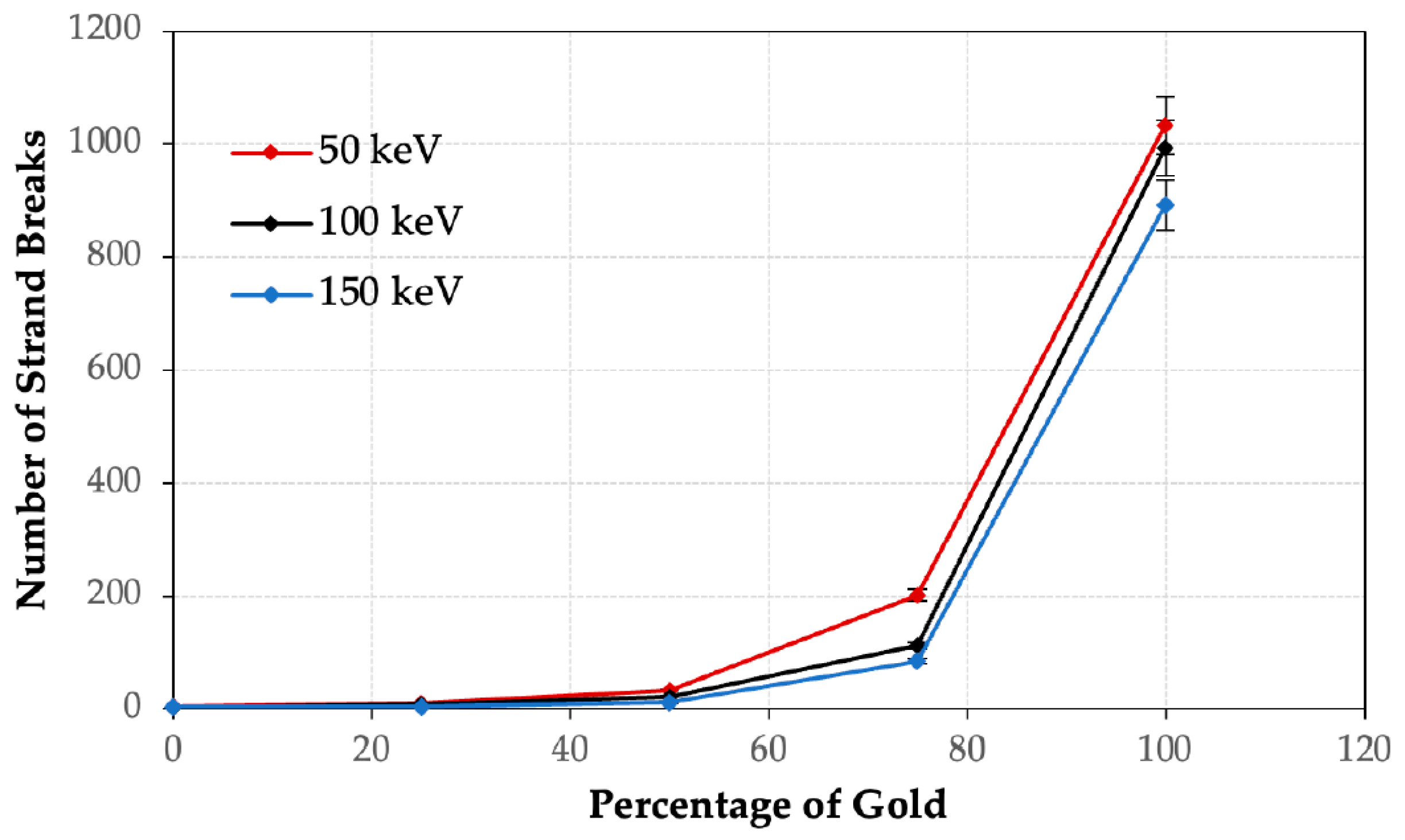
Table 2a presents the occurrence of SSBs and DSBs at different ratios of gold to iron when exposed to a 50 keV photon beam. In Figure 2, the nanoparticle configuration reveals iron as the core with gold wrapped around it. The findings indicate that when the proportion of iron exceeds that of gold within the nanoparticle, there is a decrease in the number of SSBs and DSB. In addition, it is observed that the number of DSBs is consistently smaller than that of SSBs across all photon beam energies. Similar trends can be observed in Table 2b,c for the 100 keV and 150 keV photon beams, respectively. These findings suggest that the iron serves as a core surrounded by the gold in the nanoparticle heterojunction. The volume of gold increases proportionally with the percentage ratio of gold to iron. For instance, when the percentage ratio is 100% gold and 0% iron, the nanoparticle is entirely composed of gold. Conversely, a pure iron nanoparticle is observed when the percentage ratio is 0% gold and 100% iron. Each simulation in Table 2 was conducted with 10 million histories. The relationship between the number of strand breaks and the percentage of volume of gold in the nanoparticle heterojunction is depicted in Figure 3 for various photon beam energies. The uncertainties of the results are based on the information from the Monte Carlo simulations. The results demonstrate that an increase in the percentage of volume of gold in the nanoparticle heterojunction correlates with a higher number of DNA strand breaks.
4. Discussion
4.1. Dependence of DNA Damage on the Volume of Gold or Iron
Table 2 presents the number of SSBs and DSBs for different percentages of gold to iron at various photon beam energies. Across all photon beam energies, it is evident that the number of DNA strand breaks increases with the percentage or volume of gold. For a photon beam energy of 50 keV (Table 2a), decreasing the percentage of gold from 100% to 75% results in a 5.1-fold reduction in DNA damage (SSBs and DSBs). Further decreasing the percentage of gold from 75% to 20% and from 50% to 25% leads to reductions of 5.9 times and 3.4 times, respectively. This phenomenon can be attributed to the reduction in dose enhancement resulting from the introduction of iron in the iron-gold nanoparticles. It was observed that the dose enhancement of iron was significantly lower than that of gold when subjected to kilovoltage photon beams [17]. When the volume of gold is reduced to zero and the nanoparticle heterojunction becomes purely iron, the number of DNA strand breaks is only 0.1% of that observed in the gold nanoparticle (100% gold and 0% iron). Similar trends are observed in the 100 keV (Table 2b) and 150 keV (Table 2c) photon beam energies, with the number of SSBs or DSBs decreasing with increasing beam energy.
The increase in DNA damage with a larger volume of gold can be attributed to the generation of secondary electrons at the heterojunction when irradiated by the photon beam. It is well-known that SSBs and DSBs are caused by the ionization of the DNA molecule strands due to charged particles such as secondary electrons [29]. These electrons can be generated from the heterojunction through the interaction between the photon beam and the nanoparticle. In the kV photon energy range, the photoelectric effect is dominant. The cross section or probability of photoelectric interaction is proportional to the atomic number (Z) raised to the power exponent n, where n typically ranges from 4 to 5. For a fixed wavelength, materials with higher atomic numbers, such as gold (Z = 79), have larger cross sections than iron (Z = 26) due to this relationship. It is important to note that the cross-section values can vary with different wavelengths, and the stated proportionality is valid only for a specific wavelength. This implies that a heterojunction containing a larger volume of gold can generate more secondary electrons compared to the iron volume. These findings align with the dose enhancement factors of gold and iron nanoparticles in nanoparticle-enhanced radiotherapy. The dose enhancement factor is defined as the ratio of the dose at the DNA with the addition of nanoparticles to the dose at the DNA without the addition of nanoparticles [30]. It is observed that a larger volume of gold in the heterojunction can enhance the dose at the DNA, resulting in increased DNA damage. Consequently, this enhances cancer cell kill and tumor control in patients.
In MRI-guided radiotherapy, it is essential to consider both the unique properties of gold and iron nanoparticles. While gold nanoparticles are known to be excellent radiation dose enhancers, the presence of iron nanoparticles is crucial due to their specific magnetic properties that contribute to enhancing contrast in MRI images.
Iron nanoparticles possess strong magnetic characteristics and can become magnetized when exposed to a magnetic field. This magnetization leads to local disturbances in the surrounding magnetic field, causing changes in the relaxation times of nearby water molecules. These changes can be detected by MRI scanners, enabling enhanced contrast in the resulting images [7]. Therefore, incorporating iron into the nanoparticle structure in the iron-gold heterojunction is necessary to achieve the desired contrast enhancement in MRI. On the other hand, gold nanoparticles offer various advantages in the heterojunction. They exhibit excellent biocompatibility, stability, and ease of functionalization, making them suitable for a wide range of biomedical applications [10]. In MRI-guided radiotherapy, the gold component in the heterojunction can provide additional benefits such as improved stability, enhanced biocompatibility, and potential for targeting cancer cells [11].
It is important to note that in IGRT, including MRI-guided radiotherapy, the iron component cannot be completely eliminated. This is because the unique magnetic properties of iron nanoparticles play a vital role in enhancing MRI contrast, which is essential for accurate imaging and treatment guidance. Therefore, a balance is required in the iron-gold nanoparticle heterojunction to achieve both enhanced MRI contrast and radiation dose enhancement in the context of IGRT.
4.2. Dependence of DNA Damage on the Photon Beam Energy
Figure 3 depicts the relationship between DNA damage and photon beam energy for different percentages of gold and iron in the nanoparticle heterojunction. The results reveal that higher photon beam energies result in lower levels of DNA damage caused by the irradiated nanoparticle heterojunction. Specifically, when the percentage volume of gold is 75%, decreasing the photon beam energy from 100 keV to 50 keV leads to a 1.8-fold increase in strand breaks. Further decreasing the photon beam energy from 150 keV to 50 keV results in a larger increase, with strand breaks rising 2.4 times. Similar trends can be observed when the percentage volume of gold is reduced to 50%, where strand breaks increase 1.5 times and 2.8 times when the photon beam energy is decreased from 100 keV and 150 keV to 50 keV, respectively. However, when the percentage volume of gold is equal to or less than 25%, the decrease in strand breaks with an increase in photon beam energy is not significant. This highlights the significant role played by the volume of gold in DNA damage in relation to changes in photon beam energy.
The increase in the number of DNA strand breaks with an increase in the volume of gold in the nanoparticle heterojunction can be attributed to an augmented secondary electron yield from the nanoparticle. This, in turn, enhances the radiation dose at the DNA. The observed effect is primarily due to the dominance of the photoelectric effect in the kV photon beam energy range. The cross section of the photoelectric effect is inversely proportional to the photon energy (1/E3.5), where E represents the energy of the incident photon. Consequently, lower photon beam energies result in larger photoelectric cross section and the generation of a greater number of photoelectric or secondary electrons. However, this effect is particularly evident when the percentage of gold in the nanoparticle heterojunction is substantial, as the photoelectric effect is also influenced by the atomic number of the nanoparticle material.
In radiotherapy, tissue penetration is a crucial consideration alongside dose enhancement resulting from photon beam energy. In nanoparticle-enhanced radiotherapy, the objective is to selectively enhance the radiation dose within the tumor while minimizing damage to surrounding healthy tissues. The use of kV photon beams is advantageous in terms of tissue penetration and the controlled delivery of radiation dose, enabling precise targeting of the tumor region. This makes kV photon beams suitable for intraoperative radiotherapy (IORT), a specialized form of radiotherapy conducted during surgery [31]. Unlike conventional external beam radiotherapy, which originates from outside the body, IORT involves placing a radiation source directly inside the patient’s body, usually in close proximity to the tumor. IORT finds application in the treatment of various cancers, including breast cancer, colorectal cancer, pancreatic cancer, and sarcomas [32,33]. It is typically integrated into a comprehensive treatment plan that may also involve surgery, chemotherapy, and/or external beam radiation therapy.
5. Conclusions
The Monte Carlo simulation was utilized to assess the DNA damage induced by irradiating an iron-gold nanoparticle heterojunction with kV photon beams. This heterojunction serves as both a radiation dose enhancer and a contrast agent for MRI in image-guided nanoparticle-enhanced radiotherapy. The findings indicate that DNA damage increases as the percentage of gold volume in the heterojunction rises, primarily due to photoelectric enhancement. Moreover, a lower photon beam energy of 50 keV leads to greater DNA damage compared to energies of 100 keV and 150 keV. The results suggest that to achieve effective cancer cell kill through DNA damage, the gold volume should be equal to or greater than 50% in the iron-gold nanoparticle heterojunction. These Monte Carlo results hold significance for clinicians and radiation personnel involved in designing functionalized nano-theranostic agents used in image-guided nanoparticle-enhanced radiotherapy. The forthcoming tasks involve fine-tuning the nanoparticle size and concentration to enhance the biocompatibility with healthy tissue. In addition to the current efforts, future work will involve developing a comprehensive multi-scale model that integrates simulations of the tumor, cell, and DNA. This integrated model will enable the estimation of the dose multiplication factor, which is crucial for assessing the cumulative effect of radiation on DNA damage in relation to the total dose received in radiotherapy.
Author Contributions
Conceptualization, J.C.L.C.; methodology, J.C.L.C.; software, C.A.S. and J.C.L.C.; validation, C.A.S. and J.C.L.C.; formal analysis, C.A.S.; investigation, C.A.S.; resources, C.A.S. and J.C.L.C.; data curation, C.A.S.; writing—original draft preparation, J.C.L.C.; writing—review and editing, C.A.S. and J.C.L.C.; visualization, C.A.S. and J.C.L.C.; supervision, J.C.L.C.; project administration, J.C.L.C. All authors have read and agreed to the published version of the manuscript.
Funding
This research received no external funding.
Institutional Review Board Statement
Not applicable.
Informed Consent Statement
Not applicable.
Data Availability Statement
Not applicable.
Acknowledgments
The authors would like to thank Chun He and Kaden Kujanpaa from the University of Toronto, Canada, and Mehwish Jabeen from the Ryerson University, Canada, for their assistance in the MC simulation using the Geant4-DNA code.
Conflicts of Interest
The authors declare no conflict of interest.
References
- Sung, H.; Ferlay, J.; Siegel, R.L.; Laversanne, M.; Soerjomataram, I.; Jemal, A.; Bray, F. Global cancer statistics 2020: GLOBOCAN estimates of incidence and mortality worldwide for 36 cancers in 185 countries. CA Cancer J. Clin. 2021, 1, 209–249. [Google Scholar] [CrossRef]
- Canadian Cancer Statistics Advisory Committee in Collaboration with the Canadian Cancer Society; Statistics Canada and the Public Health Agency of Canada. Canadian Cancer Statistics 2021; Canadian Cancer Society: Toronto, ON, Canada, 2021; Available online: Cancer.ca/Canadian-Cancer-Statistics-2021-EN (accessed on 5 July 2023).
- Brenner, D.R.; Weir, H.K.; Demers, A.A.; Ellison, L.F.; Louzado, C.; Shaw, A.; Turner, D.; Woods, R.R.; Smith, L.M. Canadian Cancer Statistics Advisory Committee Projected estimates of cancer in Canada in 2020. CMAJ Can. Med. Assoc. J. 2020, 192, E199–E205. [Google Scholar] [CrossRef] [PubMed] [Green Version]
- Baskar, R.; Lee, K.A.; Yeo, R.; Yeoh, K.W. Cancer and radiation therapy: Current advances and future directions. Int. J. Med. Sci. 2012, 9, 193–199. [Google Scholar] [CrossRef] [PubMed] [Green Version]
- Joiner, M.C.; van der Kogel, A.J.; Steel, G.G. Introduction: The significance of radiobiology and radiotherapy for cancer treatment. Basic Clin. Radiobiol. 2009, 13, 1. [Google Scholar]
- Chow, J.C.L. Photon and electron interactions with gold nanoparticles: A Monte Carlo study on gold nanoparticle-enhanced radiotherapy. Nanobiomaterials Med. Imaging 2016, 1, 45–70. [Google Scholar]
- Chow, J.C.L. Magnetic nanoparticles as contrast agents in magnetic resonance imaging and radiosensitizers in radiotherapy. Fundam. Ind. Appl. Magn. Nanoparticles 2022, 1, 291–316. [Google Scholar]
- Abdulle, A.; Chow, J.C.L. Contrast enhancement for portal imaging in nanoparticle-enhanced radiotherapy: A Monte Carlo phantom evaluation using flattening-filter-free photon beams. Nanomaterials 2019, 9, 920. [Google Scholar] [CrossRef] [Green Version]
- Albayedh, F.; Chow, J.C.L. Monte Carlo simulation on the imaging contrast enhancement in nanoparticle-enhanced radiotherapy. J. Med. Phys. 2018, 43, 195–199. [Google Scholar] [PubMed]
- Siddique, S.; Chow, J.C.L. Gold nanoparticles for drug delivery and cancer therapy. Appl. Sci. 2020, 10, 3824. [Google Scholar] [CrossRef]
- Moore, J.; Chow, J.C.L. Recent progress and applications of gold nanotechnology in medical biophysics using artificial intelligence and mathematical modeling. Nano Express 2021, 2, 022001. [Google Scholar] [CrossRef]
- Siddique, S.; Chow, J.C.L. Recent advances in functionalized nanoparticles in cancer theranostics. Nanomaterials 2022, 12, 2826. [Google Scholar] [CrossRef] [PubMed]
- Shen, Z.; Wu, A.; Chen, X. Iron oxide nanoparticle based contrast agents for magnetic resonance imaging. Mol. Pharm. 2017, 14, 1352–1364. [Google Scholar] [CrossRef] [PubMed]
- Danhier, F. To exploit the tumor microenvironment: Since the EPR effect fails in the clinic, what is the future of nanomedicine? J. Control. Release 2016, 244 Pt A, 108–121. [Google Scholar] [CrossRef]
- McMillan, T.J.; Tobi, S.; Mateos, S.; Lemon, C. The use of DNA double-strand break quantification in radiotherapy. Int. J. Radiat. Oncol. Biol. Phys. 2001, 49, 373–377. [Google Scholar] [CrossRef] [PubMed]
- Siddique, S.; Chow, J.C.L. Application of Nanomaterials in Biomedical Imaging and Cancer Therapy. Nanomaterials 2020, 10, 1700. [Google Scholar] [CrossRef] [PubMed]
- Zheng, X.J.; Chow, J.C.L. Radiation dose enhancement in skin therapy with nanoparticle addition: A Monte Carlo study on kilovoltage photon and megavoltage electron beams. World J. Radiol. 2017, 9, 63–71. [Google Scholar] [CrossRef] [PubMed]
- Kroese, D.P.; Brereton, T.; Taimre, T.; Botev, Z.I. Why the Monte Carlo method is so important today. Wiley Interdiscip. Rev. Comput. Stat. 2014, 6, 386–392. [Google Scholar] [CrossRef]
- Rogers, D.W. Fifty years of Monte Carlo simulations for medical physics. Phys. Med. Biol. 2006, 51, R287. [Google Scholar] [CrossRef]
- Santiago, C.A.; Chow, J.C.L. Variations in Gold Nanoparticle Size on DNA Damage: A Monte Carlo Study Based on a Multiple-Particle Model Using Electron Beams. Appl. Sci. 2023, 13, 4916. [Google Scholar] [CrossRef]
- Agostinelli, S.; Allison, J.; Amako, K.A.; Apostolakis, J.; Araujo, H.; Arce, P.; Asai, M.; Axen, D.; Banerjee, S.; Barrand, G.J.; et al. GEANT4—A simulation toolkit. Nucl. Instrum. Methods Phys. Res. Sect. A Accel. Spectrometers Detect. Assoc. Equip. 2003, 506, 250–303. [Google Scholar] [CrossRef] [Green Version]
- Arce, P.; Rato, P.; Canadas, M.; Lagares, J.I. GAMOS: A Geant4-based easy and flexible framework for nuclear medicine applications. In Proceedings of the IEEE Nuclear Science Symposium Conference Record, Dresden, Germany, 19–25 October 2008; pp. 3162–3168. [Google Scholar]
- Incerti, S.; Baldacchino, G.; Bernal, M.; Capra, R.; Champion, C.; Francis, Z.; Gueye, P.; Mantero, A.; Mascialino, B.; Moretto, P.; et al. The geant4-dna project. Int. J. Model. Simul. Sci. Comput. 2010, 1, 157–178. [Google Scholar] [CrossRef]
- Taheri, A.; Khandaker, M.U.; Moradi, F.; Bradley, D.A. A review of recent advances in the modeling of nanoparticle radiosensitization with the Geant4-DNA toolkit. Radiat. Phys. Chem. 2023, 1, 111146. [Google Scholar]
- Pitt-Francis, J.; Whiteley, J. Guide to Scientific Computing in C++; Springer: Cham, Switzerland, 2017. [Google Scholar]
- Jabeen, M.; Chow, J.C.L. Gold Nanoparticle DNA Damage by Photon Beam in a Magnetic Field: A Monte Carlo Study. In special Issue entitled “Application of Nanomaterials in Biomedical Imaging and Cancer Therapy”. Nanomaterials 2021, 11, 1751. [Google Scholar] [CrossRef]
- Chatzipapas, K.P.; Tran, N.H.; Dordevic, M.; Zivkovic, S.; Zein, S.; Shin, W.G.; Sakata, D.; Lampe, N.; Brown, J.M.; Ristic-Fira, A.; et al. Simulation of DNA damage using Geant4-DNA: An overview of the “molecularDNA” example application. Precis. Radiat. Oncol. 2023, 7, 4–14. [Google Scholar] [CrossRef]
- Delage, E.; Pham, Q.T.; Karamitros, M.; Payno, H.; Stepan, V.; Incerti, S.; Maigne, L.; Perrot, Y. PDB4DNA: Implementation of DNA geometry from the Protein Data Bank (PDB) description for Geant4-DNA Monte-Carlo simulations. Comput. Phys. Commun. 2015, 192, 282–288. [Google Scholar] [CrossRef] [Green Version]
- Chow, J.C.L. Characteristics of secondary electrons from irradiated gold nanoparticle in radiotherapy. In Handbook of Nanoparticles; Aliofkhazraei, M., Ed.; Springer International Publishing: Cham, Switzerland, 2015; Chapter 10; pp. 1–18. ISBN 978-3-319-13188-7. [Google Scholar]
- Roeske, J.C.; Nuñez, L.; Hoggarth, M.; Labay, E.; Weichselbaum, R.R. Characterization of the theorectical radiation dose enhancement from nanoparticles. Technol. Cancer Res. Treat. 2007, 6, 395–401. [Google Scholar] [CrossRef]
- Paunesku, T.; Woloschak, G.E. Future directions of intraoperative radiation therapy: A brief review. Front. Oncol. 2017, 7, 300. [Google Scholar] [CrossRef] [Green Version]
- Kyrgias, G.; Hajiioannou, J.; Tolia, M.; Kouloulias, V.; Lachanas, V.; Skoulakis, C.; Skarlatos, I.; Rapidis, A.; Bizakis, I. Intraoperative radiation therapy (IORT) in head and neck cancer: A systematic review. Medicine 2016, 95, e5035. [Google Scholar] [CrossRef]
- Sedlmayer, F.; Reitsamer, R.; Fussl, C.; Ziegler, I.; Zehentmayr, F.; Deutschmann, H.; Kopp, P.; Fastner, G. Boost IORT in breast cancer: Body of evidence. Int. J. Breast Cancer 2014, 2014, 472516. [Google Scholar]
Figure 1.
Schematic diagram showing the Monte Carlo experimental geometry for the iron-gold nanoparticle heterojunction irradiated by 50–150 keV photon beams (gamma rays).
Figure 1.
Schematic diagram showing the Monte Carlo experimental geometry for the iron-gold nanoparticle heterojunction irradiated by 50–150 keV photon beams (gamma rays).

Figure 2.
Schematic diagram of the iron-gold nanoparticle heterojunction with gold on the outer layer and the iron embedded inside.
Figure 2.
Schematic diagram of the iron-gold nanoparticle heterojunction with gold on the outer layer and the iron embedded inside.

Figure 3.
Relationship between number of strand breaks and percentage of gold in iron-gold nanoparticle heterojunction at different photon beam energies.
Figure 3.
Relationship between number of strand breaks and percentage of gold in iron-gold nanoparticle heterojunction at different photon beam energies.

Table 1.
The volume percentages of gold and iron used in the iron-gold nanoparticle simulation as shown in Figure 1.
Table 1.
The volume percentages of gold and iron used in the iron-gold nanoparticle simulation as shown in Figure 1.
| Percentage of Gold | Percentage of Iron |
|---|---|
| 0% | 100% |
| 25% | 75% |
| 50% | 50% |
| 75% | 25% |
| 100% | 0% |
Table 2.
SSBs and DSBs present at different ratios of gold to iron when irradiated by (a) 50 keV, (b) 100 keV, and (c) 150 keV photon beam, respectively.
Table 2.
SSBs and DSBs present at different ratios of gold to iron when irradiated by (a) 50 keV, (b) 100 keV, and (c) 150 keV photon beam, respectively.
| Percentage of Gold to Iron | SSBs | DSBs |
|---|---|---|
| (a) | ||
| 0% Au, 100% Fe | 4 | 0 |
| 25% Au, 75% Fe | 9 | 1 |
| 50% Au, 50% Fe | 30 | 4 |
| 75% Au, 25% Fe | 201 | 1 |
| 100% Au, 0% Fe | 1011 | 22 |
| (b) | ||
| 0% Au, 100% Fe | 2 | 0 |
| 25% Au, 75% Fe | 8 | 1 |
| 50% Au, 50% Fe | 22 | 0 |
| 75% Au, 25% Fe | 109 | 3 |
| 100% Au, 0% Fe | 982 | 11 |
| (c) | ||
| 0% Au, 100% Fe | 4 | 0 |
| 25% Au, 75% Fe | 4 | 0 |
| 50% Au, 50% Fe | 11 | 1 |
| 75% Au, 25% Fe | 83 | 2 |
| 100% Au, 0% Fe | 885 | 7 |
Disclaimer/Publisher’s Note: The statements, opinions and data contained in all publications are solely those of the individual author(s) and contributor(s) and not of MDPI and/or the editor(s). MDPI and/or the editor(s) disclaim responsibility for any injury to people or property resulting from any ideas, methods, instructions or products referred to in the content. |
© 2023 by the authors. Licensee MDPI, Basel, Switzerland. This article is an open access article distributed under the terms and conditions of the Creative Commons Attribution (CC BY) license (https://creativecommons.org/licenses/by/4.0/).
Share and Cite
MDPI and ACS Style
Chow, J.C.L.; Santiago, C.A. DNA Damage of Iron-Gold Nanoparticle Heterojunction Irradiated by kV Photon Beams: A Monte Carlo Study. Appl. Sci. 2023, 13, 8942. https://doi.org/10.3390/app13158942
AMA Style
Chow JCL, Santiago CA. DNA Damage of Iron-Gold Nanoparticle Heterojunction Irradiated by kV Photon Beams: A Monte Carlo Study. Applied Sciences. 2023; 13(15):8942. https://doi.org/10.3390/app13158942
Chicago/Turabian StyleChow, James C. L., and Christine A. Santiago. 2023. "DNA Damage of Iron-Gold Nanoparticle Heterojunction Irradiated by kV Photon Beams: A Monte Carlo Study" Applied Sciences 13, no. 15: 8942. https://doi.org/10.3390/app13158942
Note that from the first issue of 2016, this journal uses article numbers instead of page numbers. See further details here.