

Motion Technologies in Support of Fence Athletes: A Systematic Review
by
 , , , , and
Simona Aresta
1,
Mariapia Musci
2,
Francesco Bottiglione
2,
Lorenzo Moretti
3,4,
Biagio Moretti
3,4 and
Ilaria Bortone
3,*
1
Department of Electrical and Information Engineering (DEI), Politecnico di Bari, 70126 Bari, Italy
2
Department of Mechanical Mathematics and Management (DMMM), Politecnico di Bari, 70126 Bari, Italy
3
Department of Translational Biomedicine and Neuroscience (DiBraiN), School of Medicine, University of Bari “Aldo Moro”, AOU Consorziale Policlinico, Piazza Giulio Cesare 11, 70124 Bari, Italy
4
Orthopaedic & Trauma Unit, AOU Consorziale Policlinico, Piazza Giulio Cesare 11, 70124 Bari, Italy
*
Author to whom correspondence should be addressed.
Appl. Sci. 2023, 13(3), 1654; https://doi.org/10.3390/app13031654
Submission received: 2 December 2022
/
Revised: 16 January 2023
/
Accepted: 20 January 2023
/
Published: 28 January 2023
(This article belongs to the Special Issue Sports Performance and Health (in Times of COVID-19))
Abstract
:Sports biomechanics enables thorough examination of athletic movements to enhance athletic performance and/or reduce injury risk. Few studies have looked at the possibilities of cutting-edge technology in fencing, even though it presents an intriguing scenario for sports biomechanics due to the significant demands it places on the body in terms of neuromuscular coordination, strength, power, and musculoskeletal system impact. The aim of the study is to identify and summarise current evidence on the application of motion technologies in support of fence athletes and to provide a framework for the assessment and training of fencers, including performance measures and protocols. Peer-reviewed research was identified from electronic databases using a structured keyword search. Details regarding experimental design, study group characteristics, and measured outcomes were extracted from retrieved studies, summarised, and information regrouped under themes for analysis. The methodological quality of the evidence was evaluated. Thirty-five studies were included in the present review, which showed kinetic, kinematic, muscle recruitment and coordination differences among athletes as gender and athletic training differed. Findings revealed that most of the included studies investigated the lunge technique in professional athletes using Optoelectronic Systems and force platforms as preferred motion technologies. Only nine studies reported the assessment of muscle activation during task execution (%). Less than 20% of the study recurred to Artificial Intelligence/Machine Learning (AI/ML) approaches in the analysis. The potential contribution of the user’s kinematic/kinetic data and physiological measures is still underestimated. The recommendations provided in this study could help promote and support further cross-sectional and longitudinal studies in the field.
1. Introduction
In the open-skilled combat sport of fencing, no physical contact is permitted between the two competitors as they engage in an indirect struggle with their weapons. Each of the three competing weapons—foil, saber, and épée—has its regulations, and men and women compete with them. For safety purposes, fencing gear, masks, gloves, and plastrons are required [1]. This sport requires a high level of coordination, explosive strength, speed, and precision due to its asymmetrical nature.
The most common kind of assault is the lunge attack. The flèche and those derived from in-stance counterattacks are some more. Acquiring proficiency in the lunge action is one of the essential parts of fencing. Lunge starts with a sword arm extension that threatens the touch zone, followed by an explosive anterior leg extension. Aspects related to kinematics and muscle action have mainly been used to describe and research the control mechanism behind lunge performance. Additionally, the fencer’s performance is greatly influenced by his response to his opponent’s moves [2].
Technology assists in the research of fencing biomechanics because the kinetics and kinematics of the lunge have not yet been quantitatively described. Athletes’ long-term training results, physical health, and technical proficiency are assessed in sports performance analysis. Knowing how fencers behave in a match or during practice is significant because it may be helpful for them to improve their muscle coordination and strength to prevent injuries, but most importantly, to react more quickly to the opponent’s action. Coaches design a personalized training schedule to enhance trainee abilities and technique, and using a variety of instruments; they can monitor their development.
Field hockey, football, swimming, badminton, soccer, and fencing are just a few of the sports whose biomechanics are being studied thanks to growing interest in technology research, as mentioned by Adesida and colleagues [3]. Human motion analysis is one of the most precise methods for addressing the biomechanics of professional sports. Video analysis is among the most effective and often used tools for human motion analysis. Athletes receive external visual feedback from cameras during practice or competition. On the other hand, real-time tracking and recording of human motion is the goal of automatic tracking motion analysis systems, commonly referred to as motion capture. These systems use various capturing techniques, from many infrared video cameras to single-camera systems with additional sensors to get depth information from the scene [4]. Sports motion analysis using video analysis is accurate. However, it is expensive, necessitating exact calibration and a tidy workstation to conduct the analysis. On the other hand, Wearable technology is less expensive, can track real-time exertion data, does not require considerable setup, gives accurate measurements, gathers quick and real-time feedback, and provides physiological and physical information about athletes. In both team and individual sports, wearable technology has been widely used [5].
Evaluating fencers’ performance during a competition or practice session could help qualitative and quantitative studies. Fencing experts can use motion-capturing data to qualitatively improve fencers’ attack skills and mentors to pinpoint what fencers need to improve. By obtaining these kinds of results, the quantity and accuracy of the accessible data may help to build new paradigms for fencing. As a result, finding the appropriate technology to collect data becomes crucial; instrumentation is just as vital as knowing how to conduct tests. A credible scientific analysis of these devices is needed to keep up with the wearable sector’s exponential growth. A wearable device’s success depends on winning the confidence of users, stakeholders, and policymakers. These recommendations can aid in evaluating wearable sensor technologies and/or selecting suitable goods by wearable device manufacturers, athletes, coaches, team managers, insurance companies, and other stakeholders [6]. A quantitative description of motion can be provided by wearable technology such as inertial systems, surface electromyography (sEMG), and accelerometers, which can be used to track sporting activity. Motion capture systems are most commonly used to evaluate fencing movements. The demand for wearable technology in sports science is rising due to the integration of numerous devices into a single sensor, which enables a more thorough evaluation of a player’s growth. As a result, our objective was to study the available technologies for fence analysis and identify, assess, and describe them.
2. Materials and Methods
2.1. Search Strategy
A systematic search was conducted in PubMed, Scopus, IEEExplore, and Academia from the earliest record. A literature search was performed up to June 2022. This review was not registered. The search strategy included a combination of keywords [All Fields]: ‘fencing’ AND (technology*) OR (accelerometer*) OR (inertial sensor* OR inertial system) OR (‘motion capture’ AND system) OR (electromyography* OR sEMG*) AND (performance AND analysis OR ‘motion analysis’ OR ‘time motion analysis’) OR (movement OR lunge) OR (biomechanics).
2.2. Eligibility Criteria
Studies measuring the performance of fencers during practice or contests using wearables or motion capture technology were considered. The term “wearable devices” refers to any equipment worn by fencers during practice or competition, including but not limited to GPS, inertial motion units (IMUs), accelerometers, sEMG, and other devices. Cameras, markers, and Kinect are examples of motion capture systems, whereas GPS, IMU, accelerometers, sEMG, and other devices worn by fencers during practice or competitions are examples of wearable technology. There were no restrictions on the nation, age, or level of competition for male and female fencers. The analysis covered all forms of practice and competition (whether global or regional). The research included information about any physiological and/or biomechanical data made by wearable technology, such as but not limited to distance measurements, velocity/speed, acceleration, deceleration, and muscle activity. Additionally, studies that used data mining or machine learning to study footwork, biomechanics, or data mining were included. The only full-text journal articles that may be included were those written in English. The study excluded systematic or scoping reviews. Two researchers independently compared the search outcomes to the eligibility requirements (SA and MM). Disagreements were resolved through conversation.
2.3. Data Extraction
The year of publication, the type of study (longitudinal, cross-sectional), the competitive level (élite, novice), the sex (female, male), the number of participants, the study design (’experimental, pilot, observational’), the purpose of the study, the clinical scales, and the instrumentation used were all retrieved. Additionally, data about the lab’s configuration, the business, the model, the dimensions (bi-, tri-), the muscles, the key performance metrics, the presence of outside stimuli, and the application of artificial intelligence (AI) or machine learning (ML) algorithms were collected.
2.4. Data Analysis
To assess the quality of the included studies, the Mixed Methods Appraisal Tool (MMAT) has been identified as a helpful resource for the Risk of Bias Assessment [7]. The MMAT is a critical appraisal tool designed for the appraisal stage of systematic mixed studies reviews, i.e., reviews that include qualitative, quantitative, and mixed methods studies. It permits the appraisal of the methodological quality of five categories of studies: qualitative research, randomized controlled trials, non-randomized studies, quantitative descriptive studies, and mixed methods studies. We selected the “quantitative descriptive” category for the present systematic review that best represented the included studies (Table 1).
3. Results
3.1. Included Studies
The retrieved results are summarised in Figure 1. We identified 56 studies, with 35 retained for the analysis. We included as supplementary materials the PRISMA Checklist (Table 1) and the complete table with all the included works and details (Table 2).
Among the retained studies (Table 2), 25 examined the lunge maneuver, considered the core component of fencing (Figure 2). The biomechanics of the lower limbs was assessed in 6 studies and the biomechanics of the upper limbs in 2. Nine studies evaluated muscle activation and coordination in the execution of the gesture.
Expert/élite fencing athletes were the significant components of research subjects (n = 18, %), which increases the difficulty of enlarging the sample size for the recruited studies because top athletes are always rare (range of sample size: 1 to 30 athletes). These studies included all fencing weapons: foil, épée, and saber. Foils were addressed in 11 studies, épée in 19, and saber in 5. Most studies were experimental (n = 27, %) and used Optoelectronic Systems standard equipment for biomechanical assessment (n = 23), in some cases the Optoelectronic Systems were used in combination with Force Platforms (n = 13) or surface Electromyography (n = 2).
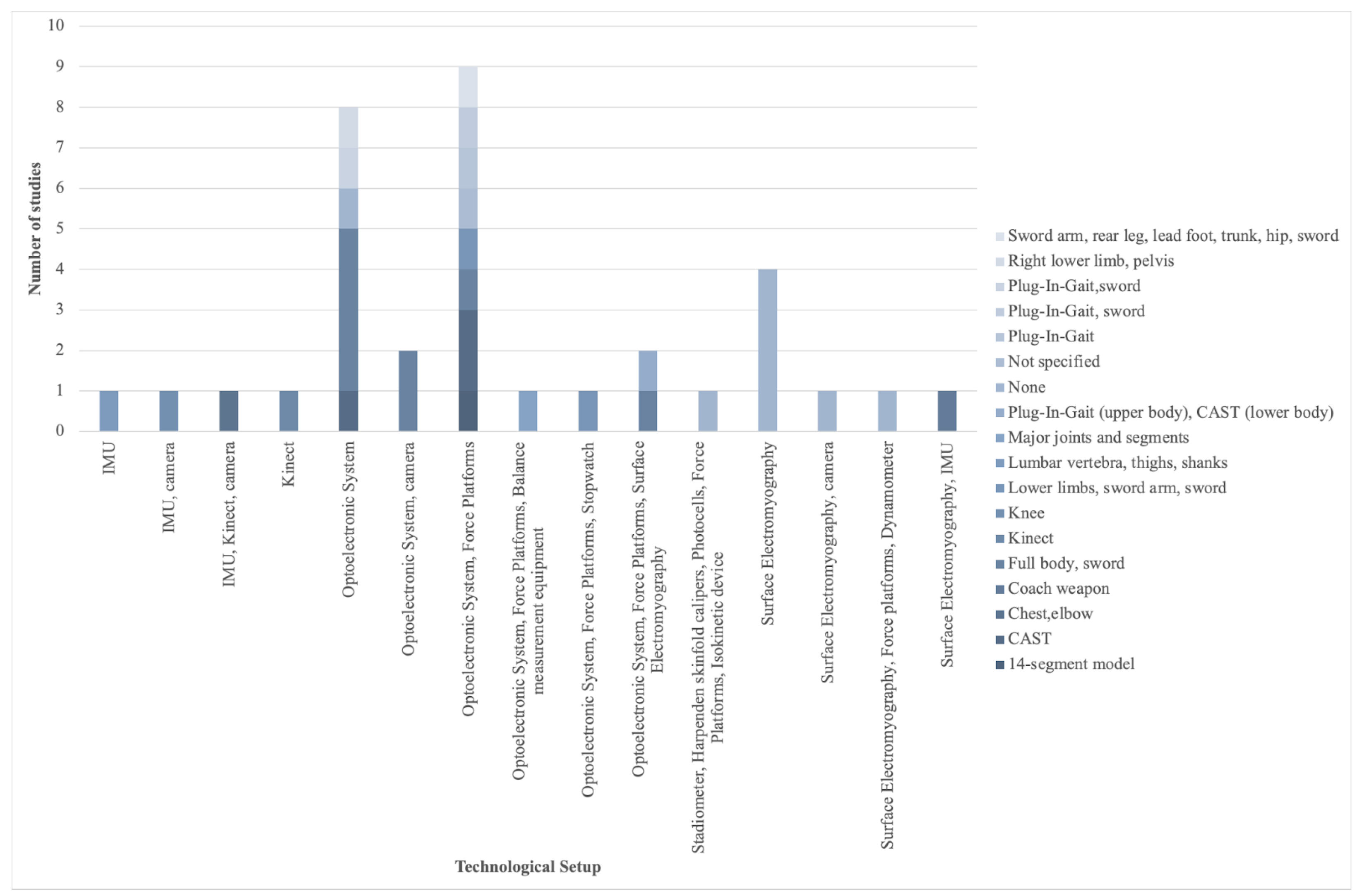
3.2. Technological Setup and Models
Based on the results of the 35 included studies, shown in Figure 3, it emerged that Optoelectronic Systems (n = 23 [2,8,9,10,11,12,13,14,15,16,17,18,19,20,21,22,23,24,25,26,27,28,29]) were the most popular technological setup in the analysis of fencers, followed by sEMG technology (n = 9 [10,15,30,31,32,33,34,35,36], IMUs (n = 4 [10,37,38,39] and markerless systems, i.e., Kinect (n = 2 [38,40]). The instrumental setups mentioned in the included articles were produced by different companies: Vicon motion capture systems (Oxford Metrics, Oxford, UK) consisted of a minimum of 6 cameras to 16 cameras, with a variable number of 2 force platforms, Kistler (Kistler Instrument Corp., Novi, MI, USA). Twenty-nine studies reported the use of tridimensional models: the most used ones were the Calibrated Anatomical System Technique (CAST) protocol (n = 4, [8,9,10,24]) and Plug-In-Gait (n = 4, [10,11,12,13]). Only one study [40] developed customized software for the Kinect sensor to compare two movement patterns and allow data to be extracted for predictive modeling purposes.
Guan and colleagues studied the biomechanical factors affecting lunge’s speed using 12 cameras, 20 anatomical markers, and 42 markers placed on the bilateral sides of the feet, ankle, leg, knee, thigh, shoulder, and trunk. Additionally, they combined two force platforms to show that élite fencers had greater peak horizontal center of gravity velocities and higher peak horizontal ground response forces exerted by the rear leg than novice fencers. The rear knee’s peak joint power, peak joint moment, and range of motion were all higher in the élite fencers than in the novice fencers. Fencing competitors at both the élite and novice levels showed joint flexion before anterior knee extension [14].
Bottoms and colleagues examined the kinematics of the lunge speed using 8 cameras, retro-reflective markers positioned in accordance with the CAST procedure, and 4 marker cluster systems [8]. The findings demonstrate that knee flexion in the rear extremity is a significant predictor, which suggests that the fencer sits low in their stance to generate power during lunge [8].
Gholipour and colleagues used reflective markers placed on the foot, shank, thigh, pelvis, trunk, and arm. Three high-speed cameras were employed, one of which captured the markers’ entire movement and course. The software then combines the information from the three cameras to produce the 3D coordinates; the speed lunge is calculated as the travel distance and time of the marker placed on the iliac spine of the armed side; this was greater for élite fencers than novice, as for the lunge distance and the trunk inclination [17].
Hassan and Klauck used an infrared device to measure the speeds of the foil, the pelvis, the elbow, the shoulder, the knee, the ankle, the heel, and the fingers on both sides. They calculated the maximum horizontal speed of the foil, the maximum vertical speed of the foil, the maximum horizontal speed of the pelvis, the horizontal speed of the foil at the time of impact, the vertical speed of the pelvis at the time of impact, and the maximum horizontal speed of the pelvis. From the maximum value, the horizontal speeds of the pelvis and foil slightly decrease over time. Additionally, time-varying marker curves were computed. Depending on the subject, the horizontal direction and angle of the foil velocity might vary by to [19]. The coordination of the upper and lower limbs was examined using the hip and lunge speeds [41].
Chuanjie and Zhengwei investigated joint angle, ground response forces, knee joint torque, and reaction force changes in the lunge movement of athletes and the causes of the knee joint using a high-speed infrared imaging system, camera, and three-dimensional lateral force platform [18]. Using a camera, the Peak Performance System, markers on the fingers, heel, ankle, knee, pelvis, shoulder, knee, wrist, hand, neck, ear, and sword, Zhang and colleagues described the biomechanics of fencing lunges. The results show that shorter subjects have trunk flexion and a greater lunge distance when their knee angle is smaller. For all subjects, the sword and leg moved at the same time. The sword’s speed is constant if it has a single peak reaction time of s, otherwise s [21].
The capacity of the fencers to attack and defend as swiftly as possible to win a tournament is another crucial component of fencing lunge. Two force platforms, a virtual plate, 24 cameras, 5 markers scattered across the screen, and 7 markings on the sword—4 on the bowl and 3 on the blade—were all utilized by Sorel and colleagues. Using a brand-new lunge assault simulator for fencing that enables the investigation of parameterizable and variable attack situations, they assessed fencer performance and reaction times under unpredictable settings [2]. According to Gutierrez (2013), the evaluation of response time can also be studied concerning the type of tactile or visual stimulus [23]; during a standard lunge, the peak force is lower, the acceleration time is longer, and the simple Reaction Time (RT), Movement Time (MT), and Reaction Response Time (RRT) are all shorter. The lunge starts with a higher force in the rear foot and a reduced force in the anterior foot when the target changes.
Using the CAST protocol, Sinclair and colleagues examined how the six available Cardan sequences affected the kinematic characteristics of the joints in the lower extremities as well as planar cross-talk in the sagittal, coronal, and transverse planes during the fencing lunge [29]. Their findings showed that the XYZ sequence was the best suitable for accurately representing the 3D kinematics of the leading leg during the fencing lunge. As a result, its ongoing use was supported. To investigate whether there were any changes in limb and joint stiffness characteristics between the sexes during the fencing lunge, Sinclair and Bottoms collected kinematic and kinetic data from 20 élite athletes [9]. Using the CAST protocol, kinematic information was quantified. An XYZ rotational sequence was used to compute the kinematics of the hip, knee, and ankle joints (where X represents sagittal plane; Y represents coronal plane and Z represents transverse plane rotations) [29]. Inverse dynamics based on Newton–Euler was also used, enabling calculation of the moments at the knee and ankle joints. The joint angular excursion, which represents the angular displacement from footstrike to peak angle, and the peak joint moment were the kinetic/kinematic parameters from the hip, knee, and ankle and were extracted for statistical analysis.

Table 2.
Analysis of the quality of studies included in the systematic review using MMAT version 2018.
Table 2.
Analysis of the quality of studies included in the systematic review using MMAT version 2018.
| Methodological Quality Criteria | |||||||
|---|---|---|---|---|---|---|---|
| S1 | S2 | 4.1 | 4.2 | 4.3 | 4.4 | 4.5 | |
| Said et al. [19] | Y | Y | Y | N | Y | Y | U |
| Klauck et al. [20] | Y | N | Y | N | Y | Y | U |
| Zhang et al. [21] | Y | N | Y | N | Y | Y | U |
| Williams et al. [32] | Y | Y | Y | N | Y | Y | Y |
| Gholipour et al. [17] | Y | Y | Y | Y | Y | Y | Y |
| Mantovani et al. [12] | Y | Y | U | U | Y | N | N |
| SuchaNwski et al. [36] | Y | U | N | N | U | Y | N |
| Morris et al. [11] | Y | N | N | N | Y | Y | N |
| Bottoms et al. [8] | Y | Y | Y | Y | Y | Y | Y |
| Gutierrez-Davila et al. [23] | Y | Y | Y | Y | Y | Y | Y |
| Gutierrez-Davila et al. [22] | Y | Y | Y | Y | Y | Y | Y |
| Borysiuk et al. [31] | Y | Y | Y | N | Y | U | Y |
| Sinclair et al. [29] | Y | Y | Y | Y | Y | Y | Y |
| Sinclair et al. [24] | Y | Y | Y | U | Y | Y | Y |
| Guilhem et al. [34] | Y | Y | Y | N | Y | Y | Y |
| Borysiuk et al. [33] | Y | Y | U | U | Y | U | N |
| Moorea et al. [16] | Y | N | N | N | N | U | N |
| Sinclair et al. [8] | Y | Y | Y | Y | Y | Y | Y |
| Kim et al. [27] | Y | Y | Y | Y | Y | Y | Y |
| Borysiuk [35] | Y | Y | U | N | Y | U | U |
| Malawski et al. [37] | Y | Y | Y | Y | Y | Y | U |
| Mawgoud et al. [40] | Y | Y | U | U | Y | U | U |
| Guan et al. [14] | Y | Y | Y | Y | Y | Y | Y |
| Plantard et al. [15] | Y | Y | Y | Y | Y | U | U |
| Chuanjie et al. [18] | Y | Y | Y | Y | Y | Y | U |
| O’Reilly et al. [39] | Y | Y | Y | Y | Y | Y | Y |
| Blazkiewicz et al. [13] | Y | Y | Y | Y | Y | Y | Y |
| Mulloy et al. [28] | Y | Y | Y | Y | Y | Y | Y |
| Sorel et al. [2] | Y | Y | Y | Y | Y | Y | Y |
| Borysiuk et al. [10] | Y | Y | Y | Y | Y | Y | Y |
| Milic et al. [26] | Y | Y | Y | Y | Y | Y | Y |
| Grontman et al. [25] | Y | U | U | U | Y | U | U |
| Borysiuk et al. [30] | Y | Y | Y | Y | Y | Y | Y |
| Chtara et al. [42] | Y | Y | Y | Y | Y | Y | Y |
| Malawski [38] | Y | Y | Y | Y | Y | U | U |
Legend: S1, S2, 4.1, 4.2, 4.3, 4.4 & 4.5 the MMAT criteria shown in Table 1; Y: Yes; N: No; U: Unable to tell.
The use of wearable sensors and/or markerless technology to acquire kinematic data from fencers was limited to just 4 separate groups. Malawski used a 2 IMU and Kinect to evaluate fencing gestures for a functional task [37]. IMUs were placed on the chest and the wrist [37]. Mawgoud and colleagues collected information about the fencing lunge using Kinect and a data mining approach to train a neural network (MLP), provide fencers features of their movement, and enhance it over time [40]. O’Reilly and colleagues employed 5 IMUS on the lumbar region, both thighs, and shanks to determine whether a lunge was executed correctly [39].
3.3. Purpose and Algorithms
The studies that were included demonstrated significant heterogeneity in both the scope of the work and the athletes studied. As shown in Figure 4, they ranged from a basic biomechanical analysis of fencing gestures, particularly the lunge movement, to identify differences in athletes from various levels or categories (n = 5) and muscle patterns (n = 6). Most studies examined the kinematics and kinetics of the gesture to understand the biomechanics of fencing. Studies investigating training activities were rare (e.g., balance [27] and change of direction [42]).
Bottoms and Sinclair studied the disparities between élite male and female athletes during the épée lunge, focusing on kinetic and kinematic elements [9,24]. Their research shed light on the causes of the different damage patterns concerning fencing among the sexes. Indeed, their findings suggest that female athletes may be more susceptible to knee injuries since the fencing lunge causes more knee abduction and hip adduction in females.
To portray the 3D kinematics, Borysiuk and colleagues integrated two biomechanical models: the Plug-In Gait model for the upper body (i.e., the left and right sides of the patient’s arms) and the CAST approach for the markers affixed to the participants’ lower limbs (on the left and right sides) [10]. To determine the fencing assault (flèche versus lunge) that is more effective in an actual competition, their investigation concentrated on the metrics reflecting sensorimotor responses (RT and MT) paired with sEMG signal and ground reaction forces.
Suchanowski and colleagues used sEMG analysis of the Rectus Femoris in both legs and the Extensor Carpi Radialis in the armed arm to define the dynamic model of the élite fencer lunge. They came to the conclusion that élite athletes begin the fencing lunge by tensing the muscles in the rear lower extremity. Additionally, the anterior lower extremity functions asynchronously, but the armed arm and rear lower extremities work synchronously [36]. By analyzing the order of arousal in the following muscles—the Rectus Femoris and the Biceps Femoris of the anterior leg, the Gastrocnemius muscle (medial and lateral head) of the rear leg, the Biceps Brachii, Triceps Brachii, and the Brachioradialis muscle of the armed arm—Borysiuk and colleagues sought to determine the dynamic structure of the fencing lunge [33]. According to their findings, seeing and anticipating visual inputs led to a decrease in muscle tension.
Plantard and colleagues used optical motion capture technologies with wearable sEMG devices to study how different muscle synergies, both upper and lower body, vary during lunge caused by visual stimulation. Two synergies were found for the lower body, whereas 3 synergies for the upper body. The task was not as discriminating as anticipated, despite slight changes based on whether the target was moving or not being identified in how the muscles were synergically recruited [15]. To ascertain the connection between muscle activation, muscular strength, and mechanical efficacy of the assault, Guilhem and colleagues examined the coordination of lower limb muscles during a particular saber assault (i.e., marché-fente) [34]. Their findings highlighted the importance of the rear extensor muscles in fencing speed performance, opening up new ideas for designing tailored training or recovery regimens for élite competitors.
3.4. Assessment of Quality
The quality of the studies incorporated into the current systematic review was evaluated using the MMAT. The research topic was stated in the introduction of every study, but less than half of them did not include a sample of the target population that was representative of it or a statistical analysis that was suitable to address the research issue.
4. Discussion
The aim of this systematic review was to provide an overview of the motion technology categories, biomechanical model categories, and algorithm categories that are utilized to assist fencers in their performances.
4.1. Technological Setup and Biomechanical Models
The goal in élite sports is often to monitor, assess, and enhance performance by giving feedback to athletes, coaches, and/or sports scientists [43]. Fencing, being a non-contact combat sport, is characterized by the presence of rapid actions, due to defensive or offensive actions, with possible changes of direction at a rapid pace, interspersed with periods of start and stop. These movements typically cause injuries in fencing athletes [44]. Harmer conducted a prospective 5-year study that showed that 52% of injuries, reported during national competitions, were strains and sprains, 12% contusions, % fractures, and the remainder involved injuries affecting knee joint the most [44]. The majority of injuries (63%) occurred in the lower extremities, primarily affecting the knee, followed by the thigh and ankle areas. The two most often affected areas above the hip were the back, notably the lumbar region, and the fingers [44]. Performing the lunge movement correctly is essential to prevent injuries. Speed, explosive strength, and movement coordination pattern all significantly affect fencers’ performances [45]. According to Turner et al., coaches typically tailor programs to a fencer’s skills, sex, and age based on their experience or personal knowledge rather than on objective data [46]. Thus, Turner et al. argued for the creation of evidence-based training programs that consider a fencer’s biomechanics, physiological needs, and physiological status to prevent injuries [46].
The injury risk is influenced by different factors such as gender and category level (élite/novice) [47]. By reporting on the influence of gender, injury rates were greater among female fencers (29–44%, average: 36%) than among male fencers (22–32%, average: 27%) [44]. In particular, female fencers may be more prone to anterior cruciate ligament injuries than male fencers due to an increased dynamic Q-angle caused by greater hip adduction and knee abduction [24]. Looking at category-level differences, élite fencers exhibit greater coordination of movement according to joint kinematics and muscle activation pattern. Considering joint kinematics, élite fencers’ lunge movement presents the proximal-to-distal coupling of upper and lower limb motion, which guarantees an efficient transformation of the joint segmental angular velocity of the lower limb into the maximal linear velocity of the center of mass. Furthermore, élites begin lunging by extending the armed arm, activating the corresponding Anterior Deltoid, and then raising the anterior foot. This is known as the proximal-to-distal sequence for muscle activation. Overall, compared to novices, élite fencers showed better-coordinated muscle synergies of the upper and lower limbs. These synergies were defined by the sequential activation of the forelimbs and then the hip and knee extensors of the rear lower limb [45]. These muscle synergies were characterized by the sequential activation of the shoulder/elbow extensors of the armed upper limb and the hip/knee extensors of the rear lower limb, followed by the activation of the anterior limb during the lunge. The élites showed the ability to maintain this activation pattern almost invariant, despite, the changing actions of the opponents, whereas the muscle activation patterns of the novice fencers were more inconsistent, with frequent pauses in movement [45]. Therefore, while élite fencers can precisely adjust muscle activation patterns to optimize attacking (lunge) efficiency without deviating from the “correct” kinematic pattern, novice fencers may not have consolidated neuromuscular strategies for complex and multi-segmental movements [45]. Indeed, studies have revealed that a routine of just fencing training does not increase muscle strength and coordination. Ballistic training is advised to increase the rate of muscular force, with the majority of the benefits appearing in the first 200 to 300 ms of a single lunge movement. Another crucial component of fencing is training for coordination and balance. When doing single-leg standing activities, fencers showed improved coordination and less body sway after undergoing focused balance training [45].
Recent developments in wearable technology enable real-time movement and load measurements of athletes during practice and training, which was previously impossible due to a lack of data. With such a wide range of data readily available, coaches can easily verify training progresses according to movement and muscle activation patterns. The key to allowing an athlete’s body to withstand the external stresses brought on by hits that may otherwise cause collisions and injuries is the athlete’s body’s capacity to quickly acquire movement patterns and initiate muscle activation [48]. Hence motion technologies can provide a series of evidence-based assessments to categorize risk status, which helps clinicians identify those athletes who are most at risk for injury, to place them on corrective exercise programs to address their weaknesses [47]. Modern research has concentrated on moving traditional sports science research from the laboratory to more realistic field settings thanks to recent advancements in hardware (IMU, markerless camera, wireless sensors), data processing, and field conditions [49,50]. Various Optoelectronic technologies from various firms have been utilized in the included investigations. Furthermore, the configuration setting and models used to examine the biomechanics of the gesture in terms of the quantity and kind of devices showed a large amount of variety.
Even though Optoelectronic Systems were used in the majority of studies, there was much variation in the laboratory setup in terms of the number of infrared cameras (1 to 24), the presence and number of force platforms (2 to 16), and the number of markerless or wearable devices (sEMG) that were used. We have determined that it is impossible to compare the results because there are 28 distinct laboratory setting combinations and 15 different biomechanical models, one of which only considers the usage of sEMG with six different combinations of muscles of interest. The outcomes of the listed research were then examined in light of the technological setup and model they used.
Compared to marker-based techniques, markerless motion capture can do movement analysis with fewer data gathering and processing times [51]. The two primary categories of camera hardware can be used in single- or multi-camera systems and use either depth or regular video cameras. Additionally, markerless techniques increase the data’s adaptability by allowing datasets to be re-analyzed using more recent posture estimation algorithms [51]. They may even enable clinicians to collect data on patients wearing regular clothing. The imprecision of joint center location detection and the need for advanced programming and computer vision understanding primarily causes the slow adoption of markerless motion capture in biomechanics [52]. Wearable technology, on the other hand, is a different strategy that could be able to get around these restrictions. Various sensor types, such as IMUs and microelectromechanical sensors (MEMS), which combine magnetometers, accelerometers, and gyroscopes, have shown promising results in research analyzing sports performance [3]. Such sensors have the advantage of being easy to install. They provide reasonably priced measurements with great accuracy.
Surface electromyography in sports science helps examine muscle coordination, identify muscle on and off, and determine how long a muscle was engaged during an action [53]. This knowledge could be invaluable for coaches to plan individualized training for the athletes to avoid injuries and strengthen their areas of weakness [46,53]. Studies looking into the muscle activation pattern reported positioning the probes according to SENIAM guidelines [54]. However, every study included in the review suggested a different way to record muscle activity during data collecting. The Triceps Brachii, the Biceps Brachii, and the Extensor Carpi Radialis were the most often mentioned muscles for the upper limb, appearing in six studies. The Anterior Deltoid has only been studied in two studies, even though the proximal-to-distal sequence was also reported for muscle activation [55], with activation of the Anterior Deltoid of the armed upper limb, with the extension of the armed arm preceding the lifting of the lead foot at the beginning of lunging in élite fencers. Eight studies out of 35 recorded the Rectus Femoris and Biceps Femoris muscle activity in the lower limb. Two investigations included the Tibialis Anterior. Five studies considered the Medial and Lateral Gastrocnemius. The sequential kinematic chain can be used in numerous propelling sporting activities because mathematical modeling shows how well this lower limb rigid body chain converts joint segment angular velocity into the linear center of mass velocity [56].
The armed side of the body commands movement during a significant portion of a competitive bout and training, making fencing a highly asymmetric sport [45]. The neuromuscular system is further burdened by the unique mobility patterns of the upper and lower extremities and the dominant effects on kinematics and kinetics [32]. The primary function of the human musculoskeletal system is to convert joint rotations into linear movement, which necessitates the careful synchronization of several skeletal muscles around numerous joints. An ideal kinematic arrangement of these joints enables successful movement. The marker system of a protocol defines the input information that the biomechanical model will obtain from the capture. Each marker applied to the subject produces an X, Y, and Z location in the capture region for each trial frame. The marker positions must be (1) recognizable on the subject, such as bone protrusions that are either palpable or visible on the skin, and (2) situated in a point that provides valuable information for the model, such as close to a joint’s center of rotation or a bone segment that maintains a measurable relationship to the location of said center of rotation [57]. The most popular techniques for characterizing and analyzing human gait were the CAST Protocol [58] and the Plug-In-Gait [59]; additionally, of the studies did not report any specific biomechanical model related to the goal of the work, and another of the included studies concentrated on muscle activations.
4.2. Purpose and Algorithms
Two investigations by Sinclair and Bottoms [9,24] both looked into gender disparities. Findings suggested that due to significantly higher hip/limb stiffness and knee moment in female fencers, they may be more prone to overuse problems than male fencers. Numerous investigations looked into the muscle movements involved in making the fencing motion. Suchanowski and colleagues [36] demonstrated that muscle activation started with the rear leg during the lunge executed by a élite athlete. According to research by Borysiuk and colleagues [31], a competitor’s required tactical actions determine the order of muscle arousal. The same authors later demonstrated that waiting for a visual stimulus and watching for visual stimuli reduce muscle tension because they cause a greater accumulation of bioelectrical potentials than tactile stimulation [33]. To determine which fencing attack would be more successful in a genuine competition, Borysiuk and colleagues analyzed two fencing attacks (the lunge and the flèche) [10]. They concluded that the flèche generated larger levels of EMG and ground response forces than the lunge, which improved the explosive force and decreased the moving phase of the complete offensive motion.
Sports informatics literature pays attention to AI/ML approaches. Examples of applications that have benefited from AI classification methods include sensor-based feedback systems [38], team-play analysis systems [39], and several pose recognition applications [41]. Less than of the included studies, however (n = 6), utilized AI/ML algorithms in their approaches, and all of them were concerned with classifying gestures in athletes, both élite and novice, based on their kinematics (mainly lunge, n = 3) and one study either kinematics or muscle activity.
Few researchers discuss how accelerometer-based algorithms can categorize fencing movement. Mantovani and colleagues could categorize the basic fencing movements by compiling a library of movements [12]. Malawski and Kwolek proposed a single accelerometer-based algorithm to categorize fundamental footwork in fencing [37]. With this evidence, it can be concluded that footwork is integral to every fencing motion. They consider it a crucial step in creating a comprehensive method and tactics analysis framework. It is feasible to examine acceleration data by extracting useful properties from it. Four separate classifiers had to be trained to comprehend fencers’ footwork. These features were to be collected from an accelerometer put on the fencer’s knee. The classifiers were Radial Basis Function-Support Vector Machine (SVM-RBF), Dynamic Time Warping (DTW), DTW-feat, and linear SVM. The DTW classifier, which achieved 70% accuracy, was the most precise [37]. O’Reilly and colleagues employed 5 IMUS on the lumbar region, both thighs, and shanks to determine whether a lunge was executed correctly [39]. To assess the accuracy of the lunge exercise categorization, Leave-One-Subject-Out-Cross-Validation (LOSOCV) and the random-forests (RF) classifier with 400 trees were utilized. A set of five lower limb IMUs may be able to distinguish between acceptable and abnormal lunge movements with 90% accuracy, 80% sensitivity, and 92% specificity, according to research. Based on data from three IMUs, a system’s accuracy, sensitivity, and specificity are 87%, 73%, and 91%, respectively. The classification scores with a reduced IMU set-up are comparable since it is assumed that the characteristics of thighs and shanks will be similar. This merely adds a small amount of data to improve categorization accuracy. Only employing data from the right thigh IMU, the approach achieved accuracy, sensitivity, and specificity of 82%, 78%, and 83%, respectively. Multi-label categorization of particular deviations is accomplished with 70% accuracy, 70% sensitivity, and 97% specificity using data from the whole 5 IMU setup. A 3 IMU setup may offer greater system functionality and efficiency when compared to a 5 IMU setup, but accuracy and sensitivity are reduced by 8–10%. A system with two IMUs did not produce good results for multi-label categorization. These results are probably influenced by the number of classes the algorithm tries to discover. Additionally, these aberrations are more pronounced at particular anatomical places, making it challenging to detect them with a smaller IMU. Furthermore, these aberrations are harder to detect with a smaller IMU setup because they are more pronounced at particular anatomical regions. If only one IMU is employed, the characteristics will likely be less discriminative between classes when using limited IMU setups to discover these specific errors [39]. Muscle activity could be included in addition to the kinematic study of the lunge to increase the discriminative power in a classification problem while maintaining a limited IMU setup. In our recent work, we acquired kinematic and electromyographic data on 21 élite and novice fencers during the lunge, step-forward lunge, and step-backward lunge. We used only wearable devices to collect biomechanical data, particularly one IMU and four sEMG probes. The IMU was placed on the lower back (on the L5/S1 vertebra). The four sEMG wearable probes were placed, according to SENIAM recommendations, the Anterior Deltoid and Rectus Femoris on the armed side, Logissimus Erector spinal muscle, and Medial Gastrocnemius on the other side. Muscles were chosen following an asymmetrical pattern, reflecting the asymmetry of the lunge gesture. The aim of the study was to find the best ML model to classify élite and novice fencers to support fencers and coaches during training. For this purpose, four models (MLP, SVM, RF, XGBoost (XGB)) were trained using kinematic and sEMG data. We found the best combination between model performances and the number of principal components (k), extracted from each biomechanical variable to classify the study population. Our results showed that the best ML algorithm was MLP with k = 50 and a training and test accuracy of 100% and 96%, respectively, [60].
4.3. Review Limitation
The results of this review should be interpreted with limitations in mind. The search was restricted to four databases, though it was supplemented by hand searches and reference lists to find additional pertinent papers. This review’s findings are also constrained by the selection of search terms and inclusion criteria, as utilizing different terms and criteria would have resulted in a different number of publications being included. However, similar reviews that have been published in the past served as a guide for the search phrases and criteria. A language bias in article selection resulted from restricting included articles to those published in English. The quality assessment checklist was developed using a standardized tool that was not previously mentioned in reviews of a similar nature.
5. Conclusions
Optoelectronic Systems are currently the most widely used motion technology for fence analysis. Most studies focused on the fencing biomechanical profile during élite-level lunge execution. However, comparisons between studies are challenging because of a need for defined performance bands. Additionally, more wearables should be employed at the non-élite level to develop effective training regimens that will boost performance and lower the chance of injury. Wearable technologies are helpful resources that can assist coaches and sports scientists in comprehending performance and making appropriate adjustments to game strategies and training regimens. Researchers and engineers should keep creating novel wearables to offer comprehensive data on players’ overall performance, well-being, and safety.
Traditional biomechanical data collection suites have become more portable and simple to deploy in field settings thanks to developments in ICT and video processing algorithms. Furthermore, various sports-related issues have been addressed using cutting-edge modeling and analysis technologies. These software programs frequently use Artificial Intelligence (AI) methods. Coaches and athletes can benefit from effective data presentation and visualization, enhancing cognitive understanding of complicated data outputs.
Author Contributions
Conceptualisation, I.B., F.B. and L.M.; methodology, I.B. and F.B.; data review, S.A., M.M. and I.B.; writing—original draft preparation, S.A. and I.B.; writing—review and editing, I.B. and F.B.; supervision, I.B. and B.M. All authors have read and agreed to the published version of the manuscript.
Funding
This research received no external funding.
Institutional Review Board Statement
Not applicable.
Informed Consent Statement
Not applicable.
Data Availability Statement
No new data were created or analysed in this study. Data sharing does not apply to this article.
Acknowledgments
The authors would like to thank the A.s.d. CLUB SCHERMA BARI and Vito Capuano for his support as professional expert in the field in the study.
Conflicts of Interest
The authors declare no conflict of interest.
References
- Bianchedi, D. The Science of Fencing. Sports Med. 2008, 38, 465–481. [Google Scholar]
- Sorel, A.; Plantard, P.; Bideau, N.; Pontonnier, C. Studying fencing lunge accuracy and response time in uncertain conditions with an innovative simulator. PLoS ONE 2019, 14, e0218959. [Google Scholar] [CrossRef] [PubMed]
- Adesida, Y.; Papi, E.; McGregor, A.H. Exploring the role of wearable technology in sport kinematics and kinetics: A systematic review. Sensors 2019, 19, 1597. [Google Scholar] [CrossRef] [PubMed] [Green Version]
- Ortega, B.P.; Olmedo, J.M.J. Application of motion capture technology for sport performance analysis. Retos Nuevas Tendencias Educ. Física Deporte Recreación 2017, 32, 241–247. [Google Scholar]
- Lim, J.Z.; Sim, A.; Kong, P.W. Wearable Technologies in Field Hockey Competitions: A Scoping Review. Sensors 2021, 21, 5242. [Google Scholar] [CrossRef]
- Düking, P.; Fuss, F.K.; Holmberg, H.C.; Sperlich, B. Recommendations for assessment of the reliability, sensitivity, and validity of data provided by wearable sensors designed for monitoring physical activity. JMIR mHealth uHealth 2018, 6, e9341. [Google Scholar] [CrossRef]
- Hong, Q.N.; Pluye, P.; Fàbregues, S.; Bartlett, G.; Boardman, F.; Cargo, M.; Dagenais, P.; Gagnon, M.P.; Griffiths, F.; Nicolau, B.; et al. VP26 A critical appraisal tool for systematic mixed studies reviews. Int. J. Technol. Assess. Health Care 2018, 34, 166. [Google Scholar] [CrossRef]
- Bottoms, L.; Greenhalgh, A.; Sinclair, J. Kinematic determinants of weapon velocity during the fencing lunge in experienced épée fencers. Acta Bioeng. Biomech. 2013, 15, 109–113. [Google Scholar]
- Bottoms, L.; Sinclair, J. Gender differences in limb and joint stiffness during the fencing lunge. Cent. Eur. J. Sport Sci. Med. 2015, 11, 39–44. [Google Scholar] [CrossRef] [Green Version]
- Borysiuk, Z.; Markowska, N.; Konieczny, M.; Kręcisz, K.; Błaszczyszyn, M.; Nikolaidis, P.T.; Knechtle, B.; Pakosz, P. Flèche versus Lunge as the Optimal Footwork Technique in Fencing. Int. J. Environ. Res. Public Health 2019, 16, 2315. [Google Scholar] [CrossRef] [Green Version]
- Morris, N.; Farnsworth, M.; Robertson, D. Kinetic analyses of two fencing attacks–lunge and fleche. In Proceedings of the ISBS-Conference Proceedings Archive, Porto, Portugal, 27 June–1 July 2011. [Google Scholar]
- Mantovani, G.; Ravaschio, A.; Piaggi, P.; Landi, A. Fine classification of complex motion pattern in fencing. Procedia Eng. 2010, 2, 3423–3428. [Google Scholar] [CrossRef]
- Błażkiewicz, M.; Borysiuk, Z.; Gzik, M. Determination of loading in the lower limb joints during step-forward lunge in fencing. Acta Bioeng. Biomech. 2018, 20, 3–8. [Google Scholar] [PubMed]
- Guan, Y.; Guo, L.; Wu, N.; Zhang, L.; Warburton, D.E. Biomechanical insights into the determinants of speed in the fencing lunge. Eur. J. Sport Sci. 2018, 18, 201–208. [Google Scholar] [CrossRef] [PubMed]
- Plantard, P.; Sorel, A.; Bideau, N.; Pontonnier, C. Motion adaptation in fencing lunges: A pilot study. Comput. Methods Biomech. Biomed. Eng. 2017, 20, S161–S162. [Google Scholar] [CrossRef] [Green Version]
- Moore, K.C.; Chow, F.M.; Chow, J.Y. Novel lunge biomechanics in modern Sabre fencing. Procedia Eng. 2015, 112, 473–478. [Google Scholar] [CrossRef] [Green Version]
- Gholipour, M.; Tabrizi, A.; Farahmand, F. Kinematics analysis of lunge fencing using stereophotogrametry. World J. Sport Sci. 2008, 1, 32–37. [Google Scholar]
- Chuanjie, Z.; Zhengwei, F. Biomechanical analysis of knee joint mechanism of the national women’s epee fencing lunge movement. Biomed. Res. (0970-938X) 2017, 28, 104–110. [Google Scholar]
- Hassan, S.E.; Klauck, J. Kinematics of lower and upper extremities motions during the fencing lunge: Results and training implications. In Proceedings of the ISBS-Conference Proceedings Archive, Konstanz, Germany, 21–25 July 1998. [Google Scholar]
- Klauck, J.; Hassan, S.E. Lower and upper extremity coordination parameters during the fencing lunge. In Proceedings of the ISBS-Conference Proceedings Archive, Konstanz, Germany, 21–25 July 1998. [Google Scholar]
- Zhang, B.; Chu, D.; Hong, Y. Biomechanical analysis of the lunge technique in the elite female fencers. In Proceedings of the ISBS-Conference Proceedings Archive, Perth, WA, Australia, 30 June–6 July 1999. [Google Scholar]
- Gutierrez-Davila, M.; Rojas, F.J.; Antonio, R.; Navarro, E. Response timing in the lunge and target change in elite versus medium-level fencers. Eur. J. Sport Sci. 2013, 13, 364–371. [Google Scholar] [CrossRef] [Green Version]
- Gutiérrez-Dávila, M.; Rojas, F.J.; Caletti, M.; Antonio, R.; Navarro, E. Effect of target change during the simple attack in fencing. J. Sport. Sci. 2013, 31, 1100–1107. [Google Scholar] [CrossRef] [Green Version]
- Sinclair, J.; Bottoms, L. Gender differences in the kinetics and lower extremity kinematics of the fencing lunge. Int. J. Perform. Anal. Sport 2013, 13, 440–451. [Google Scholar] [CrossRef]
- Grontman, A.; Horyza, Ł.; Koczan, K.; Marzec, M.; Śmiertka, M.; Trybała, M. Analysis of sword fencing training evaluation possibilities using Motion Capture techniques. In Proceedings of the 2020 IEEE 15th International Conference of System of Systems Engineering (SoSE), Budapest, Hungary, 2–4 June 2020; pp. 325–330. [Google Scholar]
- Milic, M.; Nedeljkovic, A.; Cuk, I.; Mudric, M.; García-Ramos, A. Comparison of reaction time between beginners and experienced fencers during quasi-realistic fencing situations. Eur. J. Sport Sci. 2020, 20, 896–905. [Google Scholar] [CrossRef] [PubMed]
- Kim, T.; Kil, S.; Chung, J.; Moon, J.; Oh, E. Effects of specific muscle imbalance improvement training on the balance ability in elite fencers. J. Phys. Ther. Sci. 2015, 27, 1589–1592. [Google Scholar] [CrossRef] [PubMed] [Green Version]
- Mulloy, F.; Mullineaux, D.R.; Graham-Smith, P.; Irwin, G. An applied paradigm for simple analysis of the lower limb kinematic chain in explosive movements: An example using the fencing foil attacking lunge. Int. Biomech. 2018, 5, 9–16. [Google Scholar] [CrossRef] [Green Version]
- Sinclair, J.; Taylor, P.J.; Bottoms, L. The appropriateness of the helical axis technique and six available cardan sequences for the representation of 3-d lead leg kinematics during the fencing lunge. J. Hum. Kinet. 2013, 37, 7. [Google Scholar] [CrossRef] [PubMed]
- Borysiuk, Z.; Nowicki, T.; Piechota, K.; Błaszczyszyn, M. Neuromuscular, perceptual, and temporal determinants of movement patterns in wheelchair fencing: Preliminary study. BioMed Res. Int. 2020, 2020, 6584832. [Google Scholar] [CrossRef] [PubMed]
- Borysiuk, Z.; Piechota, K.; Minkiewicz, T. Analysis of performance of the fencing lunge with regard to the difficulty level of a technical-tactical task. J. Combat. Sport. Martial Arts 2013, 4, 135–139. [Google Scholar] [CrossRef] [Green Version]
- Williams, L.; Walmsley, A. Response timing and muscular coordination in fencing: A comparison of elite and novice fencers. J. Sci. Med. Sport 2000, 3, 460–475. [Google Scholar] [CrossRef]
- Borysiuk, Z.; Markowska, N.; Niedzielski, M. Analysis of the fencing lunge based on the response to a visual stimulus and a tactile stimulus. J. Combat. Sport. Martial Arts 2014, 5, 119–124. [Google Scholar] [CrossRef] [Green Version]
- Guilhem, G.; Giroux, C.; Couturier, A.; Chollet, D.; Rabita, G. Mechanical and muscular coordination patterns during a high-level fencing assault. Med. Sci. Sports Exerc. 2014, 46, 341–350. [Google Scholar] [CrossRef]
- Borysiuk, Z. Type of perception vs. lunge in fencing technique structure. Rev. Artes Marciales Asiát. 2016, 11, 36–37. [Google Scholar] [CrossRef] [Green Version]
- Suchanowski, A.; Boryszewski, Z.; Pakosz, P. Electromyography signal analysis of the fencing lunge by Magda Mroczkiewicz, the leading world female competitor in foil. Balt. J. Health Phys. Act. 2011, 3, 4. [Google Scholar] [CrossRef]
- Malawski, F.; Kwolek, B. Classification of basic footwork in fencing using accelerometer. In Proceedings of the 2016 Signal Processing: Algorithms, Architectures, Arrangements, and Applications (SPA), Poznan, Poland, 21–23 September 2016; pp. 51–55. [Google Scholar] [CrossRef]
- Malawski, F. Depth Versus Inertial Sensors in Real-Time Sports Analysis: A Case Study on Fencing. IEEE Sens. J. 2021, 21, 5133–5142. [Google Scholar] [CrossRef]
- O’Reilly, M.A.; Whelan, D.F.; Ward, T.E.; Delahunt, E.; Caulfield, B. Classification of lunge biomechanics with multiple and individual inertial measurement units. Sports Biomech. 2017, 16, 342–360. [Google Scholar] [CrossRef]
- Mawgoud, A.A.; Abu-Talleb, A.; El Karadawy, A.I.; Eltabey, M.M. The Appliance of Artificial Neural Networks in Fencing Sport Via Kinect Sensor. Available online: https://www.researchgate.net/profile/Ahmed-A-Mawgoud/publication/343055177_The_Appliance_of_Artificial_Neural_Networks_in_Fencing_Sport_Via_Kinect_Sensor/links/5f141f25299bf1e548c365ce/The-Appliance-of-Artificial-Neural-Networks-in-Fencing-Sport-Via-Kinect-Sensor.pdf (accessed on 1 December 2020).
- Riehle, H.J.; Vieten, M.M. ISBS ’98: XVI International Symposium on Biomechanics in Sports. In Proceedings of the ISBS-Conference Proceedings Archive, Konstanz, Germany, 21–25 July 1998. [Google Scholar]
- Chtara, H.; Negra, Y.; Chaabene, H.; Chtara, M.; Cronin, J.; Chaouachi, A. Validity and reliability of a new test of change of direction in fencing athletes. Int. J. Environ. Res. Public Health 2020, 17, 4545. [Google Scholar] [CrossRef] [PubMed]
- Spencer, M.; Lawrence, S.; Rechichi, C.; Bishop, D.; Dawson, B.; Goodman, C. Time–motion analysis of elite field hockey, with special reference to repeated-sprint activity. J. Sport. Sci. 2004, 22, 843–850. [Google Scholar] [CrossRef]
- Harmer, P.A. Incidence and characteristics of time-loss injuries in competitive fencing: A prospective, 5-year study of national competitions. Clin. J. Sport Med. 2008, 18, 137–142. [Google Scholar] [CrossRef]
- Chen, T.L.W.; Wong, D.W.C.; Wang, Y.; Ren, S.; Yan, F.; Zhang, M. Biomechanics of fencing sport: A scoping review. PLoS ONE 2017, 12, e0171578. [Google Scholar] [CrossRef] [Green Version]
- Turner, A.; Miller, S.; Stewart, P.; Cree, J.; Ingram, R.; Dimitriou, L.; Moody, J.; Kilduff, L. Strength and conditioning for fencing. Strength Cond. J. 2013, 35, 1–9. [Google Scholar] [CrossRef]
- Zadeh, A.; Taylor, D.; Bertsos, M.; Tillman, T.; Nosoudi, N.; Bruce, S. Predicting sports injuries with wearable technology and data analysis. Inf. Syst. Front. 2021, 23, 1023–1037. [Google Scholar] [CrossRef]
- Bahr, R. Demise of the fittest: Are we destroying our biggest talents? Br. J. Sport. Med. 2014, 48, 1265–1267. [Google Scholar] [CrossRef] [Green Version]
- Rigozzi, C.J.; Vio, G.A.; Poronnik, P. Application of wearable technologies for player motion analysis in racket sports: A systematic review. Int. J. Sport. Sci. Coach. 2022, 17479541221138015. [Google Scholar] [CrossRef]
- Magalhaes, F.A.d.; Vannozzi, G.; Gatta, G.; Fantozzi, S. Wearable inertial sensors in swimming motion analysis: A systematic review. J. Sport. Sci. 2015, 33, 732–745. [Google Scholar] [CrossRef]
- Mündermann, L.; Corazza, S.; Andriacchi, T.P. The evolution of methods for the capture of human movement leading to markerless motion capture for biomechanical applications. J. Neuroeng. Rehabil. 2006, 3, 1–11. [Google Scholar] [CrossRef] [PubMed] [Green Version]
- Wade, L.; Needham, L.; McGuigan, P.; Bilzon, J. Applications and limitations of current markerless motion capture methods for clinical gait biomechanics. PeerJ 2022, 10, e12995. [Google Scholar] [CrossRef] [PubMed]
- Türker, H.; Sze, H. Surface electromyography in sports and exercise. Electrodiagn. New Front. Clin. Res. 2013, 175–194. [Google Scholar] [CrossRef]
- Hermens, H.J.; Freriks, B.; Merletti, R.; Stegeman, D.; Blok, J.; Rau, G.; Disselhorst-Klug, C.; Hägg, G. European recommendations for surface electromyography. Roessingh Res. Dev. 1999, 8, 13–54. [Google Scholar]
- Yiou, E.; Do, M. In fencing, does intensive practice equally improve the speed performance of the touché when it is performed alone and in combination with the lunge? Int. J. Sport. Med. 2000, 21, 122–126. [Google Scholar] [CrossRef]
- Bobbert, M.F.; van Soest, A.J.K. Why do people jump the way they do? Exerc. Sport Sci. Rev. 2001, 29, 95–102. [Google Scholar] [CrossRef] [Green Version]
- Ning, C. Design and research of motion video image analysis system in sports training. Multimed. Tools Appl. 2019, 1–19. [Google Scholar] [CrossRef]
- Cappozzo, A.; Catani, F.; Della Croce, U.; Leardini, A. Position and orientation in space of bones during movement: Anatomical frame definition and determination. Clin. Biomech. 1995, 10, 171–178. [Google Scholar] [CrossRef]
- Dempster, W.T. Space Requirements of the Seated Operator, Geometrical, Kinematic, and Mechanical Aspects of the Body with Special Reference to the Limbs; Technical Report; Michigan State Univ East Lansing: Lansing, MI, USA, 1955. [Google Scholar]
- Aresta, S.; Bortone, I.; Bottiglione, F.; Di Noia, T.; Di Sciascio, E.; Lofù, D.; Musci, M.; Narducci, F.; Pazienza, A.; Sardone, R.; et al. Combining Biomechanical Features and Machine Learning Approaches to Identify Fencers’ Levels for Training Support. Appl. Sci. 2022, 12, 12350. [Google Scholar] [CrossRef]
Figure 1.
PRISMA 2020 flow diagram for new systematic reviews, which included searches of databases, registers, and other sources.
Figure 1.
PRISMA 2020 flow diagram for new systematic reviews, which included searches of databases, registers, and other sources.

Figure 2.
Distribution of included studies according to the fencing technical gesture.
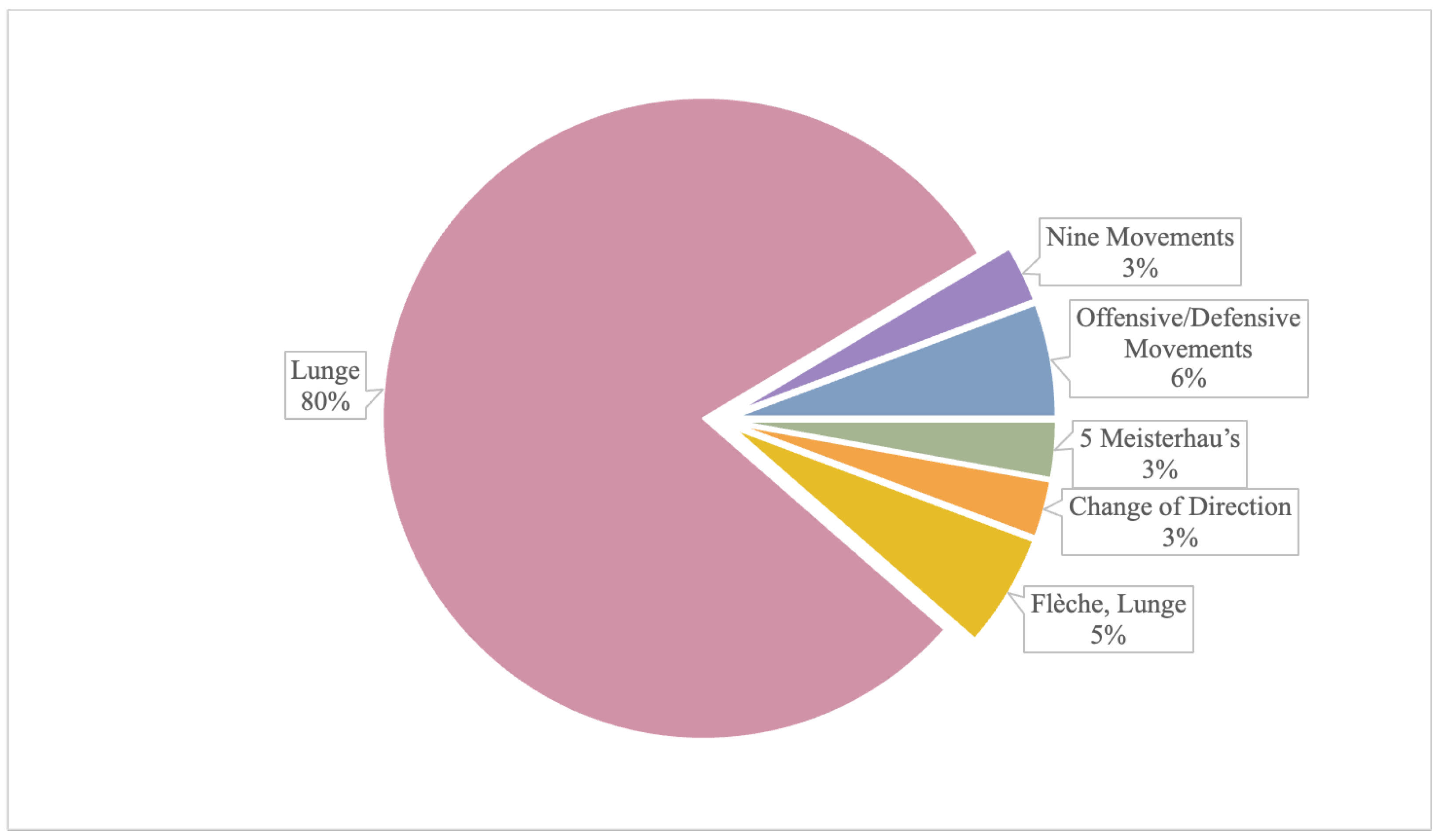
Figure 3.
Distribution of included studies according to the technological setup and the biomechanical model used for the analysis.
Figure 3.
Distribution of included studies according to the technological setup and the biomechanical model used for the analysis.

Figure 4.
Details of the included studies comparing élite versus novice athletes about the main aim and the presence of AI/ML Algorithms. DTW: Dynamic Time Warping; DTW-feat: a modification of DTW which compares a series of features; SVM: Support Vector Machine; SVM-RBF: Radial Basis Function-Support Vector Machine; K-NN: k-Nearest Neighbors; RF: Random Forest.

Table 1.
Mixed Methods Appraisal Tool (MMAT), version 2018.
| Category of Study Designs | Methodological Quality Criteria | Response | ||
|---|---|---|---|---|
| Y | N | U | ||
| Screening questions (for all types) | S1. Are there clear research questions? | |||
| S2. Do the collected data allow to address the research questions? | ||||
| Quantitative descriptive | 4.1. Is the sampling strategy relevant to address the research question? | |||
| 4.2. Is the sample representative of the target population? | ||||
| 4.3. Are the measurements appropriate? | ||||
| 4.4. Is the risk of nonresponse bias low? | ||||
| 4.5. Is the statistical analysis appropriate to answer the research question? | ||||
Y, Yes; N, No; U, Unable to tell.
Disclaimer/Publisher’s Note: The statements, opinions and data contained in all publications are solely those of the individual author(s) and contributor(s) and not of MDPI and/or the editor(s). MDPI and/or the editor(s) disclaim responsibility for any injury to people or property resulting from any ideas, methods, instructions or products referred to in the content. |
© 2023 by the authors. Licensee MDPI, Basel, Switzerland. This article is an open access article distributed under the terms and conditions of the Creative Commons Attribution (CC BY) license (https://creativecommons.org/licenses/by/4.0/).
Share and Cite
MDPI and ACS Style
Aresta, S.; Musci, M.; Bottiglione, F.; Moretti, L.; Moretti, B.; Bortone, I. Motion Technologies in Support of Fence Athletes: A Systematic Review. Appl. Sci. 2023, 13, 1654. https://doi.org/10.3390/app13031654
AMA Style
Aresta S, Musci M, Bottiglione F, Moretti L, Moretti B, Bortone I. Motion Technologies in Support of Fence Athletes: A Systematic Review. Applied Sciences. 2023; 13(3):1654. https://doi.org/10.3390/app13031654
Chicago/Turabian StyleAresta, Simona, Mariapia Musci, Francesco Bottiglione, Lorenzo Moretti, Biagio Moretti, and Ilaria Bortone. 2023. "Motion Technologies in Support of Fence Athletes: A Systematic Review" Applied Sciences 13, no. 3: 1654. https://doi.org/10.3390/app13031654
Note that from the first issue of 2016, this journal uses article numbers instead of page numbers. See further details here.