Article

Crude Polysaccharides from Wild-Growing Armillaria mellea—Chemical Composition and Antidiabetic, Anti-Inflammatory, Antioxidant, and Antiproliferative Potential

1
Department of Pharmaceutical Botany, Medical University of Lublin, ul. Chodźki 1, 20-093 Lublin, Poland
2
Sciex Europe (c/o Hach Lange Sp. z.o.o.), ul. Krakowska 119, 50-428 Wroclaw, Poland
3
Department of Molecular Biology, The John Paul II Catholic University of Lublin, ul. Konstantynów 1i, 20-708 Lublin, Poland
4
Faculty of Chemistry, University of Gdansk, ul. Wita Stwosza 63, 80-308 Gdansk, Poland
*
Authors to whom correspondence should be addressed.
Appl. Sci. 2023, 13(6), 3853; https://doi.org/10.3390/app13063853
Submission received: 22 February 2023 / Revised: 10 March 2023 / Accepted: 15 March 2023 / Published: 17 March 2023
(This article belongs to the Special Issue Functional Natural Compounds)

Abstract

:

Featured Application

Crude polysaccharide fraction from edible Armillaria mellea might be considered a functional food ingredient of natural origin.

Abstract

Armillaria mellea is a commonly harvested and consumed mushroom in Poland. Several activities of polysaccharides from this species have already been reported. However, polysaccharides from A. mellea growing in the wild in Poland have not yet been investigated. This study was conducted to obtain a crude polysaccharide fraction (AmPS) and investigate its chemical composition and biological properties. Our research suggests that A. mellea is a valuable source of polysaccharides, including β-glucans. 1H NMR and high-performance capillary electrophoresis analysis of AmPS revealed the occurrence of polysaccharides consisted of myo-inositol, mannitol, fucose, galactose, glucose, and mannose. Proteins, uronic acids, and phenolics were present only in trace amounts in the AmPS. The AmPS fraction was found to exhibit strong α-glucosidase inhibitory activity; however, no activity in relation to α-amylase was detected. The fraction was also able to inhibit pro-inflammatory enzymes, i.e., lipoxygenase, cyclooxygenase, and hyaluronidase. Moderate antioxidant and antiradical activity of AmPS was revealed by the ORAC and ABTS assay, respectively. The AmPS fraction was found to have antiproliferative potential against gastric and colorectal cancer cell lines (AGS, HT-29). These findings suggest that AmPS may be considered a health-beneficial food ingredient. Since A. mellea is a commonly known edible mushroom with a long tradition of use, its polysaccharides may be considered safe components for direct consumption or production of nutraceuticals.

1. Introduction

Armillaria mellea (Vahl) P. Kumm., also called honey mushroom, is an edible wild-growing mushroom commonly harvested and consumed in many countries, e.g., in Eastern Europe, and in China and the United Kingdom. Young fruiting bodies of A. mellea are attractive culinary products due to their flavor and texture as well as nutritional value. This species has been found to possess large amounts of consumptionally and pharmaceutically valuable compounds, including polysaccharides, proteins, terpenoids, indole compounds, sterols, minerals, and vitamins [1]. Among them, polysaccharides have been found to exhibit different pharmacological activities, such as immunomodulatory, antioxidant, and anticancer properties [2]. A study of A. mellea fruiting bodies harvested in China revealed that water-soluble polysaccharides from this species possess strong antitumor activities against the lung cancer cell line through the mitochondria-dependent pathway and activation of the caspase cascade via the release of cytochrome c [3]. Several in vivo studies have revealed an impact of polysaccharides from Chinese species of A. mellea on the immune system, i.e., enhancement of the immunological function in mice and improvement of the immune ability of mice by adjusting the immunocyte and cytokine. Additionally, protective effects against cyclophosphamide-induced damage to murine bone marrow cells have been revealed [4]. Sun et al. [5] found that polysaccharides from the honey mushroom from China were useful as a potential immunopotentiating agent, since they stimulated the murine lymphocyte proliferation induced by concanavalin A or lipopolysaccharide. Moreover, the antioxidant activities of different types of extracts from cultured mycelia of A. mellea have been evaluated. The available data indicate that water or ethanolic extracts can be used to reduce oxidative damage in the organism and prevent free radical-related diseases [6,7]. In recent years, it has been demonstrated that polysaccharides from A. mellea can be used as potential agents against diabetes. Neutral polysaccharides from this species enhanced insulin sensitivity in vitro, and lowered the blood glucose level and modulated lipid metabolism in mice [8].
All the aforementioned findings were revealed for specimens obtained in East Asia, most of which were cultivated. The knowledge of the composition and medicinal potential of polysaccharides obtained from A. mellea growing in the wild in Eastern Europe has remained limited so far. Meanwhile, it was previously observed that the geographic and climatic conditions influence chemical profile and biological effects of wild-growing mushrooms [9].
Since A. mellea is one of the most frequently collected mushrooms in Poland, the aim of our study was to investigate the broad health-beneficial activity of crude polysaccharides (AmPS) from wild-growing honey mushrooms for the first time, including their antioxidant, anti-inflammatory, antidiabetic, and antiproliferative potential. The chemical composition of AmPS was evaluated using various spectrophotometric methods. Moreover, high-performance capillary electrophoresis and 1H-nuclear magnetic resonance (1H NMR) were employed for the identification of the monosaccharide composition.

2. Materials and Methods

2.1. Materials

Armillaria mellea (Vahl) P. Kumm. fruiting bodies were harvested from the wild in the forests of Puszcza Solska, Poland (GPS: 50°28′28.6″ N 22°45′22.2″ E) in September 2019. The mushroom specimens (voucher specimen No. MSH-005) were confirmed by the authors (Prof. Renata Nowak). The collected mushrooms were freeze-dried after harvesting, then ground and stored in a freezer (−30 °C) until analysis.

2.2. Chemicals

2,2′-azinobis-(3-ethylbenzothiazoline-6-sulfonic acid) (ABTS•+), 2,2′-azobis (2-methylpropionamide) dihydrochloride (AAPH), 3,5-dinitrosalicylic acid, acetic acid, bovine serum albumin (BSA), Bradford reagent, gallic acid, hyaluronic acid, hyaluronidase, linoleic acid, p-nitrophenyl-α-D-glucopyranoside (PNPG), redistilled phenol, Rochelle salt, sodium acetate, sodium phosphate dibasic solution, sodium phosphate monobasic solution, soybean 15-lipoxygenase, Trolox, α-amylase, and α-glucosidase from Saccharomyces cerevisiae were purchased from Merck KGaA (Darmstadt, Germany). The COX (ovine) Colorimetric Inhibitor Screening Assay Kit was purchased from Cayman Chemical Company, Ann Arbor, USA. Fluorescein sodium salt was purchased from Roth (Karlsruhe, Germany). DMEM—Dulbecco’s modified Eagle medium, Dulbecco’s phosphate-buffered saline, high glucose, and L-Glutamine were purchased from Capricorn Scientific (Ebsdorfergrund, Germany). Penicillin, streptomycin, and fetal bovine serum were provided by Gibco. 3-(4,5-dimethylthiazol-2-yl)-2,5-diphenyltetrazolium bromide (MTT) and neutral red dye were provided by Sigma-Aldrich Fine Chemical Co. (St. Louis, MO, USA). Di-sodium hydrogen phosphate dihydrate (Na2HPO4·2H2O) was purchased from Chempur (Piekary Slaskie, Poland). The standard of myo-inositol was purchased from Sigma-Aldrich Fine Chemical Co. (St. Louis, MO, USA). Mannitol, xylitol, sucrose, fucose, galactose, glucose, mannose, fructose, and arabinose were provided by Dr. Ehrenstorfer (Augsburg, Germany). The Megazyme Mushroom and Yeast Beta-Glucan Assay Kit was obtained from Megazyme Ltd. (Wicklow, Ireland). Milli-Q quality (Millipore, Bedford, MA, USA) water was used throughout the HPCE analysis. Sodium hydroxide solution 1 M for HPCE was purchased from Merck (Darmstadt, Germany).
All spectrophotometric determinations were carried out using an Infinite Pro 200F microplate reader from Tecan Group Ltd. (Männedorf, Switzerland). Lyophilization was conducted using the Free Zone 1 apparatus (Labconco, Kansas City, KS, USA).

2.3. Extraction of A. mellea Crude Polysaccharides (AmPS)

Ground A. mellea (100 g) mushrooms were first macerated with ethanol (500 mL) for 24 h, and then ultrasound-assisted extraction (UAE) was applied for 30 min to remove low molecular weight compounds. The residue was extracted twice with hot water (500 mL) using UAE (30 min) as previously described [10]. The aqueous extract was purified with the use of Sevage reagent (chloroform/isoamyl alcohol, 4:1, v/v) to remove proteins. Precipitation of crude polysaccharides was performed with cold 99.8% ethanol overnight at 4 °C. Crude polysaccharides were centrifuged prior to lyophilization.

2.4. Chemical Composition of AmPS

2.4.1. Sugar Content

The investigation of the sugar content was performed using a phenol-sulfuric acid assay [11]. Glucose was used as a standard compound. The amount of sugars was calculated from the reference curve and presented as a percent of AmPS.

2.4.2. Uronic Acid Content

The amount of uronic acids was determined using previous protocol [12]. Galacturonic acid was used as a standard. The amount of uronic acids was calculated from the reference curve and presented as a percent of AmPS.

2.4.3. Protein Content

The presence of proteins in AmPS was investigated using the method of Bradford [13]. Bovine serum albumin (BSA) was utilized as a standard. The results were given as a percent of AmPS.

2.4.4. Total Phenolics Content

The presence of phenolics was evaluated with the protocol presented by Olech et al. [14]. Gallic acid was utilized as a standard compound. The results were calculated into gallic acid equivalents and presented as a percent of AmPS.

2.5. Structural Characterization

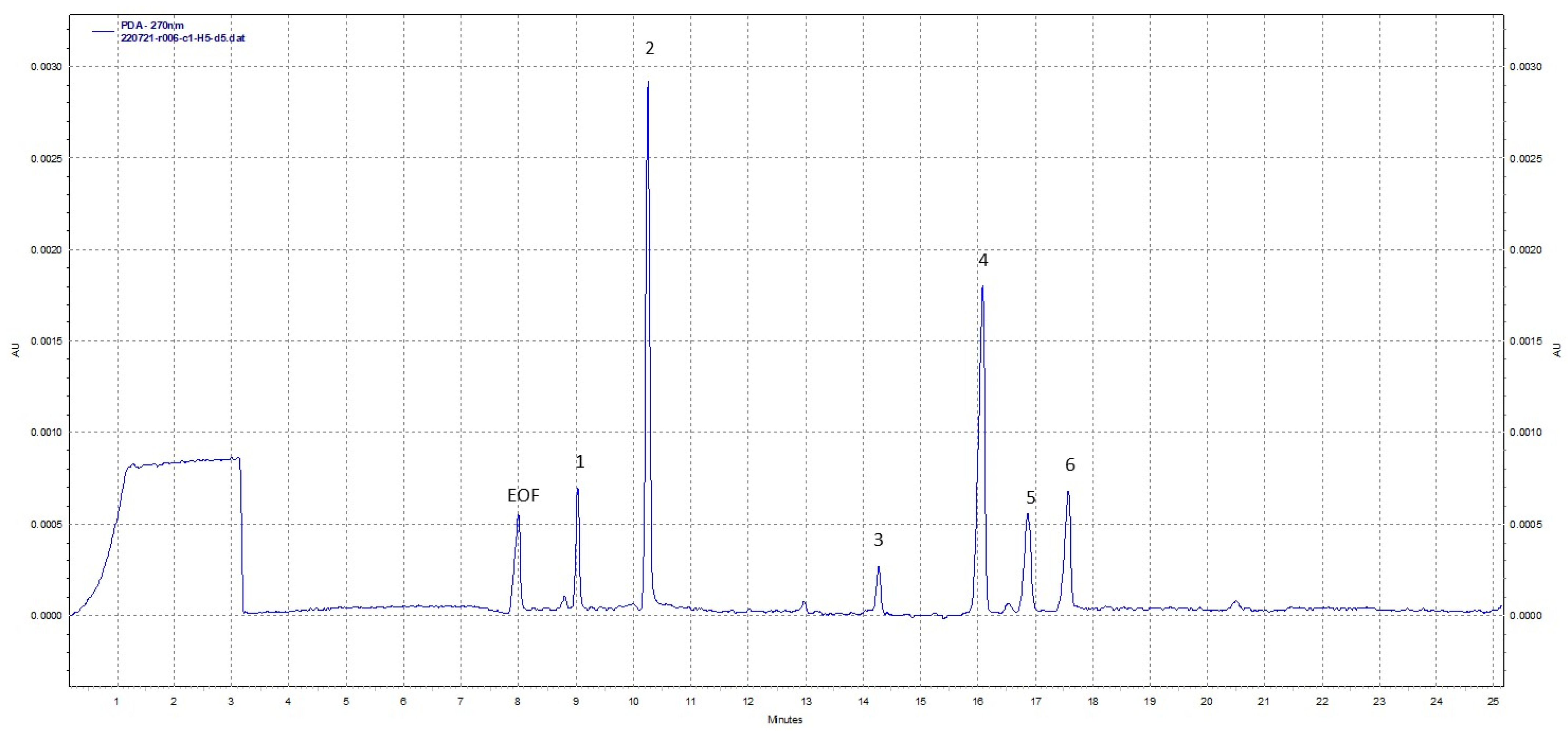
2.5.1. Monosaccharide Composition of AmPS

Prior to the determination of the AmPS sugar composition, 100 mg of the sample was hydrolyzed using 50 mL of 2 M trifluoroacetic acid (TFA) for 2 h at 100 °C. TFA was evaporated under vacuum using an IKA RV 10 (Staufen, Germany). The hydrolysate dissolved in water was centrifuged at 10,000× g for 5 min, and then the carbohydrates were analyzed using high-performance capillary electrophoresis (HPCE) with a PA 800 plus (Sciex, Framingham, MA, USA) apparatus equipped with a PDA detector according to a modified method by Rovio et al. [15]. The wavelength used for detection was 270 nm. A bare fused silica capillary (Polymicro, Phoenix, AZ, USA) with inside diameter 25 µm and a total length of 60.2 cm (50.0 cm effective length) was employed in the analysis. The samples and the capillary were maintained at 15 °C during analysis. Prior to analysis, the capillary was conditioned using 130 mM sodium hydroxide, ultrapure water, and separation buffer (130 mM NaOH and 360 mM Na2HPO4·2H2O; pH 12.6). The injections of the samples were performed under pressure (1.0 psi) for 15 s followed by the pressure injection (0.5 psi) of water for 10 s. The separation was obtained in 25 min with the voltage at the level of 30 kV. 32 Karat Software Version 9.1 was used to acquire and evaluate electropherograms. The identification of monosaccharides was achieved by comparing the migration times with corresponding standard carbohydrates analyzed under the same conditions with the use of the standard addition method.

2.5.2. NMR Spectroscopy

The AmPS sample (~8 mg) was dissolved with 1 mL of 99.5% D2O, lyophilized, and then dissolved in 0.6 mL of 99.95% D2O. The 1H NMR spectrum was acquired at 23 °C with a Bruker Avance III 700 MHz spectrometer. Bruker TopSpin 3.6.2 software was applied to acquire and process the NMR data. Acetone (δH 2.225) was used as an internal standard to reference chemical shifts.

2.6. Total and α- and β-Glucan Content

Total glucan, α-glucan, and β-glucan content in the A. mellea were investigated using the Megazyme Mushroom and Yeast Beta-Glucan Assay Kit according to the manufacturer’s instructions described in detail previously [10]. The results were shown as g per 100 g of dry weight.

2.7. Biological Activity of AmPS

2.7.1. Inhibition of α-Glucosidase

The inhibition of α-glucosidase was analyzed using the method of Telagari and Hullatti [16] described in detail by Pieczykolan et al. [17]. The crude polysaccharides from A. mellea at different concentrations were added to assay buffer (0.1 M; pH 6.8) and the enzyme solution (1 U/mL), and then maintained at 37 °C for 15 min. After that, 5 mM PNPG in 0.1 M phosphate buffer (pH 6.8) was added to start the enzymatic reaction. Incubation was conducted for 20 min at 37 °C. The reaction was terminated by the addition of 0.1 M Na2CO3. Absorbance was measured at 405 nm. The reaction system without the tested sample was the control, and without the enzyme was the blank. The percentage inhibition of α-glucosidase was calculated as shown below:
% inhibition = (1 − sample absorbance/control absorbance) × 100
The inhibition of α-glucosidase by AmPS was presented as the EC50 in mg per mL.

2.7.2. Inhibition of α-Amylase

The inhibition of α-amylase was investigated using the assay presented by Abirami et al. [18]. The AmPS sample dissolved in water (5 mg/mL) was mixed with assay buffer (0.02 M; pH 6.9) and α-amylase (4.5 U/mL/min). The mixture was maintained for 10 min at 25 °C. Then, 1% starch was added and the mixture was incubated for 30 min at 25 °C. Then, 1 mL of dinitrosalicylic acid reagent was added, and the mixture was heated at 100 °C for 5 min then cooled to 21 °C. Absorbance was read at 540 nm. The control contained the buffer instead of the sample. The percentage α-amylase inhibitory activity was calculated as shown below:
% inhibition = (1 − sample absorbance/control absorbance) × 100

2.7.3. Oxygen Radical Absorbance Capacity (ORAC) Assay

The assay was conducted using the protocol previously described [14,19]. The AmPS sample (5 mg/mL) was combined with fluorescein solution (10 nM) and incubated at 37 °C for 20 min. Then, AAPH (240 mM) was added. Fluorescence was measured at an excitation wavelength of 485 nm and an emission wavelength of 515 nm. The AmPS activity was calculated as µM of Trolox per g of crude polysaccharides based on the calibration curve of the standard compound.

2.7.4. Antiradical Activity against ABTS•+

The antiradical potential of AmPS was analyzed according to the method of Olech et al. [14]. The AmPS sample was combined with ABTS•+ at a concentration of 0.096 mg/mL. The reaction mixture was shaken and incubated at 28 °C for 6 min. Absorbance was measured at 734 nm. The control sample included ABTS•+ solution and solvent. The AmPS activity was given as µM of Trolox per g of crude polysaccharides based on the calibration curve of the standard compound.

2.7.5. Inhibition of Lipoxygenase (LOX)

The potential of AmPS to inhibit 15-lipoxygenase was evaluated using the method of Maiga et al. [20]. The AmPS sample (1 mg/mL) was combined with 0.2 M borate buffer (pH 9.00), 15-lipoxygenase solution (167 U/mL), and linoleic acid (134 µM). The absorbance was read at 234 nm. Acetylsalicylic acid (10 mM) was applied as a standard compound. The percentage inhibition of lipoxygenase was calculated in the above assay mixture system in relation to the control without the tested sample (indicated as 100%).

2.7.6. Inhibition of Cyclooxygenase (COX)

The cyclooxygenase inhibitory potential of AmPS was investigated using the COX (ovine) Colorimetric Inhibitor Screening Assay Kit according to the manufacturer’s instructions described previously in detail [10].

2.7.7. Inhibition of Hyaluronidase (HYAL)

The hyaluronidase inhibitory potential of AmPS was investigated using a previously described method [21,22]. The reaction system contained the AmPS sample, phosphate buffer (20 mM; pH 7) with 77 mM NaCl and 0.1 mg/mL of albumin, and the HYAL solution (40–100 U/mL). After 10 min of incubation at 37 °C, hyaluronic acid dissolved in sodium phosphate buffer (300 mM; pH 5.35) at a concentration of 0.5 mg/mL was added and the mixture was again incubated (45 min, 37 °C). The precipitation of undigested hyaluronic acid occurred after adding an acid albumin solution at a concentration of 2 mg/mL in 79 mM acetic acid with 24 mM sodium acetate (pH 3.75). The absorbance was measured at 600 nm. Epigallocatechin gallate (EGCG) was used as a standard. The percentage inhibition was calculated as shown below:
% inhibition = (AS − ABLK)/(AC − ABLK) × 100%
where AS is the absorbance of the tested sample, ABLK is the absorbance of the negative control (including buffer instead of the sample), and AC is the absorbance of the mixture including buffer instead of the enzyme. The inhibition of HYAL by AmPS was presented as the IC50 in µg per mL.

2.7.8. Anticancer Potential

Cell Lines

The AGS (gastric adenocarcinoma), A549 (lung carcinoma), and HT-29 (colorectal adenocarcinoma) cell lines were obtained from the American Type Culture Collection (ATCC, Manassas, VA, USA). The cells were grown in DMEM, high glucose, and L-Glutamine. The media were supplemented with 10% FBS, penicillin (100 U/mL), and streptomycin (100 U/mL). The cells were maintained in a humidified atmosphere of 95% air and 5% CO2 at 37 °C.

MTT Assay

Cells of AGS, HT-29, and A549 were seeded on 96-well microplates at a density of 2.5 × 10⁴ cells/mL in 100 μL DMEM supplemented with 10% FBS in 3 sets for various times of exposure to the AmPS. After 24 h, increasing concentrations (100–800 µg/mL) of AmPS dissolved in fresh FBS-free medium were added to the cells. Medium with sterile water was used as a control. The cell metabolic activity was assessed using the MTT assay after 72 h of incubation. Following the 72 h exposure, the control medium or the test exposure medium was removed, and the cells were rinsed with DPBS. Then, 100 μL of fresh medium (without FBS or antibiotics) with MTT solution (0.5 mg/mL) was added to the cells for 3 h. Then, the medium was removed, the cells were washed with 100 μL of PBS, and 100 μL of DMSO was added to each well to extract the dye. The plates were shaken for 5 min and the absorbance was measured at 570 nm. The results were expressed as the % of cell viability calculated based on the absorbance obtained in the tested condition versus control condition.

NRU Assay

Cells of AGS, HT-29, and A549 were seeded in 96-well microplates at a density of 2 × 10⁴ cells/mL in 100 μL DMEM supplemented with 10% FBS in 2 sets for various times of exposure to the AmPS. On the following day, increasing concentrations (50–400 µg/mL) of AmPS dissolved in fresh FBS-free medium were added to the cells for 24 and 48 h. Medium with sterile water was used as a control. After the AmPS exposure, the control medium and the test exposure medium was removed, and the cells were rinsed with PBS. Then, 100 μL of culture medium with neutral red dye solution (40 µg/mL) was added to cells. Next, the plates were incubated for 2 h under standard conditions (5% CO2, 37 °C). After 48 h, the medium was removed, the cells were quickly washed and fixed with 5% glutaraldehyde for 2 min, then washed again gently with 100 μL of PBS, and 100 μL of neutral red destain solution (50% ethanol 96%, 49% water, 1% glacial acetic acid) was added to the cells. Then, the plates were rapidly shaken for 10 min and the absorbance was read at 540 nm. The results of cytotoxicity were presented as % of cell viability calculated based on the absorbance obtained in the tested condition versus control condition.

2.8. Statistical Analysis

The results of all determinations were given as the mean ± standard deviation (SD) of 3 replicates. Calculations were carried out using STATISTICA 10.0 (StatSoft Poland, Cracow, Poland).

3. Results and Discussion

3.1. Composition of AmPS

The percentage yield of the crude polysaccharide from A. mellea was found to be 13.70% of d.w. Powdered fruiting bodies of the honey mushroom were prewashed with ethyl alcohol to remove low molecular constituents, e.g., phenolics. Then, proper extraction with hot water was carried out and the aqueous extract was deproteinized using Sevage reagent. The extraction steps were performed using the ultrasonic technique, which is recognized as an innovative, effective, and environmentally friendly method [23]. Preliminary determinations of the chemical composition of the AmPS were conducted using spectrophotometric techniques. This revealed that the AmPS was composed mostly of sugars, while uronic acids, proteins, and phenolics were present in rather small quantities (Table 1). The analysis of the glucan content in A. mellea demonstrated that this mushroom was rich in β-glucans (26.68 g/100 g d.w.), while α-glucans (1.06 g/100 g d.w.) constituted a marginal part of the total glucans (Table 1). The obtained results are in agreement with those found by Mirończuk-Chodakowska and Witkowska [24], showing that wild-growing A. mellea contained 25.09 g/100 g d.w. of β-glucans and 1.11 g/100 g d.w. of α-glucans.
To evaluate the sugar composition of AmPS, the high-performance capillary electrophoresis technique was used. This method provides high separation efficiency and is a rapid analysis with high peak capacity. It also allows the detection of even trace levels of analytes [25]. During our study, the mixture of 10 carbohydrates was analyzed under optimized separation conditions. The migration order of the tested compounds was found to be myo-inositol, xylitol, mannitol, sucrose, fucose, galactose, glucose, mannose, then arabinose, indicating that sugar alcohols migrate first, followed by disaccharides, and hexoses migrate before pentoses. The total analysis time including rinsing of the capillary before separation was 35 min. The reproducibility of analyte separation was significantly improved by adding mineral oil to the separation buffer. Ultraviolet detection at 270 nm was used for the identification of the tested compounds. These compounds are involved in reaction cascades resulting in the formation of UV-absorbing anions [15]. The described HPCE method has previously been successfully used by us for the evaluation of the sugar composition of plant polysaccharides [26]. Further modifications allowed us to also apply it for mushroom samples. The presence of six peaks was found in AmPS during the HPCE analysis (Figure 1). These peaks were identified as myo-inositol, mannitol, fucose, galactose, glucose, and mannose according to their migration times.
Our qualitative study revealed that crude polysaccharides from A. mellea contain various carbohydrates including sugar alcohols (myo-inositol and mannitol), hexoses (galactose, glucose, and mannose), and hexose deoxy sugar (fucose). Galactose was the main component of AmPS among hexoses, while mannitol was the predominant sugar alcohol. Mannose, glucose, and myo-inositol were found in smaller amounts, while fucose was detected in the lowest quantity. A previous study on polysaccharides from wild honey mushroom fruiting bodies from China showed the presence of mainly glucose as well as galactose and mannose [23]. Chang et al. [27] reported that hydrolysates of A. mellea polysaccharides obtained from cultured mycelia consisted of fucose, galactose, and xylose in a ratio of 6:5:4. Our results revealed the presence of sugar alcohols in polysaccharides from Polish A. mellea wild species for the first time, which distinguishes them from the previously studied polysaccharides of different origins. Therefore, this demonstrates that the origin of the mushroom may affect its composition, and our crude polysaccharides may possess different and new structures. The 1H NMR spectrum (Figure 2) confirms and complements the information obtained from HPCE. The main component of the sample was mannitol. Its signals with very high intensities were present in the range of 3.60–3.85 ppm. Signals characteristic of polysaccharides were also found on the spectrum. In the so-called anomeric range of the 1H NMR spectrum (4.9–5.1 ppm), there were several signals of the H1 hydrogen atoms of various monosaccharides. Moreover, in the 3.4–4.2 ppm range, there were numerous signals originating from sugar ring protons. There was also a signal at ~1.2 ppm on the spectrum, which comes from H6 hydrogen atoms of 6-deoxyhexose and confirms the presence of fucose. In addition, very low-intensity signals were identified on the spectrum at ~4.0 and 3.2 ppm. The signals were indicative of small amounts of myo-inositol in the crude polysaccharide sample, which was also identified in the electrophoretic analysis. The presented results are the first report concerning the chemical composition of the polysaccharide fraction from wild-growing A. mellea species from Poland.

3.2. Biological Activity of AmPS

3.2.1. Antidiabetic Potential

α-glucosidase and α-amylase are enzymes that participate in the degradation of starch, oligosaccharides, and disaccharides into monosaccharides, which, in turn, leads to increased blood glucose levels. Decreasing such enzymes’ activity modulates hyperglycemia after eating, which is an early metabolic disturbance that appears in diabetes [28]. Acarbose is a commonly known α-glucosidase inhibitor that reduces postprandial blood glucose spikes by delaying the digestion of carbohydrates within the small intestine; however, it exerts some side effects, mainly flatulence, abdominal distension, or diarrhea. Therefore, searching for new natural inhibitors seems reasonable. To determine the antidiabetic potential of polysaccharides from A. mellea, their impact on α-glucosidase and α-amylase were assayed. It was found that AmPS exhibited remarkable α-glucosidase inhibitory activity (EC50 0.73 mg/mL), which was even higher than in the case of the standard compound acarbose (EC50 1.37 mg/mL). However, no activity of AmPS against α-amylase was detected (Table 2). There are also previous reports available on an in vivo study of the antidiabetic activity of polysaccharides from A. mellea. Yang et al. [29] demonstrated that a polysaccharide fraction from A. mellea administered orally to type 2 diabetic rats decreased fasting blood glucose, improved glucose intolerance, and mitigated insulin resistance. Another study revealed that a neutral polysaccharide of honey mushroom, described as mannogalactoglucan, increased insulin sensitivity, lowered blood glucose, modulated lipid metabolism, and protected damaged pancreatic islets in db/db mice [8]. Our findings indicate that polysaccharides from edible A. mellea may be a potential anti-hyperglycemic food that affects postprandial blood glucose level due to the inhibition or delay of glucose production from carbohydrates. Including mushrooms such as A. mellea in the diet can be helpful in the prevention of diabetes.

3.2.2. Antioxidant Activity

The total antioxidant potential of AmPS was investigated using the ORAC assay and the ABTS•+ scavenging activity. The results of both assays are expressed in Trolox equivalents per mass of AmPS (Table 3). Crude polysaccharides from honey mushroom were found to possess antioxidant activity at a level of 414.3 μM TE/g and 94.69 μM TE/g in the ORAC and ABTS assay, respectively. Previous results for crude polysaccharides from a different edible mushroom S. crispa revealed activity at a level of 168.51 µM TE/g in the ORAC assay [10]. This demonstrates that the polysaccharide fraction from A. mellea shows significantly higher activity. The results obtained by Chen et al. [23] showed that polysaccharides from wild A. mellea demonstrated a dose-dependent antioxidant effect in various assays, including ORAC and FRAP as well as DPPH and ABTS+• scavenging activity. They found that two purified polysaccharide fractions exhibited ORAC values at levels of 2126.53 μM TE/g and 1791.05 μM TE/g. With regard to the ABTS+• scavenging activity, polysaccharides at a concentration of 2 mg/mL reduced 49.72% and 68.91% of the radical, while Trolox (a highly efficient antioxidant molecule) was found to reduce 99.02%. The reducing properties of A. mellea polysaccharides (determined with the ABTS assay) reported previously by Zhang et al. [30] revealed that IC50 values of the four polysaccharides isolated from wild sporophores and cultured products of honey mushroom varied from 885.7 to 2118 µg/mL and were lower than for positive controls (15.79 µg/mL for vitamin C and 29.82 µg/mL for BHA). The differences between our and previous results indicate that mushroom fruiting bodies of various origins possess different polysaccharide structures, which affects their biological activity. Certainly, further studies on the relationship between the composition and structure of polysaccharides in relation to their activity are needed to reveal the mechanisms of action.

3.2.3. Anti-Inflammatory Potential

The anti-inflammatory potential of the crude polysaccharides from A. mellea was evaluated using in vitro assays investigating the inhibitory effect on proinflammatory enzymes including cyclooxygenase (COX, lipoxygenase (LOX) (Table 4), and hyaluronidase (HYAL). The AmPS fraction was found to suppress the COX-1 isoform activity in a concentration-dependent manner; however, no activity against the COX-2 isoform was detected in the tested concentrations of 0.5 and 1 mg/mL. Our previous study revealed that crude polysaccharides from the edible species Cantharellus cibarius inhibited COX-1 and COX-2 activity by 43.86% and 51.59%, respectively [31]. The percentage of LOX inhibition for AmPS was 38.83%, which is rather moderate compared to that of acetylsalicylic acid used as a standard (93.97%). However, the inhibitory activity of polysaccharides from a different edible mushroom from Poland—Sparassis crispa—was estimated at 23.8% during our previous investigation [10]. The activity of AmPS in relation to hyaluronidase was determined in this study as well. This enzyme is responsible for the degradation of hyaluronic acid. When uncontrolled, the process may promote the development and progression of an inflammatory condition [32]. The AmPS fraction was shown to inhibit HYAL with an IC50 value of 590.81 ± 1.83 µg/mL. The activity of sample was two times lower than that of the standard compound EGCG (IC50 266.21 ± 1.18 µg/mL). Previous findings revealed that xylosyl galactofucan from A. mellea has significant anti-inflammatory properties via decreasing the level of tumor necrosis factor-α and cytokine monocyte chemotactic protein-1 in RAW264.7 macrophages [26]. Sun et al. [5] demonstrated that a polysaccharide from the fruiting body of honey mushroom stimulated murine lymphocyte proliferation caused by concanavalin A or lipopolysaccharide, which makes it a possible immunopotentiating agent. Similarly, our results support the assumption that polysaccharides from the honey mushroom may alleviate certain inflammatory diseases.

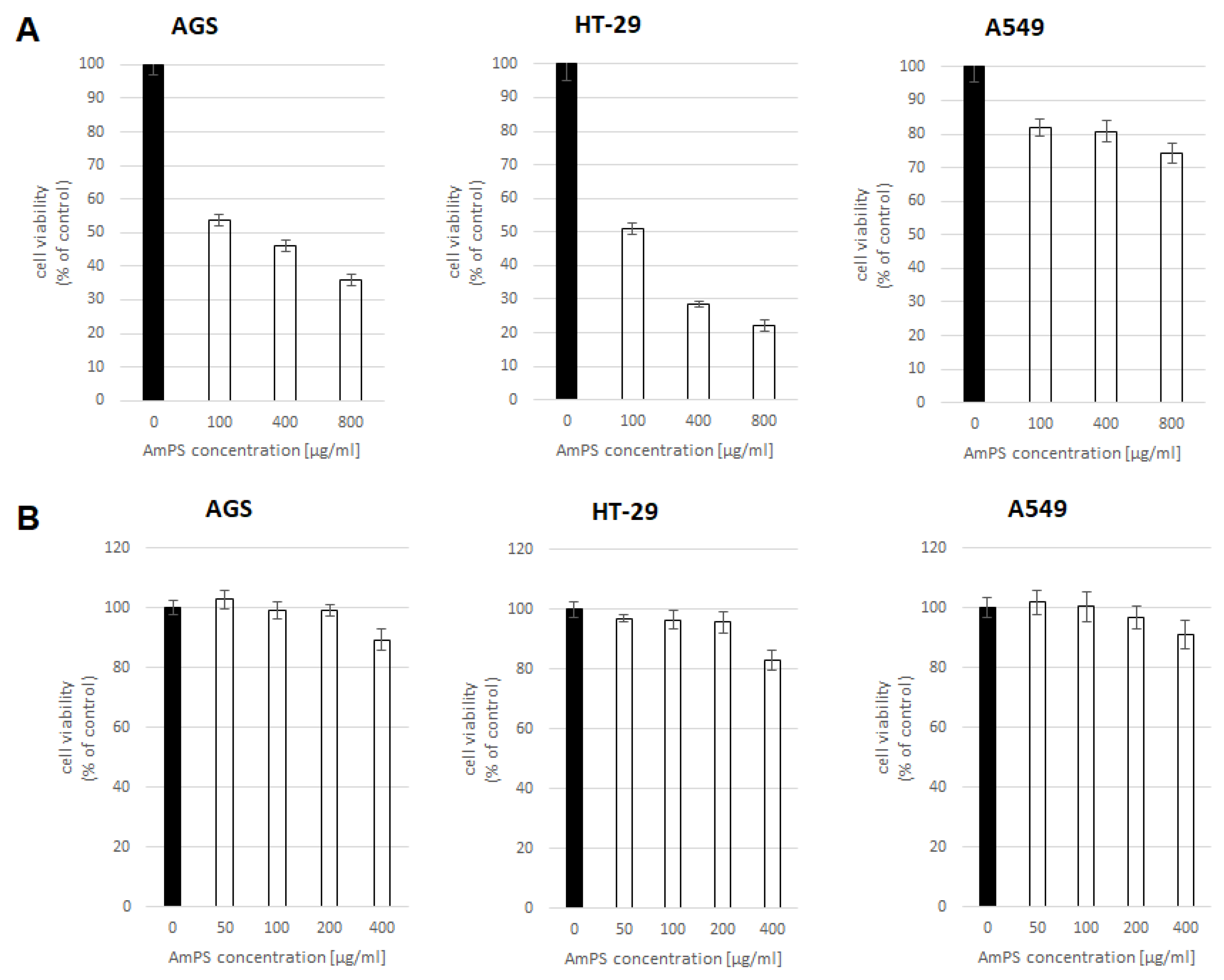
3.2.4. Anti-Cancer Potential

The antiproliferative potential of AmPS against gastric adenocarcinoma (AGS), colorectal adenocarcinoma (HT-29), and lung carcinoma (A549) was determined. These types of cancer are recognized among the five most common cancers globally according to the data from 2020 presented by the World Health Organization [33]. Therefore, searching for potential natural agents preventing the incidence of stomach, colon, and lung cancer is desirable. It was found that the crude polysaccharides from A. mellea significantly inhibited the viability of the AGS and HT-29 cell lines in a dose-dependent manner, while the viability of the A549 cell line was slightly decreased (Figure 3A). The NRU assay was used to evaluate the cytotoxicity of AmPS in relation to different cell lines. The HT-29 cell line was found to be the most susceptible to the effects of AmPS. All the analyzed concentrations slightly reduced cell viability. Only the highest concentration of AmPS (400 µg/mL) inhibited the viability of the AGS and A549 cell lines (Figure 3B). In an available study on the anticancer properties of polysaccharides from A. mellea, it was found that it exerted a potent anticancer effect on A549 cells via apoptosis-involving mechanisms [3]. Due to the fact that A. mellea is considered an edible and safe mushroom species, it can be suggested that including it in the diet may help preventing cancers of the gastrointestinal tract.

4. Conclusions

Our paper is the first describing the chemical profile and biological properties of crude polysaccharides obtained from A. mellea harvested from the wild in Poland. It revealed its broad health-beneficial potential. During the study, it was found that crude polysaccharides exert an inhibitory impact on various inflammation-related enzymes and antioxidant activity. AmPS was also shown to significantly inhibit α-glucosidase, which should encourage further exploration of its antidiabetic potential. Moreover, the antiproliferative studies demonstrated that AmPS has a significant suppressing effect on the growth of cancer cells (especially HT29) in in vitro conditions. Our study also proves that the A. mellea collected in Poland is a rich source of carbohydrates that may be a valuable part of the diet as well as a potential ingredient of functional food products. Since this species has been consumed for hundreds of years, its polysaccharides can be assumed to be safe. Our research contributes to the finding that mushrooms can ward off chronic diseases and improve everyday health. The obtained results encourage the promotion of the use of honey mushroom crude polysaccharide fraction to develop products for improving human health. Due to their common occurrence, A. mellea fruiting bodies may be considered an attractive source of health-beneficial polysaccharides for potential use as functional food ingredients or pharmaceuticals. The promising findings of various bioassays presented in this paper encourage further analysis of polysaccharides from A. mellea in preclinical studies, which may form the basis for clinical trials in the future.

Author Contributions

Conceptualization N.N.-J. and R.N; Funding acquisition, N.N.-J. and R.N.; Investigation, N.N.-J., S.K., A.M., M.M. (Marcin Moczulski) and N.S.; Methodology, N.N.-J., M.M. (Marcin Moczulski), K.K., M.M. (Maciej Masłyk), Z.K. and M.O.; Project administration, N.N.-J.; Writing—original draft, N.N.-J. and A.M.; Writing—review and editing, N.N.-J., Z.K., M.O. and R.N. All authors have read and agreed to the published version of the manuscript.

Funding

This work was supported by the Medical University of Lublin (grant No. PBmb50 and DS.46).

Institutional Review Board Statement

Not applicable.

Informed Consent Statement

Not applicable.

Data Availability Statement

Not applicable.

Acknowledgments

This paper was developed using equipment purchased within the project ‘The equipment of innovative laboratories doing research on new medicines used in the therapy of civilization and neoplastic diseases’ within the Operational Program Development of Eastern Poland 2007–2013, Priority Axis I Modern Economy, Operations I.3 Innovation Promotion.

Conflicts of Interest

The authors declare no conflict of interest. The funders had no role in the design of the study; in the collection, analyses, or interpretation of data; in the writing of the manuscript; or in the decision to publish the results.

References

  1. Muszyńska, B.; Maślanka, A.; Ekiert, H.; Sułkowska-Ziaja, K. Analysis of indole compounds in Armillaria mellea fruiting bodies. Acta Pol. Pharm. Drug Res. 2011, 68, 93–97. [Google Scholar]
  2. Stachowiak, B.; Reguła, J. Health-promoting potential of edible macromycetes under special consideration of polysaccharides: A review. Eur. Food Res. Technol. 2012, 234, 369–380. [Google Scholar] [CrossRef]
  3. Wu, J.; Zhou, J.; Lang, Y.; Yao, L.; Xu, H.; Shi, H.; Xu, S. A polysaccharide from Armillaria mellea exhibits strong in vitro anticancer activity via apoptosis-involved mechanisms. Int. J. Biol. Macromol. 2012, 51, 663–667. [Google Scholar] [CrossRef]
  4. Gao, L.W.; Li, W.Y.; Zhao, Y.L.; Wang, J.W. The cultivation, bioactive components and pharmacological effects of Armillaria mellea. Afr. J. Biotechnol. 2009, 8, 7383–7390. [Google Scholar]
  5. Sun, Y.; Liang, H.; Zhang, X.; Tong, H.; Liu, J. Structural elucidation and immunological activity of a polysaccharide from the fruiting body of Armillaria mellea. Bioresour. Technol. 2009, 100, 1860–1863. [Google Scholar]
  6. Ng, L.T.; Wu, S.J.; Tsai, J.Y.; Lai, M.N. Antioxidant activities of cultured Armillariella mellea. Appl. Biochem. Microbiol. 2007, 43, 444–448. [Google Scholar]
  7. Gao, L.W.; Wang, J.W. Antioxidant potential and DNA damage protecting activity of aqueous extract from Armillaria Mellea. J. Food Biochem. 2012, 36, 139–148. [Google Scholar] [CrossRef]
  8. Yang, S.; Yan, J.; Yang, L.; Meng, Y.; Wang, N.; He, C.; Fan, Y.; Zhou, Y. Alkali-soluble polysaccharides from mushroom fruiting bodies improve insulin resistance. Int. J. Biol. Macromol. 2019, 126, 466–474. [Google Scholar] [CrossRef]
  9. Erbiai, E.H.; da Silva, L.P.; Saidi, R.; Lamrani, Z.; da Silva, J.C.G.E.; Maouni, A. Chemical Composition, Bioactive Compounds, and Antioxidant Activity of Two Wild Edible Mushrooms Armillaria mellea and Macrolepiota procera from Two Countries (Morocco and Portugal). Biomolecules 2021, 11, 575. [Google Scholar]
  10. Nowacka-Jechalke, N.; Nowak, R.; Lemieszek, M.K.; Rzeski, W.; Gawlik-Dziki, U.; Szpakowska, N.; Kaczyński, Z. Promising potential of crude polysaccharides from Sparassis crispa against colon cancer: An in vitro study. Nutrients 2021, 13, 161. [Google Scholar] [CrossRef]
  11. Dubois, M.; Gilles, K.A.; Hamilton, J.K.; Rebers, P.A.; Smith, F. Colorimetric method for determination of sugars and related substances. Anal. Chem. 1956, 28, 350–356. [Google Scholar] [CrossRef]
  12. Van Den Hoogen, B.M.; Van Weeren, P.R.; Lopes-Cardozo, M.; Van Golde, L.M.G.; Barneveld, A.; Van De Lest, C.H.A. A microtiter plate assay for the determination of uronic acids. Anal. Biochem. 1998, 257, 107–111. [Google Scholar] [CrossRef] [PubMed]
  13. Bradford, M.M. A rapid and sensitive method for the quantitation of microgram quantities of protein utilizing the principle of protein-dye binding. Anal. Biochem. 1976, 72, 248–254. [Google Scholar] [CrossRef] [PubMed]
  14. Olech, M.; Łyko, L.; Nowak, R. Influence of accelerated solvent extraction conditions on the LC-ESI-MS/MS polyphenolic profile, triterpenoid content, and antioxidant and anti-lipoxygenase activity of rhododendron luteum sweet leaves. Antioxidants 2020, 9, 822. [Google Scholar] [CrossRef]
  15. Rovio, S.; Yli-Kauhaluoma, J.; Sirén, H. Determination of neutral carbohydrates by CZE with direct UV detection. Electrophoresis 2007, 28, 3129–3135. [Google Scholar] [CrossRef] [PubMed]
  16. Telagari, M.; Hullatti, K. In-vitro α-amylase and α-glucosidase inhibitory activity of Adiantum caudatum Linn. and Celosia argentea Linn. extracts and fractions. Indian J. Pharmacol. 2015, 47, 425–429. [Google Scholar] [PubMed] [Green Version]
  17. Pieczykolan, A.; Pietrzak, W.; Gawlik-Dziki, U.; Nowak, R. Antioxidant, anti-inflammatory, and anti-diabetic activity of phenolic acids fractions obtained from Aerva lanata (L.) juss. Molecules 2021, 26, 3486. [Google Scholar] [CrossRef]
  18. Abirami, A.; Nagarani, G.; Siddhuraju, P. In vitro antioxidant, anti-diabetic, cholinesterase and tyrosinase inhibitory potential of fresh juice from Citrus hystrix and C. maxima fruits. Food Sci. Hum. Wellness 2014, 3, 16–25. [Google Scholar] [CrossRef] [Green Version]
  19. Dienaitė, L.; Pukalskas, A.; Pukalskienė, M.; Pereira, C.V.; Matias, A.A.; Venskutonis, P.R. Phytochemical composition, antioxidant and antiproliferative activities of defatted sea buckthorn (Hippophaë rhamnoides L.) berry pomace fractions consecutively recovered by pressurized ethanol and water. Antioxidants 2020, 9, 274. [Google Scholar] [CrossRef] [Green Version]
  20. Maiga, A.; Malterud, K.E.; Diallo, D.; Paulsen, B.S. Antioxidant and 15-lipoxygenase inhibitory activities of the Malian medicinal plants Diospyros abyssinica (Hiern) F. White (Ebenaceae), Lannea velutina A. Rich (Anacardiaceae) and Crossopteryx febrifuga (Afzel) Benth. (Rubiaceae). J. Ethnopharmacol. 2006, 104, 132–137. [Google Scholar] [CrossRef]
  21. Yahaya, Y.A.; Don, M.M. Evaluation of Trametes Lactinea Extracts on the Inhibition of Hyaluronidase, Lipoxygenase and Xanthine Oxidase Activities in Vitro. J. Phys. Sci. 2012, 23, 1–15. [Google Scholar]
  22. Łyko, L.; Olech, M.; Nowak, R. LC-ESI-MS/MS characterization of concentrated polyphenolic fractions from Rhododendron luteum and their anti-inflammatory and antioxidant activities. Molecules 2022, 27, 827. [Google Scholar] [CrossRef]
  23. Chen, R.; Ren, X.; Yin, W.; Lu, J.; Tian, L.; Zhao, L.; Yang, R.; Luo, S. Ultrasonic disruption extraction, characterization and bioactivities of polysaccharides from wild Armillaria mellea. Int. J. Biol. Macromol. 2020, 156, 1491–1502. [Google Scholar] [CrossRef]
  24. Mirończuk-Chodakowska, I.; Witkowska, A.M. Evaluation of polish wild mushrooms as beta-glucan sources. Int. J. Environ. Res. Public Health 2020, 17, 7299. [Google Scholar] [CrossRef]
  25. Liu, D.; Tang, W.; Yin, J.Y.; Nie, S.P.; Xie, M.Y. Monosaccharide composition analysis of polysaccharides from natural sources: Hydrolysis condition and detection method development. Food Hydrocoll. 2021, 116, 106641. [Google Scholar] [CrossRef]
  26. Łubek-Nguyen, A.; Olech, M.; Nowacka-Jechalke, N.; Martyna, A.; Kubiński, K.; Masłyk, M.; Moczulski, M.; Kanak, S. Crude Polysaccharide Fraction from Rosa rugosa Thunb. Root—Chemical Characterisation, Enzyme Inhibitory, Antioxidant and Antiproliferative Activity. Appl. Sci. 2022, 12, 10126. [Google Scholar] [CrossRef]
  27. Chang, C.C.; Cheng, J.J.; Lee, I.J.; Lu, M.K. Purification, structural elucidation, and anti-inflammatory activity of xylosyl galactofucan from Armillaria mellea. Int. J. Biol. Macromol. 2018, 114, 584–591. [Google Scholar] [CrossRef]
  28. Krentz, A.J.; Bailey, C.J. Oral Antidiabetic Agents Current Role in Type 2 Diabetes Mellitus. Drugs 2005, 65, 385–411. [Google Scholar] [CrossRef]
  29. Yang, S.; Meng, Y.; Yan, J.; Wang, N.; Xue, Z.; Zhang, H.; Fan, Y. Polysaccharide-enriched fraction from Amillariella mellea fruiting body improves insulin resistance. Molecules 2019, 24, 46. [Google Scholar] [CrossRef] [Green Version]
  30. Zhang, S.; Liu, X.; Yan, L.; Zhang, Q.; Zhu, J.; Huang, N.; Wang, Z. Chemical Compositions and Antioxidant Activities of Polysaccharides from the Sporophores and Cultured Products of Armillaria mellea. Molecus 2015, 20, 5680–5697. [Google Scholar] [CrossRef] [Green Version]
  31. Nowacka-Jechalke, N.; Nowak, R.; Juda, M.; Malm, A.; Lemieszek, M.; Rzeski, W.; Kaczyński, Z. New biological activity of the polysaccharide fraction from Cantharellus cibarius and its structural characterization. Food Chem. 2018, 268, 355–361. [Google Scholar] [CrossRef] [PubMed]
  32. Girish, K.; Kemparaju, K.; Nagaraju, S.; Vishwanath, B. Hyaluronidase Inhibitors: A Biological and Therapeutic Perspective. Curr. Med. Chem. 2009, 16, 2261–2288. [Google Scholar] [CrossRef] [PubMed]
  33. Cancer. Available online: https://www.who.int/news-room/fact-sheets/detail/cancer (accessed on 10 February 2023).
Figure 1. Electropherogram of the crude polysaccharides from A. mellea (AmPS). EOF—electro-osmotic flow; 1—myo-inositol; 2—mannitol; 3—fucose; 4—galactose; 5—glucose; 6—mannose.
Applsci 13 03853 g001
Figure 2. 1H NMR spectrum of the crude polysaccharides from A. mellea.
Applsci 13 03853 g002
Figure 3. Viability (%) of AGS, HT-29, and A549 cell lines exposed to the tested compounds at concentrations of 100–800 µg/mL determined using the MTT assay after 72 h (A), and viability (%) of AGS, HT-29, and A549 cell lines exposed to the crude polysaccharides from A. mellea at concentrations of 50–400 µg/mL assessed with the use of the neutral red uptake assay after 48 h (B). Error bars indicate mean ± standard deviation (n = 3).
Applsci 13 03853 g003
Table 1. Chemical composition of A. mellea crude polysaccharides (AmPS); sugar, uronic acid, protein, and phenolic content expressed as a percent of AmPS, and the content of total glucans, α-glucans, and β-glucans in A. mellea given in g per 100 g of dry weight.
Results
Sugar content (% of AmPS)60.71 ± 2.35
Uronic acid content (% of AmPS)1.24 ± 0.01
Protein content (% of AmPS)3.13 ± 0.03
Phenolics content (% of AmPS)1.83 ± 0.02
Total glucan content (g/100 g d.w.)27.74 ± 0.78
α-glucan content (g/100 g d.w.)1.06 ± 0.02
β-glucan content (g/100 g d.w.)26.68 ± 0.82
Mean values from three replicate determinations with standard deviation.
Table 2. EC50 values of α-glucosidase and α-amylase inhibition by the AmPS.
Sampleα-Glucosidaseα-Amylase
EC50 (mg/mL)
AmPS0.73 ± 0.02n.d.
Acarbose1.37 ± 0.010.01 ± 0.02
Mean values from three replicate determinations with standard deviation; n.d.—activity not detected.
Table 3. Antioxidant properties of AmPS. The results of the antiradical activity with ABTS radical cation and the oxygen radical absorbance capacity (ORAC) assay are expressed as µM of Trolox equivalents per g of AmPS.
Antioxidant AssayResult
ORAC (µM TE/g AmPS)414.3 ± 2.74
ABTS (µM TE/g AmPS)94.69 ± 0.26
Mean values from three replicate determinations with standard deviation.
Table 4. Inhibition of cyclooxygenase-1 (COX-1), cyclooxygenase-2 (COX-2), and lipoxygenase (LOX) by crude polysaccharides from A. mellea (AmPS) expressed as % of inhibition. Abbreviations: ASA—acetylsalicylic acid; n.d.—not detected; n.a.—not analyzed.
COX-1COX-2LOX
(% of Inhibition)
AmPS (0.5 mg/mL)52.07 ± 1.78n.d.n.a.
AmPS (1 mg/mL)71.69 ± 2.47n.d.38.83 ± 0.47
ASA (10 mM)32.95 ± 0.6498.64 ± 0.9393.97 ± 0.76
Mean values from three replicate determinations with standard deviation.
Disclaimer/Publisher’s Note: The statements, opinions and data contained in all publications are solely those of the individual author(s) and contributor(s) and not of MDPI and/or the editor(s). MDPI and/or the editor(s) disclaim responsibility for any injury to people or property resulting from any ideas, methods, instructions or products referred to in the content.

