Article

Determination of Morphological Parameters of Supported Gold Nanoparticles: Comparison of AFM Combined with Optical Spectroscopy and Theoretical Modeling versus TEM

by
Frank Hubenthal
*,
David Blázquez Sánchez
and
Frank Träger
Institut für Physik and Center for Interdisciplinary Nanostructure Science and Technology–CINSaT, Universität Kassel, Heinrich-Plett-Str. 40, Kassel 34132, Germany
*
Author to whom correspondence should be addressed.
Appl. Sci. 2012, 2(3), 566-583; https://doi.org/10.3390/app2030566
Submission received: 25 April 2012 / Revised: 11 June 2012 / Accepted: 15 June 2012 / Published: 2 July 2012
(This article belongs to the Special Issue Feature Papers)

Abstract

:
The morphology of small gold particles prepared by Volmer–Weber growth on sapphire substrates have been investigated by two different characterization techniques. First, by non-extensive atomic force microscopy (AFM) in combination with optical spectroscopy and modeling of the optical properties using a theoretical model, recently developed in our group. Second, by extensive transmission electron microscopy (TEM). Comparing the results obtained with both techniques demonstrate that for small gold nanoparticles within the quasistatic limit, the morphological properties can be precisely determined by an appropriate theoretical modeling of the optical properties in combination with simple AFM measurements. The apparent mean axial ratio of the nanoparticles, i.e., the axial ratio that corresponds to the center frequency of the ensemble plasmon resonance, is obtained easily from the extinction spectrum. The mean size is determined by the nanoparticle number density and the amount of deposited material, measured by AFM and a quartz micro balance, respectively. To extract the most probable axial ratio of the nanoparticle ensemble, i.e., the axial ratio that corresponds to the most probable nanoparticle size in the ensemble, we apply the new theoretical model, which allows to extract the functional dependence of the nanoparticle shape on its size. The morphological parameters obtained with this procedure will be afterwards compared to extensive TEM measurements. The results obtained with both techniques yield excellent agreement. For example, the lateral dimensions of the nanoparticles after deposition of 15.2 × 10¹⁵ atoms/cm2 of gold has been compared. While a mean lateral diameter of (13 ± 2) nm has been extracted from AFM, optical spectroscopy and modeling, a value of (12 ± 2) nm is derived from TEM. The consistency of the results demonstrate the precision of our new model. Moreover, since our theoretical model allows to extract the functional dependence of the nanoparticle size and shape, a relatively simple analysis is sufficient for a full characterization of small noble metal nanoparticles.

1. Introduction

Small gold particles supported on dielectric supports have attracted considerable attention due to their extraordinary properties that are exploited in numerous applications [1,2,3,4,5,6,7,8,9,10,11]. In recent years the main foci of interest have been the optical properties, which are in the visible range and dominated by the excitation of localized surface plasmon polariton resonances, i.e., by a collective excitation of the conduction band electrons. For simplicity, we refer to this kind of excitation as plasmon resonance or simply plasmon. In contrary to the correspondent plasma oscillations in bulk material or on a smooth surface, the localized surface plasmon polariton resonances can be excited directly by an incoming electromagnetic wave. Accompanied with the excitation of a plasmon resonance is a greatly enhanced local field in the vicinity of the nanoparticle surface.
The optical properties of single spherical particles have been exactly described by Gustav Mie [12]. Mie obtained his solution after a multipole expansion of the electromagnetic fields, which yields the extinction cross section of the particles as the sum of absorption and scattering of a series of multipole excitations. For particles much smaller than the wavelength of light, the quasistatic approximation simplifies the general electrodynamic problem into electrostatic [13,14,15]. Within this approximation, scattering and higher multipole modes are neglected and the plasmon is solely due to absorption of an electric dipole. The absorption cross section σabs depends on the photon energy of the incoming electromagnetic wave and on the imaginary part of the polarizability of the nanoparticle:
Applsci 02 00566 i001
In the general case of ellipsoidal metal particles, the polarizability along the axis i is given by [13,14,15,16]:
Applsci 02 00566 i002
where V is the particle volume, Li is the depolarization factor along the i axis, ε and εm are the dielectric constants of the metal and the embedding medium, respectively. A resonance of the polarizability, i.e., of the absorption, appears, if the denominator in Equation (2) becomes zero. The resonance condition depends on the depolarization factor Li, which is a function of the particle eccentricity, i.e., of its shape. According to Equation (2), for ellipsoidal nanoparticles three plasmon resonances can be excited. Since the plasmon resonance is highly sensitive to the nanoparticle morphology, optical spectroscopy has widely been used as a characterization tool [17,18,19,20,21,22]. While the position of the Plasmon resonance is sensitive to the particle morphology and environment, its width is usually a superposition of homogeneous and inhomogeneous broadening. The homogeneous broadening is related by the dielectric constant to the electronic properties of the metal and eventually to the surrounding, while inhomogeneous broadening is caused by the width of the size and shape distribution of a nanoparticle ensemble. Although theoretical modeling is widely used to characterize nanoparticle ensembles, the full potential of the optical properties has not been exploited for a long time, due to a lack of adequate modeling, as pointed out in [23].
Since for plenty applications of noble metal nanoparticles the optical and, thus, the morphological parameters are of enormous interest, one would like to determine the size and shape of the nanoparticles as precisely as possible. Different techniques are employed to characterize the particle morphology [24,25,26,27,28,29,30,31,32,33,34,35,36,37,38,39]. One direct and powerful technique is transition electron microscopy (TEM) [15,28,29,30,31,32,33,34,35,36,37,40], but TEM suffers from the fact that sample preparation for three-dimensional TEM investigations is rather complicated and cannot be easily accomplished in situ. In addition, obtaining sufficient statistical data from TEM measurements is extremely time consuming.
On the other hand, optical spectroscopy combined with atomic force microscopy has been used in the past to characterize the morphology of noble metal nanoparticle ensembles [6,17,18,20,41,42]. However, for a detailed modeling of the optical properties of supported nanoparticle ensembles, three important issues have to be overcome:
  • the asymmetric dielectric surrounding,
  • the possibly broad size and shape distribution, and
  • the particle-particle interaction within an ensemble.
As explained in reference [23], under certain circumstances, which are fulfilled for the nanoparticles investigated here, an appropriate modeling of the optical properties in quasistatic approximation is possible and all relevant morphological parameters of a sample can be obtained easily.
In this paper, we demonstrate that the morphology of supported noble nanoparticles can be precisely extracted by means of AFM combined with optical spectroscopy and an appropriate modeling. For this purpose, we have compared the morphological data obtained with the aforementioned relatively simple method with extensive TEM investigations. We found an excellent agreement, which demonstrates the accuracy of the indirectly extracted morphology of the nanoparticle ensemble by rigorous modeling the optical spectra.

2. Experimental

Gold nanoparticle ensembles were prepared at room temperature under ultrahigh vacuum conditions (base pressure < 5 × 10⁻⁸ mbar during atom deposition). An electron-beam evaporator (Omicron, EFM-3) equipped with a charge filter was used to obtain a thermal beam of atoms. The beam is directed onto sapphire substrates (Crystec, orientation (0001)) and the nanoparticles were grown by adsorption with subsequent diffusion and nucleation, i.e., Volmer–Weber growth. The flux of atoms was monitored by a quartz crystal microbalance (Inficon, 6 MHz, 008·010-G10) and set to a constant value of 1.3 ×10¹³ atoms/(cm2s). The coverage Θ has been extracted by the deposition time and the sticking coefficient of unity for gold atoms on sapphire [43]. After a residence time of 24 h in the UHV, diffusion processes are completed and the nanoparticles are stable for at least several weeks.
The particle shape is determined by the surface energy of the nanoparticles and the substrate. As a consequence, nanoparticles with an oblate shape are generated, which can be approximated by rotational ellipsoids [15,17,44]. We emphasize that due to the surface energies, the nanoparticles are truncated at the contact plane to the substrate. Nevertheless, the approximation is justified since experimentally the low energetic plasmon mode, which corresponds to the electron excitation along the long axis of the nanoparticles, is only marginally, if at all, affected by the truncated shape. An effect on the high energetic plasmon resonance cannot be observed since this resonance is completely damped by the nearby interband transition. Hence, the nanoparticle shape can be described by the axial ratio x = a/b, where a denotes the short half axis perpendicular and b the long half axis parallel to the substrate surface.
The mean particle size ⟨Req⟩ of the nanoparticles was derived from the coverage Θ, the particle density np, defined as the number of particles per cm2, and assuming the density of bulk material. The particle density was measured by a scanning force microscope (Thermo-Microscopes, Autoprobe CP, AP-0100) operated under ambient conditions and in non-contact mode. Although an accurate determination of the particle morphology cannot be achieved by AFM due to tip convolution, the relative size distribution is reproduced correctly, because the nanoparticle dimensions are linearly enlarged by the AFM tip [45].
The extinction spectrum of the nanoparticle ensemble has been measured in situ using light of a xenon arc lamp (Osram, XBO 450 W/1) in combination with a monochromator (AMKO, 1,200 lines/mm, blaze: 250 nm). The light was linearly polarized by a rotating Glan–Thompson prism (Halle Nachfolger). The samples were illuminated under an angle of incidence of ϑ = 45° with respect to the surface normal with photon energies between = 1.3 eV and = 4.5 eV (276 nm–954 nm). The transmitted light was detected by a photodiode and normalized to a reference signal recorded in front of the sample, to account for intensity variations of the xenon arc lamp. In addition, for background subtraction we have measured the optical spectrum of the blank sample prior to nanoparticle preparation. Due to their symmetry, oblate nanoparticles exhibit usually two plasmon modes, a high energetic (1,0)-mode, which corresponds to an electron excitation along the short axis a and a low energetic (1,1)-mode, which corresponds to an electron excitation along the long axis b. However, for gold nanoparticles, the (1,0)-mode is completely damped by the nearby interband transition [13,14,15] and only the (1,1)-mode is excited.
To extract the relevant morphological parameters, such as the shape and size correlation of the nanoparticles within the ensemble or the mean axial ratio, we apply a new theoretical model [23]. The background of the model, which has been recently developed in our group, is briefly explained in Section 3.3. The model takes advantage of the strong shape and size correlation of nanoparticles generated in Volmer–Weber growth and is based on a precise modeling of the inhomogeneously broadened extinction spectrum of a nanoparticle ensemble. The rigorous application of the model yields the functional dependence between the axial ratio and the radius of the nanoparticles within the ensemble. It also permits to determine the most probable axial ratio on the sample and to calculate the amount of material on the substrate if—for any reason—unknown. For the latter purpose the size of the nanoparticles is used as a free parameter [23]. Hence, the modeling reveals important morphological parameters of a nanoparticle ensemble, which otherwise are difficult to obtain.
Complementary to the previous technique, high resolution TEM (HRTEM) measurements have been performed to extract directly the morphology of the nanoparticle ensemble. All HRTEM images were recorded with a Jeol 4000EX HREM microscope operating at an acceleration voltage of 400 kV with a point-to-point resolution of 0.17 nm. The TEM samples were prepared in the conventional way, by mechanical thinning of the substrate followed by an ion milling process. To avoid sample damage or diffusion of surface atoms due to heating from the electron bombardment, a low electron flux has been applied during the TEM measurements. The results obtained with HRTEM have been afterwards compared to the previously obtained results. We note that due to the handling of the samples in air, no measurable shape changes have been observed. This has been proven previously with samples which have been exposed to air and whose optical properties remained the same after back transfer in the UHV.

3. Results and Discussions

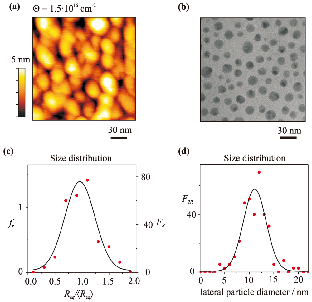
3.1. Determination of the Mean Particle Size and the Apparent Mean Shape

Figure 1(a) depicts an AFM image of the gold nanoparticle ensemble recorded after deposition of Θ = (1.5 ± 0.2) × 1016 atoms/cm2 on a sapphire support, demonstrating the three-dimensional island growth.
Figure 1. (a) AFM image of gold particles on a quartz support for a coverage of Θ = (1.5 ± 0.2) × 1016 atoms/cm2 and (c) extracted size distribution; (b) TEM image of a similar sample and (d) extracted size distribution.
Applsci 02 00566 g001
The particle density has been extracted from the AFM image and amounts to nAFMp = (2.9 ± 0.3) × 1011 particles/cm2. With this particle density, we derive a mean equivalent nanoparticle radius of ⟨Req⟩ = (5.5 ± 0.3) nm. In addition, a HRTEM image of a sample prepared under the same experimental conditions and with the same optical properties as the sample shown in Figure 1(a) is depicted in Figure 1(b). From a set of HRTEM images we have extracted a particle density of nSTMp = (3.1 ± 0.3) × 1011 particles/cm2, in good agreement with the data obtained by AFM. In addition, we have extracted the mean lateral diameter of the ensemble from the HRTEM images, which amounts to ⟨2b⟩ = (12 ± 2) nm. This value will be later compared to the mean lateral diameter determined with our new modeling technique.
However, neither AFM nor TEM is capable to extract the precise size or shape of the nanoparticles. While in the AFM images, the apparent nanoparticle size and shape is convoluted with the AFM tip, the TEM images reveal only the lateral dimensions, but no information on the nanoparticle height. Therefore, a direct comparison of the nanoparticle morphology is not possible. However, the width of the relative size distribution Req/Req⟩, obtained with AFM (Figure 1(c)) and the width of the lateral particle diameter distribution, obtained by HRTEM (Figure 1(d)) can be compared. In both cases, the width of the size distribution is in the same range of 30% to 35%.
To extract the apparent mean axial ratio, i.e., the axial ratio that corresponds to nanoparticles which exhibit the same plasmon resonance frequency as the nanoparticle ensemble, optical spectroscopy in combination with simple quasistatic modeling of the optical properties of single nanoparticles is applied. Figure 2(a) depicts extinction spectra of gold nanoparticles for a coverage ranging from Θ = 0.1 × 10¹⁶ atoms/cm2 to Θ= 1.5 × 10¹⁶ atoms/ cm2. It can be clearly observed that for increasing coverage the (1,1)-mode of the plasmon resonance increases in amplitude and shifts to lower photon energies. The latter effect is due to the flattening of the nanoparticles during growth [15,17,42]. Although atoms from the vapor phase can be directly deposited on top of the already grown nanoparticles, this process is rather unlikely. In practice, atoms are deposited on the substrate and attach from the side to the nanoparticles. A subsequent upstepping, i.e., an upward movement of the atoms, is necessary to obtain the equilibrium shape. However, upstepping is unfavorable compared to diffusion along the substrate surface. As a consequence, the mass transport across the surface is faster than in the direction normal to the surface and the nanoparticles grow in a non-thermodynamical equilibrium [14,15], i.e., with oblate shape.
Since surface diffusion takes place also after deposition of atoms, a shape change towards more spherical shape occurs after preparation. To allow the nanoparticle ensembles to complete their morphological changes, the samples are rested after preparation in the UHV chamber for 24 h. As a consequence of the shape changes towards more spherical, the center position of the plasmon resonance is slightly back-shifted to higher photon energies. To demonstrate this effect, an optical spectrum after a residence time of 24 h is depicted in Figure 2(b) (solid line). The photon energy where the Plasmon resonance has its maximum, i.e., at =1.87 eV, will be considered as the resonance frequency Applsci 02 00566 i012 
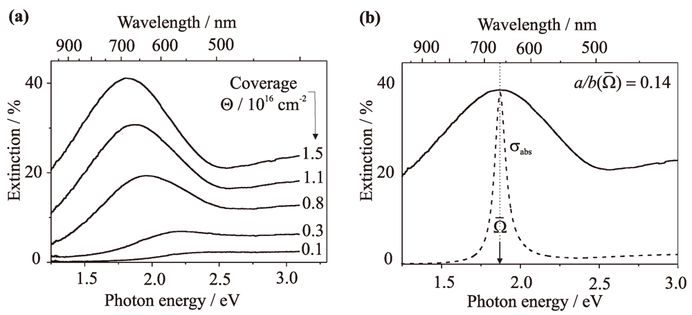
of the nanoparticle ensemble. In addition to the measured spectrum, an absorption cross section in arbitrary units of a single gold nanoparticle with the same resonance frequency as the nanoparticle ensemble is depicted. Since the generated nanoparticles are well separated and exhibit radii smaller than R = 15 nm, the center frequency of its plasmon resonance depends only on the nanoparticle shape and the dielectric surrounding. Hence, performing a simple quasistatic modeling, taking the inhomogeneous environment into account by an effective dielectric constant [14,15], we obtain the apparent mean axial ratio of the nanoparticle ensemble, which amounts to Applsci 02 00566 i013 = 0.14.
Figure 2. (a) Extinction spectra during growth of gold on sapphire at different coverage Θ; (b) Optical extinction spectrum of gold particles 24 h after preparation (solid line) and calculated absorption cross section of a single particle with the same resonance frequency as the ensemble (dashed line).
Applsci 02 00566 g002
Note, the observed broad size distribution (Figure 1(b)) results in a broad shape distribution, which has significant consequences. The plasmon mode is inhomogeneously broadened (cf. Figure 2) and, most importantly, the spectrum is dominated by large nanoparticles [15]. As a consequence, neither the mean size of the nanoparticles in the ensemble nor their mean shape can be extracted solely from the extinction spectrum.
Nevertheless, as we will demonstrate in Section 3.3 the strong correlation between the shape, size, and plasmon resonance position of single nanoparticles in the quasistatic regime can be exploited to characterize precisely the morphology of the nanoparticles.

3.2. Support Interaction

In general, the support may influence the optical properties in two ways.
  • First, the support changes the dielectric environment and, thus, shifts the plasmon resonance to longer wavelengths. This effect can be taken into account by including the Yamaguchi theory [46] in the quasistatic approximation, using a mixing factor m [15]. The mixing factor corresponds to the part of the nanoparticle surface in contact with the substrate and has a value between 0 and 1 [15]. For oblate nanoparticles on substrates m can be determined either from the energetic position of both plasmon modes [47] or due to a modeling of the nanoparticle geometry using the adhesion energy on the substrate and the surface energy of the metal [43,48]. The latter method is necessary for gold nanoparticles, because they exhibit only the (1,1)-mode. The Yamaguchi theory has been proven to be sufficient for most applications, although the dielectric environment is reduced to an average of the dielectric functions of the contributing environments [14,15].
  • Second, due to the surface energies, an oblate growth is expected, which cause also a red-shift of the plasmon resonance. However, neither the TEM nor the AFM images yield a clear evidence of an oblate nanoparticle shape. Moreover, since for gold nanoparticles the (1,0)-mode is damped by the interband transition, also from the extinction spectrum the shape cannot be extracted directly.
In the following, we demonstrate that indeed oblate nanoparticles are grown. As explained in the introduction, the polarizabilities of an oblate nanoparticle along its principal axes a and b are determined by αa and αb. Hence, the absorption cross section depends on the relative orientation of the particles’ axes with respect to the angle of incidence and polarization of the incoming light. Consequently, the absorption cross section σabs of the (1,1)-mode of gold nanoparticles is a function of the angle of incidence ϑ and polarization angle φ. Thus, σabs in the range of the (1,1) mode can be written as [13]:
Applsci 02 00566 i003
This procedure is valid since αa and αb peak at different frequency ranges. Equation (3) reveals, that for normal incidence (ϑ = 0°) the absorption is independent of the polarization angle. This is easily understood due to the symmetry of the system. In contrast, for an angle of incidence of ϑ = 45°, the particle absorption follows the dependence 1 + cos2 φ, due to the oblate shape. In other words, the nanoparticles appear round only under normal incidence, and the symmetry is lost for ϑ ≠ 0°. To demonstrate that the nanoparticles are indeed oblate, the amplitude of the (1,1)-mode has been measured as a function of the polarization angle for ϑ = 0and ϑ = 45. The experimental results together with the theoretical curves are depicted in Figure 3.
Figure 3. Amplitude of the (1,1)-mode as a function of the polarization angle φ for gold particles with a ⟨Req⟩ = (5.5 ± 0.3) nm. (a) Angle of incidence ϑ= 0°and (b) angle of incidence ϑ= 45°.
Applsci 02 00566 g003
It is obvious that the experimental data is in accord with the theoretical curve, which demonstrates that the nanoparticles can be approximated as rotational ellipsoids. The small deviation of the experimental data from the theoretical curve is due to experimental uncertainties.

3.3. Single Particle Characterization

Yet, only the apparent mean axial ratio Applsci 02 00566 i013 = 0.14 of the nanoparticle ensemble has been obtained. However, the apparent mean axial ratio corresponds to the shape of particles that have a dominant impact on the optical spectrum. Since the absorption cross section is proportional to R3 [15], Applsci 02 00566 i013 = 0.14 is not the most probable axial ratio in the nanoparticle distribution [23].
To obtain the most probable axial ratio, by taking the actual shape and size distribution into account, we use the following assumptions and simplifications:
  • The particles are small compared to the wavelength of light, i.e., the electromagnetic field within the particles is spatially nearly constant [13,14]. A comparison between calculations performed with the quasistatic approximations and the T-matrix method for gold nanoparticles can be found in reference [15], Figure 2.11. The figure demonstrates that the quasistatic approximation is fairly sufficient for the nanoparticles with Req ≤ 15 nm.
  • The nanoparticles can be approximated by ellipsoidal nanoparticles with a Gaussian size distribution. This is usually fulfilled for particles grown on surfaces or deposited from gas phase [49,50,51].
  • The optical properties of bulk materials ε(ω) without modifications due to quantum size effects can be used. This simplification is justified, because previous observations demonstrate that major disagreements may be anticipated for particles smaller than 1 nm [14]. Furthermore, the resonance condition is only slightly influenced by variations of ε(ω) due to the reduced dimensions for nanoparticles in our range of interest [52].
  • Chemical interface damping (CID) is neglected. This assumption is justified, because for gold nanoparticles on sapphire, no CID has been observed [41]. On the other hand, for samples where CID is expected, it can be included in the theory by using an appropriate damping parameter in the size-dependent dielectric function [15].
  • The influence of the substrate on the plasmon resonance can be described by the effective medium theory [14,15,53,54,55,56,57,58,59.
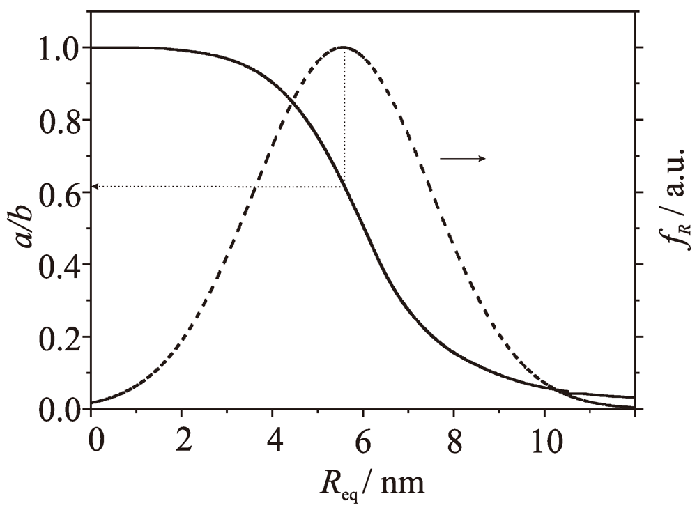
  • A strong correlation between the nanoparticle size and shape exists, which is fulfilled for nanoparticles grown in Volmer–Weber growth. Hence, the nanoparticle ensemble can be characterized by a functional dependence of the shape and size.
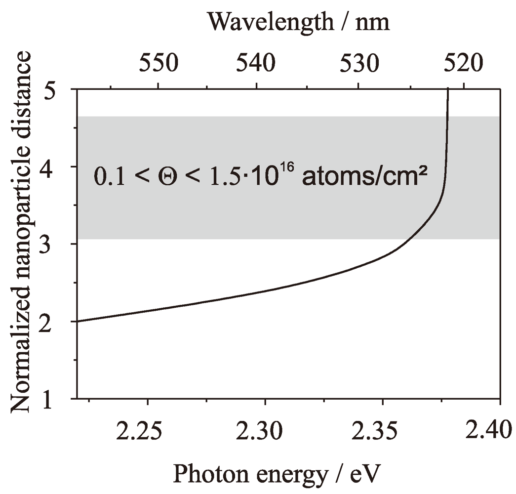
  • Diploe-dipole interaction between the nanoparticles can be neglected. Note, particle-particle interaction might also cause a red shift of the plasmon resonance, but it has been demonstrated in a previous publication that a significant red-shift occurs only for very small distances between the particles [15]. The assumption is justified, because the broad shape distribution of the particles leads, in turn, to the broad distribution of plasmon frequencies, which reduces the mutual interaction between the particles. In other words, the adjacent particles are most probable of different shape and, hence, are not in resonance with each other.
    To demonstrate this, it is useful to treat the nanoparticles independently from their size. Thus, we define a dimensionless normalized distance d between the nanoparticles by
    Applsci 02 00566 i004
    where Applsci 02 00566 i014 the average distance between the nanoparticles that can be calculated from the particle number density measured by AFM. The functional dependence between d and the position of the plasmon resonance is displayed in Figure 4. The grey shaded area indicates the normalized distances of our nanoparticle ensembles for the coverages used in our experiments. Hence, the red-shift of the plasmon resonance due to a dipole-dipole interaction between the nanoparticles can be neglected for nanoparticles with ⟨Req⟩ = (5.5 ± 0.3) nm and a particle density of about np = 3 × 10¹¹ particles/cm2, as investigated here.
Figure 4. Photon energy of the plasmon resonance versus normalized nanoparticle distance. The shaded area indicates the coverages used in this study.
Applsci 02 00566 g004
To obtain the most probable axial ratio, a probability distribution function of the axial ratios fa/b(x) has to be introduced. For simplicity, we have defined the axial ratio as a/b = x. fa/b(x) has to satisfy the normalization condition [23]:
Applsci 02 00566 i005
The integration limits correspond to spherical particles (upper limit) and infinite flat particles (lower limit). Since the optical spectrum is a linear superposition of the extinction spectra of single particles, the extinction spectrum of the ensemble can be expressed as:
Applsci 02 00566 i006
where np is the density of particles. V (x)· s(x) is the extinction spectrum of a single particle, with axial ratio x and volume V (x) [23]. Note that the volume of the particle is not an independent variable but rather a function of the axial ratio V (x).
It is useful to define the function Applsci 02 00566 i015 as Applsci 02 00566 i015 = fa/b(x)V (x) , which describes the axial ratio distribution and the impact of the particle volume on the spectrum, thus:
Applsci 02 00566 i007
From reference [23] on can obtain s(x) to be:
Applsci 02 00566 i008
where L(x) is the depolarization factor known for any axial ratio x, c is the speed of light, and ε(ω) = ε1(ω)m. ε1(ω) and εm being the dielectric permittivities of the particle material and the surrounding medium, respectively.
To find Applsci 02 00566 i015
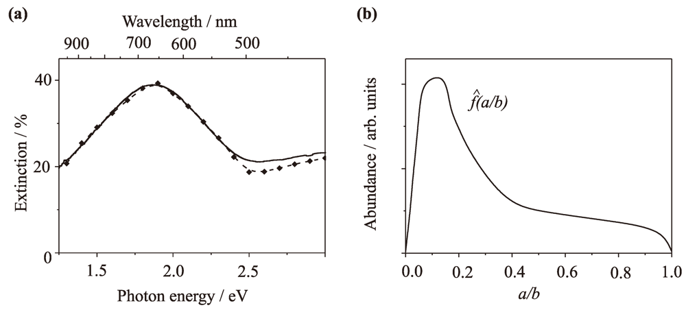
, Equation (7) is fitted to the experimental data by a spline with about ten to twenty sampling points, and the mean error squares are minimized [60]. This procedure holds, since the spectral dependence of the plasmon resonance on the axial ratio follows a steady and monotonic function [13]. Figure 5(a) demonstrates that the modeled spectrum Sa/b ideally reproduces the experimental one. The function Applsci 02 00566 i015, which has been extracted from the modeling, is depicted in Figure 5(b). It should be stressed here that the value of x = 0.14 at which Applsci 02 00566 i015 peaks is the axial ratio previously denoted as apparent mean axial ratio Applsci 02 00566 i013 It is neither the most probable axial ratio nor the mean axial ratio of the particle ensemble. Due to the convolution with the volume, the maximum of Applsci 02 00566 i015 peaks at lower values compared to fa/b(x) [23]. Nevertheless, the agreement to the simple modeling (cf. Section 3.1) is perfect.
Figure 5. (a) Optical extinction spectrum of the gold nanoparticle ensemble depicted in Figure 1(a) (solid line) and the modeled spectrum (squares); (b) Calculated function Applsci 02 00566 i016.
Applsci 02 00566 g005
To extract the axial ratio probability distribution fa/b(x), the function V (x) is needed. For this purpose, the following Equation
Applsci 02 00566 i009
has to be numerically integrated, as demonstrated in reference [23]. Req is the equivalent radius of a nanoparticle and fR(Req) the size distribution, obtained from AFM measurements. The numerical integration of Equation (9) readily provides both functions x(Req) and Req(x), i.e., the functional dependence between nanoparticle size and shape. These functions are very powerful, because they permit to determine the morphology of certain particles within the nanoparticle ensemble. The function x(Req) together with the size distribution for the nanoparticle ensemble already displayed in Figure 1 are depicted in Figure 6.
Figure 6. Axial ratio as a function of the equivalent radius (solid line) and size distribution (dashed line). The dotted arrow indicates the axial ratio for the most probable nanoparticle size in the ensemble.
Applsci 02 00566 g006
To compare the size of the nanoparticles extracted by AFM and optical spectroscopy to the TEM data, the mean lateral diameter must be obtained. For this purpose, the most probable axial ratio has to be known. It can be obtained either by numerical integration of Equation 9 and calculating the maximum of fa/b(x), or it can be obtained directly from Figure 6. The dotted arrow correlates the maximum of fR(x) to the corresponding axial ratio, as indicated in Figure 6. For the investigated nanoparticle ensemble, the most probable axial ratio amounts to x(⟨Req⟩) = 0.61. Applying the definition of the equivalent radius
Applsci 02 00566 i010
the mean lateral dimension, i.e., ⟨2b⟩, can be easily calculated. For the given parameters (⟨Req⟩ = 5.5 nm and x(⟨Req⟩) = 0.61) it amounts to ⟨2b⟩ = (13 ± 2) nm, which is in excellent agreement with ⟨2b⟩ = (12 ± 2) nm extracted from the TEM images. All determined morphological parameters are summarized in Table 1.
Table 1. AFM and optical spectroscopy versus TEM.
np / 1011 cm-2Req⟩ / nm⟨2b⟩ / nm
TEM3.1 ± 0.3 12 ± 2
AFM2.9 ± 0.35.5 ± 0.3
Opt. Spec. + Modeling3.4 ± 0.35.5 ± 0.313 ± 2
We emphasize that although several approximations have been made, the agreement between the TEM data and the nanoparticle morphology extracted by theoretical modeling combined with AFM is excellent. The shape and size of the nanoparticles within the ensemble is well reproduced by the rigorous modeling of the inhomogeneously broadened extinction spectrum in quasistatic approximation, using only the number density of nanoparticles as input parameter.
Note, Equation (9) relates the optical spectrum to the morphology of the nanoparticles. Hence, the optical spectrum can be fitted also by the morphological parameters:
Applsci 02 00566 i011
Inserting x(Req) in Equation (11), the spectrum can be modeled again, now using the morphological parameters. Hence, Equation (11) together with Equation (7) allows to proof if the optical data are consistent with the morphological data. For this purpose, the equivalent radius and the nanoparticle density are used as free parameters for modeling the optical spectrum with Equation (11). The best fit has been obtained for ⟨Req⟩ ≅ 5.5 nm and np ≅ 3.4 × 1011 cm2. The extracted particle density is in fair agreement with the TEM data and only slightly larger than the nanoparticle density obtained by AFM. The latter fact can be explained by the limited resolution of the AFM, whereby very small nanoparticles might be not observed. However, the derived equivalent radius is in perfect agreement with the AFM data (cf. Section 3.1). Hence, the optical measurements combined with an adequate modeling yield the same morphological parameters as the AFM and TEM analysis.
We emphasize that other samples have been previously modeled with our technique [23]. For these samples indirect comparisons of the experimentally and theoretically obtained morphological parameters have been made. In all cases a good agreement of the modeled and experimental data have been found, demonstrating that our model is generally applicable to supported noble metal nanoparticles under the assumptions made in Section 3.3.

4. Conclusions

Two techniques to characterize the morphology of supported gold nanoparticles prepared by Volmer–Weber growth have been applied and the extracted results have been compared. First, we applied simple AFM measurements in combination with a rigorous modeling of the inhomogeneously broadened extinction spectrum of the nanoparticle ensemble. With AFM we have measured the nanoparticle density and extracted the mean equivalent radius of the nanoparticles. This data has been afterwards used as input parameter in our new model to determine the functional dependence between the size and shape of the nanoparticles. Using this functional dependence, we have determined fundamental morphological parameters of the nanoparticle ensemble.
In addition, we have demonstrated that the morphological data can be related to the optical spectrum. Hence, theoretical modeling of the inhomogeneously broadened extinction spectrum using the mean equivalent radius and the nanoparticle density as free parameters can be performed. By obtaining the best fit to the experimental data, we have extracted again the nanoparticle density and the mean nanoparticle size, but now based on the extinction spectrum. The comparison of the modeled data with the experimentally determined data was in excellent agreement.
Second, we performed extensive TEM measurements to extract again the nanoparticle density and the mean lateral diameter of the nanoparticles. Afterwards, we have compared the morphological data measured by TEM with data extracted from AFM analysis and rigorous modeling of the inhomogeneously broadened extinction spectrum. The comparison reveals that the results obtained with both methods show a perfect agreement and demonstrates that our new theoretical model can precisely characterize the morphology of noble metal nanoparticles ensembles. For example, the lateral size of the nanoparticles has been determined to be 2b = (13 ± 2) nm by modeling the extinction spectrum in combination with AFM, while it amounts to 2b = (12 ± 2) nm if measured with TEM. However, our modeling technique allows to extract much more morphological parameters that are not easily accessible by TEM or AFM measurements. Most importantly, the functional dependence between the shape and the size has been extracted. This functional dependence has been used to determine the most probable axial ratio of the supported nanoparticles, which amounts to a/b = 0.61. Hence, rigorous theoretical modeling of the extinction spectra in combination with AFM reveals important morphological data of noble metal nanoparticle ensembles with high precision, which can not or, at least, can not easily be extracted by other techniques.

Acknowledgements

We thank the group of Gustaaf van Tendeloo, University of Antwerp, for performing the TEM measurements. Financial support by the European Commission under contract HPRN-CT-2002-00328 NANOCLUSTER and the Deutsche Forschungsgemeinschaft is gratefully acknowledged.

References

  1. Valden, M.; Lai, X.; Goodman, D. Onset of catalytic activity of gold clusters on titania with the appearance of nonmetallic properties. Science 1998, 281, 1647–1650. [Google Scholar] [CrossRef]
  2. Chen, M.; Goodman, D. The structure of catalytically active gold on titania. Science 2004, 306, 252–255. [Google Scholar] [CrossRef]
  3. Zijlstra, P.; Chong, J.; Gu, M. Five-dimensional optical recording mediated by surface plasmons in gold nanorods. Nature 2009, 459, 410–413. [Google Scholar] [CrossRef]
  4. Krenn, J. Nanoparticle waveguides: Watching energy transfer. Nat. Mater. 2003, 2, 210–211. [Google Scholar] [CrossRef]
  5. Morarescu, R.; Englert, L.; Kolaric, B.; Damman, P.; Vallée, R.; Baumert, T.; Hubenthal, F.; Träger, F. Tuning nanopatterns on fused silica substrates: A theoretical and experimental approach. J. Mater. Chem. 2011, 21, 4076–4081. [Google Scholar]
  6. Hubenthal, F.; Blázquez Sánchez, D.; Borg, N.; Schmidt, H.; Kronfeldt, H.D.; Träger, F. Tailor-made metal nanoparticles as SERS substrates. Appl. Phys. B 2009, 95, 351–359. [Google Scholar] [CrossRef]
  7. Hubenthal, F.; Morarescu, R.; Englert, L.; Haag, L.; Baumert, T.; Träger, F. Parallel generation of nanochannels in fused silica with a single femtosecond laser pulse: Exploiting the optical near fields of triangular nanoparticles. Appl. Phys. Lett. 2009, 95, 063101-1–063101-3. [Google Scholar]
  8. Morarescu, R.; Englert, L.; Kolaric, B.; Damman, P.; Vallée, R.; Baumert, T.; Hubenthal, F.; Träger, F. Tuning nanopatterns on fused silica substrates: A theoretical and experimental approach. J. Mater. Chem. 2011, 21, 4076–4081. [Google Scholar]
  9. Ossig, R.; Kwon, Y.-H.; Hubenthal, F.; Kronfeldt, H.-D. Naturally grown Ag nanoparticles on quartz substrates as SERS substrate excited by a 488 nm diode laser system for SERDS. Appl. Phys. B 2012, 106, 835–839. [Google Scholar] [CrossRef]
  10. Morarescu, R.; Träger, F.; Hubenthal, F. Surface plasmon resonance spectroscopy for in situ sensing of molecular nanowire formation. In Recent Researches in Communications, Automation, Signal Processing, Nanotechnology, Astronomy and Nuclear Physics; Bojkovic, Z., Kacprzyk, J., Mastorakis, N., Mladenov, V., Revetria, R., Zadeh, L.A., Zemliak, A., Eds.; WSEAS Press: Cambridge, UK, 2011; pp. 308–313. [Google Scholar]
  11. Kwon, Y.-H.; Ossig, R.; Hubenthal, F.; Kronfeldt, H.-D. Influence of surface plasmon resonance wavelength on SERS activity of naturally grown silver nanoparticle ensemble. J. Raman Spec. 2012. [Google Scholar]
  12. Mie, G. Beiträge zur Optik trüber Medien, speziell kolloidaler Metallösungen. Annalen der Physik 1908, 25, 377–445. [Google Scholar] [CrossRef]
  13. Bohren, C.; Huffman, D. Absorption and Scattering of Light by Small Particles; Wiley: New York, NY, USA, 1983. [Google Scholar]
  14. Kreibig, U.; Vollmer, M. Optical Properties of Metal Clusters; Springer: Berlin, Germany, 1995. [Google Scholar]
  15. Hubenthal, F. Noble metal nanoparticles: Synthesis and applications. In Comprehensive Nanoscience and Technology; Andrews, D.L., Scholes, G.D., Wiederrecht, G.P., Eds.; Oxford Academic Press: New York, NY, USA, 2011; Volume 1, pp. 375–435. [Google Scholar]
  16. Gans, R. Über die F]orm ultramikroskopischer Goldteilchen. Ann. Physik 1912, 37, 881–900. [Google Scholar] [CrossRef]
  17. Hubenthal, F. Nanoparticles and their tailoring with laser light. Eur. J. Phys. 2009, 30, S49–S61. [Google Scholar] [CrossRef]
  18. Hubenthal, F.; Borg, N.; Weidner, T.; Siemeling, U.; Träger, F. Gold nanoparticle growth on self-assembled monolayers of ferrocenyl-substituted terpyridine on graphite. Appl. Phys. A 2009, 94, 11–17. [Google Scholar] [CrossRef]
  19. Hubenthal, F.; Borg, N.; Träger, F. Optical properties and ultrafast electron dynamics in gold-silver alloy and core-shell nanoparticles. Appl. Phys. B 2008, 93, 39–45. [Google Scholar] [CrossRef]
  20. Hilger, A.; Tenfelde, M.; Kreibig, U. Silver nanoparticles deposited on dielectric surfaces. Appl. Phys. B 2001, 73, 361–372. [Google Scholar] [CrossRef]
  21. Kreibig, U. Interface-induced dephasing of Mie plasmon polaritons. Appl. Phys. B 2008, 93, 79–89. [Google Scholar] [CrossRef]
  22. Morarescu, R.; Blázquez Sánchez, D.; Borg, N.; Vartanyan, T.; Träger, F.; Hubenthal, F. Shape tailoring of hexagonally ordered triangular gold nanoparticles with nanosecond-pulsed laser light. Appl. Surf. Sci. 2009, 225, 9822–9825. [Google Scholar]
  23. Hubenthal, F.; Hendrich, C.; Vartanyan, T.; Träger, F. Determination of fundamental morphological parameters of supported nanoparticle ensembles: Extracting the functional dependence between nanoparticle shape and size. Plasmonics 2012. [Google Scholar]
  24. Hicks, E.; Zou, S.; Schatz, G.; Spears, K.; van Duyne, R.; Gunnarsson, L.; Rindzevicius, T.; Kasemo, B.; Käll, M. Controlling plasmon line shapes through diffractive coupling in linear arrays of cylindrical nanoparticles fabricated by electron beam lithography. Nano Lett. 2005, 5, 1065–1070. [Google Scholar] [CrossRef]
  25. Wang, H.; Brandl, D.; Nordlander, P.; Halas, N.J. Plasmonic nanostructures: Artificial molecules. Acc. Chem. Res. 2007, 40, 53–62. [Google Scholar] [CrossRef]
  26. Hu, M.; Petrova, H.; Sekkinen, A.; Chen, J.; McLellan, J.; Li, Z.; Marquez, M.; Li, X.; Xia, Y; Hartland, G. Optical properties of Au-Ag nanoboxes studied by single nanoparticle spectroscopy. J. Phys. Chem. B 2006, 110, 19923–19928. [Google Scholar]
  27. Wu, W.; Dey, D.; Memis, O.; Katsnelson, A.; Mohseni, H. Fabrication of large area periodic nanustructures using nanosphere photolithograpy. Nanoscale Res. Lett. 2008, 3, 351–354. [Google Scholar] [CrossRef]
  28. Marty, R.; Arbouet, A.; Girard, C.; Margueritat, J.; Gonzalo, J.; Afonso, C. Sculpting nanometer-sized light landscape with plasmonic nanocolumns. J. Chem. Phys. 2009, 131, 224707:1–224707:8. [Google Scholar]
  29. Resta, V.; Siegel, J.; Bonse, J.; Gonzalo, J.; Afonso, C.; Piscopiello, E.; Van Tenedeloo, G. Sharpening the shape distribution of gold nanoparticles by laser irradiation. J. Appl. Phys. 2006, 100, 084311:1–084311:6. [Google Scholar]
  30. Gonzalo, J.; Perea, A.; Babonneau, D.; Afonso, C.; Beer, N.; Barnes, J.P.; Petford-Long, A.; Hole, D.; Townsend, P. Competing processes during the production of metal nanoparticles by pulsed laser deposition. Phys. Rev. B 2005, 71, 125420-1–125420-8. [Google Scholar]
  31. Barnes, J.P.; Petford-Long, A.; Doole, R.; Serna, R.; Gonzalo, J.; Suárez-García, A.; Afonso, C.; Hole, D. Structural studies of Ag nanocrystals embedded in amorphous Al2O3 grown by pulsed laser deposition. Nanotechnology 2002, 13, 465–470. [Google Scholar] [CrossRef]
  32. Novo, C.; Gomez, D.; Perez-Juste, J.; Zhang, Z.; Petrova, H.; Reismann, M.; Mulvaney, P.; Hartland, G. Contributions from radiation damping and surface scattering to the linewidth of the longitudinal plasmon band of gold nanorods: a single particle study. Phys. Chem. Chem. Phys. 2006, 8, 3540–3546. [Google Scholar]
  33. Pyatenko, A.; Shimokawa, K.; Yamaguchi, M.; Nishimura, O.; Suzuki, M. Synthesis of silver nanoparticles by laser ablation in pure water. Appl. Phys. A 2004, 79, 803–806. [Google Scholar]
  34. Dadosh, T.; Sperling, J.; Bryant, G.; Breslow, R.; Shegai, T.; Dyshel, M.; Haran, G.; Bar-Joseph, I. Plasmonic control of the shape of the raman spectrum of a single molecule in a silver nanoparticle dimer. ACS Nano 2009, 3, 1988–1994. [Google Scholar]
  35. Pastoriza-Santos, I.; Liz-Marzán, L. Colloidal silver nanoplates. State of the art and future challenges. J. Mater. Chem. 2008, 18, 1724–1737. [Google Scholar] [CrossRef]
  36. Stalmashonak, A.; Seifert, G.; Graener, H. Optical three-dimensional shape analysis of metallic nanoparticles after laser-induced deformation. Optics Lett. 2007, 32, 3215–3217. [Google Scholar] [CrossRef]
  37. Vogel, F.; Träger, F.; Hubenthal, F. A new route for mass production of uniform metal nanoparticles in water by means of laser light induced processes. J. Nanosci. Nanotechnol. 2011, 11, 2368–2375. [Google Scholar]
  38. Watanabe, K.; Menzel, D.; Nilius, N.; Freund, H.J. Photochemistry on metal nanoparticles. Chem. Rev. 2006, 106, 4301–4320. [Google Scholar] [CrossRef]
  39. MacDonald, K.; Fedotov, V.; Zheludev, N. Optical nonlinearity resulting from a light-induced structural transition in gallium nanoparticles. Appl. Phys. Lett. 2003, 82, 1087–1089. [Google Scholar] [CrossRef]
  40. Hubenthal, F. Ultrafast dephasing time of localized surface plasmon polariton resonance and the involved damping mechanisms in colloidal gold nanoparticles. Prog. Surf. Sci. 2007, 82, 378–387. [Google Scholar] [CrossRef]
  41. Hubenthal, F.; Hendrich, C.; Träger, F. Damping of the localized surface plasmon polariton resonance of gold nanoparticles. Appl. Phys. B 2010, 100, 225–230. [Google Scholar] [CrossRef]
  42. Ouacha, H.; Hendrich, C.; Hubenthal, F.; Träger, F. Laser-assisted growth of gold nanoparticles: Shaping and optical characterization. Appl. Phys. B 2005, 81, 663–668. [Google Scholar] [CrossRef]
  43. Campbell, C. Ultrathin metal films and particles on oxide surfaces: structural, electronic and chemisorptive properties. Surf. Sci. Rep. 1997, 27, 1–111. [Google Scholar] [CrossRef]
  44. Stietz, F. Lasermanipulation of the size and shape of supported nanoparticles. Appl. Phys. A 2001, 72, 381–394. [Google Scholar] [CrossRef]
  45. Grabar, K.; Brown, K.; Keating, C.; Stranick, S. Nanoscale characterization of gold colloidmonolayers: a comparison of four techniques. Anal. Chem. 1997, 69, 471–477. [Google Scholar] [CrossRef]
  46. Yamaguchi, T.; Yoshida, S.; Kinbara, A. Optical effect of the substrate on the anomalous absorption of aggregated silver films. Thin Solid Films 1974, 21, 173–187. [Google Scholar] [CrossRef]
  47. Hendrich, C. Untersuchung der Elektronendynamik in Goldnanoteilchen durch Messung der Dephasierungszeit des Oberflächenplasmons mit spektralem Lochbrennen.
  48. Ziegler, T. Dephasierungszeit des Oberflächenplasmon-Polaritons: Spektrales Lochbrennen an Edelmetall-Nanoteilchen.
  49. Wenzel, T.; Bosbach, J.; Stietz, F.; Träger, F. In situ determination of the shape of supported metal clusters during growth. Surf. Sci. 1999, 432, 257–264. [Google Scholar] [CrossRef]
  50. Xu, G.; Tazawa, M.; Jin, P.; Nakao, S. Surface plasmon resonance of sputtered Ag films: substrate and mass thickness dependence. Appl. Phys. A 2005, 80, 1535–1540. [Google Scholar] [CrossRef]
  51. Bouwen, W.; Kunnen, E.; Temst, K.; Thoen, P.; van Bael, M.; Vanhoutte, F.; Weidele, H.; Lievens, P.; Silverans, R. Characterization of granular Ag films grown by low-energy cluster beam deposition. Thin Solid Films 1999, 354, 87–92. [Google Scholar] [CrossRef]
  52. Hövel, H.; Becker, T.; Bettac, A.; Reihl, R.; Tschudy, M.; Williams, E. Controlled cluster condensation into preformed nanometer-sized pits. J. Appl. Phys. 1997, 81, 154-1–154-5. [Google Scholar] [CrossRef]
  53. Maxwell-Garnett, J. Colours in metal glasses and in metallic films. Philos. Trans. Roy. Soc. 1904, 203, 385–420. [Google Scholar] [CrossRef]
  54. Maxwell-Garnett, J. Colours in metal glasses, in metallic films, and in metallic solution. II. Philos. Trans. Roy. Soc. 1906, 205, 237–288. [Google Scholar] [CrossRef]
  55. Lichtenecker, K. Die Dielektrizitätskonstante natürlicher und künstlicher MischkÖrper. Physik Z 1926, 27, 115–158. [Google Scholar]
  56. Sihvola, A. Electromagnetic Mixing Formulas and Applications; IEE Press: Stevenage, UK, 1999. [Google Scholar]
  57. Landau, L.; Lifschitz, E. Lehrbuch der theoretischen Physik VIII; Akademie Verlag: Berlin, Germany, 1974. [Google Scholar]
  58. Beer, A. Einleitung in die höhere Optik; Vieweg: Braunschweig, Germany, 1853. [Google Scholar]
  59. Li, X.; Tamada, K.; Baba, A.; Knoll, W.; Hara, M. Estimation of dielectric function of biotin-capped gold nanoparticles via signal enhancement on surface plasmon resonance. J. Chem. Phys. B 2006, 110, 15755–15762. [Google Scholar] [CrossRef]
  60. Press, W.; Teukolsky, S.; Vetterling, W.; Flannery, B. Numerical Recipes in C, 2nd ed; Cambridge University Press: New York, NY, USA, 1992. [Google Scholar]

