A 1064 nm Dispersive Raman Spectral Imaging System for Food Safety and Quality Evaluation

1
Environmental Microbial and Food Safety Laboratory, United States Department of Agriculture/Agricultural Research Service, Bldg. 303, Beltsville Agricultural Research Center East, 10300 Baltimore Ave., Beltsville, MD 20705-2350, USA
2
National R&D Centre for Agro-Processing, China Agricultural University, 17 Qinghua East Road, Beijing 100083, China
*
Author to whom correspondence should be addressed.
Appl. Sci. 2018, 8(3), 431; https://doi.org/10.3390/app8030431
Submission received: 20 February 2018
/
Revised: 6 March 2018
/
Accepted: 9 March 2018
/
Published: 13 March 2018
(This article belongs to the Special Issue Hyper- and Multi-Spectral Imaging)
Abstract
:Raman spectral imaging is an effective method to analyze and evaluate the chemical composition and structure of a sample, and has many applications for food safety and quality research. This study developed a 1064 nm dispersive Raman spectral imaging system for surface and subsurface analysis of food samples. A 1064 nm laser module is used for sample excitation. A bifurcated optical fiber coupled with Raman probe is used to focus excitation laser on the sample and carry scattering signal to the spectrograph. A high throughput volume phase grating disperses the incoming Raman signal. A 512 pixels Indium-Gallium-Arsenide (InGaAs) detector receives the dispersed light signal. A motorized positioning table moves the sample in two-axis directions, accumulating hyperspectral image of the sample by the point-scan method. An interface software was developed in-house for parameterization, data acquisition, and data transfer. The system was spectrally calibrated using naphthalene and polystyrene. It has the Raman shift range of 142 to 1820 cm−1, the spectral resolution of 12 cm−1 at full width half maximum (FWHM). The spatial resolution of the system was evaluated using a standard resolution glass test chart. It has the spatial resolution of 0.1 mm. The application of the system was demonstrated by surface and subsurface detection of metanil yellow contamination in turmeric powder. Results indicate that the 1064 nm dispersive Raman spectral imaging system is a useful tool for food safety and quality evaluation.
1. Introduction
Incidents of foodborne illness outbreaks have necessitated the development of a reliable technique for food safety and quality evaluation. Advanced sensing techniques, such as optical methods, hold promise for food authentication [1,2,3]. Among the optical techniques, Raman spectroscopy has an advantage due to its higher specificity and sensitivity. Simple spectral measurement method and insensitivity to the presence of water in the sample (eliminating the need for the removal of water from the sample) are additional advantages of Raman spectroscopic technique. Unique fingerprints for chemicals in Raman spectra allow analyses of a wide range of chemicals. It has been widely used to evaluate the chemical properties of food samples [4,5,6].
Raman spectra can be used to identify and evaluate one or more chemicals in a mixture. Raman spectroscopy has a broad application in food processing [7,8,9,10]. It has also been applied to food quality and safety evaluation [11,12,13,14,15,16,17,18]. Commercially available backscattering Raman spectroscopy, transmission Raman spectroscopy, and Fourier transform Raman (FT-Raman) spectroscopy have been used to evaluate food samples. Backscattering Raman spectroscopy is suitable for surface analysis, such as the detection of food adulterants [19], determination of fatty acids in pork adipose tissue [20], and detection of pesticide on fruit surface [14]. Transmission Raman spectroscopy is used for analysis of internal layers of the sample by placing the sample in between the laser and the detector. Examples include evaluation of protein content in packed corn kernels [21] and the evaluation of protein and oil composition in single soybeans [22]. FT-Raman spectroscopy, using 1064 nm laser, has a negligible fluorescence effect. It has been used to detect foodborne microorganisms on food surface [23], screening deoxynivalenol toxin in cereals [24], and detecting chemical adulteration in food and feeds [25].
Commercial Raman spectroscopic systems use 532 nm, 633 nm or 785 nm laser and FT-Raman spectroscopy uses 1064 nm laser source for sample excitation [26]. In Raman spectroscopic systems, the sample is held and adjusted in the sample compartment to focus the laser beam on the sample surface for spectral measurement [27,28,29]. This technique is useful for sample measurement at the microscopic level only. In FT-Raman measurement, the sample is generally filled in NMR tube and held vertically in the sample compartment to adjust laser focus on the sample for measurement [30,31]. This method requires sample adjustment for each measurement. FT-Raman technique is frequently used for analysis of liquid and paste [32,33,34,35]. These commercial systems can measure food samples from selected spots only. Due to the necessity of adjusting the sample for each subsequent measurement, these systems cannot be used to measure the large surface area of the sample. The commercial Raman spectroscopic systems are designed for general use, and cannot be used for non-destructive subsurface detection by spatially offset Raman spectroscopic (SORS) technique. In SORS measurement, a series of spatially offset Raman spectra is collected as the sample is moved at a spatial distance away from the laser source [36]. Increasing the offset distance enhances the Raman signal of deep subsurface layer enabling detection of the inside material.
Hyperspectral imaging system combines spectroscopic and imaging technique to acquire spectral image of the sample. Qin et al. (2010) developed a 785 nm Raman spectral imaging system for food safety and quality evaluation [37]. The system consists of a 785 nm laser module for sample excitation. A dispersive Raman spectrograph, especially designed for a 785 nm laser excitation is mounted with a 16-bit charged couple device (CCD) camera for spectral imaging. The scattering light from sample is guided to reflection grating of the spectrograph through a narrow input slit. The reflection grating disperses the light beam into different wavelengths. The dispersed light is reflected to the CCD camera to form continuous spectra. Raman spectra of the sample are collected by point-scan method, accumulating hyperspectral image of the entire surface area of the sample. The system has been used for qualitative and quantitative evaluation of ingredients and chemical contaminants in food powders [38,39,40,41,42]. However, the system fails to measure food samples emitting high fluorescence.
Currently available 1064 nm Raman systems use interferometer-based FT-Raman spectroscopy for spectral measurement of high fluorescence emitting samples [32,43,44,45]. The FT-Raman cannot collect hyperspectral Raman image of the large surface area of the sample. Reports on 1064 nm dispersive hyperspectral macro-scale Raman imaging system is lacking in literatures. This study used state-of-art 1064 nm Raman spectrograph to develop a 1064 nm dispersive Raman spectral imaging system for food safety and quality evaluation. The system was used to detect chemical adulteration in turmeric powder. Turmeric has culinary as well as medicinal value. Encapsulated turmeric is used as dietary supplement. Turmeric is reportedly adulterated with metanil yellow for color and appearance [32]. The 1064 nm dispersive Raman spectral imaging system was used to acquire hyperspectral Raman image of turmeric-metanil yellow mixture samples at different concentrations for the detection of metanil yellow contamination in turmeric powder. The system was also used for subsurface identification of turmeric/metanil yellow packed inside different capsule layers. The system can acquire Raman spectral image of large surface area of sample for food inspection application. The system is also capable of SORS measurement for non-destructive detection of subsurface layer material. The primary objectives of this study are to:
- develop a 1064 nm dispersive Raman spectral imaging system for food safety and quality evaluation;
- present the detailed description of the system, hardware components and interface software;
- demonstrate application example of the 1064 nm dispersive Raman spectral imaging system for detection of metanil yellow contamination in turmeric powder at different concentrations; and,
- demonstrate application for non-destructive subsurface detection and identification of gelatin-encapsulated pure and mixed samples of metanil yellow and turmeric powder by SORS method.
2. Raman Spectral Imaging System
2.1. System Design
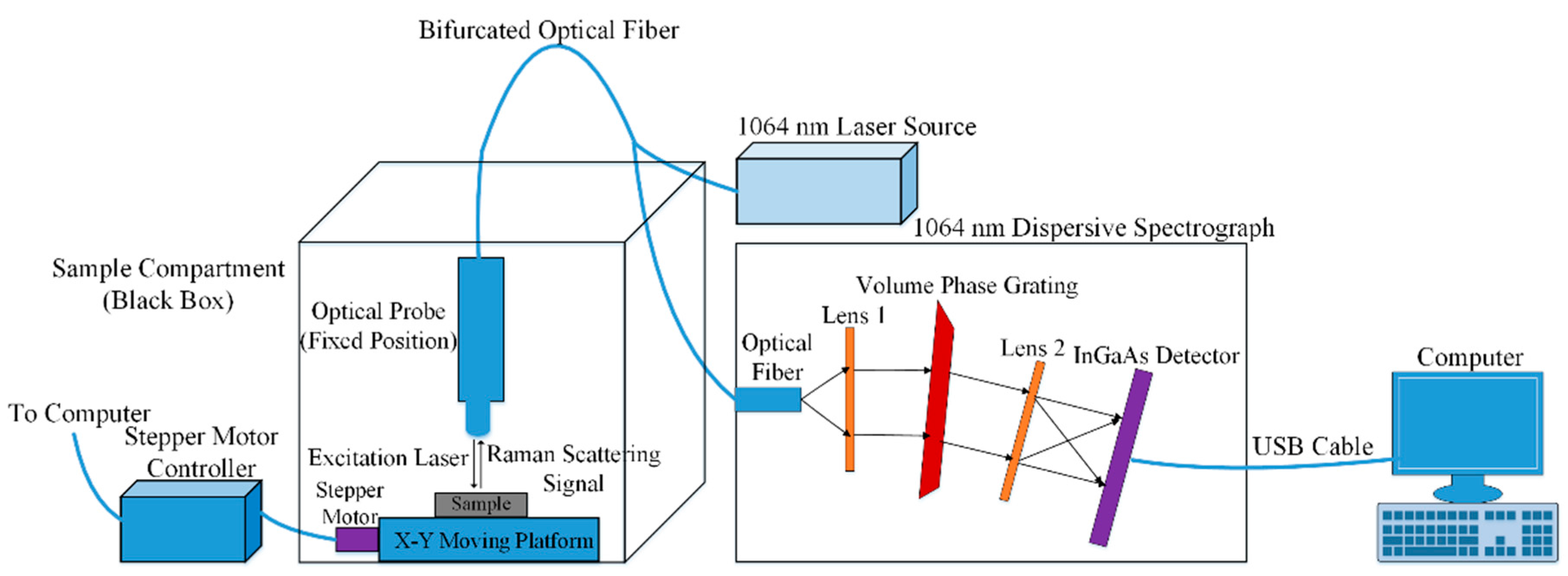
Figure 1 shows a schematic of the 1064 nm dispersive Raman spectral imaging system. The Raman spectrograph uses a high throughput volume phase grating (VPG) (BaySpec, Inc., San Jose, CA, USA) optimized for 1064 nm laser excitation. A concave lens (Lens 1) in the spectrograph guides the incoming light to the VPG, where the light is diffracted into different angular output paths. The concave lens (Lens 2) reflects the dispersed light to the 512 pixels Indium-Gallium-Arsenide (InGaAs) detector (Nunavut, BaySpec, Inc., San Jose, CA, USA). The detector is thermoelectrically deep cooled to −55 °C during spectral acquisition to minimize the dark current. The detector is connected to the computer using a USB cable for detector control and data transfer.
A 1064 nm laser module (MiniLite, BaySpec, Inc., San Jose, CA, USA) with a power of 600 mW is used for sample excitation. A fiber-optic Raman probe (RPB, InPhotonics, Inc., Norwood, MA, USA) is used to focus the laser on the sample surface and acquire the scattering signal. A long-pass filter assembled in the probe eliminates the light at and below 1064 nm. The probe is coupled to the Raman spectrograph via a bifurcated optical fiber that consists of excitation fiber and collection fiber. The excitation fiber (diameter 105 µm) carries the laser from the source to the Raman probe, is connected to the output of laser source by angled physical contact (APC) connector. The laser is focused on the sample surface by an extension tip of the probe. The end face polished at an 8-degree angle in the APC connector allows any light that is reflected back to the source to reflect out into the fiber cladding, achieving a better return loss. The collection fiber (diameter 200 µm) transfers the acquired scattering Raman signal to the spectrometer, is connected to the spectroscopy by fiber-optic connector (FC). The Raman probe uses bandpass, diachronic and edge filters for separating the excitation light and the scattered signal. The probe has a focal length of 7.5 mm and spot size of 158 µm. A two-axis motorized positioning table (MAXY4009W1-S4, Velmex, Bloomfield, NY, USA) is used to hold and move the sample in two perpendicular directions. The positioning table can move through an area of 127 mm × 127 mm with a displacement resolution of 6.35 µm. Stepper motor controller (Velmex, Bloomfield, NY, USA) is programmed to control its movement. Sample movement at an adjustable step size below the fixed position Raman probe allows accumulation of Raman spectral image of the entire surface area of the sample, which can be analyzed both spectrally and spatially.
2.2. Software
Interface software of the Raman system was developed on a LabView platform (National Instruments, Austin, TX, USA) for parameter setup and data transfer. The interface software controls system operational parameters, such as initialization, exposure time, spectral acquisition and display, sample movement, and data transfer. The software development kits (SDKs) that were provided by manufacturers of InGaAs detector and positioning table were used to develop the interface software. The acquired hyperspectral data are stored in the band interleaved by pixel (BIP) format, which can be commercially analyzed by available software, such as ENVI (4.5, ITT Visual Information Solutions, Boulder, CO, USA) and Matlab (R2013a, MathWorks, Natick, MA, USA).
2.3. Spectral Calibration and Resolution
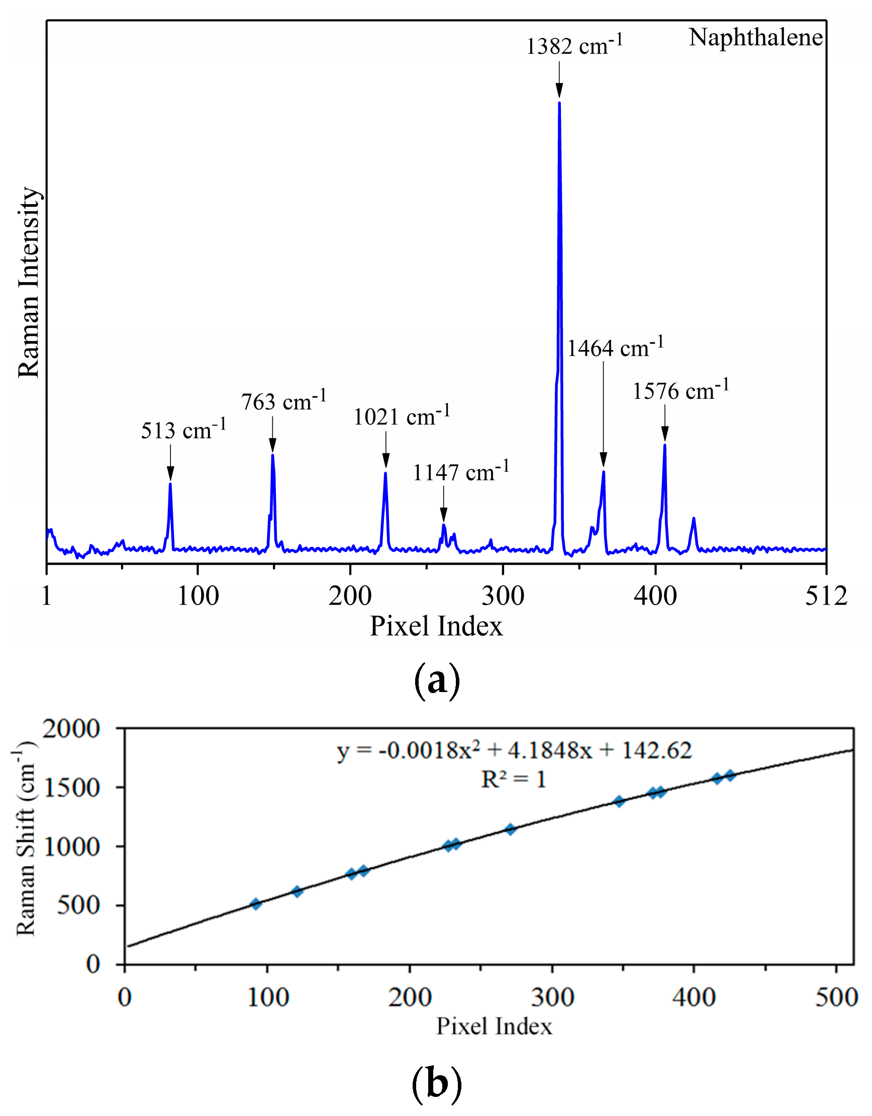
The X-axis in the Raman spectral measurement, termed as Raman shift (wavenumber, cm−1), is the change in the energy state from the excitation source. Typically, chemicals with known wavenumber shifts are used to assign a wavenumber to the corresponding pixels along the spectral dimension. The process is called spectral calibration. In this study, spectral calibration was done using polystyrene and naphthalene. American Society for Testing and Materials (ASTM) International enlists six chemicals (polystyrene, naphthalene, 1,4-bis(2-methylstyryl) benzene, sulfur, toluene-acetonitrile (concentration 50/50 by volume), 4-acetamidophenol, benzonitrile, and cyclohexane) as Raman shift standards for spectrometer calibration [46]. Polystyrene and naphthalene were packaged separately in two Petri-dishes and scanned covering the surface area of 10 mm × 10 mm with a step size of 1 mm for X- and Y-axes, collecting a total of 100 spectra from each Petri-dish. Figure 2a shows an average Raman spectrum of naphthalene (polystyrene spectrum not shown). Seven spectral peaks were identified for naphthalene between 513 to 1576 cm−1. Similarly, five peaks were identified for polystyrene in the wavenumber range of 620 to 1602 cm−1. Spectral calibration was performed using the twelve peaks covering the wavenumber range of 513 to 1602 cm−1. Quadratic fitting was used to develop spectral calibration model. The quadratic model was used to determine the wavenumbers of corresponding pixels. The Raman system covered the wavenumber range of 142 to 1820 cm−1 (Figure 2b) after spectral calibration. The spectral resolution of the Raman spectrometer is 12 cm−1 at full width half maximum (FWHM).
2.4. Spatial Resolution
The spatial resolution of the 1064 nm Raman spectral imaging system was evaluated using a standard resolution glass test chart (Edmund Optics Inc., Barrington, NJ, USA). The test chart was placed on the top of a Petri-dish containing naphthalene. The 1064 nm laser source illuminated the test chart. A step size of 0.1 mm was used to scan X and Y directions. Figure 3 shows the single band image of the resolution test chart. The laser spot on the white space penetrated through the glass test chart to the naphthalene, while the illumination on the black dots suppressed the Raman spectra of naphthalene, creating a contrast between white space and the black dot. In the Figure 3, the small inner dots have the diameter of 0.25 mm with 0.5 mm distance between each dot. The outer larger dots have the diameter of 0.5 mm with 1 mm dot spacing. Both the dots and their dot spacing can be distinguished in the image due to the small step size used to scan the chart. The step size used to scan the X and Y direction is the spatial resolution of the Raman image. Figure 3 shows that the system can achieve the spatial resolution of 0.1 mm.
3. Application to Authenticate Turmeric Powder
Turmeric (Curcuma long L.), which is a yellow colored herbaceous root from ginger family, is commonly used for food seasoning, for medicinal purpose and as dietary supplements [32]. Anti-inflammatory, anticarcinogenic, antioxidant, wound-healing effects are some of the medicinal value that is attributed to turmeric [47,48,49,50]. The medicinal value of turmeric is associated with the yellow color pigment “curcumin” (diferuloyol methane) content in it. Curcumin content varies in the range of 0.3% to 8.6% [47,51,52,53,54], due to nutrients and acidity content in soil [55,56], fertilizer, soil type and cultivar [57,58,59]. Due to the isolation of curcumin from turmeric for medicinal and cosmetic purposes, it is adulterated with color dyes as a substitute for curcumin [60]. Metanil yellow (C18H14N3NaO3S), a yellow color toxic azo dye, is added to turmeric for brighter color and appealing appearance [32]. Toxicologically, metanil yellow is classified as a CII category substance by the Joint Food and Agricultural Organization of the United Nations (FAO) and World Health Organization (WHO) expert committee on Food Additives [61]. Study on rats shows that the consumption of metanil yellow causes neurotoxicity [62], hepatocellular carcinoma [63], tumor development [64], deleterious effect on gastric mucin [65], and lymphocytic leukemia [66]. This study demonstrates the application of the 1064 nm dispersive Raman spectral imaging system for the detection of metanil yellow contamination in turmeric powder and subsurface detection and identification of gelatin-encapsulated pure and mixed samples of metanil yellow and turmeric powder. For each sample, a dark current spectrum was acquired with the laser off and a cap covering the probe and subtracted from the Raman spectrum at each pixel during the spectral acquisition. Five-point moving average filter was used to smooth the spectra.
3.1. Detection of Metanil Yellow Contamination in Turmeric Powder
Metanil yellow (70% dye, Aldrich, Carson City, NV, USA) and organic turmeric powder (Frontier Natural Products CO-OP, Norway, IA, USA) were mixed together in a vortex mixer (Scientific Industries Inc., Bohemia, NY, USA) for ten minutes to prepare sample mixture at 1%, 3%, 5%, 7%, and 10% metanil yellow concentration (w/w). The weight of each mixture sample was 0.27 g. Each mixture sample was packed into a shallow nickel-plated sample container (internal volume 25 mm × 25 mm × 1 mm), and its surface was leveled flush with the edge of the container. A Raman spectral image of each sample was obtained at 1 s exposure time and 80 mW laser power, covering a spatial area of 25 mm × 25 mm with a step size of 0.25 mm, collecting a total of 10,000 spectral pixels.
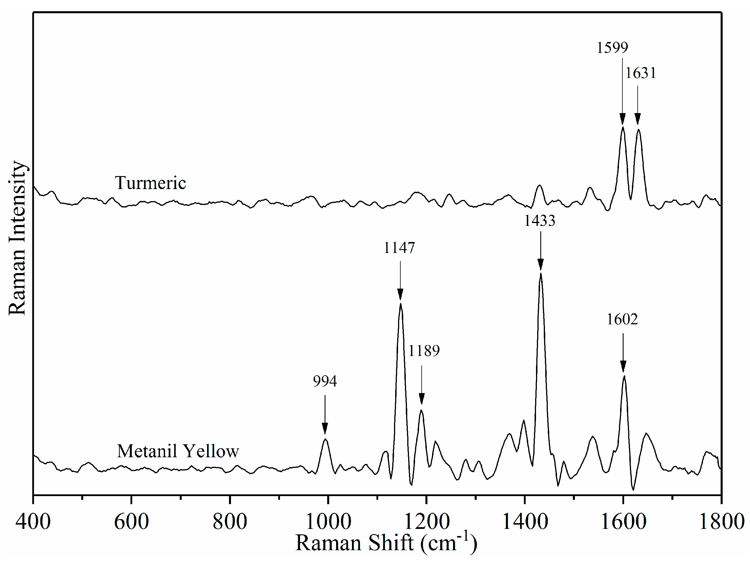
Figure 4 shows the Raman spectra of turmeric powder and metanil yellow. The yellow color appearance of turmeric and metanil yellow is due to the similarly extended conjugation in both the chemicals [32]. However, a different chemical composition results in different vibrational modes specific to their chemical structure. The sharp peaks of metanil yellow at 1147 cm−1 and 1433 cm−1, due to the N = N site are most definitive to its identification and quantification [67,68,69]. The 1631 cm−1 peak in turmeric spectrum is due to the carbonyl group. The carbonyl group is not present in metanil yellow [32]. Therefore, the 1631 cm−1 peak is most definitive for turmeric identification.
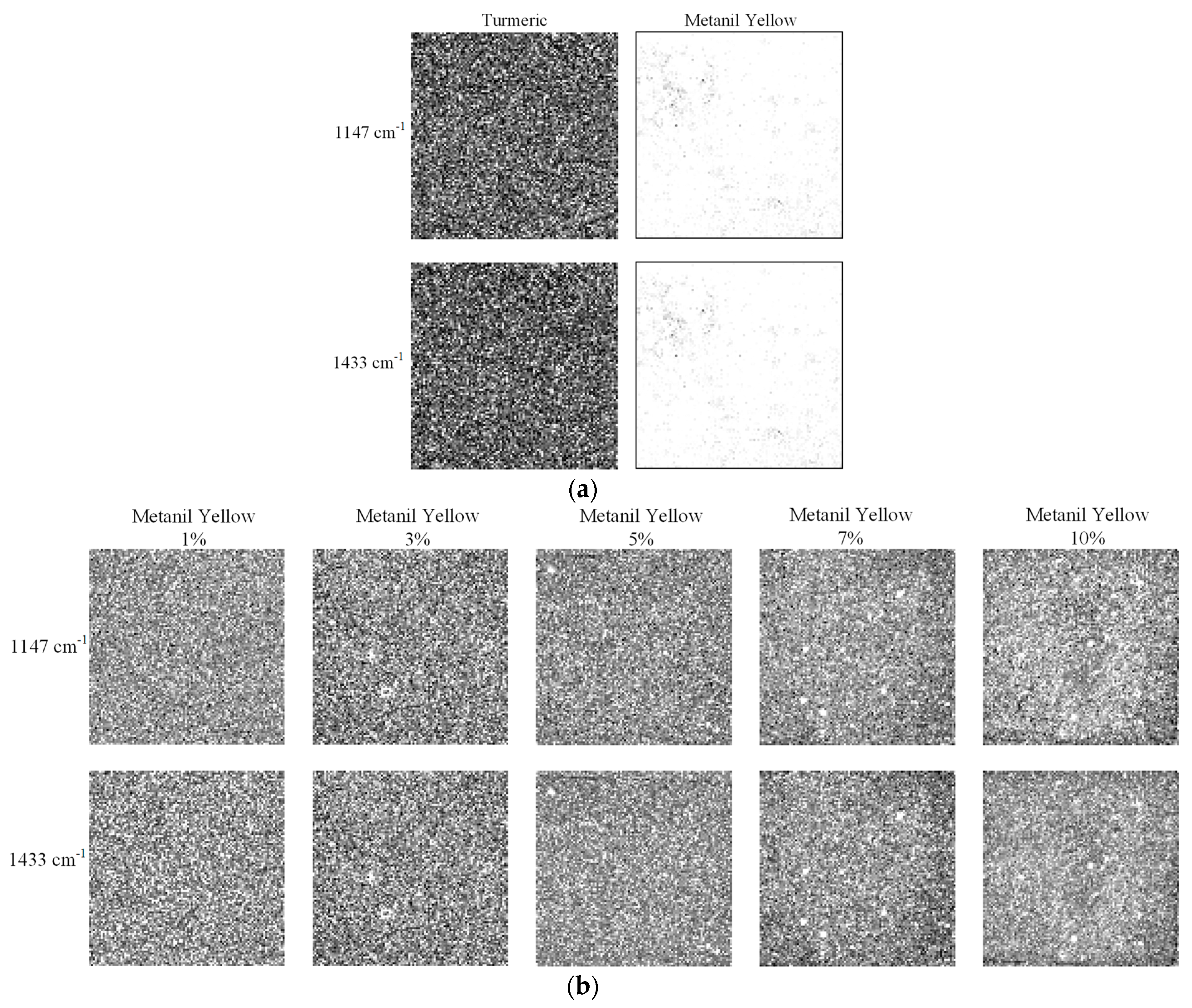
Figure 5 shows the Raman spectral images of turmeric, metanil yellow, and turmeric-metanil yellow mixtures across all of the concentration at 1147 cm−1 and 1433 cm−1. In Figure 5a, the dark pixels in the spectral images of turmeric show low spectral intensity of turmeric at 1147 cm−1 and 1433 cm−1. However, spectral images of metanil yellow have white pixels revealing high spectral intensity at 1147 cm−1 and 1433 cm−1 (Figure 5a). The 1147 cm−1 and 1433 cm−1 are the highest intensity metanil yellow peaks. Figure 5b is the raw spectral images of turmeric-metanil yellow samples at different concentrations. The white pixels in the spectral images are due to the high spectral peak intensities at 1147 cm−1 and 1433 cm−1.
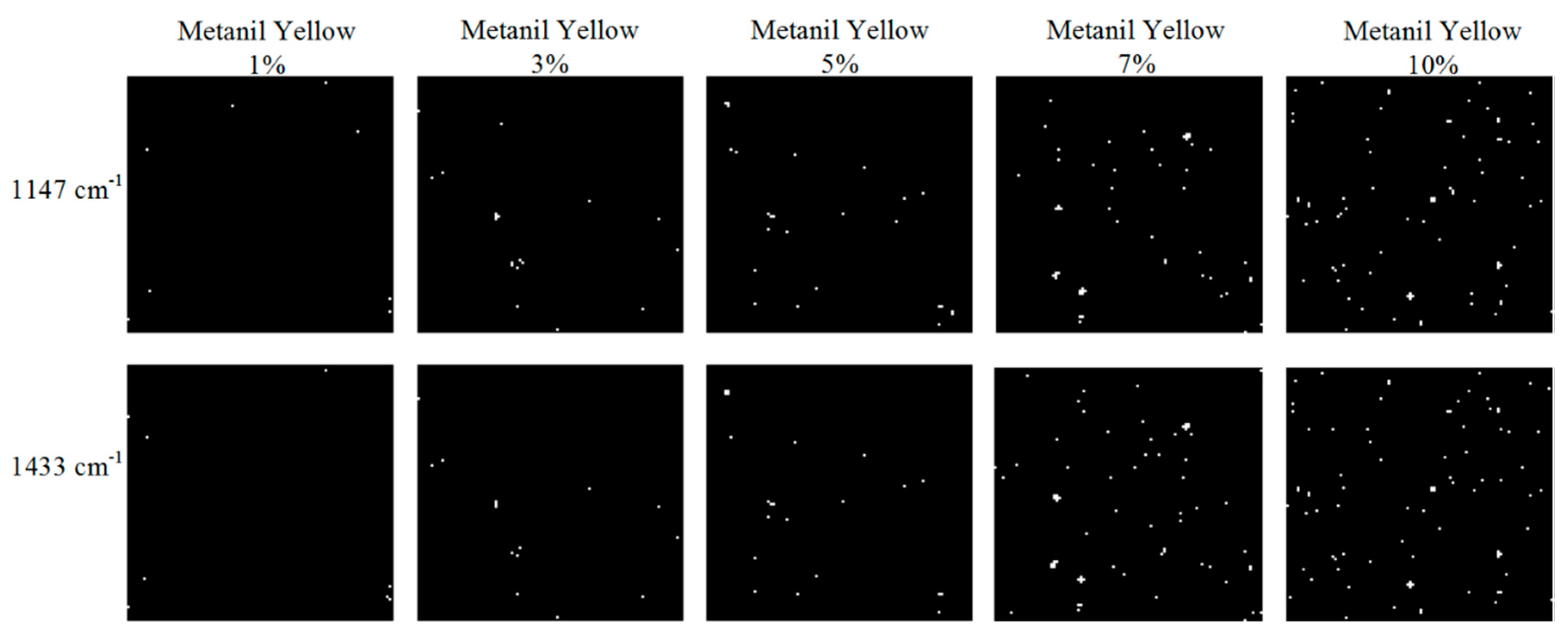
For identification of metanil yellow pixels, binary detection images were created. The 1147 cm−1 and 1433 cm−1 peaks were used to convert the Raman spectral images into two separate single-band binary images. An intensity threshold was used to convert all the pixels below the threshold value into background pixels of turmeric. It was observed that the intensities of some of the metanil yellow pixels were lower than the set threshold value (false negative), while the intensities of some turmeric pixels were higher than the set threshold value (false positive). To avoid the false-positive and false-negative cases, a pixel-to-pixel analysis was done to select the most appropriate threshold value. Each pixel in the binary image was evaluated with its corresponding spectrum. A final intensity threshold value of 550 was set. All the pixels below the threshold value were converted to background pixels, and the remaining pixels represented metanil yellow. Figure 6 shows the 1147 cm−1 and 1433 cm−1 binary detection images of the samples, in which the white pixels represent the detected metanil yellow particles. A gradual increase in the number of white pixels at higher concentration shows more metanil yellow particles were detected at increasing concentration. A total of 8, 19, 35, 55, and 80 metanil yellow pixels were detected in the 1147 cm−1 binary images of 1%, 3%, 5%, 7%, and 10% concentration samples (Figure 6). A similar number of metanil yellow pixels were detected in the 1433 cm−1 binary images. The metanil yellow pixels have a similar spatial distribution in both the binary images.
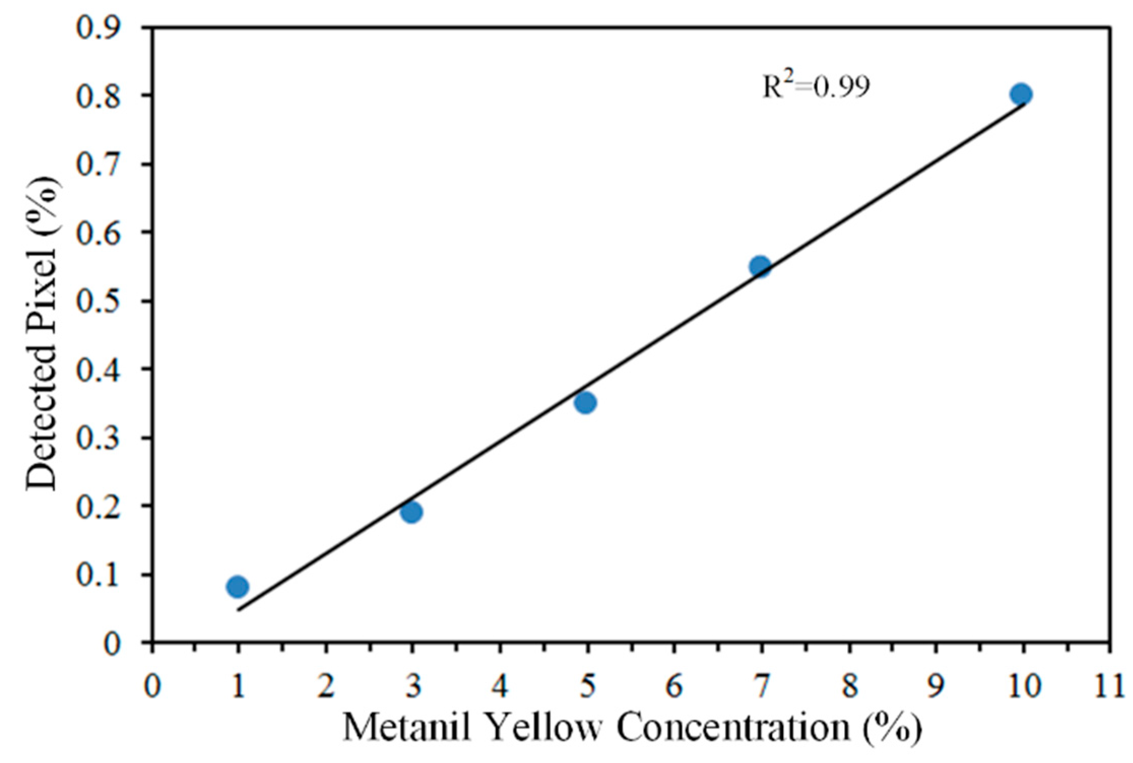
Out of ten thousand pixels that were acquired from 10% concentration sample, 80 detected metanil yellow pixels correspond to 0.80% of total pixels. Similarly, in 1% concentration sample, 0.08% of total pixels were detected as metanil yellow. The percentage of detected metanil yellow pixels were correlated with its actual concentration in the sample with a correlation coefficient of 0.99 (Figure 7). The result shows a linear relationship between the metanil yellow concentration in the sample and the percentage of its detected pixels.
3.2. Subsurface Detection of Turmeric Powder and Metanil Yellow
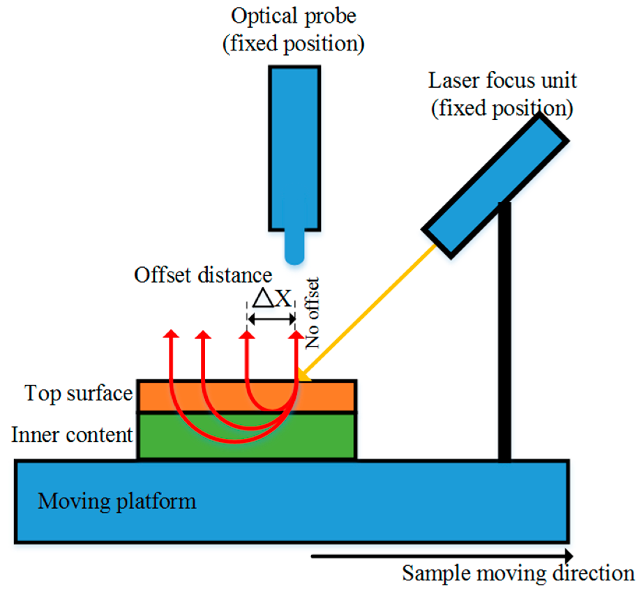
Spatially offset Raman spectroscopic (SORS) technique retrieves Raman information of the inner content of the sample from diffusely scattering media [36]. The SORS technique collects scattering signal from a series of offset distance from the laser excitation spot [70]. It uses a laser focus unit at a fixed 45° incident angle to deliver laser light onto the sample surface. An optical probe is fixed perpendicular to the sample to collect Raman scattering signal. Figure 8 shows the SORS technique to acquire Raman information of the inner content. First, Raman spectrum of sample at the no-offset position is measured by overlapping the focal point of laser and the optical probe. Then, the sample and the laser focus unit is moved at an incremental step away from the probe creating an increased offset distance between the probe and the laser focus point on the sample. As the offset distance is increased, the laser penetrates deeper through the thick top surface layer. At an increased offset distance, the signal contribution from the deep subsurface layer increases, gradually outweighing the signal contribution of the top surface layer, enabling the detection of subsurface layer material. The SORS signal can be used to retrieve Raman information of multiple layers from the same sample. The SORS method has been applied for diagnosis of breast cancer by measuring calcification composition through varying tissue thickness [71]. It has been applied for evaluation of internal maturity of tomatoes by evaluating the carotenoids change in them [72]. Although, the SORS method can be used for detection and analysis of subsurface layer material, it cannot be used to measure the depth of the sample.
Empty gelatin capsules of size 000 (length 26.1 mm, single-wall thickness 0.11 mm) were obtained from Capsuline (Pompano Beach, FL, USA) to prepare capsule-turmeric samples. A one-layer capsule sample was prepared by packing turmeric tightly in a capsule and capping it. A two-layer capsule sample was prepared by inserting the turmeric packed capsule inside an empty capsule and capping the outer capsule only. Similarly, three-, four-, and five-layer capsule samples were prepared by inserting the turmeric packed capsule inside empty capsules and capping the outermost capsule. The wall thickness of the capsule layers ranged from 0.11 mm (one-layer capsule) to 0.55 mm (five-layer capsule).
Each capsule sample was held immobile in the positioning platform. The 1064 nm laser module focused a laser spot on the capsule body near the edge of its cap. First, Raman spectrum of capsule-turmeric sample was obtained. Then, the positioning table moved the sample and the laser focus unit in incremental steps in the right direction away from the Raman probe, creating an increased offset distance for each subsequent spectral acquisition. A total of 31 spectra along the longitudinal flat surface body of each capsule sample proceeding away from the capped end were collected using 20 s exposure time, 400 mW laser power, and a step size of 0.1 mm from no offset to 3 mm offset range. The spatially offset Raman spectra of samples were subtracted with the empty capsule spectrum to eliminate the influence of capsule signal in the SORS data.
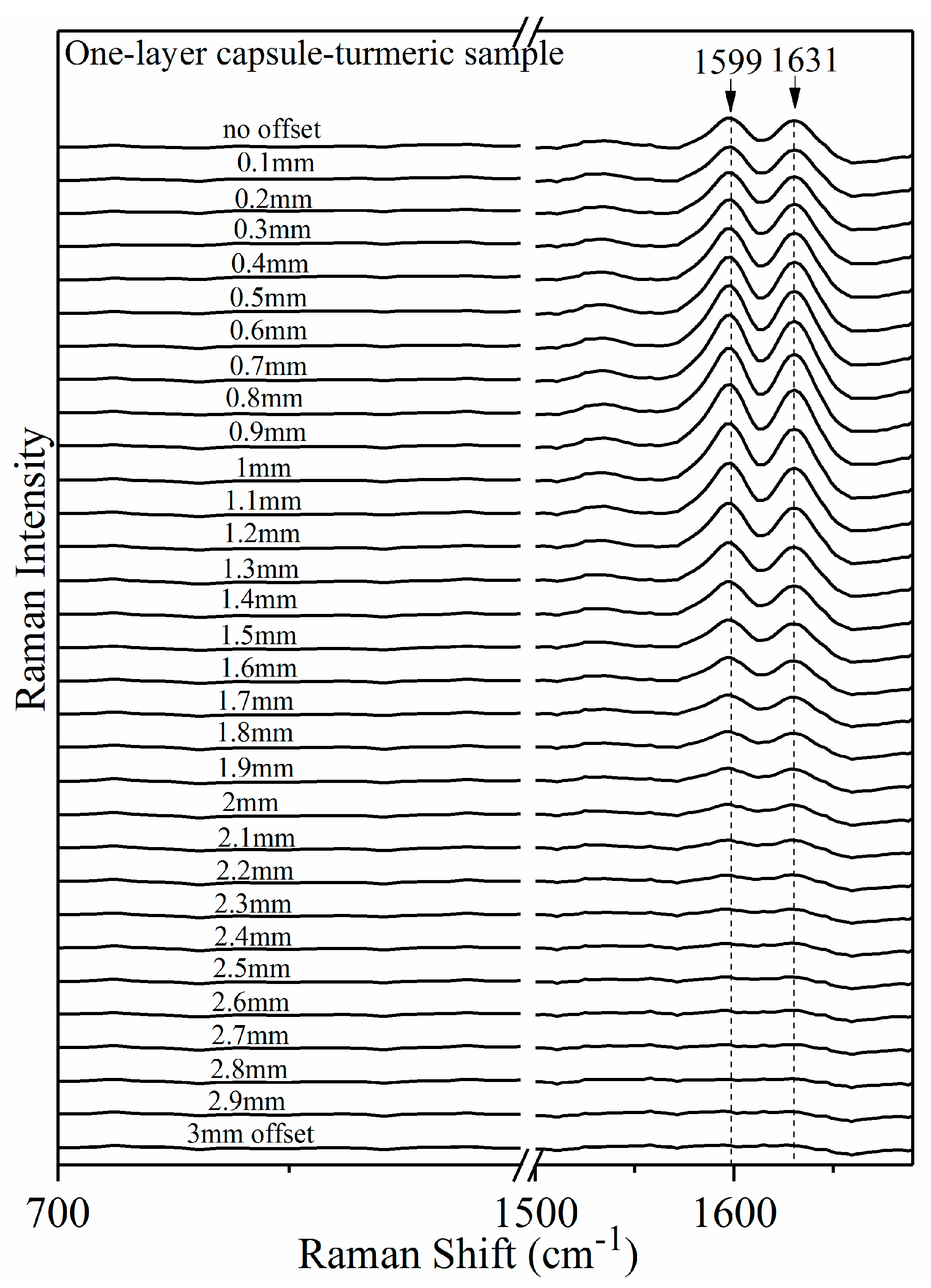
Figure 9 shows the spatially offset Raman spectra of the one-layer capsule-turmeric sample. The 1599 cm−1 and 1631 cm−1 are turmeric peaks. The pattern of increasing 1599 cm−1 and 1631 cm−1 peak intensity from no offset up to 1.0 mm offset shows that the signal contribution of the subsurface material (turmeric) gradually increases as the offset distance increases. After 1.0 mm offset, the signal contribution from turmeric layer gradually decreased.
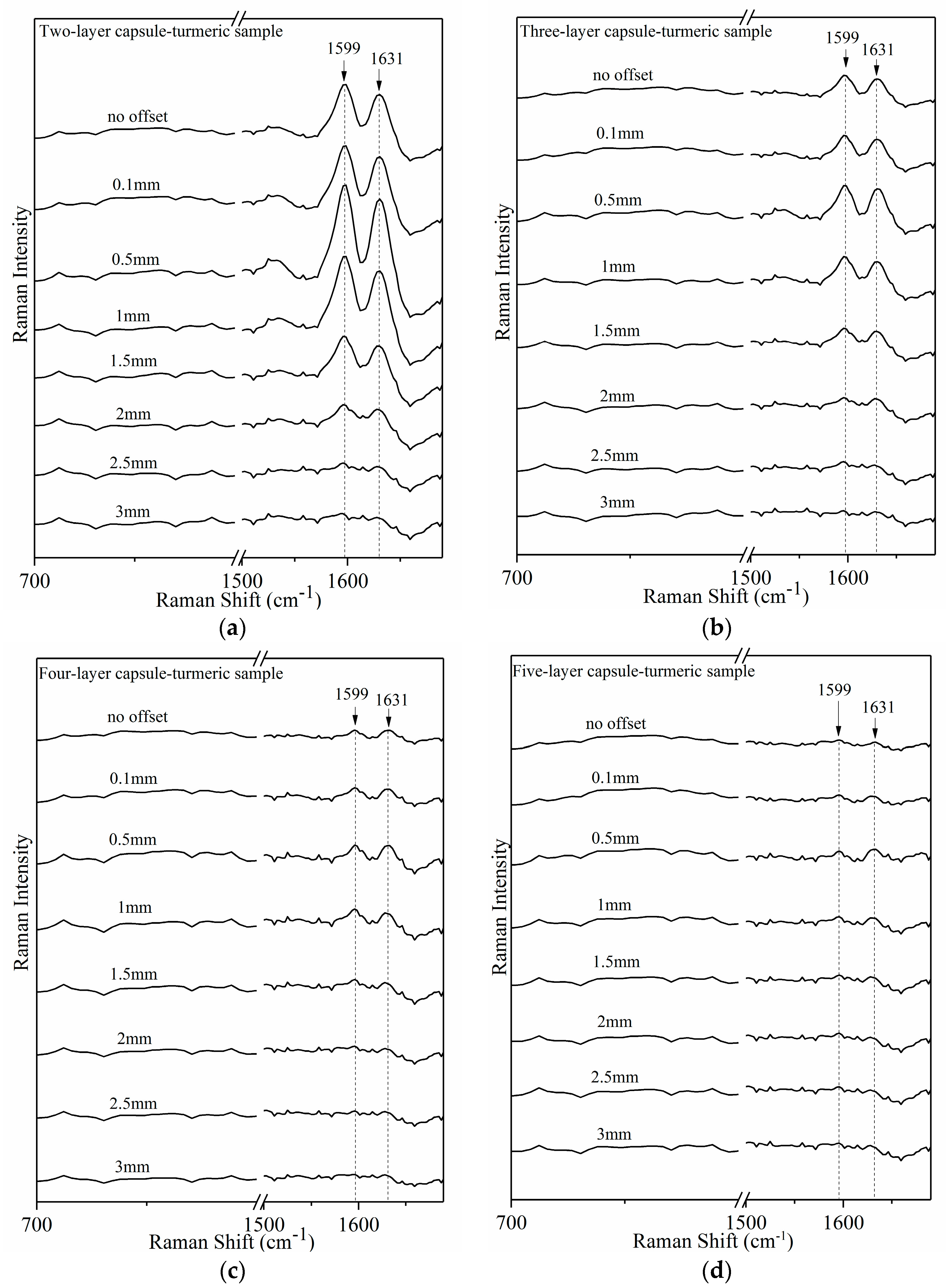
Figure 10 shows the spatially offset Raman spectra from capsule-turmeric sample at two-, three-, four-, and five-capsule layer at no offset, 0.1 mm, 0.5 mm, 1.0 mm, 1.5 mm, 2.0 mm, 2.5 mm, and 3.0 mm offset. Steep increase in the 1599 cm−1 and 1631 cm−1 intensity with increasing offset distance is readily visible in Figure 10a for two-layer capsule-turmeric sample. Similarly, for three-, four-, and five-layer capsule samples, enhancement of 1599 cm−1 and 1631 cm−1 peak is evident as the offset distance is increased (Figure 11b–d).
At no offset, the turmeric peaks evident in one- and two-layer capsule samples are attenuated as the capsule layer is increased (Figure 10c,d) because the capsule layer suppresses the deep subsurface turmeric signal. In the five-layer capsule sample (Figure 10d), 1599 cm−1 and 1631 cm−1 peaks are not evident at no offset. This prevents identification of turmeric inside the five-layer capsule. The no offset measurement measures backscattering Raman signal. This indicates that the backscattering Raman measurement technique is not useful to analyze the subsurface material through the thick top surface layer. However, increasing the offset distance enhanced the 1599 cm−1 and 1631 cm−1 peaks (Figure 10d), allowing for the identification of subsurface turmeric despite the presence of thick gelatin capsule layers.
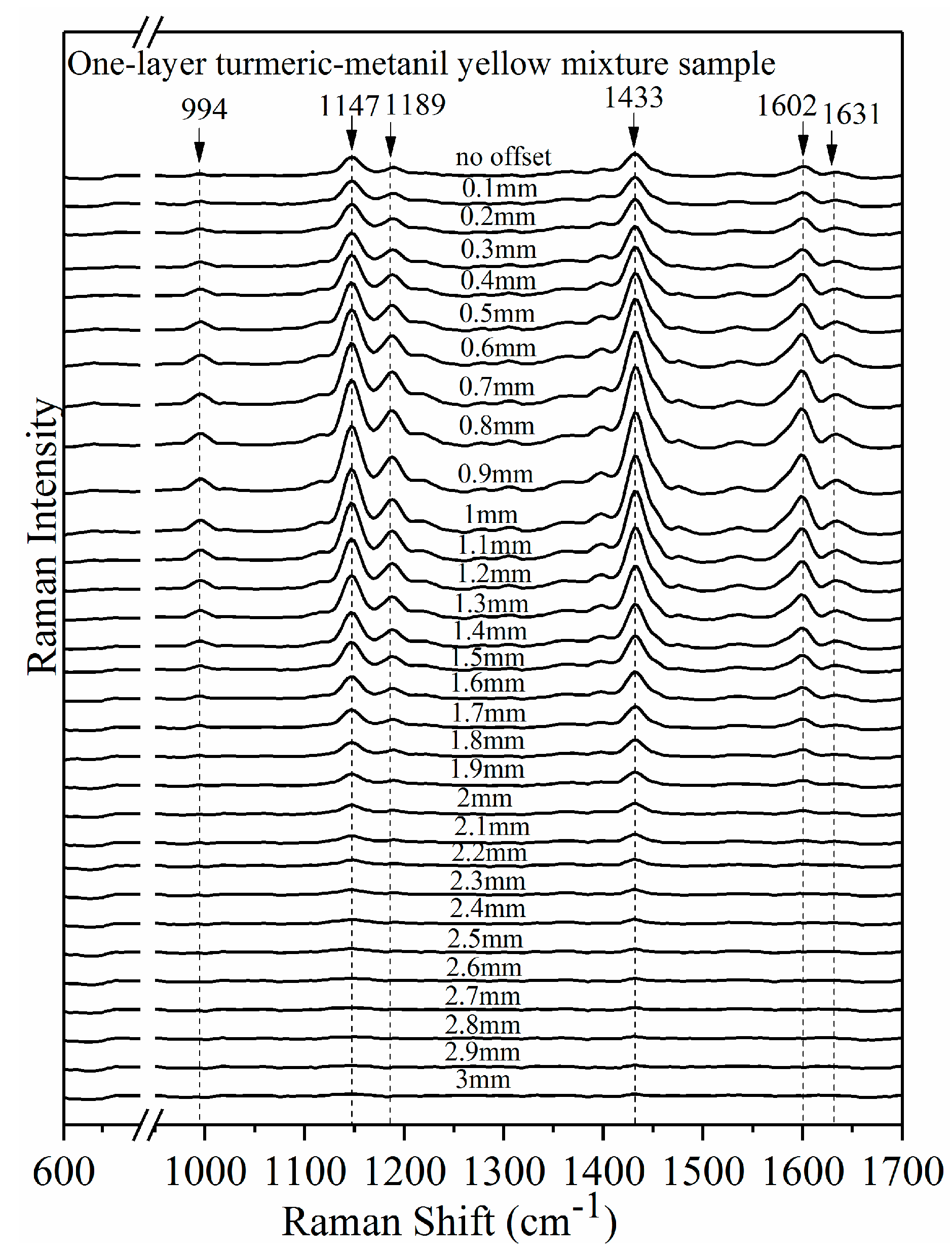
The effectiveness of the SORS method was further validated by identifying metanil yellow and turmeric powder packed inside one up to five-layer of capsule. Turmeric-metanil yellow mixture sample at 50% concentration (w/w) was prepared and packed inside gelatin capsule to obtain one-, two-, three-, four-, and five-layer samples. A series of spatially offset Raman spectra were collected from no offset to 3 mm offset using a 0.1 mm increment along the longitudinal flat surface of capsule sample. Figure 11 shows the SORS spectra from the one-layer capsule sample. The 994 cm−1, 1147 cm−1, 1189 cm−1, 1433 cm−1, and 1602 cm−1 are metanil yellow peaks. The 1631 cm−1 is a turmeric peak. The turmeric peak at 1599 cm−1 is not resolved from the 1602 cm−1 peak of metanil yellow. Metanil yellow peaks at 1147 cm−1, 1189 cm−1, and 1433 cm−1 are evident at no offset, and are enhanced by increasing the offset distance. The 994 cm−1 metanil yellow peak is not evident at no offset. Increasing the offset distance enhanced the 994 cm−1 peak. The 1631 cm−1 turmeric peak is not evident at no offset. Increasing the offset distance gradually increased the 1631 cm−1 peak intensity.
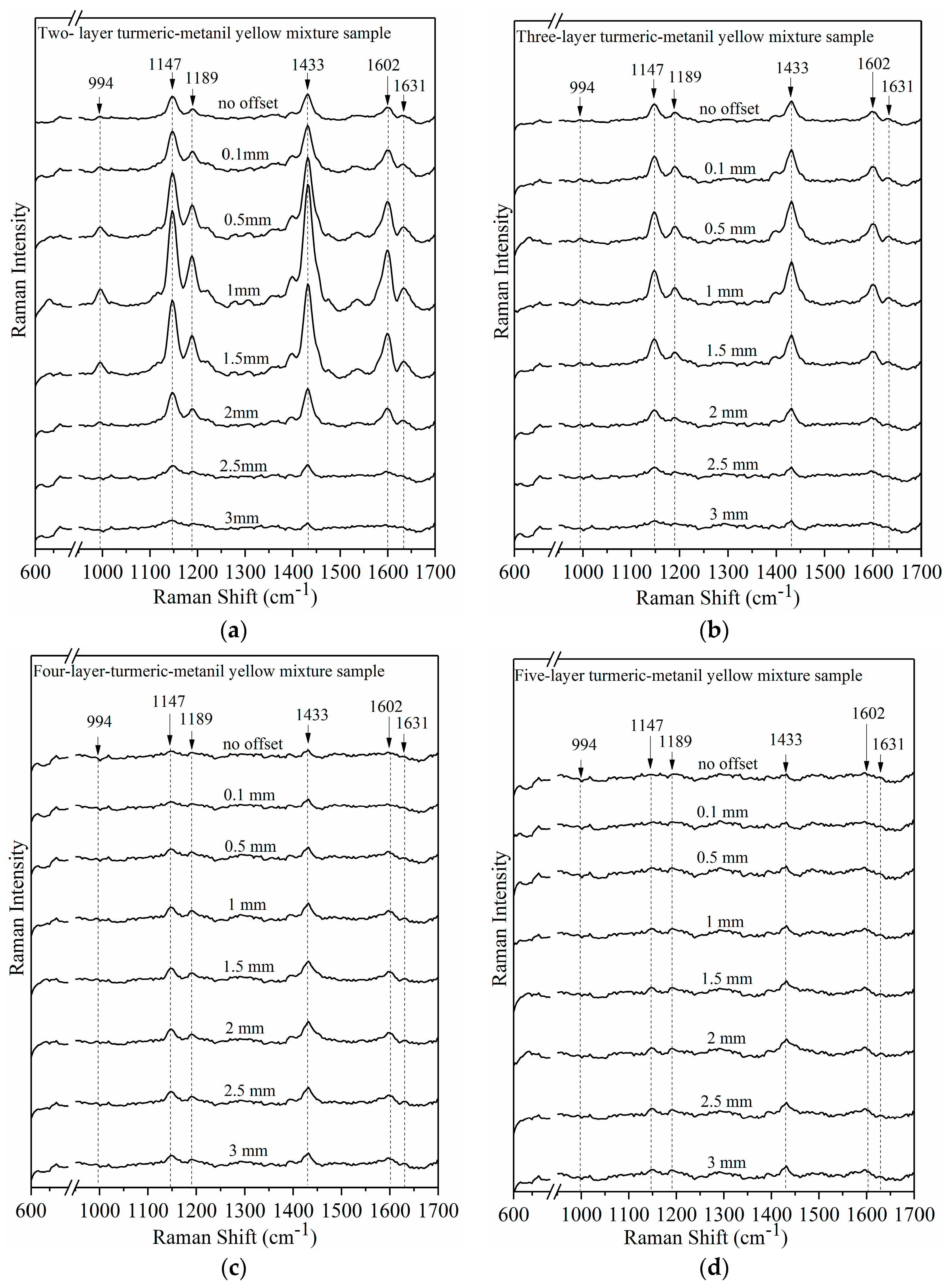
Figure 12 shows the SORS spectra from two-, three-, four-, and five-layer capsule samples at no offset, 0.1 mm, 0.5 mm, 1.0 mm, 1.5 mm, 2.0 mm, 2.5 mm, and 3.0 mm offset. Increasing the capsule layer attenuated the metanil yellow and turmeric peaks at no offset. The metanil yellow peaks at 1147 cm−1 and 1433 cm−1 were evident at no offset in the two- and three-layer capsule samples (Figure 12a,b). The 1147 cm−1 peak is not evident at no offset in the four- and five-layer capsule samples (Figure 12c,d). Increasing the offset distance enhanced the 1147 cm−1 peak (Figure 12c,d). The 1433 cm−1 peak is evident at no offset in the two-, three-, and four-layer capsule samples (Figure 12a–c). An increase in the peak intensity at 1433 cm−1 was observed from no offset to 1.0 mm offset in two-, three-, and four-layer capsule samples (Figure 12a–c). After 1.0 mm offset, the 1433 cm−1 peak intensity gradually decreased. In the five-layer capsule sample, the 1433 cm−1 peak is not evident at no offset (Figure 12d). Increasing the offset distance enhanced the 1433 cm−1 peak in the five-layer capsule sample.
The turmeric peak at 1631 cm−1 is not evident at no offset in the two-, three-, four-, and five-layer capsule samples (Figure 12a–d). Increasing the offset distance enhanced the 1631 cm−1 peak in the multiple layer capsule samples. The 1631 cm−1 peak can be used to identify the turmeric packed inside gelatin capsule. The results demonstrate this SORS measurement technique has a potential for non-destructive subsurface detection and the identification of gelatin-encapsulated pure and mixed samples of turmeric and metanil yellow powder.
4. Conclusions
A 1064 nm dispersive Raman spectral imaging system was developed for food safety and quality evaluation. The system is capable of hyperspectral Raman imaging and spatially offset Raman spectroscopic (SORS) measurement of the food samples. The application of the hyperspectral Raman imaging system was demonstrated by detecting metanil yellow contamination in turmeric powder at five concentration levels. A hyperspectral Raman image of the mixture sample at each concentration was collected covering the sample surface area of 25 mm × 25 mm, using a step size of 0.25 mm along the X and Y direction. The system could detect metanil yellow at concentrations as low as 1%. The detected metanil yellow pixels linearly correlated with its concentration in the mixture sample (R2 = 0.99). Application of the system for SORS measurement was demonstrated by subsurface detection of gelatin-encapsulated pure and mixed samples of turmeric and metanil yellow powder. This system is a versatile platform capable of 1064 nm hyperspectral Raman imaging and SORS measurement for a wide variety of food and pharmaceutical samples.
Author Contributions
Kuanglin Chao and Sagar Dhakal conceived and designed the experiments; Kuanglin Chao and Sagar Dhakal performed the experiments; Kuanglin Chao and Sagar Dhakal analyzed the data, with other co-authors Jianwei Qin, Moon Kim, and Yankun Peng also participating in the discussion of the results; Kuanglin Chao and Sagar Dhakal wrote the paper.
Conflicts of Interest
The authors declare no conflict of interest.
Disclaimer
Mention of specific products is for identification only and does not imply endorsement by the US Department of Agriculture to the exclusion of other suitable products or suppliers.
References
- Huang, H.; Liu, L.; Ngadi, M.O. Recent development in hyperspectral imaging for assessment of food quality and safety. Sensors 2014, 14, 7248–7276. [Google Scholar] [CrossRef] [PubMed]
- Cen, H.; He, Y. The theory and application of near infrared reflectance spectroscopy in determination of food quality. Trends Food Sci. Technol. 2007, 18, 72–83. [Google Scholar] [CrossRef]
- Chen, Q.; Zhang, C.; Zhao, J.; Ouyang, Q. Recent advances in emerging imaging techniques for non-destructive detection of food quality and safety. Trends Anal. Chem. 2013, 52, 261–274. [Google Scholar] [CrossRef]
- Herrero, A.M. Raman spectroscopy a promising technique for quality assessment of meat and fish: A review. Food Chem. 2008, 107, 1642–1651. [Google Scholar] [CrossRef]
- Li-Chan, E.C.Y. The applications of Raman spectroscopy in food science. Trends Food Sci. Technol. 1996, 7, 361–370. [Google Scholar] [CrossRef]
- Yang, D.; Ying, Y. Applications of Raman spectroscopy in agricultural products and food analysis: A review. Appl. Spectrosc. Rev. 2011, 46, 539–560. [Google Scholar] [CrossRef]
- Wang, Q.; Li, Z.; Ma, Z.; Liang, L. Real time monitoring of multiple components in wine fermentation using an on-line auto-calibration Raman spectroscopy. Sens. Actuators B Chem. 2014, 202, 426–432. [Google Scholar] [CrossRef]
- Uysal, R.S.; Soykut, E.A.; Boyaci, I.H.; Topcu, A. Monitoring multiple components in vinegar fermentation using Raman spectroscopy. Food Chem. 2013, 141, 4333–4343. [Google Scholar] [CrossRef] [PubMed]
- Ozbalci, B.; Boyaci, I.H.; Topcu, A.; Kadilar, C.; Tamer, U. Rapid analysis of sugars in honey by processing Raman spectrum chemometric methods and artificial neural networks. Food Chem. 2013, 136, 1444–1452. [Google Scholar] [CrossRef] [PubMed]
- Camerlingo, C.; Zenone, F.; Delfino, I.; Diano, N.; Mita, D.G.; Lepore, M. Investigation on clarified fruit juice composition by using visible light micro-Raman spectroscopy. Sensors 2007, 7, 2049–2061. [Google Scholar] [CrossRef] [PubMed]
- Wang, Q.; Lonergan, S.M.; Yu, C. Rapid determination of pork sensory quality using Raman spectroscopy. Meat Sci. 2012, 91, 232–239. [Google Scholar] [CrossRef] [PubMed]
- Pedersen, D.K.; Morel, S.; Andersen, H.J.; Engelsen, S.B. Early prediction of water-holding capacity in meat by multivariate vibrational spectroscopy. Meat Sci. 2003, 65, 581–592. [Google Scholar] [CrossRef]
- Schmidt, H.; Scheier, R.; Hopkins, D.L. Preliminary investigation on the relationship of Raman spectra of sheep meat with shear force and cooking loss. Meat Sci. 2013, 93, 138–143. [Google Scholar] [CrossRef] [PubMed]
- Dhakal, S.; Li, Y.; Peng, Y.; Chao, K.; Qin, J.; Guo, L. Prototype instrument development for non-destructive detection of pesticide residue in apple surface using Raman technology. J. Food Eng. 2014, 123, 94–103. [Google Scholar] [CrossRef]
- Lopez-Diez, E.C.; Bianchi, G.; Goodacre, R. Rapid and quantitative assessment of the adulteration of virgin olive oils with hazelnut oils using Raman spectroscopy and chemometrics. J. Agric. Food Chem. 2003, 51, 6145–6150. [Google Scholar] [CrossRef] [PubMed]
- Okazaki, S.; Hiramatsu, M. Rapid nondestructive screening for melamine in dried milk by Raman spectroscopy. Forensic Toxicol. 2009, 27, 94–97. [Google Scholar] [CrossRef]
- Sowoidnich, K.; Schmidt, H.; Kronfeldt, H.D.; Schwagele, F. A portable 671 nm Raman sensor system for rapid meat spoilage identification. Vib. Spectrosc. 2012, 62, 70–76. [Google Scholar] [CrossRef]
- Meisel, S.; Stockel, S.; Rosch, P.; Popp, J. Identification of meat-associated pathogens via Raman microspectroscopy. Food Microbiol. 2014, 38, 36–43. [Google Scholar] [CrossRef] [PubMed]
- Cheng, Y.; Dong, Y.; Wu, J.; Yang, X.; Bai, H.; Zheng, H.; Ren, D.; Zou, Y.; Li, M. Screening melamine adulterant in milk powder with laser Raman spectrometry. J. Food Compos. Anal. 2010, 23, 199–202. [Google Scholar] [CrossRef]
- Olsen, E.F.; Rukke, E.O.; Flatten, A.; Isaksson, T. Quantitative determination of saturated-, monosaturated- and polysaturated fatty acids in pork adipose tissue with non-destructive Raman spectroscopy. Meat Sci. 2007, 76, 628–634. [Google Scholar] [CrossRef] [PubMed]
- Shin, K.; Chung, H.; Kwak, C.W. Transmission Raman measurement directly through packed corn kernels to improve sample representation and accuracy of compositional analysis. Analyst 2012, 137, 3690–3696. [Google Scholar] [CrossRef] [PubMed]
- Schulmerich, M.V.; Walsh, M.J.; Gelber, M.K.; Kong, R.; Kole, M.R.; Harrison, S.K.; McKinney, J.; Thompson, D.; Kull, L.S.; Bhargava, R. Protein and oil composition predictions of single soybeans by transmission Raman spectroscopy. J. Agric. Food Chem. 2012, 60, 8097–8102. [Google Scholar] [CrossRef] [PubMed]
- Yang, H.; Irudayaraj, J. Rapid detection of foodborne microorganisms on food surface using Fourier transform Raman spectroscopy. J. Mol. Struct. 2003, 646, 35–43. [Google Scholar] [CrossRef]
- Liu, Y.; Delwiche, S.R.; Dong, Y. Feasibility of FT-Raman spectroscopy for rapid screening for DON toxin in ground wheat and barley. Food Addit. Contam. Part A 2009, 26, 1396–1401. [Google Scholar] [CrossRef]
- Liu, Y.; Chao, K.; Kim, M.; Tuschel, D.; Olkhovyk, O.; Priore, R.J. Potential of Raman spectroscopy and imaging methods for rapid and routine screening of the presence of melamine in animal feed and foods. Appl. Spectrosc. 2009, 63, 477–480. [Google Scholar] [CrossRef] [PubMed]
- Qin, J.; Chao, K.; Kim, M. Raman scattering for food quality and safety assessment. In Light Scattering Technology for Food Property, Quality and Safety Assessment; Lu, R., Ed.; CRC Press: Boca Raton, FL, USA, 2016; pp. 387–428. [Google Scholar]
- Baeten, V.; Hourant, P.; Morales, M.T.; Aparicio, R. Oil and fat classification by FT-Raman spectroscopy. J. Agric. Food Chem. 1998, 46, 2638–2646. [Google Scholar] [CrossRef]
- Silveira, F.L.; Silverira, L.; Villaverde, A.B.; Pacheco, M.T.T.; Pasqualucci, C.A. Use of dispersive Raman spectrograph in the determination of unsaturated fat in commercial edible oil- and fat-containing industrialized foods. Instrum. Sci. Technol. 2009, 38, 107–123. [Google Scholar] [CrossRef]
- Sato-Berru, R.Y.; Medina-Valtierra, J.; Medina-Gutierrez, C.; Frausto-Reyes, C. Quantitative NIR-Raman analysis of methyl-parathion pesticide microdroplets on aluminum substrates. Spectrochim. Acta Part A 2004, 60, 2231–2234. [Google Scholar] [CrossRef] [PubMed]
- Langkilde, F.W.; Sjoblom, J.; Tekenbergs-Hjelte, L.; Mrak, J. Quantitative FT-Raman analysis of two crystal forms of a pharmaceutical compound. J. Pharm. Biomed. Anal. 1997, 15, 687–696. [Google Scholar] [CrossRef]
- Baeten, V.; Peirna, J.A.F.; Dardenne, P.; Meurens, M.; Garcia-Gonzalex, D.L.; Aparicio-Ruiz, R. Detection of the presence of hazelnut oil in olive oil by FT-Raman and FT-MIR spectroscopy. J. Agric. Food Chem. 2005, 53, 6201–6206. [Google Scholar] [CrossRef] [PubMed]
- Dhakal, S.; Chao, K.; Schmidt, W.; Qin, J.; Kim, M.; Chan, D. Evaluation of turmeric powder adulterated with metanil yellow using FT-Raman and FT-IR spectroscopy. Foods 2016, 5, 36. [Google Scholar] [CrossRef] [PubMed]
- Yang, H.; Irudayaraj, J.; Paradkar, M.M. Discriminant analysis of edible oils and fats by FTIR, FT-NIR and FT-Raman spectroscopy. Food Chem. 2005, 93, 25–32. [Google Scholar] [CrossRef]
- Anibal, C.V.D.; Marsal, L.F.; Callao, M.P.; Ruisanchez, I. Surface enhanced Raman spectroscopy (SERS) and multivariate analysis as a screening tool for detecting Sudan I dye in culinary spices. Spectrochim. Acta A 2012, 87, 135–141. [Google Scholar] [CrossRef] [PubMed]
- Kamil, M.M.; Mohamed, G.F.; Shaheen, M.S. Fourier transformer infrared spectroscopy for quality assurance of tomato products. J. Am. Sci. 2011, 7, 559–572. [Google Scholar]
- Matousek, P.; Clark, I.P.; Draper, E.R.C.; Morris, M.D.; Goodship, A.E.; Everall, N.; Towrie, M.; Finney, W.F.; Parker, A.W. Subsurface probing in diffusely scattering media using spatially offset Raman spectroscopy. Appl. Spectrosc. 2005, 59, 393–400. [Google Scholar] [CrossRef] [PubMed]
- Qin, J.; Chao, K.; Kim, M.S. Raman chemical imaging system for food safety and quality inspection. Trans. ASABE 2010, 53, 1873–1882. [Google Scholar] [CrossRef]
- Dhakal, S.; Chao, K.; Qin, J.; Kim, M.; Peng, Y.; Chan, D. Identification and evaluation of composition in food powder using point-scan Raman spectral imaging. Appl. Sci. 2017, 7, 1. [Google Scholar] [CrossRef]
- Dhakal, S.; Chao, K.; Qin, J.; Kim, M.S.; Chan, D. Raman spectral imaging for quantitative contaminants evaluation in skim milk powder. J. Food Meas. Charact. 2016, 10, 374–386. [Google Scholar] [CrossRef]
- Dhakal, S.; Chao, K.; Qin, J.; Kim, M.S.; Schmidt, W.; Chan, D.E. Parameter selection for Raman spectroscopy-based detection of chemical contaminants in food powders. Trans. ASABE 2016, 59, 751–763. [Google Scholar]
- Qin, J.; Chao, K.; Kim, M. Simultaneous detection of multiple adulterants in dry milk using macro-scale Raman chemical imaging. Food Chem. 2013, 138, 998–1007. [Google Scholar] [CrossRef] [PubMed]
- Qin, J.; Chao, K.; Kim, M.; Lee, H.; Peng, Y. Development of a Raman chemical imaging detection method for authenticating skim milk powder. J. Food Meas. Charact. 2014, 8, 122–131. [Google Scholar] [CrossRef]
- Schrader, B.; Schulz, H.; Andreev, G.N.; Klump, H.H.; Sawatzki, J. Non-destructive NIR-FT-Raman spectroscopy of plant and animal tissues, of food and works of art. Talanta 2000, 53, 35–45. [Google Scholar] [CrossRef]
- Lerma-Garcia, M.J.; Ramis-Ramis, G.; Herrero-Martinex, J.M.; Simo-Alfonso, E.F. Authentication of extra virgin olive oils by fourier-transform infrared spectroscopy. Food Chem. 2010, 118, 78–83. [Google Scholar] [CrossRef]
- Rubayiza, A.B.; Meurens, M. Chemical Discrimination of Arabica and Robusta coffees by fourier transform Raman spectroscopy. J. Agric. Food Chem. 2005, 53, 4654–4659. [Google Scholar] [CrossRef] [PubMed]
- ASTM Standards. E1840-96: Standard Guide for Raman Shift Standards for Spectrometer Calibration; ASTM: West Conshohocken, PA, USA, 2007. [Google Scholar]
- Joe, B.; Vijakumar, M.; Lokesh, B.R. Biological properties of curcumin-cellular and molecular mechanisms of action. Crit. Rev. Food Sci. Nutr. 2004, 44, 97–111. [Google Scholar] [CrossRef] [PubMed]
- Duvoix, A.; Blasius, R.; Delhalle, S.; Schnekenburger, M.; Morceau, F.; Henry, E.; Dicato, M.; Diederich, M. Chemopreventive and therapeutic effects of curcumin. Cancer Lett. 2005, 223, 181–190. [Google Scholar] [CrossRef] [PubMed]
- Ruby, A.J.; Kuttan, G.; Babu, K.D.; Rajasekharan, K.N.; Kuttan, R. Anti-tumor and antioxidant activity of natural curcuminoids. Cancer Lett. 1995, 94, 79–83. [Google Scholar] [CrossRef]
- Sidhu, G.S.; Singh, A.K.; Thaloor, D.; Banaudha, K.K.; Patnaik, G.K.; Srimal, R.C.; Maheswari, R.K. Enhancement of wound healing by curcumin in animals. Wound Repair Regen. 1998, 6, 167–177. [Google Scholar] [CrossRef] [PubMed]
- Jayaprakasha, G.K.; Rao, L.J.M.; Sakariah, K.K. Improved HPLC Method for the Determination of Curcumin, Demethoxycurcumin, and Bisdemethoxycurcumin. J. Agric. Food Chem. 2002, 50, 3668–3672. [Google Scholar] [CrossRef] [PubMed]
- Velagudhan, K.C.; Muralidharan, V.K.; Amalraj, V.A.; Gautam, P.L.; Mandal, S.; Kumar, D. Curcuma Genetic Resources; National Bureau of Plant Genetic Resources, ICAR, Regional Station: Thrissur, India, 1999. [Google Scholar]
- Milobedzka, J.; Kostanecki, S.V.; Lampe, V. Zur Kenntnis des Curcumins. Ber. Dtsch. Chem. Ges. 1910, 43, 2163–2170. [Google Scholar] [CrossRef]
- Heath, D.D.; Khwaja, F.; Rock, C.L. Curcumin content of turmeric and curry powders. FASEB J. 2004, 18, A125. [Google Scholar]
- Hossain, M.A.; Ishimine, Y. Growth, yield and quality of turmeric (Curcuma long L.) cultivated on dark-red soil, gray soil and red soil in Okinawa, Japan. Plant Prod. Sci. 2005, 8, 482–486. [Google Scholar] [CrossRef]
- Sasikumar, B. Genetics resources of Curcuma: Diversity, characterization and utilization. Plant Genet. Resour. Charact. Util. 2005, 3, 230–251. [Google Scholar] [CrossRef]
- Akamine, H.; Hossain, M.; Ishimine, Y.; Yogi, K.; Hokama, K.; Iraha, Y.; Aniya, Y. Effects of application of N, P and K alone or in combination on growth, yield and curcumin content of turmeric (Curcumin long L.). Plant Prod. Sci. 2007, 10, 151–154. [Google Scholar] [CrossRef]
- Govindararajan, V.S.; Stahl, W.H. Turmeric-chemistry, technology, and quality. Crit. Rev. Food Sci. Nutr. 1980, 12, 199–301. [Google Scholar] [CrossRef] [PubMed]
- Siviero, A.; Gallo, E.; Maggini, V.; Gori, L.; Mugelli, A.; Firenzuoli, F.; Vannacci, A. Curcumin, a golden spice with a low bioavailability. J. Herb. Med. 2015, 5, 57–70. [Google Scholar] [CrossRef]
- Sasikumar, B.; Syamkumar, S.; Remya, R.; Zachariah, T.J. PCR based detection of adulteration in the market samples of turmeric powder. Food Biotechnol. 2004, 18, 299–306. [Google Scholar] [CrossRef]
- Srivastava, L.P.; Khanna, S.K.; Singh, G.B.; Krishna Murti, C.R. In vitro studies on the biotransformation of metanil yellow. Environ. Res. 1982, 27, 185–189. [Google Scholar] [CrossRef]
- Nagaraja, T.N.; Desiraju, T. Effects of chronic consumption of metanil yellow by developing and adult rats on brain regional levels of noradrenaline, dopamine and serotonin, on acetylcholine esterase activity and on operant conditioning. Food Chem. Toxicol. 1993, 31, 41–44. [Google Scholar] [CrossRef]
- Fernandes, C.; Lalitha, V.S.; Rao, K.V.K. Enhancing effect of malachite green on the development of hepatic preneoplastic lesion induced by N-nitrosodiethylamine in rats. Carcinogenesis 1991, 12, 839–845. [Google Scholar] [CrossRef] [PubMed]
- Gupta, G.; Sundarrajan, M.; Rao, K.V.K. Tumor promotion by metanil yellow and malachite green during rat hepatocarcinogenesis is associated with dysregulated expression of cell cycle regulatory proteins. Teratog. Carcinog. Mutagen. 2003, 1, 301–312. [Google Scholar] [CrossRef] [PubMed]
- Raza, H.; Khanna, S.K.; Singh, G.B. Metanil yellow and gastric mucin. Indian J. Exp. Biol. 1978, 16, 383–384. [Google Scholar] [PubMed]
- Prasad, O.M.; Rastogi, P.M. Haematological changes induced by feeding a common food color, metanil yellow in Albino mice. Toxicol. Lett. 1983, 16, 103–107. [Google Scholar] [CrossRef]
- Zheng, Y.B.; Payton, J.L.; Chung, C.H.; Liu, R.; Cheunkar, S.; Pathem, B.K.; Yang, Y.; Jensen, L.; Weiss, P.S. Surface-enhanced Raman spectroscopy to probe reversible photoswitchable azobenzene in controlled nanoscale environments. Nano Lett. 2011, 11, 3447–3452. [Google Scholar] [CrossRef] [PubMed]
- Sett, P.; De, A.K.; Chattopadhyay, S.; Mallick, P.K. Raman excitation profile of diphenylamine. Chem. Phys. 2002, 276, 211–224. [Google Scholar] [CrossRef]
- Yamada, O.; Hiura, H.; Igarashi, T.; Kaneko, N.; Takahashi, H. Configuration-sensitive infrared bands and vibrational assignments of S-alkyldithizones based on isotopic substitutions. Spectrochim. Acta 1988, 44, 1409–1415. [Google Scholar] [CrossRef]
- Chao, K.; Dhakal, S.; Qin, J.; Peng, Y.; Schmidt, W.; Kim, M.; Chan, D. A spatially offset Raman spectroscopy method for non-destructive detection of gelatin-encapsulated powders. Sensors 2017, 17, 618. [Google Scholar] [CrossRef] [PubMed]
- Stone, N.; Baker, R.; Rogers, K.; Parker, A.W.; Matousek, P. Subsurface probing if calcification with spatially offset Raman spectroscopy (SORS): Future possibilities for the diagnosis of breast cancer. Analyst 2007, 132, 899–905. [Google Scholar] [CrossRef] [PubMed]
- Qin, J.; Chao, K.; Kim, M. Nondestructive evaluation of internal maturity of tomatoes using spatially offset Raman spectroscopy. Postharvest Biol. Technol. 2016, 47, 437–443. [Google Scholar] [CrossRef]
Figure 1.
Schematic of 1064 nm dispersive Raman spectral imaging system.

Figure 2.
Raman spectrum of naphthalene for spectral calibration (a); quadratic fitting of naphthalene and polystyrene Raman peaks for spectral calibration (b).
Figure 2.
Raman spectrum of naphthalene for spectral calibration (a); quadratic fitting of naphthalene and polystyrene Raman peaks for spectral calibration (b).

Figure 3.
Image of a resolution test chart acquired by 1064 nm Raman spectral imaging system.

Figure 4.
Raman spectra of turmeric and metanil yellow.

Figure 5.
Raman spectral images of: turmeric and metanil yellow (a); turmeric-metanil yellow mixture samples (b).
Figure 5.
Raman spectral images of: turmeric and metanil yellow (a); turmeric-metanil yellow mixture samples (b).

Figure 6.
Single-band binary detection images of turmeric-metanil yellow mixture samples.

Figure 7.
Relationship between actual metanil yellow concentration and percentage of its detected pixels in 1147 cm−1 binary image.
Figure 7.
Relationship between actual metanil yellow concentration and percentage of its detected pixels in 1147 cm−1 binary image.

Figure 8.
Spatially offset Raman spectroscopic technique for subsurface detection.

Figure 9.
Spatially offset Raman spectra of turmeric powder packaged inside one-layer capsule.

Figure 10.
Spatially offset Raman spectra of turmeric powder packaged inside: two-layer (a); three-layer (b); four-layer (c); and, five-layer (d) capsule.
Figure 10.
Spatially offset Raman spectra of turmeric powder packaged inside: two-layer (a); three-layer (b); four-layer (c); and, five-layer (d) capsule.

Figure 11.
Spatially offset Raman spectra of turmeric-metanil yellow mixture packaged inside one-layer capsule.
Figure 11.
Spatially offset Raman spectra of turmeric-metanil yellow mixture packaged inside one-layer capsule.

Figure 12.
Spatially offset Raman spectra of turmeric-metanil yellow mixture packaged inside: two-layer (a); three-layer (b); four-layer (c); and, five-layer (d) capsule.
Figure 12.
Spatially offset Raman spectra of turmeric-metanil yellow mixture packaged inside: two-layer (a); three-layer (b); four-layer (c); and, five-layer (d) capsule.

© 2018 by the authors. Licensee MDPI, Basel, Switzerland. This article is an open access article distributed under the terms and conditions of the Creative Commons Attribution (CC BY) license (http://creativecommons.org/licenses/by/4.0/).
Share and Cite
MDPI and ACS Style
Chao, K.; Dhakal, S.; Qin, J.; Kim, M.; Peng, Y. A 1064 nm Dispersive Raman Spectral Imaging System for Food Safety and Quality Evaluation. Appl. Sci. 2018, 8, 431. https://doi.org/10.3390/app8030431
AMA Style
Chao K, Dhakal S, Qin J, Kim M, Peng Y. A 1064 nm Dispersive Raman Spectral Imaging System for Food Safety and Quality Evaluation. Applied Sciences. 2018; 8(3):431. https://doi.org/10.3390/app8030431
Chicago/Turabian StyleChao, Kuanglin, Sagar Dhakal, Jianwei Qin, Moon Kim, and Yankun Peng. 2018. "A 1064 nm Dispersive Raman Spectral Imaging System for Food Safety and Quality Evaluation" Applied Sciences 8, no. 3: 431. https://doi.org/10.3390/app8030431
Note that from the first issue of 2016, this journal uses article numbers instead of page numbers. See further details here.