Article

Sildenafil Citrate Liposomes for Pulmonary Delivery by Ultrasonic Nebulization

by
María José De Jesús Valle
1,2,
Pablo Gil González
1,
Maximiano Prata Ribeiro
3,4,
André R. T. S. Araujo
3,5 and
Amparo Sánchez Navarro
1,2,*
1
Department of Pharmaceutical Sciences, Faculty of Pharmacy, University of Salamanca, 37007 Salamanca, Spain
2
Institute of Biopharmaceutical Sciences of the University of Salamanca (IBSAL), 37007 Salamanca, Spain
3
CPIRN-IPG-Center of Potential and Innovation of Natural Resources, Polytechnic Institute of Guarda, 6300-559 Guarda, Portugal
4
CICS-UBI-Health Sciences Research Centre, University of Beira Interior, 6201-001 Covilha, Portugal
5
LAQV, REQUIMTE, Department of Chemical Sciences, Laboratory of Applied Chemistry, Faculty of Pharmacy, Porto University, 4050-313 Porto, Portugal
*
Author to whom correspondence should be addressed.
Appl. Sci. 2018, 8(8), 1291; https://doi.org/10.3390/app8081291
Submission received: 26 June 2018 / Revised: 12 July 2018 / Accepted: 19 July 2018 / Published: 2 August 2018

Abstract

:
Technological advances in lipid vesicles facilitate optimization of their properties to achieve therapeutic goals and promote alternative drug administration routes. Sildenafil citrate (SC) is orally administered for the treatment of pulmonary hypertension, but local release would be advantageous in terms of efficacy and safety. In the present study, liposomes from egg phosphatidylcholine and cholesterol loaded with SC, with and without d-α-tocopheryl polyethylene glycol 1000 succinate (Vit E TPGS), were prepared by sonication of the components. A transmembrane pH gradient was applied for active loading of liposomes, and the size, zeta potential, and entrapment efficiency (EE%) were determined. The liposomes were lyophilized and then nebulized. The nebulized samples were collected and the EE% was determined. The transmembrane pH gradient produced a significant increase in the EE% (from 17.68 ± 4.25% to 89.77 ± 7.64%) and, after lyophilization, the EE% remained the same as that of the originals, but the size and zeta potential were modified. EE% of liposomes decreased upon nebulization, particularly for those with Vit E TPGS. Thus, the additives used for lyoprotection reduced the impact of nebulization. Additional studies are essential, but according to these results, SC-loaded liposomes can be considered as suitable and safe carriers for the local release of sildenafil in the pulmonary system.

Graphical Abstract

1. Introduction

Pulmonary arterial hypertension (PAH) is a chronic disorder characterized by a progressive increase in pulmonary vascular resistance, leading to right heart failure and premature death [1].
Therapies for PAH target the prostacyclin, endothelin, or nitric oxide pathways, and are believed to be effective by reversing or diminishing vasoconstriction, vascular endothelial cell proliferation, smooth muscle cell proliferation, and endothelial dysfunction [2]. Approved drugs currently used in the treatment of PAH include the orally administered 5-phosphodiesterase (PDE-5) inhibitors—sildenafil and tadalafil. Sildenafil was first approved in 1998 for erectile dysfunction, but additional uses for the drug have since been found [3]. In 2005, its use was approved for PAH in adults, and, in 2011, sildenafil received approval for the treatment of pediatric patients aged 1–17 years [4]. Intravenous injection of sildenafil is also approved for patients who are unable to take sildenafil orally [5]. Sildenafil increases the level of cyclic guanosine monophosphate (cGMP) in the body, where the accumulation of cGMP leads to a series of cellular changes that cause decreased intracellular calcium levels and the relaxation of smooth muscles [6,7]. PDE-5 inhibitors have been associated with ocular side effects [8], but other unwanted effects, such as systemic hypotension, are expected, since PDE-5 is found in the corpus cavernosum, retina, platelets, smooth muscles of the vascular system, and pulmonary circulation [7]. In 2012, the FDA issued a warning against the use of sildenafil in pediatric patients, based on the results of a clinical trial showing a higher risk of mortality after 2 years of treatment among children randomized to high-dose treatment, versus those receiving low doses [9]. Pulmonary drug delivery is an efficient method for passive drug targeting, with relevant advantages compared to oral or intravenous administration. Direct access to the respiratory system and the avoidance of extensive systemic exposure are some of the most interesting features of this route for PDE-5 inhibitors used in PAH treatment. Antibiotics are among the drugs considered for pulmonary administration [10,11], and nanocarrier-mediated drug delivery to the lungs proved beneficial over conventional inhalation in handling various pulmonary diseases [12]. Studies with isolated rat lung proved that vancomycin nebulization produced much higher drug levels in respiratory tissue and bronchoalveolar fluids than those achieved in systemic fluid [13]. Also, nebulization of liposomes facilitated the drug uptake in the lungs, compared to the drug solution [14]. Among colloidal carriers, liposomes have been shown to be safe for pulmonary administration in animals and humans. Inhalation of hydrogenated soy phosphatidylcholine liposomes did not cause pathological effects on alveolar macrophages or physiological abnormalities in the lungs of sheep, even after prolonged administration. Also, liposomal insulin formulations delivered to the lungs by nebulization have been reported to be safe in animal models [15]. Moreover, some studies have established that the inhalation of liposomes is safe for humans, and that inhaled liposome-entrapped beclometasone is well tolerated by humans when administered in therapeutic doses [16]. Arikace®, an anti-pseudomonal liposome formulation, has been shown to be safe and suitable for inhalation by humans suffering from cystic fibrosis [17]. Liposomes are known for their sustained drug release capability, as shown by Li et al. [18]. These authors found that an aerosolized liposomal formulation of terbutaline produced a prolonged anti-asthmatic effect, in comparison to the solution aerosol. Sustained drug release avoids the high peaks associated with side effects and reduces the dose frequency required for maintaining therapeutic levels. Accordingly, the inhalation of drugs entrapped in liposomes is likely to produce safer and more efficacious kinetic profiles than the inhalation of free drugs. Sildenafil formulations based on solid lipid nanoparticles [19,20] and polymeric biodegradable nanoparticles [21,22,23,24,25] have been recently developed and are currently being assessed as potential pulmonary delivery systems. However, liposomal formulations of sildenafil have only been proposed for vaginal delivery [26]. The results of an in vitro study on the stability of nebulized sildenafil citrate loaded liposomes [27] are the only reported data related to the use of liposomes for the pulmonary administration of sildenafil.
Among the commercially available inhaler systems, ultrasonic nebulizers are frequently used for liquid formulations. The shearing provided during nebulization to convert the aqueous liposome dispersions into aerosol droplets may exert physical stress, causing drug leakage or changes in liposome morphology [28]. Lehofer et al. [29] investigated the impact of atomization techniques on the stability and transport efficiency of liposomes showing different surface characteristics. The authors found that conventional liposomes were the most stable, while polymer-coated and positively charged liposomes were more prone to aggregation and drug leakage.
Regardless of therapeutic aims, in most cases the methods used to prepare liposomes involve the use of organic solvents to obtain a lipid solution. From the original method of Banghan [30] to those based on simple or multiple emulsification [31,32,33], as well as modified ethanol injection methods [34], all require the use and later removal of an organic solvent. Little attention has been paid to solvent-free procedures, although high-pressure homogenization and supercritical fluid methods have been applied to prepare liposomes [35,36,37,38]. Sonication is used as an additional step to homogenize and reduce the size of previously formed liposomes, but recent studies have probed that the sonication of components, as a single step, is a procedure suitable for the preparation of liposomes [39].
The aim of present study was to optimize sildenafil citrate-loaded liposomes for pulmonary drug delivery, and to evaluate their stability after lyophilization and ultrasonic nebulization. The liposomes were prepared by a solvent-free procedure based on direct sonication of components. A pH transmembrane gradient was applied to increase entrapment efficiency and drug loading. Sucrose and trehalose were used as lyoprotective agents, and the influence of these agents on the characteristics of liposomes before and after nebulization was evaluated.

2. Materials and Methods

2.1. Reagents

Egg l-α-phosphatidylcholine (EPC), lanolin cholesterol (Ch), d-α-tocopheryl polyethylene glycol 1000 succinate (Vit E TPGS), and formic acid were purchased from Sigma-Aldrich Quimica S.A. Sildenafil citrate (SC) was obtained from Fagron Ibérica SAU and sucrose and trehalose from Guinama. Acetonitrile HPLC reagent was purchased from Fisher Chemical. Ultrapure water was obtained using a Milli-Q A10 system (Merk, Darmstadt, Germany).

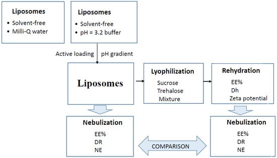
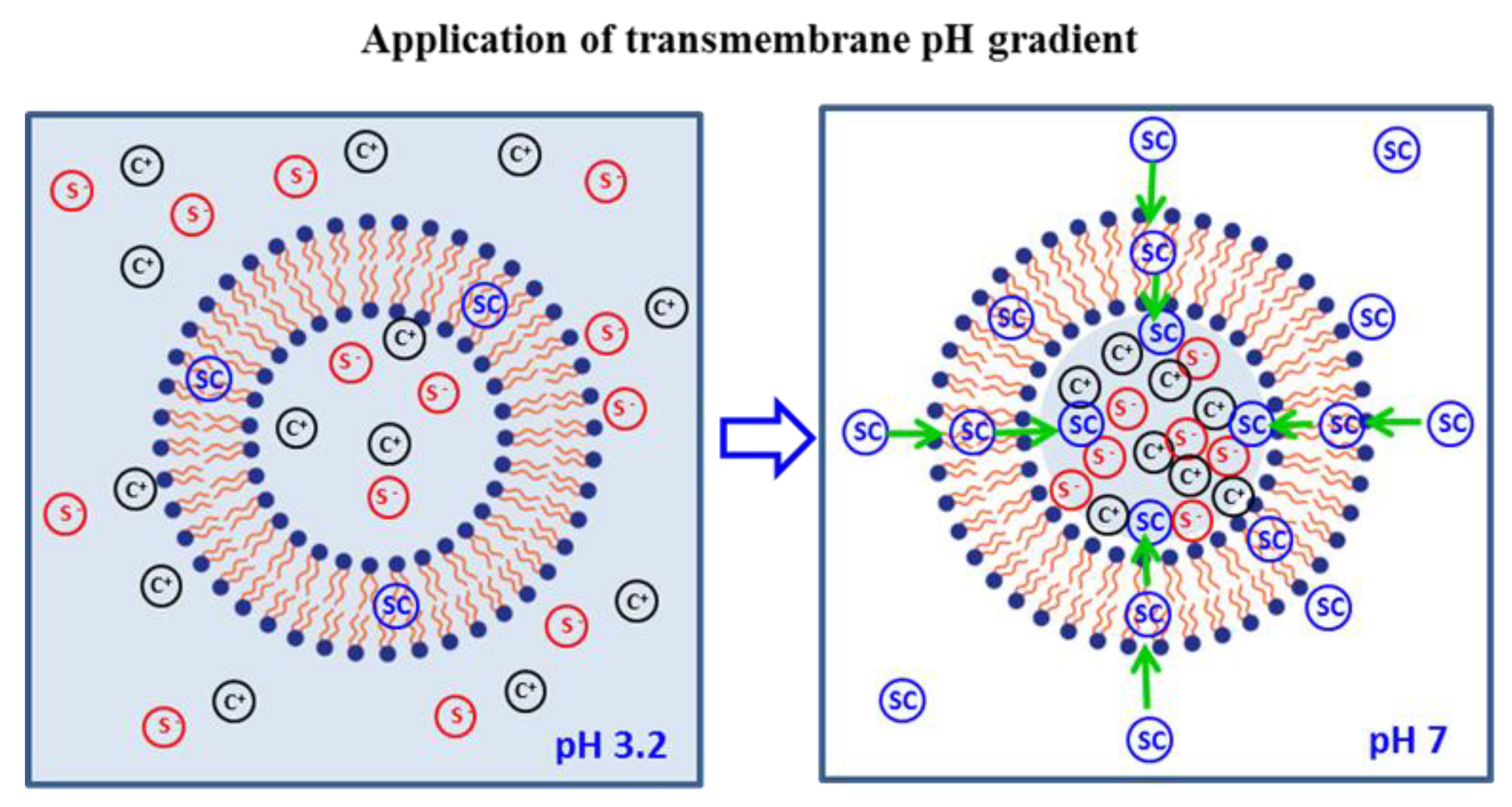
2.2. Preparation of Liposomes

SC-loaded liposomes with bilayers composed of EPC and Ch, with and without Vit E TPGS, were prepared by direct sonication of the components according to a previously described method [39]. Briefly, EPC and Ch were gently mixed with a 1 mg/mL SC solution in water or citrate buffer (pH = 3.2), with or without Vit E TPGS (0.1% w/w). The mixtures were sonicated for 30 min in a Fisher Scientific FB 15061 ultrasonic bath (50 Hz) at 50 ± 2 °C. The sonicated samples were kept at room temperature for 60 min for liposome stabilization and then stored at 4 °C until active loading by transmembrane pH gradient.
For active loading, the liposome suspensions prepared with citrate buffer were adjusted to pH = 7.0 with NaOH 0.1 N. The resulting suspensions were maintained for 20 h at 25 ± 2 °C under mechanical agitation to facilitate the diffusion of the drug across the lipid bilayer and its accumulation in the liposome core (pH = 3.2), according to the pH-dependent solubility profile of sildenafil [40]. The marked influence of pH on sildenafil solubility facilitates the accumulation of the drug in the liposomal acidic core. The adjustment of the external medium to pH = 7.0 maximizes the unionized fraction of the drug, which shows high lipophilicity and permeability. Unionized molecules are able to cross the lipid bilayer to reach the acidic aqueous core, where they are trapped as ionized species. Figure 1 illustrates the mechanism of SC active loading in liposomes by transmembrane pH gradient.

2.3. Lyophilization

Four percent sucrose, 4% trehalose, or 4% mixture of sucrose and trehalose (1 w/w ratio) was added to the liposome suspensions as the lyoprotective agent. The mixtures were frozen at −80 °C (Nuaire −86 °C Ultralow Freezer) and then lyophilized (Ehrisa Beta Freeze-Drying and Varian DS 102 vacuum pump).

2.4. Nebulization

The liposomes were nebulized using an ultrasonic aerosol generator (700700-UV system TSE, HF-Frequency: 1.70 MHz) connected to an artificial ventilator (7025 Rodent Ventilator) set at 60 respirations per min and 2 mL of tidal volume. The liposome suspension (10 g) was placed into the nebulizer container at room temperature. After 5, 10 and 15 min of nebulization, the temperature and the amount of solution remaining in the container were determined, and samples were collected for determination of entrapment efficiency (EE%) of liposomes. Discharged samples were also collected, and the volume and concentration of SC were quantified to determine the discharge rate (DR) and the nebulization efficiency (NE), according to the following expressions:
DR (mL/min) = V/t
where V is the volume of discharged sample and t the duration of the nebulization.
NE% = (Cd/Cn) × 100
where Cd and Cn are the SC concentrations measured in the discharged sample and the nebulized liposome suspension, respectively.

2.5. Characterization of Liposomes

The liposomes were characterized in terms of size (hydrodynamic diameter = Dh), polydispersity index (PDI), zeta potential, drug entrapment efficiency (EE%), and drug loading (mg of SC/g lipid). The Dh, PDI, and zeta potential were determined by dynamic light scattering (DLS) in a Zetasizer Nano ZS (Malvern Instruments, CO., UK). The analysis was performed at 25 °C and with a scattering angle of 173° after the appropriate dilution (×100 or ×1000) with Milli-Q water or buffer solution (pH = 3.2) to avoid the phenomenon of multiple scattering. Liposome morphology was characterized through transmission electron microscopy (TEM) using a Hitachi HT7700, Japan. The samples were diluted in Milli-Q water (1:100) and then were placed on a formvar-coated copper grid and allowed to dry at room temperature overnight. The images were captured using an accelerating voltage of 80 kV at the magnification of ×10,000 to ×20,000.
To determine the EE%, the liposome suspensions were centrifuged at 14.000 rpm for 45 min at 6 °C to separate the unentrapped drug in the supernatant from the drug loaded into liposomes. The amount of drug in the supernatant was determined by high pressure liquid chromatography (HPLC), using a Purosphere STAR rpE18, with a 3 µm column of 50 cm × 4.0 mm. A mixture of 0.1% formic acid in water and acetonitrile (70/30 v/v) adjusted to pH 4.2 with triethanolamine was used as the mobile phase at a flow rate of 1.5 mL/min. The UV detector was set at 292 nm (HPLC system with Waters Alliance 2695 separation module, 2998 photodiode array detector, and empower processor system), and the calibration range was 25−500 µg/mL. EE (%) was estimated from the following equation:
EE (%) = [(QtQs)/Qt] × 100,
where Qt is the total drug amount in the liposome suspension and Qs is the drug amount quantified in the supernatant.
Drug loading (DL) was estimated from the Qsc/Qlip ratio, where Qsc and Qlip represent the amount of SC (mg) and the amount of lipids (g) in the liposomes, respectively.
The Dh, PDI, zeta potential, and EE% were determined for the liposomes obtained without and with applying the transmembrane pH gradient. In the latter case, these characteristics were determined for fresh, lyophilized, and nebulized liposomes.
The density (ρ) and kinematic viscosity (η) of the liposome suspensions at 25 °C were measured before and after adding the lyoprotective agent. A capillary viscometer was used, and η (mm2/s) was estimated by measuring the time (second) it took for the sample to flow through the capillary under the influence of gravity. The instrument was calibrated with Milli-Q water (ρ = 1 g/mL; η = 1 mm2/s) and the viscometer constant (K, mm2/s2) was estimated. Then, the viscosity of liposome suspensions was calculated as follows:
η = K × t,
where t is the time it took for the liposome suspension to flow from the lower to upper mark.
The following scheme summarizes the above-described methodology (Figure 2).

2.6. Statistical Analysis

Data were presented as the mean and standard deviation (m ± sd). The difference between groups was regarded to be statistically significant when the p value was lower than 0.05, using Student’s t-tests or analysis of variance (ANOVA) to compare two or more groups, respectively.

3. Results

As reported in previous studies [39,41,42], the direct sonication of the components produced drug-loaded liposomes without the use of organic solvents, which results in an environmentally friendly approach. The characteristics of the SC-loaded liposomes obtained in this study are summarized in Table 1.
EE% values under 18% were obtained when Milli-Q water was used as the hydration medium, and this value was moderately increased when citrate buffer (pH = 3.2) was used instead. However, the most relevant increase in the EE% was observed after applying the transmembrane pH gradient, irrespective of whether the liposomes contained Vit E TPGS or not (EE = 89.77 ± 7.64% and 80.30 ± 11.03%, respectively). The difference between these two values was not statistically significant (p = 0.1495). However, when these values were compared to those obtained prior to the application of the pH gradient, the differences were statistically significant for both types of liposomes (p = 0.0039).
The particle size and charge were determined using the Zetasizer Nano ZS device, as described in the Material and Methods Section. The results showed the effects of pH on liposome size and zeta potential. At pH = 3.2, the Dh values were 304.3 nm (PDI = 0.413) and 303.2 nm (PDI = 0.452) for liposomes without and with Vit E TPGS, respectively. Smaller liposomes were obtained when the pH was adjusted to 7.0 for the transmembrane gradient (209.7 nm; PDI = 0.537 or 219.8 nm; PDI = 0.534 for liposomes without or Vit E TPGS, respectively). Changes were also observed for the zeta potential, which showed values of −2.10 mV or −2.05 mV at the acidic pH and −20.90 mV or −21.30 mV at pH = 7.0.
The liposomes prepared by active loading were lyophilized with sucrose, trehalose, or sucrose and trehalose, and homogeneous cakes were obtained in all cases. After the cakes were hydrated, the aspect and characteristics of the fresh suspensions were immediately restored, and the EE% values determined for rehydrated liposomes did not show statistical differences when compared to fresh liposomes (p = 0.1390). By contrast, the lyophilized liposomes exhibited a greater diameter and more negative charge as compared to the fresh liposomes (see data in Table 2), irrespective of whether sucrose, trehalose, or sucrose and trehalose was used as the lyoprotective agent.
The morphology observed under the TEM showed that the liposomes present a circular or elliptical shape typical of the classical vesicular bilayer structure. Although the lyophilization increased the Dh of the liposomes, they maintained their typical morphology, as illustrated in Figure 3.
The effect of ultrasonic nebulization on the liposomes was evaluated, and the change of the EE% throughout the period of nebulization was measured for fresh and lyophilized liposomes. Table 3 shows the results of this assay. A progressive decrease in the EE% was observed as the nebulization period increased for both types of fresh liposomes. After 15 min of nebulization, the EE% decreased from 89.77 ± 7.64% to 77.78 ± 0.89% and from 80.30 ± 11.03% to 59.24 ± 1.61% for liposomes without and with Vit E TPGS, respectively. The differences in the EE% values before and after nebulization were significant in both cases (p = 0.0389 and p = 0.0201, respectively). In the case of the liposomes without Vit E TPGS, the lyophilized and fresh samples behaved similarly during the nebulization, and the presence of the lyoprotective agents did not affect the results obtained for the EE%. In contrast, the lyophilized samples with liposomes containing Vit E TPGS remained more stable than the fresh ones during nebulization, and only slight differences in the EE% were observed. As shown in Table 3, the EE% values were greater than 80% after 15 min of nebulization for this type of liposomes, irrespective of whether the suspension contained sucrose, trehalose, or sucrose and trehalose (EE = 82.2 ± 1.13%, 81.12 ± 1.43%, and 81.82 ± 0.89%, respectively). The comparison of these values to the mean value obtained for fresh liposomes (59.24 ± 1.61%) revealed statistically significant differences (p = 0.0126).
Finally, the influence of the density and viscosity of the samples on the performance of the nebulizer was analyzed and the results are shown in Table 4. It was found that the addition of the lyoprotective agent did not modify the density of the liposome suspensions, but it did increase viscosity in all cases. The viscosity values after the addition of 4% sucrose, trehalose, or sucrose and trehalose were higher (η = 1.24 ± 0.01 mm2/s) than those observed for the samples without the additives (η = 1.15 ± 0.05 mm2/s), the differences being statistically significant (p = 0.0052).
The viscosity of the samples did affect nebulizer performance. A higher DR coupled with a lower NE was observed for samples with a lower viscosity (fresh liposomes without additives) as compared to those with a higher viscosity (lyophilized liposomes with sucrose, trehalose, or sucrose and trehalose). Statistical differences among samples were only found for NE (p = 0.0126).

4. Discussion

SC-loaded liposomes composed of natural phospholipids (EPC and Ch), with or without Vit E TPGS, were prepared using a procedure that guarantees the absence of unwanted residues and the avoidance of organic solvents. In contrast, most reported methods use organic solvents to obtain liposomes, which are then sonicated to reduce and homogenize their size. We have found that the first step, consisting of dissolving the lipids in an organic solvent and its subsequent evaporation, is not necessary for obtaining liposomes with a high PDI. Direct sonication of the components allowed us to obtain liposomes loaded with different drugs showing different Dh and PDI [39,41,42]. In the present study, liposomes with high PDI were obtained, and extrusion is recommended to reduce size polydispersity. Since optimization of the liposome drug loading was the main objective of this work, extrusion was not carried out. The EE% of the liposomes was significantly increased by applying a transmembrane pH gradient (see Table 1 in Results). The marked influence of pH on sildenafil solubility facilitates the accumulation of the drug in the liposomal acidic core, leading to optimum EE% values.
The separation of the liposomes by centrifugation allowed us to determine that approximately 8% of the drug solution volume was retained in the liposome core. Therefore, only about 8% of the drug is trapped inside the water core, as long as an active loading is not achieved. Since SC solubility at pH = 3 has been reported to be 6.96 mg/mL [40], and a 1 mg/mL dissolution was used in the present study, approximately 56% (0.08 × 6.96 = 0.56) of the drug is expected to be trapped inside the aqueous core using pH gradient active loading. The EE% values obtained here (>80%) support this hypothesis and confirm that SC is also trapped in the lipid bilayer due to the favorable partition coefficient of its unionized species.
Although lyophilization has been proposed as the best approach for ensuring the long-term stability of liposomes, many factors should be controlled in order to obtain an acceptable level of encapsulated drug retention after lyophilization [43]. In the present study, sucrose, trehalose, or their mixture were used as lyoprotective agents, and differences among the samples containing these agents were not found when rehydrated samples were compared. Excellent results were obtained regarding SC retention in the liposomes after lyophilization, and EE% values above 88% were found in all cases, irrespective of the lyoprotectant agent used or if the liposomes contained Vit E TPGS or not. In contrast, Dh significantly increased after lyophilization, and the liposomes containing Vit E TPGS and lyophilized with the mixture exhibited the highest Dh and PDI values (688.4 nm, PDI = 0.682). The zeta potential was also affected by lyophilization, likely due to the negative charge of disaccharides.
Ultrasonic nebulizers are frequently used for pulmonary delivery of liquid formulations; therefore, the impact of this type of nebulization on the stability of SC-loaded liposomes was studied. Very interesting results were obtained when comparing the behavior of liposomes with or without Vit E TPGS, and these are illustrated in the next figure. On one hand, in absence of lyoprotectants, ultrasonic nebulization slightly affected the EE% of the liposomes without Vit E TPGS (Figure 4A) but did produce a relevant effect on the liposomes with Vit E TPGS (Figure 4B), with 12.39% versus 26.23% reduction observed, respectively. The surfactant properties of Vit E TPGS may promote the lipid bilayer destabilization induced by the ultrasonic vibration involved in aerosol generation. On the other hand, the additives used as lyoprotectants affect both types of liposomes in different ways. The presence of sucrose, trehalose, or the mixture did not modify the results of the EE% registered during nebulization of the liposomes without Vit E TPGS, but it did produce a beneficial effect on liposomes containing Vit E TPGS (see Figure 4).
Reductions of 7.40%, 8.41% and 7.43% in EE were observed when samples of Vit E TPGS liposomes containing sucrose, trehalose, or the mixture, respectively, were nebulized. The viscous damping force in samples with additives might counteract the synergic effects of Vit E TPGS and ultrasonic nebulization, contributing to the maintenance of the integrity of these type of liposomes during nebulization.
Regarding the influence of viscosity on the nebulizer performance, opposing effects on DR and NE were observed, as shown in Figure 5 and Figure 6.
The influence of sample viscosity on nebulizer performance has been previously reported [44,45], and the results showing that higher viscosity leads to lower DR were predictable, since ultrasonic nebulizers have been found to be less efficient for use with viscous solutions [44]. The beneficial effects of viscosity on NE, however, have not been reported before. Samples containing less SC than the nebulized suspensions (NE < 100%) were discharged by the ultrasonic nebulizer, irrespective of whether the suspension contained a lyoprotective agent or not, and irrespective of whether the liposomes contained Vit E TPGS or not. Nevertheless, lower values of NE were obtained for suspensions without the additives. Viscous damping force might be considered again to explain the positive effect of the additives on NE. For liposome suspensions without the lyoprotective agent, the less viscous external medium facilitates its own nebulization, and the result is higher discharged volume (higher DR) of samples with lower liposome content (lower NE). When lyoprotectants were added, the viscosity of the external medium increased and its nebulization became slower, resulting in lower discharged volume (lower DR) of samples with higher liposome content (higher NE). Despite the additives being beneficial, the discharged samples contained, in all cases, fewer liposomes and less SC than original liposome suspensions.

5. Conclusions

The liposomes studied in this work were prepared without the use of organic solvents, from EPC, Ch and Vit E TPG. Therefore, they can be considered to be biocompatible and safe for pulmonary administration. The high EE% obtained by applying the transmembrane pH gradient led to drug loading values high enough for the liposomes to be used as drug carriers of therapeutic doses of SC. The lyoprotectants sucrose, trehalose, or the mixture preserved liposome drug loading after lyophilization but did not maintain the size and zeta potential of the original liposomes. Although a progressive decrease in the EE% was observed when the liposomes were nebulized, the magnitude of change was not relevant, and this was counteracted by the lyoprotectants used, which also produced a beneficial effect on the performance of the nebulizer. Additional studies are still essential, but according to our results, SC-loaded liposomes can be considered suitable and safe carriers for the local release of sildenafil in the pulmonary system by ultrasonic nebulization.

Author Contributions

Conceptualization, A.S.N.; Methodology, M.J.J.V., P.G.G., MP.R. and A.R.T.S.A.P.; Investigation, A.S.N. and M.J.J.V.; Validation, M.J.J.V.; Resources, M.P.R. and A.R.T.S.A.P.; Visualization, M.J.J.V.; Writing-Original Draft Preparation, A.S.N.; Writing-Review & Editing M.J.J.V., M.P.R and A.R.T.S.A.P.; Supervision, A.S.N.

Funding

This research received no external funding.

Conflicts of Interest

The authors declare no conflict of interest.

References

  1. Barst, R.J.; Ertel, S.I.; Beghetti, M.; Ivy, D.D. Pulmonary arterial hypertension: A comparison between children and adults. Eur. Respir. J. 2011, 37, 665–677. [Google Scholar] [CrossRef] [PubMed]
  2. Frumkin, L.R. The Pharmacological Treatment of Pulmonary Arterial Hypertension. Pharmacol. Rev. 2012, 64, 583–620. [Google Scholar] [CrossRef] [PubMed] [Green Version]
  3. Ghofrani, H.A.; Osterloh, I.H.; Grimminger, F. Sildenafil: from angina to erectile dysfunction to pulmonary hypertension and beyond. Nat. Rev. Drug Discov. 2006, 5, 689–702. [Google Scholar] [CrossRef] [PubMed]
  4. Simonca, L.; Tulloh, R. Sildenafil in Infants and Children. Children 2017, 4, 60. [Google Scholar] [CrossRef] [PubMed]
  5. Hill, K.D.; Sampson, M.R.; Li, J.S.; Tunks, R.D.; Schulman, S.R.; Cohen-Wolkowiez, M. Pharmacokinetics of intravenous sildenafil in children with palliated single ventricle heart defects: Effect of elevated hepatic pressures. Cardiol. Young 2016, 26, 354–362. [Google Scholar] [CrossRef] [PubMed]
  6. Weerateerangkul, P.; Palee, S.; Chinda, K.; Chattipakorn, S.C.; Chattipakorn, N. Effects of Kaempferia parviflora Wall. Ex. Baker and sildenafil citrate on cGMP level, cardiac function, and intracellular Ca2+ regulation in rat hearts. J. Cardiovasc. Pharmacol. 2012, 60, 299–309. [Google Scholar] [CrossRef] [PubMed]
  7. Corbin, J.D.; Francis, S.H. Cyclic GMP phosphodiesterase-5: Target of sildenafil. J. Biol. Chem. 1999, 274, 13729–13732. [Google Scholar] [CrossRef] [PubMed]
  8. Moschos, M.M.; Nitoda, E. Pathophysiology of visual disorders induced by phosphodiesterase inhibitors in the treatment of erectile dysfunction. Drug Des. Dev. Ther. 2016, 8, 3407–3413. [Google Scholar] [CrossRef] [PubMed]
  9. Samiee-Zafarghandy, S.; Smith, P.B.; van den Anker, J.N. Safety of Sildenafil in Infants. Pediatr. Crit. Care Med. 2014, 15, 362–368. [Google Scholar] [CrossRef] [PubMed]
  10. Chono, S.; Tanino, T.; Seki, T.; Morimoto, K. Efficient drug targeting to rat alveolar macrophages by pulmonary administration of ciprofloxacin incorporated into mannosylated liposomes for treatment of respiratory intracellular parasitic infections. J. Control Release 2008, 127, 50–58. [Google Scholar] [CrossRef] [PubMed]
  11. Pinto-Alphandary, H.; Andremont, A.; Couvreur, P. Targeted delivery of antibiotics using liposomes and nanoparticles: Research and applications. Int. J. Antimicrob. Agents 2000, 13, 155–168. [Google Scholar] [CrossRef]
  12. Kurmi, B.D.; Kayat, J.; Gajbhiye, V.; Tekade, R.K.; Jain, N.K. Micro- and nanocarrier-mediated lung targeting. Expert Opin. Drug Deliv. 2010, 7, 781–794. [Google Scholar] [CrossRef] [PubMed]
  13. De Jesús Valle, M.J.; González López, F.; Domínguez-Gil Hurlé, A.; Sánchez Navarro, A. Pulmonary versus systemic delivery of antibiotics: Comparison of vancomycin dispositions in the isolated rat lung. Antimicro. Agents Chemother. 2007, 51, 3771–3774. [Google Scholar] [CrossRef] [PubMed]
  14. De Jesús Valle, M.J.; Garavís González, J.; González López, F.; Sánchez Navarro, A. Pulmonary disposition of vancomycin nebulized as lipid vesicles in rats. J. Antibiot. 2013, 66, 447–451. [Google Scholar] [CrossRef] [PubMed]
  15. Rudokas, M.; Najlah, M.; Alhnan, M.A.; Elhissi, A. Liposome Delivery Systems for Inhalation: A Critical Review Highlighting Formulation Issues and Anticancer Applications. Med. Princ. Pract. 2016, 25 (Suppl. 2), 60–72. [Google Scholar] [CrossRef] [PubMed] [Green Version]
  16. Saari, M.; Vidgren, M.T.; Koskinen, M.O.; Turjanmaa, V.M.; Nieminen, M.M. Pulmonary distribution and clearance of two beclomethasone liposome formulations in healthy volunteers. Int. J. Pharm. 1999, 181, 1–9. [Google Scholar] [CrossRef]
  17. Clancy, J.P.; Dupont, L.; Konstan, M.W.; Billings, J.; Fustik, S.; Goss, C.H.; Lymp, J.; Minic, P.; Quittner, A.L.; Rubenstein, R.C.; et al. Phase II studies of nebulised Arikace in CF patients with Pseudomonas aeruginosa infection. Thorax 2013, 68, 818–825. [Google Scholar] [CrossRef] [PubMed]
  18. Li, Q.; Zhan, S.; Liu, Q.; Su, H.; Dai, X.; Wang, H.; Beng, H.; Tan, W. Preparation of a Sustained-Release Nebulized Aerosol of R-terbutaline Hydrochloride Liposome and Evaluation of Its Anti-asthmatic Effects via Pulmonary Delivery in Guinea Pigs. AAPS Pharm. Sci. Tech. 2018, 19, 232–241. [Google Scholar] [CrossRef] [PubMed]
  19. Makled, S.; Nafee, N.; Boraie, N. Nebulized solid lipid nanoparticles for the potential treatment of pulmonary hypertension via targeted delivery of phosphodiesterase-5-inhibitor. Int. J. Pharm. 2017, 517, 312–321. [Google Scholar] [CrossRef] [PubMed]
  20. Paranjpea, M.; Finkea, J.H.; Richterc, C.; Gothschb, T.; Kwadeb, A.; Büttgenbachc, S.; Müller-Goymanna, C.C. Physicochemical characterization of sildenafil-loaded solid lipid nanoparticle dispersions (SLN) for pulmonary application. Int. J. Pharm. 2014, 476, 41–49. [Google Scholar] [CrossRef] [PubMed]
  21. Beck-Broichsitter, M.; Schmehl, T.; Gessler, T.; Seeger, W.; Kissel, T. Development of a biodegradable nanoparticle platform for sildenafil: Formulation optimization by factorial design analysis combined with application of charge-modified branched polyesters. J. Control. Release 2012, 157, 469–477. [Google Scholar] [CrossRef] [PubMed]
  22. Beck-Broichsitter, M.; Kleimann, P.; Gessler, T.; Seeger, W.; Kissel, T.; Schmehl, T. Nebulization performance of biodegradable sildenafil-loaded nanoparticles using the Aeroneb Pro: Formulation aspects and nanoparticle stability to nebulization. Int. J. Pharm. 2012, 422, 398–408. [Google Scholar] [CrossRef] [PubMed]
  23. Beck-Broichsitter, M.; Hecker, A.; Kosanovic, D.; Schmehl, T.; Gessler, T.; Weissmann, N.; Ghofrani, H.A.; Kissel, T.; Seeger, W.; Schermuly, R.T. Prolonged vasodilatory response to nanoencapsulated sildenafil in pulmonary hypertension. Nanomedicine 2016, 12, 63–68. [Google Scholar] [CrossRef] [PubMed]
  24. Beck-Broichsitter, M.; Stoisiek, K.; Bohr, A.; Aragão-Santiago, L.; Gessler, T.; Seeger, W.; Kissel, T. Potential of the isolated lung technique for the examination of sildenafil absorption from lung-delivered poly(lactide-co-glycolide) microparticles. J. Control Release 2016, 226, 15–20. [Google Scholar] [CrossRef] [PubMed]
  25. Ghasemian, E.; Vatanara, A.; Rouini, M.R.; Rouholamini Najafabadi, A.; Gilani, K.; Lavasani, H.; Mohajel, N. Inhaled sildenafil nanocomposites: Lung accumulation and pulmonary pharmacokinetics. Pharm. Dev. Technol. 2016, 21, 961–971. [Google Scholar] [CrossRef] [PubMed]
  26. Refai, H.; Hassan, D.; Abdelmonem, R. Development and characterization of polymer coated liposomes for vaginal delivery of sildenafil citrate. Drug Deliv. 2017, 24, 278–288. [Google Scholar] [CrossRef] [PubMed]
  27. De Jesús Valle, M.J.; de la Cuesta Melgar, E.; Martín Rebellado, S.; López Díaz, D.; Velázquez Salicio, M.; Sánchez Navarro, A. Sildenafil citrate-loaded liposomes and albusomes as drug carriers for pulmonary delivery. Stability after nebulization. In Proceedings of the 11th World Meeting on Pharmaceutics, Biopharmaceutics and Pharmaceutical Technology, Granada, Spain, 19–22 March 2018. [Google Scholar]
  28. Elhissi, A. Liposomes for Pulmonary Drug Delivery: The Role of Formulation and Inhalation Device Design. Curr. Pharm. Des. 2017, 23, 362–372. [Google Scholar] [PubMed]
  29. Lehofer, B.; Bloder, F.; Jain, P.P.; Marsh, L.M.; Leitinger, G.; Olschewski, H.; Leber, R.; Olschewski, A.; Prassl, R. Impact of atomization technique on the stability and transport efficiency of nebulized liposomes harbouring different surface characteristics. Eur. J. Pharm. Biopharm. 2014, 88, 1076–1085. [Google Scholar] [CrossRef] [PubMed]
  30. Bangham, J.A.; Lea, E.J. The interaction of detergents with bilayer lipid membranes. Biochim. Biophys. Acta 1978, 551, 388–396. [Google Scholar] [CrossRef]
  31. Wang, T.; Wang, N.; Wang, T.; Sun, W.; Li, T. Preparation of submicron liposomes exhibiting efficient entrapment of drugs by freeze-drying water-in-oil emulsions. Chem. Phys. Lipids 2011, 164, 151–157. [Google Scholar] [CrossRef] [PubMed]
  32. Zawada, Z.H. Liposomes from hydrogenated soya lecithin formed in sintered glass pores. Acta Pol. Pharm. 2012, 69, 107–111. [Google Scholar] [PubMed]
  33. Mozafari, M.R. Liposomes: an overview of manufacturing techniques. Cell Mol. Biol. Lett. 2005, 10, 711–719. [Google Scholar] [PubMed]
  34. Tang, S.; Hao, J.; Gao, D.; Duan, J.; Liu, Z. Preparation and characterization of oleanolic acid nanoparticles. Curr. Pharm. Anal. 2013, 9, 177–182. [Google Scholar] [CrossRef]
  35. Otake, K.; Shimomura, T.; Goto, T.; Imura, T.; Furuya, T.; Furuya, T.; Yoda, S.; Takebayashi, Y.; Sakai, H.; Abe, M. Preparation of liposomes using an improved supercritical reverse phase evaporation method. Langmuir 2006, 22, 2543–2550. [Google Scholar] [CrossRef] [PubMed]
  36. Salba, Z.; Navarro, I.; Troconiz, I.F.; Tros de Llarduya, C.; Garrido, M.J. Application of different methods to formulate PEGliposomes of oxaliplatin: Evaluation in vitro and in vivo. Eur. J. Pharm. Biopharm. 2012, 81, 273–280. [Google Scholar]
  37. Geho, W.B.; Geho, H.C.; Lau, J.R.; Gana, T.J. Hepatic-Directed Vesicle Insulin: A Review of Formulation Development and Preclinical Evaluation. J. Diabetes Sci. Technol. 2009, 3, 1451–1459. [Google Scholar] [CrossRef] [PubMed] [Green Version]
  38. Ranjan Karn, P.; Cho, W.; Park, H.-J.; Hwang, S.-J. Characterization and stability studies of a novel liposomal cyclosporine A prepared using the supercritical fluid method: Comparison with the modified conventional Bangham method. Int. J. Nanomedicine 2013, 8, 365–377. [Google Scholar]
  39. De Jesús Valle, M.J.; Sánchez Navarro, A. Liposomes prepared in absence of organic solvents: Sonication versus lipid film hydration method. Curr. Pharm. Anal. 2015, 11, 86–91. [Google Scholar] [CrossRef]
  40. Wang, Y.; Chow, M.S.; Zuo, Z. Mechanistic analysis of pH dependent solubility and trans-membrane permeability of anphoreric compounds: application to sildenafil. Int. J. Pharm. 2008, 352, 217–224. [Google Scholar] [CrossRef] [PubMed]
  41. De Jesús Valle, M.J.; López Díaz, D.; Velazquez, M.; Sánchez Navarro, A. Development and In Vitro Evaluation of a Novel Drug Delivery System (Albumin Microspheres Containing Liposomes) Applied to Vancomycin. J. Pharm. Sci. 2016, 105, 2180–2187. [Google Scholar] [CrossRef] [PubMed]
  42. De Jesús Valle, M.J.; Maderuelo Martín, C.; Zarzuelo Castañeda, A.; Sánchez Navarro, A. Albumin micro/nanoparticles entrapping liposomes for itraconazole green formulation. Eur. J. Pharm. Sci. 2017, 106, 159–165. [Google Scholar] [CrossRef] [PubMed]
  43. Chen, C.; Han, D.; Cai, C.; Tang, X. An overview of liposome lyophilization and its future potential. J. Control Release 2010, 142, 299–311. [Google Scholar] [CrossRef] [PubMed]
  44. Yeo, L.Y.; Friend, J.R.; McIntosh, M.P.; Meeusen, E.N.; Morton, D.A. Ultrasonic nebulization platforms for pulmonary drug delivery. Expert Opin. Drug Deliv. 2010, 7, 663–679. [Google Scholar] [CrossRef] [PubMed]
  45. Elphick, M.; von Hollen, D.; Pritchard, J.N.; Nikander, K.; Hardaker, L.E.; Hatley, R.H. Factors to consider when selecting a nebulizer for a new inhaled drug product development program. Expert Opin. Drug Deliv. 2015, 12, 1375–1387. [Google Scholar] [CrossRef] [PubMed]
Figure 1. Schematic illustration of the active loading mechanism by transmembrane pH gradient.
Applsci 08 01291 g001
Figure 2. Schematic description of applied methodology.
Applsci 08 01291 g002
Figure 3. TEM images of the fresh (A) and the rehydrated liposomes (B).
Applsci 08 01291 g003
Figure 4. Influence of the lyoprotective agents on the EE% of the nebulized liposomes without Vit E TPGS (A) and with Vit E TPGS (B).
Applsci 08 01291 g004
Figure 5. Effect of viscosity on DR for the nebulized liposomes with and without Vit E TPGS.
Applsci 08 01291 g005
Figure 6. Effect of viscosity on NE for the nebulized liposomes with and without Vit E TPGS.
Applsci 08 01291 g006
Table 1. Characteristics of the liposomes prepared using Milli-Q water (pH = 6.7), citrate buffer (pH = 3.2), or the transmembrane gradient (pH gradient). Abbreviations: Vit E TPGS: d-α-tocopheryl polyethylene glycol 1000 succinate; EE: entrapment efficiency; Dh: hydrodynamic diameter; PDI: polydispersity index; DL: drug loading.
Influence of the Transmembrane pH Gradient on the Characteristics of the Liposomes
LiposomesDh (nm)PDIZeta Potential (mV)EE (%)DL (mg/g lipid)
Without
Vit
E TPGS
pH = 6.7---<18%<11
pH = 3.2304.30.413−2.1049.47 ± 9.7829.71 ± 5.87
pH gradient209.70.537−20.9089.77 ± 7.6453.92 ± 4.59
With
Vit
E TPGS
pH = 6.7---<15%<9
pH = 3.2303.20.452−2.0522.67 ± 11.3213.62 ± 11.00
pH gradient219.80.534−21.3080.30 ± 11.0348.23 ± 6.62
Table 2. Characteristics of the liposomes before (fresh liposomes; red curves) and after lyophilization with sucrose (S; green curves), trehalose (T; black curves), or sucrose and trehalose (S + T; blue curves).
Influence of Lyophilization on the Characteristics of the Liposomes
LiposomesDh (nm)PDIZeta Potential (mV)EE%DL (mg/g Lipid)
Without Vit E TPGSFresh209.70.537−20.9089.77 ± 7.6453.32 ± 4.59
S408.40.572−21.089.65 ± 1.0253.84 ± 0.61
T433.10.577−46.689.95 ± 2.1554.02 ± 1.29
S + T471.70.701−38.988.95 ± 0.3053.42 ± 0.18
Applsci 08 01291 i001
LiposomesDh (nm)PDIZeta Potential (mV)EE%DL (mg/g Lipid)
With Vit E TPGSFresh219.80.534−21.3080.30 ± 11.0348.23 ± 0.13
S367.50.642−34.088.77 ± 0.2253.32 ± 0.35
T826.90.642−36.688.57 ± 0.5953.20 ± 0.35
S + T688.40.682−31.288.39 ± 0.2553.09 ± 0.15
Applsci 08 01291 i002
Table 3. EE% of nebulized liposomes before lyophilization (Fresh), and after lyophilization with sucrose (S), with trehalose (T), or with the mixture (S + T).
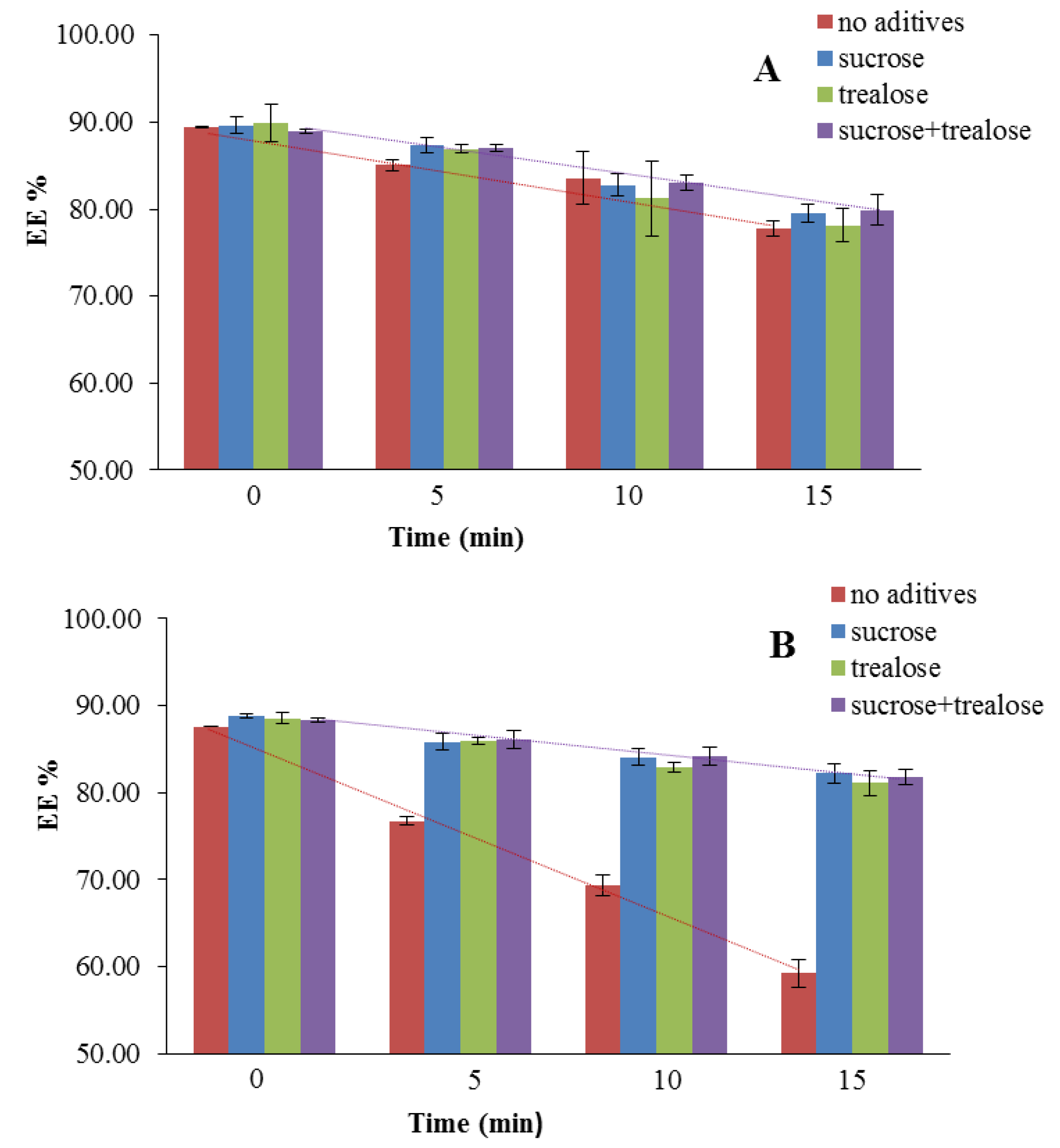
Influence of Nebulization on the Liposomes Stability (EE%)
Liposomes 0 min5 min10 min15 min
Without Vit E TPGSFresh89.77 ± 7.6485.10 ± 0.6483.56 ± 3.0477.78 ± 0.89
S89.65 ± 1.0287.35 ± 0.8882.73 ± 1.2879.47 ± 1.02
T89.95 ± 2.1586.90 ± 0.5081.23 ± 4.2978.15 ± 1.91
S + T88.95 ± 0.3087.06 ± 0.3983.04 ± 0.8879.89 ± 1.79
With Vit E TPGSFresh80.30 ± 11.0376.76 ± 0.5269.34 ± 1.1659.24 ± 1.61
S88.77 ± 0.2285.84 ± 0.9284.06 ± 0.9782.20 ± 1.13
T88.57 ± 0.5985.94 ± 0.4382.86 ± 0.5381.12 ± 1.43
S + T88.39 ± 0.2586.05 ± 1.0584.15 ± 1.0481.82 ± 0.89
Table 4. Performance of the ultrasonic nebulizer based on the discharge rate (DR) and the nebulization efficiency (NE) for liposomes before (Fresh) and after lyophilization with sucrose (S), trehalose (T), or the mixture (S + T).
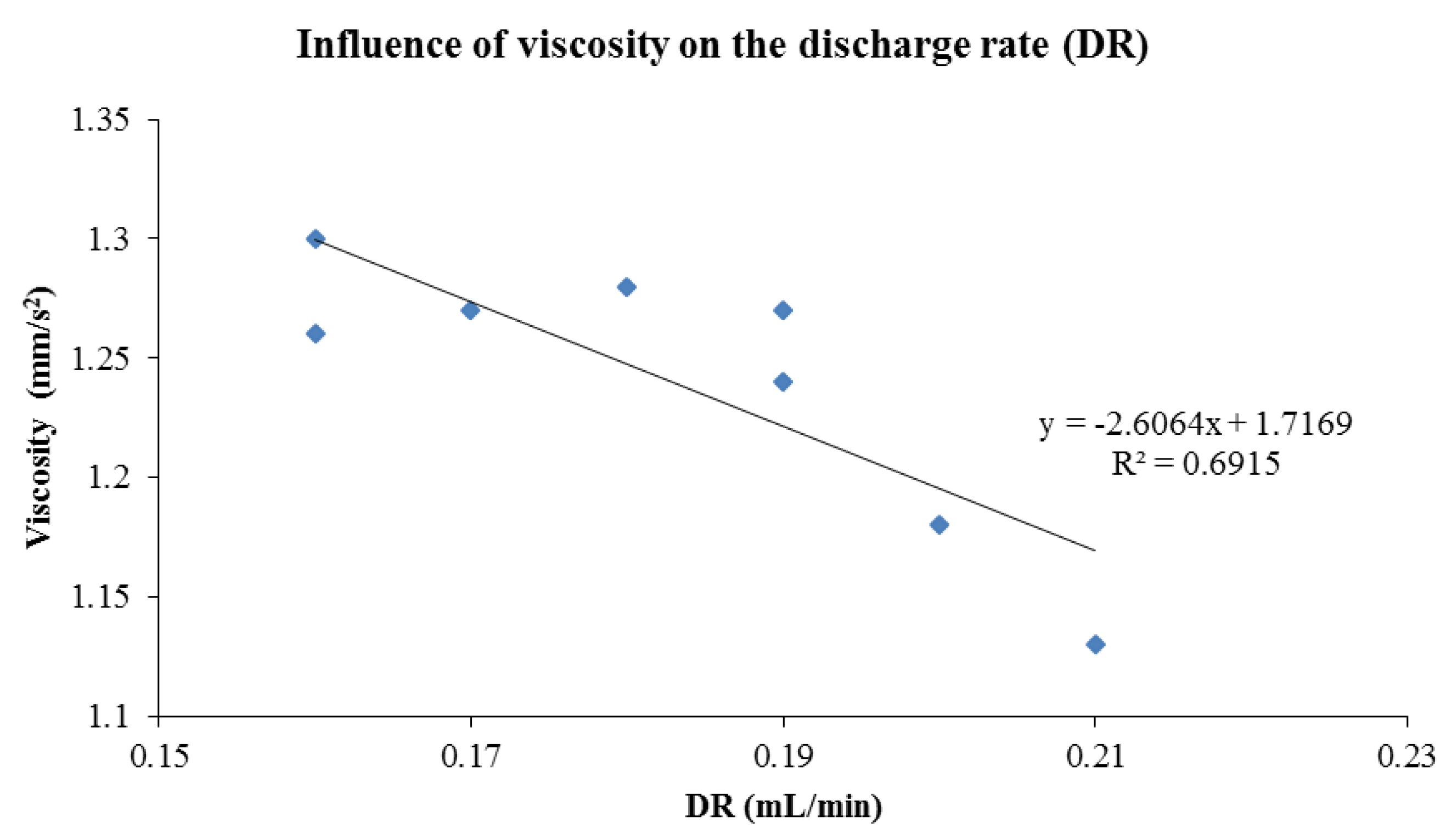
Nebulizer Performance
Liposomes DR (mL/min)NE (%)η (mm2/s)ρ (g/mL)
Without Vit E TPGSFresh0.21 ± 0.0150.51 ± 2.471.13 ± 0.041.01 ± 0.01
S0.17 ± 0.0165.19 ± 2.981.27 ± 0.031.02 ± 0.01
T0.19 ± 0.0275.22 ± 6.081.24 ± 0.011.01 ± 0.01
S + T0.19 ± 0.0268.00 ± 2.481.27 ± 0.011.02 ± 0.01
With Vit E TPGSFresh0.20 ± 0.0150.87 ± 1.901.18 ± 0.061.01 ± 0.01
S0.16 ± 0.0276.99 ± 2.691.30 ± 0.041.02 ± 0.01
T0.18 ± 0.0178.41 ± 0.221.28 ± 0.031.00 ± 0.00
S + T0.16 ± 0.0181.24 ± 2.121.26 ± 0.041.02 ± 0.01

