Spatial Modes of Laser-Induced Mass Transfer in Micro-Gaps


 , and
, and 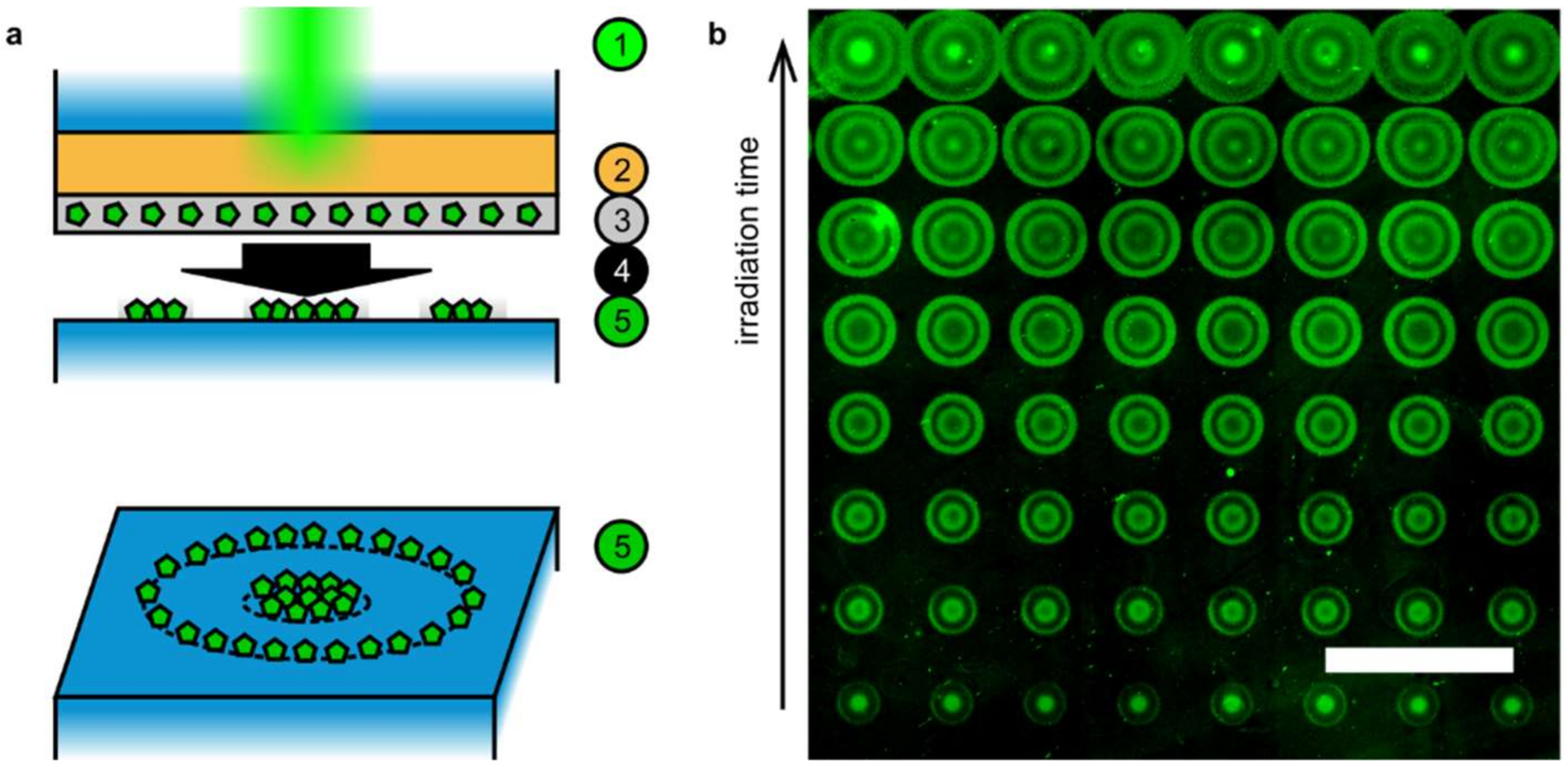
Abstract
:1. Introduction
2. Experimental Setup Details and General Procedure
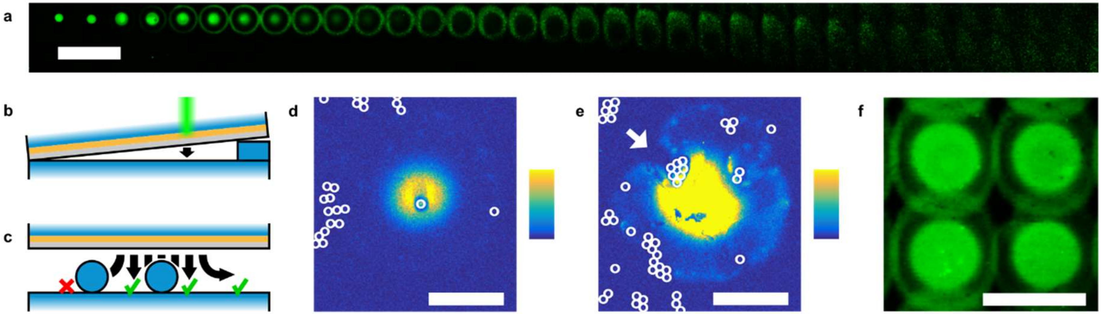
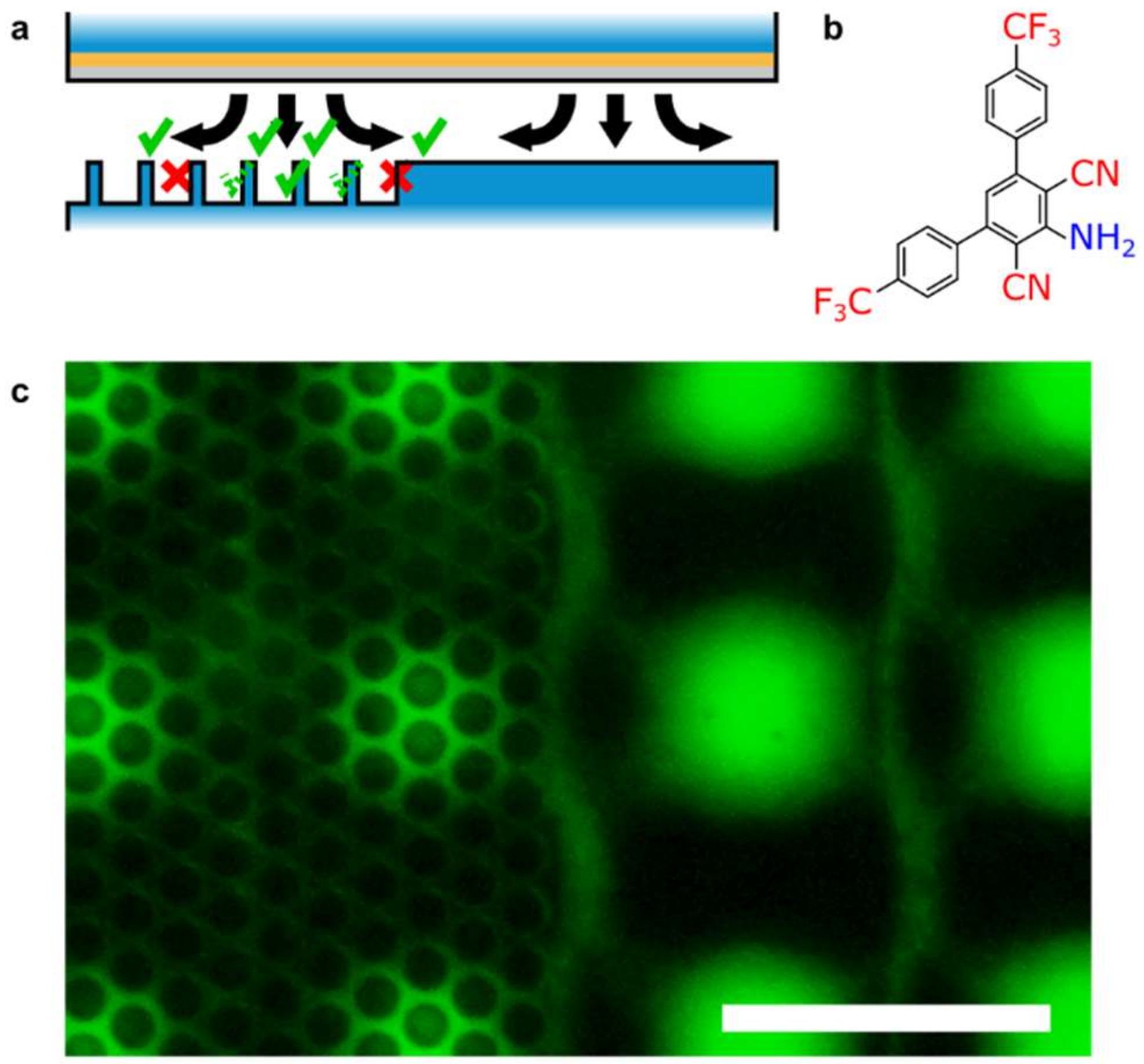
2.1. LIFT Surfaces
2.2. Laser System
2.3. Chemical Processing
2.4. Staining and Imaging
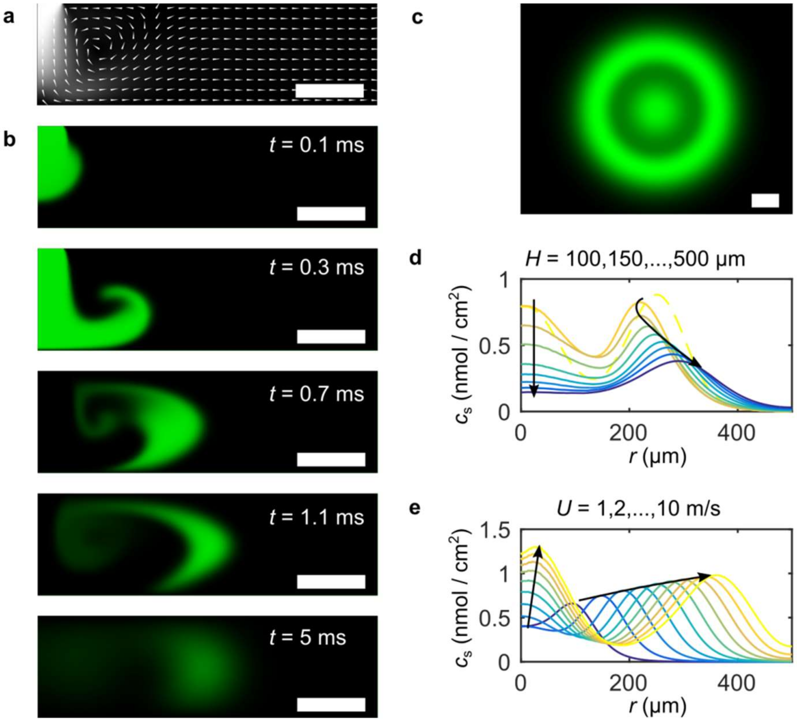
3. Details of the Numerical Simulation
3.1. Geometry
3.2. System of Equations
3.3. Boundary Conditions
3.4. Solver Details
4. Results
5. Discussion and Conclusions
Author Contributions
Funding
Acknowledgments
Conflicts of Interest
References
- Loeffler, F.F.; Foertsch, T.C.; Popov, R.; Mattes, D.S.; Schlageter, M.; Sedlmayr, M.; Ridder, B.; Dang, F.-X.; von Bojničić-Kninski, C.; Weber, L.K.; et al. High-flexibility combinatorial peptide synthesis with laser-based transfer of monomers in solid matrix material. Nat. Commun. 2016, 7, 11844. [Google Scholar] [CrossRef] [PubMed]
- Piqué, A.; Serra, P. Laser Printing of Functional Materials: 3D Microfabrication, Electronics and Biomedicine; Wiley-Vch: Weinheim, Germany, 2018. [Google Scholar]
- Bohandy, J.; Kim, B.F.; Adrian, F.J. Metal deposition from a supported metal film using an excimer laser. J. Appl. Phys. 1986, 60, 1538–1539. [Google Scholar] [CrossRef]
- Pohl, R.; Visser, C.W.; Römer, G.-W.; Lohse, D.; Sun, C.; Huis in’t Veld, B. Ejection in Picosecond Laser-Induced Forward Transfer of Metals. Phys. Rev. Appl. 2015, 3, 024001. [Google Scholar] [CrossRef]
- Fernández-Pradas, J.M.; Colina, M.; Serra, P.; Domínguez, J.; Morenza, J.L. Laser-induced forward transfer of biomolecules. Thin Solid Film 2004, 453-454, 27–30. [Google Scholar] [CrossRef]
- Brown, M.S.; Kattamis, N.T.; Arnold, C.B. Time-resolved dynamics of laser-induced micro-jets from thin liquid films. Microfluid. Nanofluidics 2011, 11, 199–207. [Google Scholar] [CrossRef]
- Merrifield, R.B. Solid Phase Peptide Synthesis. I. The Synthesis of a Tetrapeptide. J. Am. Chem. Soc. 1963, 85, 2149–2154. [Google Scholar] [CrossRef]
- Von Bojnicic-Kninski, C.; Bykovskaya, V.; Maerkle, F.; Popov, R.; Palermo, A.; Mattes, D.S.; Weber, L.K.; Ridder, B.; Foertsch, T.C.; Welle, A.; et al. Selective Functionalization of Microstructured Surfaces by Laser-Assisted Particle Transfer. Adv. Funct. Mater. 2016, 26, 7067–7073. [Google Scholar] [CrossRef]
- Held, F.E.; Guryev, A.A.; Fröhlich, T.; Hampel, F.; Kahnt, A.; Hutterer, C.; Steingruber, M.; Bahsi, H.; von Bojničić-Kninski, C.; Mattes, D.S.; et al. Facile access to potent antiviral quinazoline heterocycles with fluorescence properties via merging metal-free domino reactions. Nat. Commun. 2017, 8, 15071. [Google Scholar] [CrossRef] [PubMed]
- Tosun, I.; Uner, D.; Ozgen, C. Critical Reynolds number for Newtonian flow in rectangular ducts. Ind. Eng. Chem. Res. 1988, 27, 1955–1957. [Google Scholar] [CrossRef]
- Chatterjee, A. Newtonian Radial Entrance Flow. Aiche J. 2000, 46, 462–475. [Google Scholar] [CrossRef]
- Adrian, F.J.; Bohandy, J.; Kim, B.F.; Jette, A.N.; Thompson, P. A study of the mechanism of metal deposition by the laser-induced forward transfer process. J. Vac. Sci. Technol. Bnanotechnol. Microelectron. Mater. Process. Meas. Phenom. 1987, 5, 1490. [Google Scholar] [CrossRef]


© 2019 by the authors. Licensee MDPI, Basel, Switzerland. This article is an open access article distributed under the terms and conditions of the Creative Commons Attribution (CC BY) license (http://creativecommons.org/licenses/by/4.0/).
Share and Cite
Foertsch, T.C.; Davis, A.T.; Popov, R.; von Bojničić-Kninski, C.; Held, F.E.; Tsogoeva, S.B.; Loeffler, F.F.; Nesterov-Mueller, A. Spatial Modes of Laser-Induced Mass Transfer in Micro-Gaps. Appl. Sci. 2019, 9, 1303. https://doi.org/10.3390/app9071303
Foertsch TC, Davis AT, Popov R, von Bojničić-Kninski C, Held FE, Tsogoeva SB, Loeffler FF, Nesterov-Mueller A. Spatial Modes of Laser-Induced Mass Transfer in Micro-Gaps. Applied Sciences. 2019; 9(7):1303. https://doi.org/10.3390/app9071303
Chicago/Turabian StyleFoertsch, Tobias C., Alex T. Davis, Roman Popov, Clemens von Bojničić-Kninski, Felix E. Held, Svetlana B. Tsogoeva, Felix F. Loeffler, and Alexander Nesterov-Mueller. 2019. "Spatial Modes of Laser-Induced Mass Transfer in Micro-Gaps" Applied Sciences 9, no. 7: 1303. https://doi.org/10.3390/app9071303