
Lead Break during Extraction: Predisposing Factors and Impact on Procedure Complexity and Outcome: Analysis of 3825 Procedures
 , , , , ,
Abstract
:1. Introduction
1.1. Goal of the Study
1.2. Novel Elements
2. Methods
2.1. Study Population
2.2. Lead Extraction Procedure
2.2.1. Procedure Complexity
2.2.2. Unexpected Technical Problems during TLE
2.2.3. Procedure Information
2.2.4. Definition of Lead Break during Extraction
2.2.5. Extraction of Distal Fragments of the Broken Lead
2.3. Dataset and Statistical Methods
2.3.1. Creation of Patient Subgroups for Analysis
2.3.2. Statistical Analysis
2.4. Approval of the Bioethics Committee
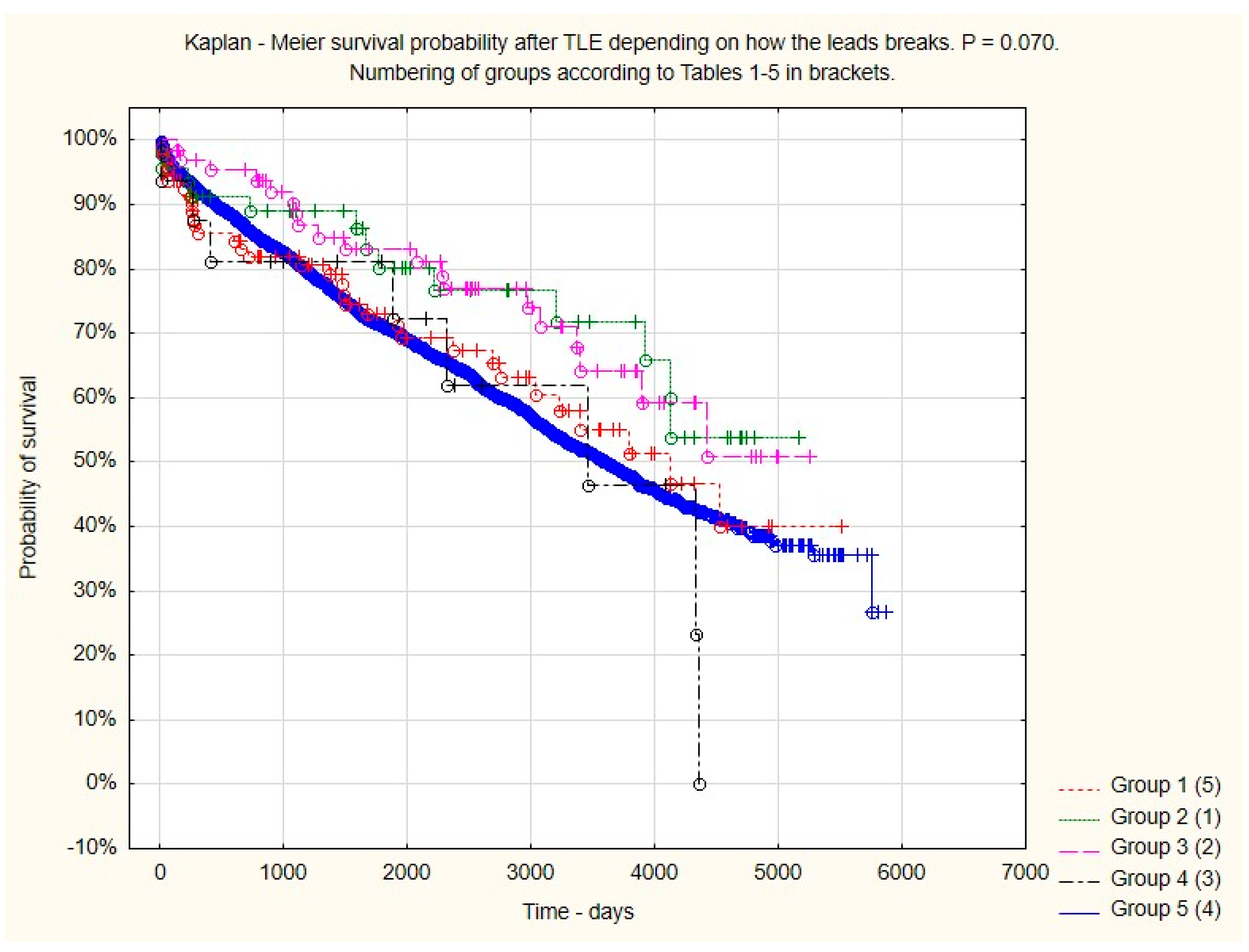
3. Results
4. Results Summary
5. Discussion
6. Conclusions
- Lead break during lead extraction occurs in 6.04% of cases, but broken lead fragments can be removed in 35.50% of patients, or the length of these fragments can be significantly reduced in 12.99% of patients.
- A larger sum of lead dwell times, a higher left ventricular ejection fraction, a higher number of previous CIED-related procedures, and the extraction of passive fixation leads were the strongest predictors of lead break during extraction. Some scores (LED index and LECOM score) predicted an increased risk of lead break.
- Lead break increases procedure difficulty and complexity, as well as the risk of TLE-related major complications. Thus, the possibility of lead break should be taken into account when planning the removal strategy and training in regards to transvenous lead extraction.
7. Study Limitations
Author Contributions
Funding
Institutional Review Board Statement
Informed Consent Statement
Data Availability Statement
Conflicts of Interest
References
- Wilkoff, B.L.; Love, C.J.; Byrd, C.L.; Bongiorni, M.G.; Carrillo, R.G.; Crossley, G.H., 3rd; Epstein, L.M.; Friedman, R.A.; Kennergren, C.E.; Mitkowski, P.; et al. Transvenous lead extraction: Heart Rhythm Society expert consensus on facilities, training, indications, and patient management: This document was endorsed by the American Heart Association (AHA). Heart Rhythm 2009, 6, 1085–1104. [Google Scholar] [CrossRef] [PubMed]
- Kusumoto, F.M.; Schoenfeld, M.H.; Wilkoff, B.; Berul, C.I.; Birgersdotter-Green, U.M.; Carrillo, R.; Cha, Y.M.; Clancy, J.; Deharo, J.C.; Ellenbogen, K.A.; et al. 2017 HRS expert consensus statement on cardiovascular implantable electronic device lead management and extraction. Heart Rhythm 2017, 14, e503–e551. [Google Scholar] [CrossRef] [PubMed]
- Bongiorni, M.G.; Burri, H.; Deharo, J.C.; Starck, C.; Kennergren, C.; Saghy, L.; Rao, A.; Tascini, C.; Lever, N.; Kutarski, A.; et al. 2018 EHRA expert consensus statement on lead extraction: Recommendations on definitions endpoints research trial design data collection requirements for clinical scientific studies registries: Endorsed by APHRS/HRS/LAHRS. Europace 2018, 20, 1217. [Google Scholar] [CrossRef] [PubMed]
- Smith, H.J.; Fearnot, N.E.; Byrd, C.L.; Wilkoff, B.L.; Love, C.J.; Sellers, T.D. Five-years experience with intravascular lead extraction. U.S.Lead Extraction Database. Pacing Clin. Electrophysiol. 1994, 17, 2016–2020. [Google Scholar] [CrossRef] [PubMed]
- Byrd, C.L.; Wilkoff, B.L.; Love, C.J.; Sellers, T.D.; Turk, K.T.; Reeves, R.; Young, R.; Crevey, B.; Kutalek, S.P.; Freedman, R.; et al. Intravascular extraction of problematic or infected permanent pacemaker leads: 1994–1996. U.S. Extraction Database, MED Institute. Pacing Clin. Electrophysiol. 1999, 22, 1348–1357. [Google Scholar] [CrossRef] [PubMed]
- Bongiorni, M.G.; Giannola, G.; Arena, G.; Soldati, E.; Bartoli, C.; Lapira, F.; Zucchelli, G.; Di Cori, A. Pacing and implantable cardioverter-defibrillator transvenous lead extraction. Ital. Heart J. 2005, 6, 261–266. [Google Scholar] [PubMed]
- Gomes, S.; Cranney, G.; Bennett, M.; Li, A.; Giles, R. Twenty-year experience of transvenous lead extraction at a single centre. Europace 2014, 16, 1350–1355. [Google Scholar] [CrossRef]
- Diemberger, I.; Migliore, F.; Biffi, M.; Cipriani, A.; Bertaglia, E.; Lorenzetti, S.; Massaro, G.; Tanzarella, G.; Boriani, G. The “Subtle” connection between development of cardiac implantable electrical device infection and survival after complete system removal: An observational prospective multicenter study. Int. J. Cardiol. 2018, 250, 146–149. [Google Scholar] [CrossRef] [PubMed]
- Kennergren, C.; Bucknall, C.A.; Butter, C.; Charles, R.; Fuhrer, J.; Grosfeld, M.; Tavernier, R.; Morgado, T.B.; Mortensen, P.; Paul, V.; et al. Laser-assisted lead extraction: The European experience. Europace 2007, 9, 651–656. [Google Scholar] [CrossRef] [PubMed]
- Roux, J.F.; Pagé, P.; Dubuc, M.; Thibault, B.; Guerra, P.G.; Macle, L.; Roy, D.; Talajic, M.; Khairy, P. Laser lead extraction: Predictors of success and complications. Pacing Clin. Electrophysiol. 2007, 30, 214–220. [Google Scholar] [CrossRef] [PubMed]
- Wazni, O.; Epstein, L.M.; Carrillo, R.G.; Love, C.; Adler, S.W.; Riggio, D.W.; Karim, S.S.; Bashir, J.; Greenspon, A.J.; DiMarco, J.P.; et al. Lead extraction in the contemporary setting: The LExICon study: An observational retrospective study of consecutive laser lead extractions. J. Am. Coll. Cardiol. 2010, 55, 579–586. [Google Scholar] [CrossRef] [PubMed]
- Brunner, M.P.; Cronin, E.M.; Duarte, V.E.; Yu, C.; Tarakji, K.G.; Martin, D.O.; Callahan, T.; Cantillon, D.J.; Niebauer, M.J.; Saliba, W.I.; et al. Clinical predictors of adverse patient outcomes in an experience of more than 5000 chronic endovascular pacemaker and defibrillator lead extractions. Heart Rhythm 2014, 11, 799–805. [Google Scholar] [CrossRef] [PubMed]
- Hussein, A.A.; Tarakji, K.G.; Martin, D.O.; Gadre, A.; Fraser, T.; Kim, A.; Brunner, M.P.; Barakat, A.F.; Saliba, W.I.; Kanj, M.; et al. Cardiac Implantable Electronic Device Infections: Added Complexity and Suboptimal Outcomes with Previously Abandoned Leads. JACC Clin. Electrophysiol. 2017, 3, 1–9. [Google Scholar] [CrossRef] [PubMed]
- Artus, A.; Mansourati, J.; Fatemi, M.; Pierre, B.; Schatz, A.; Badoz, M.; Laurent, G.; Guenancia, C.; Garnier, F. Efficacy and safety of the new TightRail™ mechanical sheath for transvenous lead extraction: Results of a French multicenter study. J. Cardiovasc. Electrophysiol. 2022, 33, 731–737. [Google Scholar] [CrossRef]
- Starck, C.T.; Gonzalez, E.; Al-Razzo, O.; Mazzone, P.; Delnoy, P.P.; Breitenstein, A.; Steffel, J.; Eulert-Grehn, J.; Lanmüller, P.; Melillo, F.; et al. Results of the Patient-Related Outcomes of Mechanical lead Extraction Techniques (PROMET) study: A multicentre retrospective study on advanced mechanical lead extraction techniques. Europace 2020, 22, 1103–1110. [Google Scholar] [CrossRef] [PubMed]
- Yap, S.C.; Bhagwandien, R.E.; Theuns, D.A.M.J.; Yasar, Y.E.; de Heide, J.; Hoogendijk, M.G.; Kik, C.; Szili-Torok, T. Efficacy and safety of transvenous lead extraction using a liberal combined superior and femoral approach. J. Interv. Card. Electrophysiol. 2021, 62, 239–248. [Google Scholar] [CrossRef] [PubMed]
- Zhou, X.; Ze, F.; Li, D.; Wang, L.; Duan, J.; Yuan, C.; He, J.; Guo, J.; Li, X. Transfemoral extraction of pacemaker and implantable cardioverter defibrillator leads using Needle’s Eye Snare: A single-center experience of more than 900 leads. Heart Vessels 2020, 35, 825–834. [Google Scholar] [CrossRef] [PubMed]
- Nof, E.; Bongiorni, M.G.; Auricchio, A.; Butter, C.; Dagres, N.; Deharo, J.C.; Rinaldi, C.A.; Maggioni, A.P.; Kutarski, A.; Kennergren, C.; et al. Comparison of outcomes in infected cardiovascular implantable electronic devices between complete, partial, and failed lead removal: An ESC-EHRA-EORP ELECTRa (European Lead Extraction ConTrolled) registry. Europace 2019, 21, 1876–1889. [Google Scholar] [CrossRef] [PubMed]
- Calvagna, G.M.; Romeo, P.; Ceresa, F.; Valsecchi, S. Transvenous retrieval of foreign objects lost during cardiac device implantation or revision: A 10-year experience. Pacing Clin. Electrophysiol. 2013, 36, 892–897. [Google Scholar] [CrossRef] [PubMed]
- Kutarski, A.; Pietura, R.; Czajkowski, M. Breakage of extracted leads: Another management option. Kardiol. Pol. 2012, 70, 307–312. [Google Scholar] [PubMed]
- Tanawuttiwat, T.; Cheng, A.; Rickard, J.; Chow, G.V.; Sciortino, C.M.; Brinker, J. Successful extraction of right ventricular lead remnants using the FlexCath® steerable sheath. J. Interv. Card. Electrophysiol. 2016, 45, 107–110. [Google Scholar] [CrossRef] [PubMed]
- Raatikainen, M.J.; Perälä, J.; Lahtinen, J. Successful defibrillator lead remnant extraction from right ventricle using a steerable transseptal sheath and a basket retriever. Europace 2009, 11, 1238–1240. [Google Scholar] [CrossRef] [PubMed]
- Kutarski, A.; Chudzik, M.; Tomaszewski, A.; Pietura, R.; Oszczygiel, A.; Czajkowski, M.; Wranicz, J.K. Difficult dual-stage transcutaneous multiple lead extraction with loss of external silicone tube of broken lead. Cardiol. J. 2013, 20, 94–99. [Google Scholar] [CrossRef] [PubMed]
- Golzio, P.G.; Bongiorni, M.G.; Giuggia, M.; Vinci, M.; Gazzera, C.; Breatta, A.D. Retrieval of pacemaker lead tip embolized into the distal pulmonary arterial bed during extraction procedure. Pacing Clin. Electrophysiol. 2007, 30, 1558–1561. [Google Scholar] [CrossRef]
- Robinson, T.; Oliver, J.; Sheridan, P.; Sahu, J.; Bowes, R. Fragmentation and embolization of pacemaker leads as a complication of lead extraction. Europace 2010, 12, 754–755. [Google Scholar] [CrossRef] [PubMed]
- Kim, D.; Baek, Y.S.; Lee, M.; Uhm, J.S.; Pak, H.N.; Lee, M.H.; Joung, B. Remnant Pacemaker Lead Tips after Lead Extractions in Pacemaker Infections. Korean Circ. J. 2016, 46, 569–573. [Google Scholar] [CrossRef] [PubMed]
- Jacheć, W.; Nowosielecka, D.; Ziaja, B.; Polewczyk, A.; Kutarski, A. LECOM (Lead Extraction COMplexity): A New Scoring System for Predicting a Difficult Procedure. J. Clin. Med. 2023, 12, 7568. [Google Scholar] [CrossRef] [PubMed]
- Bontempi, L.; Vassanelli, F.; Cerini, M.; Inama, L.; Salghetti, F.; Giacopelli, D.; Gargaro, A.; Raweh, A.; Curnis, A. Predicting the difficulty of a transvenous lead extraction procedure: Validation of the LED index. J. Cardiovasc. Electrophysiol. 2017, 28, 811–818. [Google Scholar] [CrossRef] [PubMed]
- Bontempi, L.; Curnis, A.; Della Bella, P.; Cerini, M.; Radinovic, A.; Inama, L.; Melillo, F.; Salghetti, F.; Marzi, A.; Gargaro, A.; et al. The MB score: A new risk stratification index to predict the need for advanced tools in lead extraction procedures. Europace 2020, 22, 613–621. [Google Scholar] [CrossRef] [PubMed]
- Mazzone, P.; Tsiachris, D.; Marzi, A.; Ciconte, G.; Paglino, G.; Sora, N.; Sala, S.; Vergara, P.; Gulletta, S.; Della Bella, P. Predictors of advanced lead extraction based on a systematic stepwise approach: Results from a high volume center. Pacing Clin. Electrophysiol. 2013, 36, 837–844. [Google Scholar] [CrossRef] [PubMed]
- Tułecki, Ł.; Jacheć, W.; Polewczyk, A.; Czajkowski, M.; Targońska, S.; Tomków, K.; Karpeta, K.; Nowosielecka, D.; Kutarski, A. Assessment of the impact of organisational model of transvenous lead extraction on the effectiveness and safety of procedure: An observational study. BMJ Open 2022, 12, e062952. [Google Scholar] [CrossRef] [PubMed]
- Jacheć, W.; Polewczyk, A.; Polewczyk, M.; Tomasik, A.; Kutarski, A. Transvenous Lead Extraction SAFeTY Score for Risk Stratification and Proper Patient Selection for Removal Procedures Using Mechanical Tools. J. Clin. Med. 2020, 9, 361. [Google Scholar] [CrossRef] [PubMed]
- Sidhu, B.S.; Ayis, S.; Gould, J.; Elliott, M.K.; Mehta, V.; Kennergren, C.; Butter, C.; Deharo, J.C.; Kutarski, A.; Maggioni, A.P.; et al. Risk stratification of patients undergoing transvenous lead extraction with the ELECTRa Registry Outcome Score (EROS): An ESC EHRA EORP European lead extraction ConTRolled ELECTRa registry analysis. Europace 2021, 23, 1462–1471. [Google Scholar] [CrossRef] [PubMed]
- Morita, J.; Yamaji, K.; Nagashima, M.; Kondo, Y.; Sadohara, Y.; Hirokami, J.; Kuji, R.; Korai, K.; Fukunaga, M.; Hiroshima, K.; et al. Predictors of lead break during transvenous lead extraction. J. Arrhythm. 2021, 37, 645–652. [Google Scholar] [CrossRef]
- Gianni, C.; Elchouemi, M.; Helmy, R.; Spinetta, L.; La Fazia, V.M.; Pierucci, N.; Asfour, I.; Della Rocca, D.G.; Mohanty, S.; Bassiouny, M.A.; et al. Safety and feasibility of same-day discharge following uncomplicated transvenous lead extraction. J. Cardiovasc. Electrophysiol. 2024, 35, 278–287. [Google Scholar] [CrossRef] [PubMed]


| Patient-Related Predictors of TLE Complexity, Major Complications and Coexisting Indications for TLE | Lead Break with a Long Fragment | Lead Break with a Short Fragment | Lead Break of the Tip of the Lead Only | Lead Break with Loss of a Free-Floating Fragment | All Extractions with Lead Break | All Extractions without Lead Break |
|---|---|---|---|---|---|---|
| Mean ± SD N (%) M–W U test, Chi2 test | Mean ± SD N (%) M–W U test, Chi2 test | Mean ± SD N (%) M–W U test, Chi2 test | Mean ± SD N (%) M–W U test, Chi2 test | Mean ± SD N (%) M–W U test, Chi2 test | Mean ± SD N (%) | |
| Group number/number of patients (%) | 1/95 (2.48) | 2/46 (1.20) | 3/68 (1.78) | 4/22 (0.57) | 5/231 (6.04) | 6/3594 (93.96) |
| Patient age during TLE (years) | 59.29 ± 19.36 p < 0.001 | 56.67 ± 21.57 p = 0.001 | 60.40 ± 19.79 p = 0.022 | 66.09 ± 14.03 p = 0.724 | 59.75 ± 19.55 p < 0.001 | 66.44 ± 15.34 |
| Patient age at first system implantation (years) | 43.65 ± 20.39 p < 0.001 | 41.93 ± 22.28 p < 0.001 | 47.74 ± 21.35 p < 0.001 | 50.14 ± 17.71 p = 0.015 | 45.14 ± 20.87 p < 0.001 | 58.14 ± 15.34 |
| Female | 48 (50.53) p = 0.016 | 17 (36.96) p = 0.970 | 25 (36.76) p = 0.964 | 6 (27.27) p = 0.427 | 96 (41.56) p = 0.282 | 1358 (37.79) |
| Ischemic heart disease | 31 (32.63) p < 0.001 | 18 (39.13) p < 0.001 | 37 (54.41) p = 785 | 11 (50.00) p = 0.669 | 97 (41.99) p < 0.001 | 2042 (56.82) |
| NYHA FC III or IV | 12 (12.63) p = 0.491 | 4 (8.70) p = 0.268 | 11 (16.18) p = 0.938 | 4 (18.18) p < 0.830 | 34 (14.72) p = 0.738 | 567 (15.78) |
| LVEF (%) | 55.14 ± 11.98 p < 0.001 | 55.41 ± 10.58 p = 0.003 | 54.04 ± 12.53 p = 0.015 | 52.59 ± 13.49 p = 0.266 | 54.63 ± 11.98 p < 0.001 | 49.14 ± 15.44 |
| Charlson co-morbidity index (points) | 4.12 ± 4.14 p < 0.001 | 3.04 ± 3.11 p < 0.001 | 3.53 ± 3.15 p = 0.003 | 3.96 ± 3.15 p = 0.421 | 3.71 ± 3.59 p < 0.001 | 4.81 ± 3.69 |
| Indications for TLE | ||||||
| Infective endocarditis, with or without pocket infection | 14 (14.74) p = 0.125 | 12 (26.09) p = 0.608 | 14 (20.59) p = 0.978 | 7 (31.82) p = 0.385 | 47 (20.35) p = 0.651 | 785 (21.84) |
| Local (isolated) pocket infection | 11 (11.58) p = 0.958 | 3 (6.52) p = 0.676 | 10 (14.71) p = 0.208 | 2 (9.09) p = 0.868 | 26 (11.26) p = 0.425 | 339 (9.43) |
| Mechanical lead damage (electrical failure) | 42 (44.21) p < 0.001 | 18 (39.13) p = 0.076 | 20 (29.41) p = 0.676 | 4 (18.18) p = 0.529 | 84 (36.36) p = 0.001 | 949 (26.41) |
| Lead dysfunction (exit/entry block, dislodgement, perforation, extracardiac pacing) | 11 (11.58) p = 0.013 | 8 (17.39) p = 0.474 | 13 (19.12) p = 0.548 | 3 (13.64) p = 0.434 | 35 (15.15) p = 0.008 | 825 (22.96) |
| Change of pacing mode/upgrading, downgrading | 5 (5.26) p = 0.751 | 0 (0.00) p = 0.132 | 2 (2.94) p = 0.334 | 0 (0.00) p = 0.414 | 7 (3.03) p = 0.043 | 238 (6.62) |
| Other non-infectious indications * | 12 (12.63) p = 0.902 | 5 (10.87) p = 0.876 | 9 (13.24) p = 0.950 | 6 (27.27) p = 0.087 | 32 (13.85) p = 0.698 | 458 (12.74) |
| System-Related Risk Factors for TLE Complexity and Major Complications | Lead Break with a Long Fragment | Lead Break with a Short Fragment | Lead Break of the Tip of the Lead Only | Lead Break with Loss of a Free-Floating Fragment | All Extractions, with Lead Break | All Extractions, without Lead Break |
|---|---|---|---|---|---|---|
| Mean ± SD N (%) M–W U test, Chi2 test | Mean ± SD N (%) M–W U test, Chi2 test | Mean ± SD N (%) M–W U test, Chi2 test | Mean ± SD N (%) M–W U test, Chi2 test | Mean ± SD N (%) M–W U test, Chi2 test | Mean ± SD N (%) | |
| Group number/number of patients (%) | 1/95 (2.48) | 2/46 (1.20) | 3/68 (1.78) | 4/22 (0.57) | 5/231 (6.04) | 6/3594 (93.96) |
| System and history of pacing | ||||||
| Oldest lead before TLE (months) | 188.8 ± 86.14 p < 0.001 | 177.7 ± 85.46 p < 0.001 | 151.7 ± 90.68 p < 0.001 | 191.7 ± 106.0 p < 0.001 | 176.0 ± 90.26 p < 0.001 | 97.66 ± 72.71 |
| Global lead dwell time (years) before TLE | 29.86 ± 17.69 p < 0.001 | 28.36 ± 13.37 p < 0.001 | 22.76 ± 13.00 p < 0.001 | 35.51 ± 21.89 p < 0.001 | 28.01 ± 16.46 p < 0.001 | 14.62 ± 12.28 |
| Abandoned leads before TLE | 27 (28.42) p < 0.001 | 10 (21.74) p = 0.008 | 14 (20.59) p = 0.004 | 12 (54.55) p < 0.001 | 63 (27.27) p < 0.001 | 354 (9.85) |
| Unnecessary (large) lead loop in the heart | 10 (10.53) p = 0.005 | 6 (13.04) p = 0.005 | 3 (4.41) p = 1.000 | 7 (31.82) p < 0.001 | 26 (11.26) p < 0.001 | 158 (4.40) |
| Number of leads in the heart before TLE | 2.16 ± 0.84 p = 0.004 | 2.20 ± 0.62 p = 0.005 | 2.10 ± 0.79 p = 0.071 | 2.68 ± 1.21 p = 0.027 | 2.20 ± 0.84 p < 0.001 | 1.94 ± 0.73 |
| ≥4 leads in the heart before TLE | 8 (8.42) p < 0.001 | 0 (0.00) p = 0.260 | 5 (7.35) p < 0.001 | 7 (31.82) p < 0.001 | 20 (8.66) p < 0.001 | 98 (2.73) |
| Leads on both sides of the chest before TLE | 7 (7.37) p = 0.003 | 0 (0.00) p = 0.280 | 6 (8.82) p < 0.001 | 5 (22.73) p < 0.001 | 18 (7.79) p < 0.001 | 89 (2.48) |
| Number of procedures before lead extraction | 2.90 ± 1.67 p < 0.001 | 2.54 ± 1.24 p < 0.001 | 2.45 ± 1.41 p < 0.001 | 2.76 ± 1.14 p < 0.001 | 2.68 ± 1.47 p < 0.001 | 1.81 ± 1.03 |
| Two or more CIED procedures before TLE | 84 (83.42) p < 0.001 | 37 (80.43) p < 0.001 | 51 (75.00) p = 0.001 | 19 (86.36) p = 0.001 | 191 (82.68) p = 0.001 | 1834 (51.03) |
| Risk scores for prediction of major complications or increased procedure complexity. | ||||||
| SAFeTY-TLE score (points) | 10.75 ± 4.55 p < 0.001 | 9.62 ± 4.11 p < 0.001 | 6.36 ± 3.82 p < 0.001 | 10.07 ± 5.03 p < 0.001 | 9.75 ± 4.59 p < 0.001 | 5.67 ± 4.07 |
| SAFeTY-TLE score (%) | 5.80 ± 8.89 p < 0.001 | 3.69 ± 3.00 p < 0.001 | 3.36 ± 4.75 p < 0.001 | 3.89 ± 3.31 p < 0.001 | 4.48 ± 6.64 p < 0.001 | 1.54 ± 2.43 |
| EROS score (points) | 2.15 ± 0.92 p < 0.001 | 2.11 ± 0.82 p < 0.001 | 1.81 ± 0.87 p < 0.014 | 2.27 ± 0.83 p < 0.001 | 2.05 ± 0.89 p < 0.001 | 1.50 ± 0.71 |
| MB score (points) | 3.53 ± 0.68 p < 0.001 | 3.50 ± 0.78 p < 0.001 | 3.32 ± 0.89 p < 0.001 | 3.46 ± 0.91 p < 0.001 | 3.46 ± 0.79 p < 0.001 | 2.53 ± 1.25 |
| LED index (points) | 17.37 ± 7.11 p < 0.001 | 16.46 ± 7.08 p < 0.001 | 13.52 ± 7.46 p < 0.001 | 18.32 ± 9.04 p < 0.001 | 16.44 ± 7.48 p < 0.001 | 9.65 ± 6.10 |
| Advanced TLE (Mazzone) scale (points) | 2.53 ± 0.70 p < 0.001 | 2.50 ± 0.69 p = 0.006 | 2.49 ± 0.70 p = 0.001 | 2.41 ± 0.80 p = 0.154 | 2.50 ± 0.70 p < 0.001 | 2.12 ± 0.93 |
| LECOM score (points) | 12.97 ± 4.20 p < 0.001 | 12.45 ± 3.50 p < 0.001 | 11.20 ± 4.07 p < 0.001 | 14.18 ± 5.32 p < 0.001 | 12.36 ± 4.23 p < 0.001 | 7.94 ± 3.96 |
| LECOM score (%) | 41.81 ± 22.15 p < 0.001 | 39.76 ± 20.58 p < 0.001 | 33.07 ± 21.00 p < 0.001 | 50.34 ± 29.69 p < 0.001 | 39.64 ± 22.76 p < 0.001 | 19.08 ± 17.43 |
| LECOM score (patients with very high expected complexity of the procedure) | 79 (83.16) p < 0.001 | 33 (71.54) p < 0.001 | 45 (66.18) p < 0.001 | 18 (81.83) p < 0.001 | 175 (76.76) p < 0.001 | 1135 (31.58) |
| Potential procedure-related risk factors for major complications and increased procedure complexity. | ||||||
| Number of extracted leads per patient | 2.01 ± 0.88 p < 0.001 | 1.96 ± 0.70 p < 0.001 | 1.87 ± 0.69 p = 0.002 | 2.50 ± 1.10 p < 0.004 | 2.00 ± 0.82 p < 0.001 | 1.63 ± 0.72 |
| Extraction of abandoned lead(s) (any) | 26 (27.37) p < 0.001 | 10 (21.74) p = 0.003 | 13 (19.12) p = 0.004 | 11 (50.00) p < 0.001 | 60 (25.97) p < 0.001 | 322 (8.96) |
| Extraction of old model UP leads (excluding LV leads) | 29 (30.53) p < 0.001 | 9 (19.57) p < 0.001 | 10 (14.71) p = 0.002 | 8 (36.36) p < 0.001 | 56 (24.24) p < 0.001 | 210 (5.84) |
| Extraction of passive fixation leads (excluding LV leads) | 88 (92.63) p < 0.001 | 45 (97.83) p < 0.001 | 62 (91.18) p < 0.001 | 20 (90.91) p = 0.001 | 215 (93.07) p < 0.001 | 2008 (55.87) |
| Extraction of VDD leads | 7 (7.37) p = 0.005 | 1 (2.17) p = 0.860 | 1 (1.47) p = 0.564 | 1 (4.55) p = 0.565 | 10 (4.33) p = 0.113 | 93 (2.59) |
| Extraction of leads with redundant loop in the heart | 11 (11.58) p = 0.002 | 6 (13.04) p = 0.007 | 2 (2.94) p = 0.518 | 8 (36.36) p < 0.001 | 32 (13.85) p < 0.001 | 165 (4.59) |
| Oldest lead extracted per patient (months) | 185.3 ± 83.85 p < 0.001 | 176.7 ± 85.69 p < 0.001 | 152.0 ± 90.68 p < 0.001 | 191.7 ± 106.04 p < 0.001 | 174.3 ± 89.25 p < 0.001 | 95.90 ± 71.68 |
| Sum of dwell times of extracted leads (years) | 28.46 ± 17.41 p < 0.001 | 26.13 ± 14.48 p < 0.001 | 21.29 ± 13.15 p < 0.001 | 35.51 ± 21.89 p < 0.001 | 26.56 ± 16.62 p < 0.001 | 12.96 ± 11.95 |
| Procedure Complexity | Lead Break with a Long Fragment | Lead Break with a Short Fragment | Lead Break of the Tip of the Lead Only | Lead Break with Loss of a Free-Floating Fragment | All Extractions, with Lead Break | All Extractions, without Lead Break |
|---|---|---|---|---|---|---|
| Mean ± SD N (%) M–W U test, Chi2 test | Mean ± SD N (%) M–W U test, Chi2 test | Mean ± SD N (%) M–W U test, Chi2 test | Mean ± SD N (%) M–W U test, Chi2 test | Mean ± SD N (%) M–W U test, Chi2 test | Mean ± SD N (%) | |
| Group number/number of patients (%) | 1/95 (2.48) | 2/46 (1.20) | 3/68 (1.78) | 4/22 (0.57) | 5/231 (6.04) | 6/3594 (93.96) |
| TLE complexity and outcomes | ||||||
| Extraction time (sheath-to-sheath) (minutes) | 69.80 ± 49.46 p < 0.001 | 27.20 ± 14.47 p < 0.001 | 24.74 ± 28.10 p < 0.001 | 92.73 ± 77.15 p < 0.001 | 50.23 ± 49.44 p < 0.001 | 12.77 ± 18.01 |
| * Average time of single lead extraction (minutes) | 38.75 ± 31.64 p < 0.001 | 15.70 ± 9.51 p < 0.001 | 12.83 ± 12.76 p < 0.001 | 44.09 ± 50.76 p < 0.001 | 27.04 ± 29.65 p < 0.001 | 7.75 ± 9.55 |
| Overall number of technical problems in the group of patients | 95 (100.0) p < 0.001 | 32 (69.57) p < 0.001 | 26 (38.24) p < 0.001 | 22 (100.0) p < 0.001 | 175 (75.76) p < 0.001 | 552 (15.36) |
| Number of technical problems per patient | 2.21 ± 1.02 p < 0.001 | 1.53 ± 0.62 p < 0.001 | 1.54 ± 0.71 p < 0.001 | 2.73 ± 1.03 p < 0.001 | 2.05 ± 0.99 p < 0.001 | 1.24 ± 0.50 |
| Two or more technical problems | 69 (72.63) p = 0.001 | 15 (32.61) p = 0.001 | 12 (17.65) p = 0.001 | 22 (100.0) p = 0.001 | 118 (51.08) p = 0.001 | 115 (3.20) |
| Utilitization of additional tools and techniques | ||||||
| Evolution (old and new) or TightRail | 15 (15.79) p < 0.001 | 4 (8.70) p < 0.001 | 6 (8.82) p < 0.001 | 4 (18.18) p < 0.001 | 29 (12.55) p < 0.001 | 29 (0.81) |
| Metal sheaths | 29 (30.53) p < 0.001 | 9 (19.57) p < 0.001 | 12 (17.65) p = 0.001 | 5 (22.73) p = 0.006 | 55 (23.81) p < 0.001 | 261 (7.26) |
| Lasso catheters/snares, basket catheters | 91 (95.79) p < 0.001 | 12 (26.09) p < 0.001 | 4 (5.88) p = 0.014 | 22 (100.0) p < 0.001 | 129 (55.84) p < 0.001 | 65 (1.81) |
| Need to use an alternative approach | 36 (37.89) p < 0.001 | 2 (4.35) p = 0.281 | 5 (7.35) p = 0.003 | 10 (45.45) p < 0.001 | 53 (22.94) p < 0.001 | 74 (2.06) |
| Procedure difficulty score | ||||||
| ** Retrospective TLE combined difficulty score (points) | 3.58 ± 0.79 p < 0.001 | 1.98 ± 1.34 p < 0.001 | 1.04 ± 1.43 p < 0.001 | 3.82 ± 0.73 p < 0.001 | 2.54 ± 1.61 p < 0.001 | 0.43 ± 0.95 |
| Retrospective TLE combined difficulty score of two or more points | 95 (100.0) p < 0.001 | 33 (71.74) p < 0.001 | 23 (33.82) p < 0.001 | 22 (100.0) p < 0.001 | 173 (74.89) p < 0.001 | 556 (15.47) |
| Procedure Complications and Long-Term Outcomes | Lead Break with a Long Fragment | Lead Break with a Short Fragment | Lead Break of the Tip of the Lead Only | Lead Break with Loss of a Free-Floating Fragment | All Extractions, with Lead Break | All Extractions, without Lead Break |
|---|---|---|---|---|---|---|
| N (%) Chi2 test | N (%) Chi2 test | N (%) Chi2 test | N (%) Chi2 test | N (%) Chi2 test | N (%) | |
| Group number/number of patients (%) | 1/95 (2.48) | 2/46 (1.20) | 3/68 (1.78) | 4/22 (0.57) | 5/231 (6.04) | 6/3594 (93.96) |
| TLE efficacy and complications | ||||||
| Major complications (any) | 7 (7.37) p < 0.001 | 7 (15.22) p < 0.001 | 5 (7.35) p < 0.001 | 4 (18.18) p < 0.001 | 23 (9.96) p < 0.001 | 55 (1.53) |
| Hemopericardium | 4 (4.21) p = 0.002 | 5 (10.87) p < 0.001 | 3 (4.41) p = 0.004 | 1 (4.55) p = 0.080 | 13 (5.63) p < 0.001 | 33 (0.92) |
| Hemothorax | 1 (1.05) p = 0.014 | 0 (0.00) p = 0.821 | 0 (0.00) p = 0.783 | 0 (0.00) p = 0.877 | 1 (0.43) p = 0.190 | 4 (0.11) |
| Tricuspid valve damage during TLE (severe) | 2 (2.11) p = 0.017 | 1 (2.17) p = 0.074 | 2 (2.94) p = 0.002 | 3 (13.64) p < 0.001 | 8 (3.46) p < 0.001 | 15 (0.42) |
| Rescue cardiac surgery | 4 (4.21) p = 0.001 | 4 (8.70) p < 0.001 | 2 (2.94) p = 0.057 | 1 (4.55) p = 0.054 | 11 (4.76) p < 0.001 | 29 (0.81) |
| Death, procedure-related (intra-,post-procedural) | 0 (0.00) p = 0.691 | 0 (0.00) p = 0.783 | 0 (0.00) p = 0.736 | 0 (0.00) p = 0.847 | 0 (0.00) p = 0.534 | 6 (0.17) |
| Clinical success | 84 (88.42) p < 0.001 | 34 (73.91) p < 0.001 | 43 (63.24) p < 0.001 | 19 (86.36) p < 0.001 | 180 (77.92) p < 0.001 | 3567 (99.25) |
| Complete procedural success | 58 (61.05) p < 0.001 | 6 (13.04) p < 0.001 | 1 (1.47) p < 0.001 | 12 (54.55) p < 0.001 | 77 (33.33) p < 0.001 | 3565 (99.19) |
| Long-term mortality after TLE | ||||||
| Survivors | 64 (67.37) p = 0.549 | 33 (71.74) p = 0.300 | 49 (72.06) p = 0.190 | 11 (50.00) p = 0.160 | 157 (67.97) p = 0.270 | 2314 (64.39) |
| One-year mortality | 12 (12.63) p = 0.115 | 4 (8.70) p = 0.890 | 2 (2.94) p = 0.119 | 3 (13.64) p = 0.347 | 21 (9.09) p = 0.604 | 292 (8.13) |
| Overall follow-up mortality | 31 (32.63) p = 0.549 | 13 (28.26) p = 0.300 | 19 (27.94) p = 0.190 | 11 (50.00) p = 0.160 | 74 (32.036) p = 0.270 | 1280 (35.62) |
| Management of Lead Break | Lead Break with a Long Fragment | Lead Break with a Short Fragment | Lead Break of the Tip of the Lead Only | Lead Break with Loss of Broken Lead Fragment or Lead Tip | All Extractions, with Lead Break |
|---|---|---|---|---|---|
| N (%) | N (%) | N (%) | N (%) | N (%) | |
| Group number/number of patients (%) | 1/95 (2.48) | 2/46 (1.20) | 3/68 (1.78) | 4/22 (0.57) | 5/231 (6.04) |
| Grasped remnant entirely removed | 63 (66.32) | 4 (8.70) | 0 (0.00) | 15 (68.18) | 82 (35.50) |
| Shortening the lead remnant, with a retained fragment < 4 cm | 26 (27.37) | 1 (2.17) | 0 (0.00) | 3 (13.64) | 30 (12.99) |
| Unsuccessful attempt at retrieval of the fragment | 3 (3.16) | 10 (21.74) | 0 (0.00) | 4 (18.18) | 17 (7.36) |
| No attempt made to grasp the fragment (no chance) | 0 (0.00) | 29 (63.04) | 68 (100.0) | 0 (0.00) | 97 (41.99) |
| Lead remnant removal during emergency or planned cardiac surgery | 3 (3.16) | 1 (2.17) | 0 (0.00) | 0 (0.00) | 4 (1.73) |
| Procedure aborted due to major complications and ultimately, death of the patient | 0 (0.00) | 1 (2.17) | 0 (0.00) | 0 (0.00) | 1 (0.43) |
| Univariable Regression Model | Multivariable Regression Model | |||||
|---|---|---|---|---|---|---|
| OR | 95% CI | p | OR | 95% CI | p | |
| Patient age during TLE (by 1 year) | 0.977 | 0.970–0.984 | <0.001 | |||
| Patient age at first system implantation (by 1 year) | 0.965 | 0.959–0.971 | <0.001 | 0.971 | 0.961–0.981 | <0.001 |
| Sum of all lead dwell times (by 1 year) | 1.126 | 1.107–1.145 | <0.001 | 1.018 | 1.005–1.031 | 0.006 |
| Ischemic heart disease (y/n) | 0.567 | 0.432–0.743 | <0.001 | 1.125 | 0.803–1.577 | 0.494 |
| LVEF (by 1% p) | 1.027 | 1.017–1.038 | <0.001 | 1.014 | 1.003–1.026 | 0.013 |
| Charlson co-morbidity index (by 1 point) | 0.912 | 0.875–0.951 | <0.001 | 1.033 | 0.983–1.086 | 0.203 |
| Non-infectious indications for TLE (y/n) | 0.979 | 0.788–1.216 | 0.846 | |||
| Abandoned leads before TLE (y/n) | 3.510 | 2.575–4.786 | <0.001 | 1.107 | 0.712–1.720 | 0.652 |
| Lead loops before TLE (y/n) | 2.678 | 1.765–4.062 | <0.001 | 1.347 | 0.856–2.120 | 0.198 |
| Number of the leads in the heart before TLE (by 1) | 1.526 | 1.296–1.798 | <0.001 | 1.021 | 0.804–1.298 | 0.862 |
| Leads on both sides of the chest (y/n) | 1.020 | 0.980–1.062 | 0.335 | |||
| Number of CIED-related procedures before TLE (by 1) | 1.692 | 1.537–1.863 | <0.001 | 1.187 | 1.033–1.364 | 0.016 |
| UP lead extraction (y/n) | 4.627 | 3.377–6.341 | <0.001 | 1.197 | 0.808–1.771 | 0.370 |
| Extraction of passive fixation lead (y/n) | 10.677 | 6.397–17.82 | <0.001 | 6.354 | 3.679–10.97 | <0.001 |
| Sum of dwell times of all extracted leads (by 1 year) | 1.056 | 1.048–1.065 | <0.001 | |||
| Number of extracted leads (by 1) | 1.784 | 1.526–2.085 | <0.001 | |||
| Extraction of abandoned lead/leads (y/n) | 3.671 | 2.681–5.026 | <0.001 | |||
| Extraction of looped leads (y/n) | 1.889 | 1.384–2.578 | <0.001 | |||
| Dwell time of the oldest extracted lead (by 1 year) | 1.129 | 1.110–1.148 | <0.001 | |||
| Univariable Regression Model | Multivariable Regression Model | |||||
|---|---|---|---|---|---|---|
| OR | 95%CI | p | OR | 95%CI | p | |
| SAFeTY-TLE (by 1% p) | 1.198 | 1.160–1.237 | <0.001 | 1.029 | 0.988–1.071 | 0.171 |
| EROS (by 1 point) | 2.396 | 2.036–2.819 | <0.001 | 1.081 | 0.860–1.358 | 0.505 |
| MB (by 1 point) | 2.130 | 1.847–2.455 | <0.001 | 1.128 | 0.895–1.422 | 0.307 |
| LED (by 1 point) | 1.129 | 1.110–1.148 | <0.001 | 1.058 | 1.024–1.094 | <0.001 |
| Advanced TLE (by 1 point) | 1.605 | 1.379–1.868 | <0.001 | 1.128 | 0.898–1.416 | 0.300 |
| LECOM (by 1% point) | 1.041 | 1.035–1.047 | <0.001 | 1.019 | 1.009–1.029 | <0.001 |
Disclaimer/Publisher’s Note: The statements, opinions and data contained in all publications are solely those of the individual author(s) and contributor(s) and not of MDPI and/or the editor(s). MDPI and/or the editor(s) disclaim responsibility for any injury to people or property resulting from any ideas, methods, instructions or products referred to in the content. |
© 2024 by the authors. Licensee MDPI, Basel, Switzerland. This article is an open access article distributed under the terms and conditions of the Creative Commons Attribution (CC BY) license (https://creativecommons.org/licenses/by/4.0/).
Share and Cite
Kutarski, A.; Jacheć, W.; Czajkowski, M.; Stefańczyk, P.; Kosior, J.; Tułecki, Ł.; Nowosielecka, D. Lead Break during Extraction: Predisposing Factors and Impact on Procedure Complexity and Outcome: Analysis of 3825 Procedures. J. Clin. Med. 2024, 13, 2349. https://doi.org/10.3390/jcm13082349
Kutarski A, Jacheć W, Czajkowski M, Stefańczyk P, Kosior J, Tułecki Ł, Nowosielecka D. Lead Break during Extraction: Predisposing Factors and Impact on Procedure Complexity and Outcome: Analysis of 3825 Procedures. Journal of Clinical Medicine. 2024; 13(8):2349. https://doi.org/10.3390/jcm13082349
Chicago/Turabian StyleKutarski, Andrzej, Wojciech Jacheć, Marek Czajkowski, Paweł Stefańczyk, Jarosław Kosior, Łukasz Tułecki, and Dorota Nowosielecka. 2024. "Lead Break during Extraction: Predisposing Factors and Impact on Procedure Complexity and Outcome: Analysis of 3825 Procedures" Journal of Clinical Medicine 13, no. 8: 2349. https://doi.org/10.3390/jcm13082349