Article

Assessing the Preservation of Lumbar Lordotic Curvature in Everyday Sitting Conditions Assessed with an Inertial Measurement System

1
Department of Rehabilitation Medicine, National Traffic Injury Rehabilitation Hospital, Yangpyeong 12564, Republic of Korea
2
Department of Rehabilitation Medicine, Armed Forces Yangju Hospital, Yangju 11429, Republic of Korea
3
Department of Physical and Rehabilitation Medicine, Chung-Ang University Gwangmyeong Hospital, Gwangmyeong 14353, Republic of Korea
4
Department of Rehabilitation Medicine, Seoul National University Hospital, Seoul 03080, Republic of Korea
5
Department of Orthopaedic Surgery, Seoul National University Hospital, Seoul 03080, Republic of Korea
6
Mellowing Factory Co., Ltd., Seoul 06053, Republic of Korea
7
Department of Rehabilitation Medicine, Seoul National University College of Medicine, Seoul 03080, Republic of Korea
8
Institute of Aging, Medical Research Center, Seoul National University, Seoul 03080, Republic of Korea
*
Author to whom correspondence should be addressed.
These authors contributed equally to this work.
J. Clin. Med. 2024, 13(9), 2728; https://doi.org/10.3390/jcm13092728
Submission received: 27 March 2024 / Revised: 25 April 2024 / Accepted: 2 May 2024 / Published: 6 May 2024
(This article belongs to the Section Clinical Rehabilitation)

Abstract

:
Background/Objectives: Lumbar lordotic curvature (LLC), closely associated with low back pain (LBP) when decreased, is infrequently assessed in clinical settings due to the spatiotemporal limitations of radiographic methods. To overcome these constraints, this study used an inertial measurement system to compare the magnitude and maintenance of LLC across various sitting conditions, categorized into three aspects: verbal instructions, chair type, and desk task types. Methods: Twenty-nine healthy participants were instructed to sit for 3 min with two wireless sensors placed on the 12th thoracic vertebra and the 2nd sacral vertebra. The lumbar lordotic angle (LLA) was measured using relative angles for the mediolateral axis and comparisons were made within each sitting category. Results: The maintenance of LLA (LLAdev) was significantly smaller when participants were instructed to sit upright (−3.7 ± 3.9°) compared to that of their habitual sitting posture (−1.2 ± 2.4°) (p = 0.001), while the magnitude of LLA (LLAavg) was significantly larger with an upright sitting posture (p = 0.001). LLAdev was significantly larger when using an office chair (−0.4 ± 1.1°) than when using a stool (−3.2 ± 7.1°) (p = 0.033), and LLAavg was also significantly larger with the office chair (p < 0.001). Among the desk tasks, LLAavg was largest during keyboard tasks (p < 0.001), followed by mouse and writing tasks; LLAdev showed a similar trend without statistical significance (keyboard, −1.2 ± 3.0°; mouse, −1.8 ± 2.2°; writing, −2.9 ± 3.1°) (p = 0.067). Conclusions: Our findings suggest that strategies including the use of an office chair and preference for computer work may help preserve LLC, whereas in the case of cueing, repetition may be necessary.

1. Introduction

The lumbar lordotic curvature (LLC), or the inward curving of the lumbar spine, is one of the most prominent morphological characteristics in the human spinal column and is considered a key structural adaptation to bipedalism [1]. There has been ongoing debate over the relationship between LLC and low back pain (LBP), as LLC has both biomechanical advantages and disadvantages [2,3,4,5]. Preservation of LLC contributes to neutralizing shear loads in the soft tissues surrounding the spine [2], enhancing the capacity to bear gravitational force [3], and preventing the posterior migration of the nucleus pulposus [4]. Conversely, an overly increased LLC may lead to excessive pressure on the posterior ligaments and facet joints [5]. Despite this controversy, recent studies strongly support that a decreased LLC is closely related to LBP [6,7]. The harmful effects of flexed lumbar posture, including increased muscle activity and passive strain on the posterior elements of the spine [8,9,10,11,12], are well documented. However, it is the damage to the posterior annulus of the lumbar disc [13], caused by increased disc pressure and posterior migration of the nucleus pulposus [14,15,16,17], that plays a critical role in pain induction.
While the causes of LBP are multifactorial [18], prolonged sitting is closely associated with LBP [19,20], with studies showing that awkward postures significantly increase the risk of developing LBP [21]. As a flexed lumbar posture, characterized by decreased LLC, is a common problematic posture while sitting, numerous studies have investigated how LLC is affected by various sitting conditions. Furthermore, studies have assessed how LLC is influenced by specific instructions to maintain good posture [22,23,24,25,26,27,28], such as erect/comfortable/slouch, ideal/habitual, and upright/relaxed. These studies have consistently shown that specific instructions to maintain good posture result in a significantly larger LLC compared to that of habitual sitting postures [23,27,28]. Similarly, numerous studies have compared LLC differences across different types of chairs [26,27,28,29,30,31,32,33,34], including office chairs, standard chairs, stools, or kneeling chairs. Most findings suggest that office chairs, especially those with lumbar support, shows the largest LLC [26,27,28,31], whereas stools tend to show the smallest LLC [31,33]. Only a few studies have examined LLC across various task types [34,35], finding less lumbar flexion during typing compared to using a mouse. Despite the extensive research on the impact of sitting conditions on LLC, these existing studies have not focused on the preservation of LLC over time. While sitting is relatively stable, LLC can change over time if the posture is maintained for an extended period. Considering this, investigating not only the magnitude of LLC but also the degree of change over time may be a critical aspect for developing strategies for spinal health maintenance in everyday sitting.
In clinical settings, evaluating LLC has primarily been conducted using radiographic methods [36,37,38,39], which provide the most accurate, skeleton-based assessment. However, radiographic assessment has inherent limitations, such as spatiotemporal constraints and radiation hazards, which prevent its application to various activities over an extended period. Recent advancements in inertial sensors have offered a more accessible solution for evaluating LLC [25,28,40,41,42,43,44,45,46]. Due to their compact size and wireless capability, inertial measurement systems have gained popularity for assessing spinal curvatures in various clinical scenarios [27,43,44,45] and have been validated against traditional gold-standard measures like radiological and optoelectronic motion capture systems [25,40,41,42,43]. The Attitude and Heading Reference System (AHRS), an advanced inertial sensor system, utilizes complex algorithms to provide more accurate and comprehensive data than conventional inertial sensors [47]. By employing AHRS, this study enables effective real-time monitoring of LLC, presenting a promising method for assessing the preservation of LLC in daily sitting conditions.
In this in vivo study on a healthy population, an AHRS was used to investigate the dynamics of LLC over a duration of 3 min during various sitting conditions, categorized into three groups: verbal instructions, chair type, and desk task types. Our hypothesis was that the preservation of LLC, along with its magnitude, might differ significantly across these conditions. The primary aim of this study was to assess the feasibility of using AHRS for evaluating LLC preservation. Additionally, we sought to determine which postures or tasks within each category are more beneficial for preserving LLC.

2. Methods

2.1. Design

This study is an analytical cross-sectional study conducted in the Biomechanics laboratory of the Seoul National University Hospital. All participants followed the same test protocols conducted over a single day, which included 10 different sitting conditions. This study is reported following the STROBE (Strengthening the Reporting of Observational Studies in Epidemiology) guidelines [48].

2.2. Participants

A total of 29 healthy volunteers, comprising 15 men and 14 women, participated in this study. The mean ages for men and women were 32.2 ± 12.6 years and 39.1 ± 14.1 years, respectively. Anthropomorphic characteristics including the age, weight, height, and body mass index (BMI) of each participant were collected and are summarized in Table 1. Participants recruited for this study had neither experienced LBP nor radicular pain radiating beyond the gluteal fold for 12 weeks leading up to the study, and had no history of back disorders, implying that they were expected to have normal ranges of LLA. Exclusion criteria included skin complications at the sensor attachment site, individuals with psychological disorders such as depression requiring regular medication, pregnant individuals, and those unable to participate in the experiment due to external factors. This study was performed in accordance with the Declaration of Helsinki and was approved by the Institutional Review Board of Seoul National University Hospital (IRB No. 1703-174-842). All participants provided informed consent to participate in the study.

2.3. Experimental Set-Up

To evaluate LLC, we used an AHRS based on the wireless inertial measurement unit (IMU, MTw Awinda; Xsens Technologies B.V., Enschede, Netherlands), which uses Strap-Down Integration and the Xsens Kalman Filter to provide more accurate data [47]. Using medical tape, two IMU sensors were affixed on the skin over the spinous process of the 12th thoracic vertebra, palpated along the 12th rib, and over the 2nd sacral vertebra at the midpoint between the posterior superior iliac spines (Figure 1) [49,50]. Prior to data collection, each sensor was calibrated in parallel in the corner of the experiment room to ensure accurate angle measurements. The three output angles were initialized to zero, such that when the two sensors were subtracted after calibration, the angular differences were near zero. Data from the sensors were sampled at a rate of 40 Hz and wirelessly transmitted to a data-processing computer.

2.4. Data Processing

The lumbar lordotic angle (LLA) served as a measure for evaluating LLC. Conventionally, the LLA is derived from radiographic images using the Cobb angle, formed between the lines at the inferior endplate of T12 and the superior endplate of S1 (Figure 1d) [50]. When measuring with inertial sensors, the placement can vary slightly among studies; we adhered to the protocols of previously related studies [42,51].
Data processing was conducted in accordance with the methods of our group’s previous biomechanical study [43,46]. The sensor output data provided the 3-dimensional orientations relative to the rotation angles around the X (upward longitudinal axis), Y (mediolateral axis), and Z (anteroposterior axis) axes (Figure 1c). Lumbar posture was determined by subtracting the angles between the thoracic sensor (T12) and the sacral sensor (S2) along each of these axes, and the calculated relative angles along the X, Y, and Z axes correspond to the degrees of axial twist, kyphosis–lordosis, and lateral bending, respectively. Specifically, the Y-axis angle, which measures the degree of kyphosis–lordosis, was used as the LLA (Figure 1d). Positive and negative values indicated lordotic and kyphotic alignment of the lumbar spine, respectively. Instead of calculating 3-dimensional Euler angles between the two sensors, a simple subtraction method was utilized to calculate relative angles because the sensors were expected to be moving mainly within the sagittal plane during the test conditions in this study.

2.5. Test Protocol

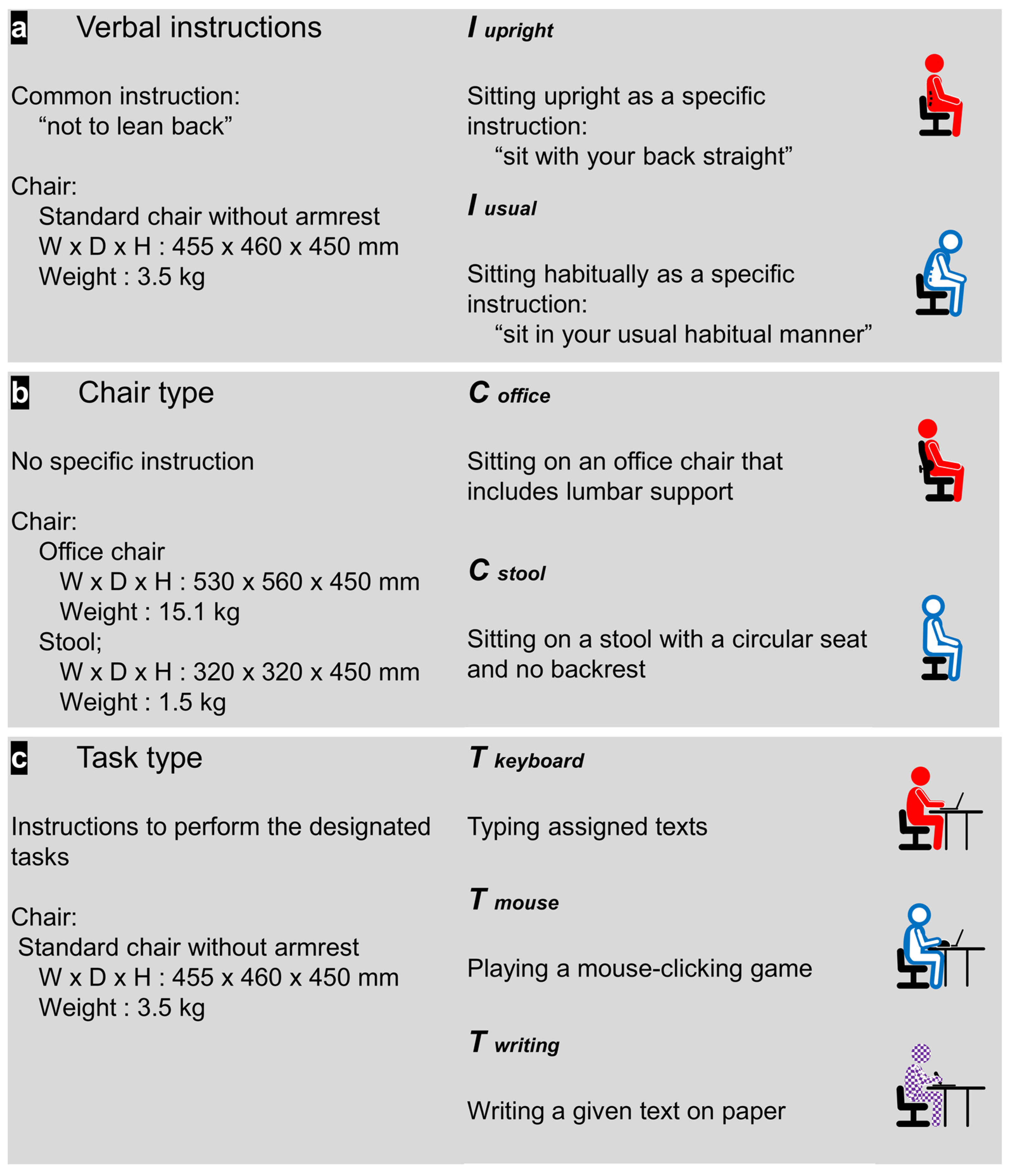
With the sensors attached to the bony landmarks, participants were asked to undergo seven test conditions, categorized into three aspects: verbal instructions, chair type, and type of desk tasks (Figure 2). We primarily aimed to include postures frequently used in daily life within each category, specifically selecting those where the magnitude of LLA is clearly distinct, to investigate whether maintenance of LLA shows similar trends across these postures.
For the verbal instructions, we chose to compare postures following good posture instructions to those in their usual, habitual state, selecting common directives likely to be encountered in daily life. Specifically, the static sitting postures were performed with the common directive of not leaning back, following two different instructions: to ‘sit with your back straight’ (Iupright) or to ‘sit in your usual habitual manner’ (Iusual). The chair type category included two static sitting postures using different chairs: an office chair (Coffice) or a stool (Cstool), both frequently used in daily settings and selected for their potential to show the greatest differences in the magnitude of LLA [31]. Due to limited research on task type, we incorporated a writing task in addition to the tasks utilized from past studies [35]. Desk tasks were performed, including keyboard typing, during which participants typed assigned texts (Tkeyboard); computer mouse manipulation, in which participants played a mouse-clicking game (Tmouse); and writing on paper, in which participants transcribed a given text (Twriting). All sitting conditions, except for the chair type category, were performed on a standard chair.
All sitting conditions, except for the chair type category, were performed on a standard chair. The standard chair used was 45 cm in height without armrests. The office chair included lumbar support and was height-adjustable, set to match the height of a standard chair. The stool was 45 cm high with a circular seat and lacked a backrest.

2.6. Outcome Variables

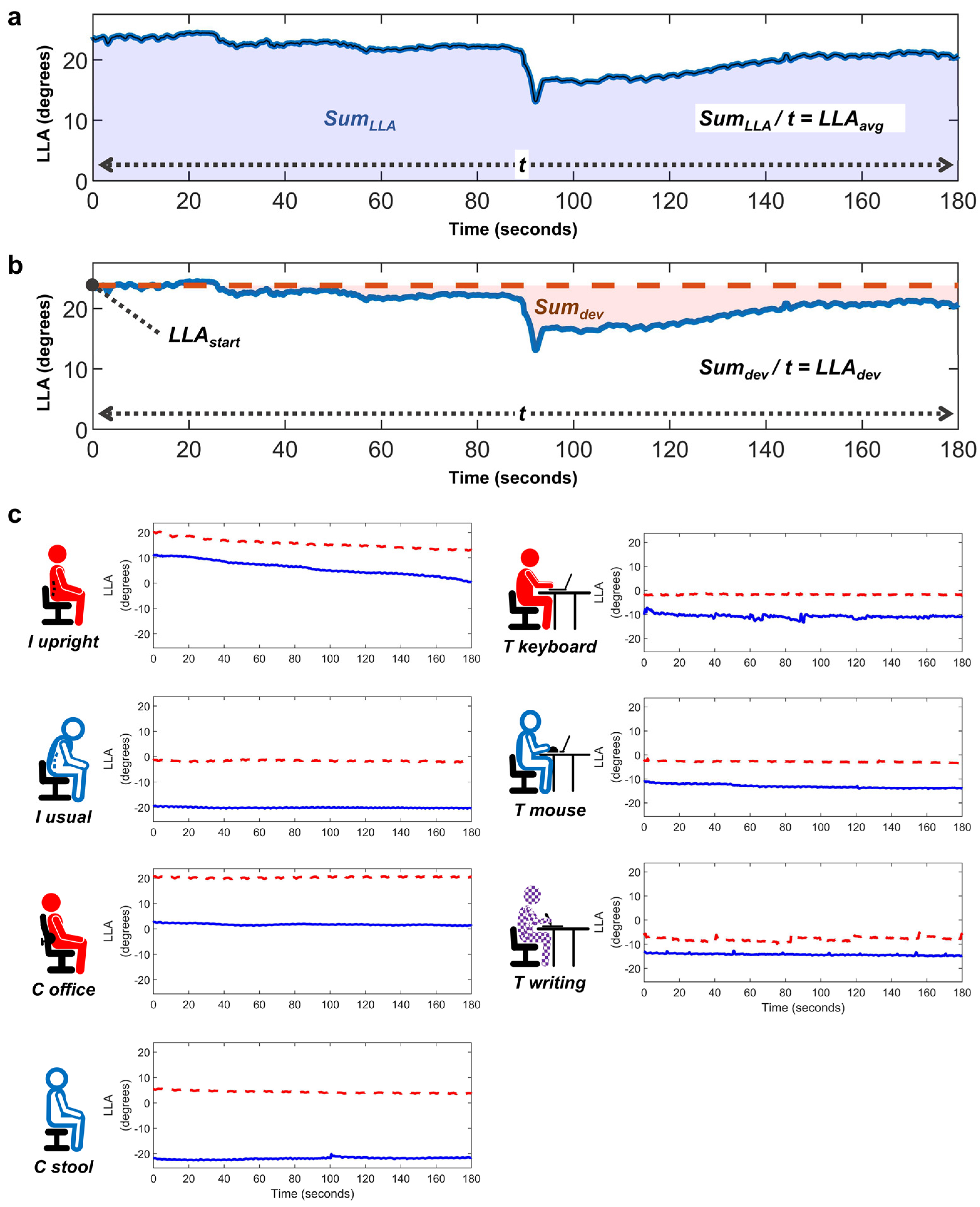
To assess the magnitude and preservation of LLA, data were tracked for 3 min and displayed as graphs (Figure 3). The primary outcomes included two key variables derived from LLA: LLAavg, representing the overall magnitude of LLA, and LLAdev, representing the preservation of LLA. LLAavg was computed by dividing SumLLA by the test duration, where SumLLA represents a time-weighted integration of LLA obtained using the trapezoidal rule (Figure 3a). LLAdev was defined as the deviation from LLAstart, the initial LLA value at the start of the test. It was calculated by integrating the difference between each time point’s LLA value and LLAstart over the total time and then dividing this sum of deviations (Sumdev) by the test duration. LLAdev reflects the extent of change in LLA throughout the test duration (Figure 3b). Since most tests showed a decreasing tendency over time, a larger LLAdev indicates good maintenance of LLC, while a smaller value suggests a greater shift towards a kyphotic position.

2.7. Statistical Analysis

The primary analysis involved comparing the primary outcomes (LLAavg and LLAdev) within three sitting categories. Based on the results of the Shapiro–Wilk normality tests, parametric methods were applied for LLAavg, and non-parametric methods were used for LLAdev. Each of the two static sitting postures, with different instructions and those on different chairs, were compared using either paired t-tests or Wilcoxon signed-rank tests. Repeated Measures ANOVA (RM-ANOVA) or Generalized Estimating Equations (GEEs) were used to compare each of the three desk tasks, with post hoc analysis performed using Bonferroni correction.
As a secondary analysis, we aimed to determine if these changes varied by sex, necessitating the use of statistical methods appropriate for repeated measures within two groups. Accordingly, RM-ANOVA or GEEs were used for between-group comparisons and interaction effect analysis for all sitting categories. Within each sex group, comparisons across sitting conditions were made using paired t-tests, Wilcoxon signed rank tests, RM-ANOVA, or Friedman tests, as appropriate. Data are presented as mean ± standard deviation. Statistical analyses were performed using statistical software (SPSS Version 19.0, SPSS Inc., Chicago, IL, USA).

3. Results

3.1. Comparison within Verbal Instruction Category

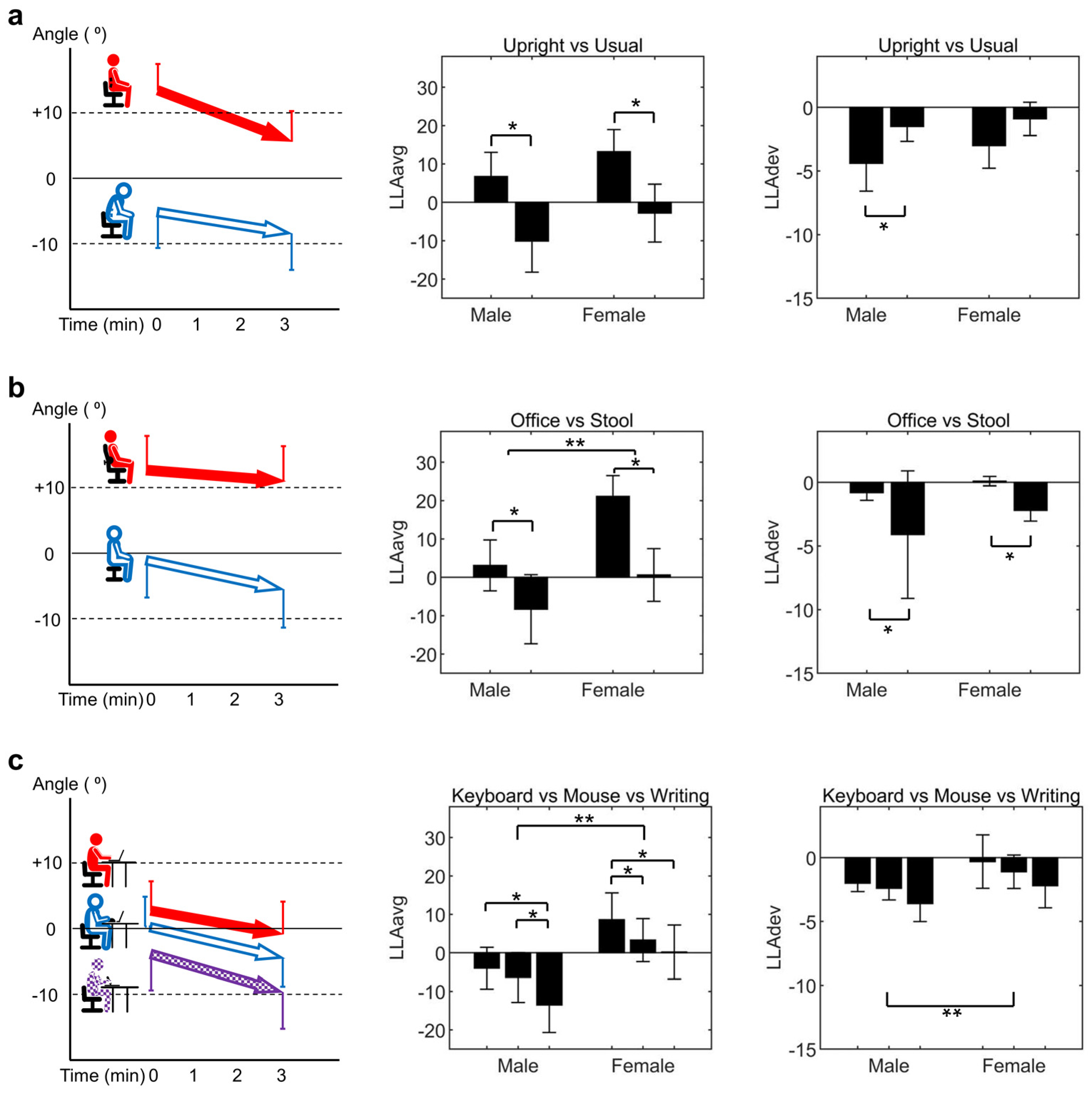
Figure 4a illustrates the comparison between upright sitting (Iupright) and habitual sitting (Iusual). The LLAavg of Iupright (9.9 ± 12.0°) was significantly larger compared to that of Iusual (−6.6 ± 15.4°) (p < 0.001, Cohen’s d = 1.6, power = 1.00). The LLAdev of Iupright (−3.7 ± 3.9°) was significantly smaller compared to that of Iusual (−1.2 ± 2.4°) (p = 0.001, r = 0.59, power = 0.85), indicating poorer preservation of LLA in the Iupright compared to Iusual.

3.2. Comparison within Chair Type Category

Figure 4b displays the comparison between sitting in an office chair (Coffice) and on a stool (Cstool). The LLAavg of Coffice (11.8 ± 14.8°) was significantly larger than that of Cstool (−4.0 ± 16.0°) (p < 0.001, Cohen’s d = 1.1, power = 0.99). The LLAdev of Coffice (−0.4 ± 1.1°) was significantly larger than that of Cstool (−3.2 ± 7.1°) (p < 0.001, r = 0.44, power = 0.73).

3.3. Comparison within Task Type Category

Figure 4c shows the comparison between the three desk tasks. There were significant differences among the three tasks on the desk in LLAavg (p < 0.001, η p 2 = 0.388, power = 1.00). Post hoc analysis revealed significant differences for all three comparisons, with Tkeyboard (2.0 ± 13.4°) being larger than Tmouse (−1.7 ± 12.6°) (p = 0.009), Tkeyboard being larger than Twriting (−6.9 ± 15.3°) (p < 0.001), and Tmouse being larger than Twriting (p = 0.014). The LLAdev trends for the three desk tasks were similar to those observed for LLAavg, but the differences were not statistically significant (p = 0.067). Specifically, LLAdev was −1.2 ± 3.0° for Tkeyboard, −1.8 ± 2.2° for Tmouse, and −2.9 ± 3.1° for Twriting.

3.4. Sex-Related Differences in LLA across Sitting Conditions

Table 2 presents detailed data on LLA variables, while Table 3 summarizes the results from the comparative analysis conducted using RM-ANOVA or GEE. In the verbal instruction category, no sex-related differences or interaction effects were found for either LLAavg or LLAdev. Within each sex group, Iupright showed a significantly larger LLAavg compared to Iusual in both groups (p < 0.001, Cohen’s d = 1.4, power = 1.00 for male, and p < 0.001, Cohen’s d = 2.0, power = 1.00 for female). The trend of smaller LLAdev in Iupright was consistent across sexes but was significant only in the male group (p = 0.009, r = 0.78, power = 0.87).
In the chair type category, females exhibited a significantly larger LLAavg than males (p = 0.006, η p 2 = 0.251, power = 0.83), with no interaction effects observed. Meanwhile, no sex-related differences or interaction effects were found in LLAdev. The LLAavg of Coffice was significantly larger than that of Cstool in both sexes (p = 0.013, Cohen’s d = 0.73, power = 0.85 for male and p < 0.001 Cohen’s d= 1.72, power = 1.00 for female), and the LLAdev of Coffice was also significantly larger than that of Cstool (p = 0.05, r = 0.35, power = 0.35 for male and p = 0.001, r = 1.66, power = 1.00 for female).
In the task type category, female participants had a significantly larger LLAavg and LLAdev than male participants (p = 0.010, η p 2 = 0.221, power = 0.76 for LLAavg and p = 0.032, Wald χ2 = 4.6, power = 0.52 for LLAdev), with no interaction effects observed for either variable. The difference in LLAavg across the three desk tasks was consistent within each sex group, though statistical significance was only confirmed in some comparisons.

4. Discussion

Utilizing the inertial measurement system, this study aimed to investigate the differences in the magnitude and preservation of LLC within three sitting categories: verbal instructions, chair type, and task type. The analysis revealed that the instructed upright sitting posture resulted in a greater magnitude of LLC compared to that of the usual sitting posture, yet the preservation of LLC was better with the usual sitting posture than that of the upright sitting posture. Regarding the chair type, using an office chair, compared to a stool, contributed to a larger and better maintained LLC. In the task type category, the magnitude of LLC was significantly larger in the order of keyboard use, mouse manipulation, and writing tasks, with a similar pattern noted for the preservation.
The study’s results show that LLA changes with different instructions, as participants displayed a significantly larger LLA when instructed to sit upright compared to their usual sitting posture. This result is similar to those of previous studies, which showed less flexed positions with the instruction to sit upright [22,26,28]. Particularly, O‘Sullivan et al.’s study, which used a strain gauge-based device, compared habitual and upright sitting [23]. They found that the habitual sitting posture had about a 20% more flexed range of motion compared to upright sitting. It was reasonably expected that a larger LLA would be observed when sitting upright as opposed to habitual sitting. But interestingly, our results suggest that a single instruction to sit upright may not be sufficient to maintain LLA for an extended period, as the decreasing tendency of LLA was significantly larger. While the LLAavg was significantly different over the 3 min duration used in this study, the difference between the two postures might not have been as distinct if the research had been conducted over a much longer duration. These findings imply that maintaining LLA in daily life may require repetitive feedback rather than a one-time instruction, and they support the potential need for biofeedback devices to aid in preserving LLC [52,53].
Our results, upon comparing the sitting postures involving different chairs, demonstrated that the office chair led to a larger LLA than the stool, a finding consistent with previous research [26,28,32,33]. Particularly, similar outcomes were observed in a radiographic study of 30 healthy males across five sitting positions, which showed the largest LLA when sitting on a chair with back support (36.2 ± 8.4°) and the smallest when sitting on a stool (0.6 ± 3.6°) [31]. A study by Alamin et al., involving 20 asymptomatic subjects, also found significantly smaller LLA when sitting on a stool (16.6 ± 15.6°) compared to when seated on a hard-back chair (28.6 ± 14.3°), similar to our study’s standard chair [33]. One notable aspect of the LLA pattern in the office chair was that it resulted in the largest LLAdev among all the tests, suggesting that it may be the most advantageous during prolonged sitting.
While several studies have linked prolonged sitting among office workers to LBP [19,21,54], there have been very few that have compared LLA with different types of desk work. Our study revealed that keyboard typing resulted in a larger LLA compared to mouse tasks, a finding consistent with previous research [35]. Additionally, our results showed that both computer tasks, typing and using a mouse, exhibited larger LLA than the writing task. This difference in LLA between tasks can be attributed to the nature of the tasks themselves. In computer tasks, the gaze direction typically involves looking at a monitor, which may encourage a more upright posture, while in writing tasks, the gaze direction is generally directed downwards towards the desk, potentially encouraging more flexion. Another key finding of our study is that there was similar tendency in LLAdev among the three tasks, with computer work better maintaining LLA compared to writing. This finding is consistent with previous research that suggested postural changes were more prevalent during reading compared to computer work [34].
Numerous previous studies, along with a recent systematic review, have established an association between LLA and sex [55,56,57,58], demonstrating that LLA tends to be larger in females. In our study, a similar tendency was observed across all sitting conditions, with significant differences particularly noted in the chair and task type categories. Notably, some research has indicated that the tendency for a larger LLA in females is posture-dependent, being significant while sitting but not standing [59]. Furthermore, a study focusing on sex-related differences in spine curvature during prolonged sitting on various chairs observed that males exhibited a more flexed lumbar angle than females [35]. However, our research did not find any interaction effects, indicating that being female did not result in disproportionately larger differences across instructions, chair types, or tasks. In terms of preserving LLA, females showed a tendency to better maintain LLA, significantly when performing different type of tasks. This may suggest that the sex-related differences in LLA during sitting are not solely due to skeletal structure differences.
In our study, the preservation of LLA varied across different sitting conditions, with significant differences observed in some categories. This variation in LLA maintenance could primarily be attributed to the inherent characteristics of each sitting condition. However, it may also be influenced by biomechanical factors such as paraspinal muscle activity, which includes considerations of muscle fatigue or the unconscious effort to maintain posture with minimal energy expenditure. Paraspinal muscles, including the erector spinae and multifidus, primarily function as lumbar extensors and play a crucial role in spinal stabilization and motion [60]. Notably, a reduction in the multifidus muscle mass is known to be associated with LBP [61]. O‘Sullivan et al. have demonstrated increased muscle activity during upright sitting compared to slump sitting, which may relate to our findings of insufficient maintenance in upright postures [62,63]. The improved maintenance observed in an office chair in our study could be related to findings from Makhsous et al. indicating that paraspinal activity decreases with the application of lumbar support [27,64]. However, regarding different task types, previous studies have shown lower paraspinal activity during reading compared to computer work, contradicting our hypothesis [34,65,66]. This suggests that the nature of the task itself has a greater influence, indicating that the preservation of LLA results from the complex interplay of various factors. Future research should involve between-subject comparisons to identify biomechanical parameters correlated with LLA maintenance, including paraspinal muscle activity.
Maintaining LLC may be a crucial biomechanical correction to LBP. Several studies have confirmed that modifying sitting posture can directly reduce LBP [67,68,69]. Research, such as the work by Williams et al., highlights that a lordotic posture, when compared to a kyphotic one, can significantly mitigate pain intensity in individuals experiencing LBP [67]. Similarly, studies by Aota et al. and Pillastrini et al. have demonstrated that lumbar supports and ergonomic interventions that help preserve lumbar lordosis significantly reduce the incidence and severity of LBP [68,69]. These studies suggest that strategies to maintain LLC during sitting postures may help prevent LBP. However, they did not directly assess LLA. Therefore, understanding how changes in pain directly relate to variations in LLA, i.e., the relationship between LLC and LBP, has been difficult. Further research utilizing our study methods to directly investigate how specific changes in LLA can prevent LBP is necessary.
Our study has some limitations. First, the short test duration of 3 min is considerably brief compared to that of typical daily sitting activities. Although we incorporated everyday activities into our research for comparison, this short duration makes it challenging to fully generalize our results to actual daily activities. The experimental design, which includes serial conduction of multiple conditions without randomization and the brief resting periods used in our experiment, also make it difficult to confidently apply these findings outside the laboratory setting. These design choices were made to incorporate a variety of test conditions. Future research should consider including fewer test conditions for longer testing durations or multiple trials, sufficient resting periods, and randomization of test condition order. Second, given the cross-sectional nature of our study, it is challenging to establish a direct causal relationship between sitting conditions and LLA changes. Additionally, since the study involved only healthy individuals and did not measure LBP as an outcome, it cannot conclusively prove the preventive effects on LBP. Future research should undertake longitudinal studies or clinical trials to determine whether these strategies can effectively preserve LLC and prevent LBP in real-world applications.

5. Conclusions

Consistent patterns and significant differences in magnitude and preservation of lumbar lordotic curvature (LLC) were observed across various everyday sitting conditions among healthy participants using an AHRS. Specifically, within different instructions, the magnitude of LLC was larger in upright sitting, whereas preservation was better in habitual sitting. Comparing chair types, the use of an office chair showed a larger and better maintained LLC compared to using a stool. Within the different task types, the magnitude of LLC was significantly larger in computer tasks than in writing tasks, with a similar trend observed for preservation. These findings suggest that utilizing an office chair and favoring computer work may contribute to the preservation of LLC; however, cueing for upright sitting may require repetition to effectively maintain LLC. Given the association between LLC and low back pain (LBP), these practices could potentially serve as preventive measures against LBP. To further refine our understanding and practical applications of these findings, future research should focus on longitudinal studies or clinical trials, including participants with LBP, that examine the effects of sitting conditions over extended periods to validate the efficacy of interventions aimed at preserving LLC and preventing LBP. Additionally, there is a need for research into the biomechanical factors that influence the preservation of LLC.

Author Contributions

Conceptualization, J.C.K., J.-G.K., B.S.K., C.K.K., M.C., J.L. and S.G.C.; Investigation, J.C.K., J.-G.K., B.S.K., C.K.K. and M.C.; Methodology, J.-G.K., M.C. and J.L.; Software, J.L.; Writing—original draft, J.C.K. and J.-G.K.; Writing—review and editing, J.C.K.; Writing—critical review and editing, supervision, S.G.C. All authors have read and agreed to the published version of the manuscript.

Funding

This research was funded by the Bio and Medical Technology Development Programs of the National Research Foundation (NRF), funded by the Ministry of Science and ICT, Republic of Korea, grant number 2016M3A9F1941984, and by a grant from the Patient-Centered Clinical Research Coordinating Center (PACEN), funded by the Ministry of Health & Welfare, Republic of Korea, grant number HC21C0064.

Institutional Review Board Statement

This study was performed in accordance with the Declaration of Helsinki and was approved by the Institutional Review Board of Seoul National University Hospital (IRB No. 1703-174-842).

Informed Consent Statement

Informed consent was obtained from all subjects involved in the study. Written informed consent was also obtained from the student researcher featured in Figure 2 for the publication of images in an online open access publication.

Data Availability Statement

The data sets for this study are available on request. The raw data supporting the conclusions of this manuscript will be made available by the authors to any qualified researcher. Contact: Ju Chan Kim (ORCID 0000-0003-4015-6096).

Conflicts of Interest

Author Joonnyong Lee was employed by the company Mellowing Factory Co., Ltd. The remaining authors declare that the research was conducted in the absence of any commercial or financial relationships that could be construed as a potential conflict of interest.

References

  1. Williams, S.A.; Russo, G.A. Evolution of the hominoid vertebral column: The long and the short of it. Evol. Anthropol. 2015, 24, 15–32. [Google Scholar] [CrossRef] [PubMed]
  2. Farfan, H.F. The biomechanical advantage of lordosis and hip extension for upright activity. Man as compared with other anthropoids. Spine 1978, 3, 336–342. [Google Scholar] [CrossRef] [PubMed]
  3. Aspden, R. The Spine as an Arch A New Mathematical Model. Spine 1989, 14, 266–274. [Google Scholar] [CrossRef]
  4. Edmondston, S.J.; Song, S.; Bricknell, R.V.; Davies, P.A.; Fersum, K.; Humphries, P.; Wickenden, D.; Singer, K.P. MRI evaluation of lumbar spine flexion and extension in asymptomatic individuals. Man. Ther. 2000, 5, 158–164. [Google Scholar] [CrossRef]
  5. Adams, M.A.; Hutton, W.C. The effect of posture on the role of the apophysial joints in resisting intervertebral compressive forces. J. Bone Jt. Surg. Br. Vol. 1980, 62, 358–362. [Google Scholar] [CrossRef] [PubMed]
  6. Adams, M.A.; Mannion, A.F.; Dolan, P. Personal risk factors for first-time low back pain. Spine 1999, 24, 2497–2505. [Google Scholar] [CrossRef]
  7. Chun, S.W.; Lim, C.Y.; Kim, K.; Hwang, J.; Chung, S.G. The relationships between low back pain and lumbar lordosis: A systematic review and meta-analysis. Spine J. Off. J. N. Am. Spine Soc. 2017, 17, 1180–1191. [Google Scholar] [CrossRef]
  8. Adams, M.A.; Dolan, P. Time-dependent changes in the lumbar spine’s resistance to bending. Clin. Biomech. 1996, 11, 194–200. [Google Scholar] [CrossRef]
  9. McGill, S.M.; Brown, S. Creep response of the lumbar spine to prolonged full flexion. Clin. Biomech. 1992, 7, 43–46. [Google Scholar] [CrossRef]
  10. Solomonow, M.; Zhou, B.H.; Baratta, R.V.; Burger, E. Biomechanics and electromyography of a cumulative lumbar disorder: Response to static flexion. Clin. Biomech. 2003, 18, 890–898. [Google Scholar] [CrossRef]
  11. Twomey, L.; Taylor, J. Flexion creep deformation and hysteresis in the lumbar vertebral column. Spine 1982, 7, 116–122. [Google Scholar] [CrossRef] [PubMed]
  12. Andersson, B.J.; Ortengren, R.; Nachemson, A.; Elfstrom, G. Lumbar disc pressure and myoelectric back muscle activity during sitting. IV. Studies on a car driver’s seat. Scand. J. Rehabil. Med. 1974, 6, 128–133. [Google Scholar] [CrossRef] [PubMed]
  13. Wade, K.R.; Robertson, P.A.; Thambyah, A.; Broom, N.D. How healthy discs herniate: A biomechanical and microstructural study investigating the combined effects of compression rate and flexion. Spine 2014, 39, 1018–1028. [Google Scholar] [CrossRef] [PubMed]
  14. Shah, J.S.; Hampson, W.G.; Jayson, M.I. The distribution of surface strain in the cadaveric lumbar spine. J. Bone Jt. Surg. Br. Vol. 1978, 60, 246–251. [Google Scholar] [CrossRef] [PubMed]
  15. Andersson, G.B.; Schultz, A.B. Effects of fluid injection on mechanical properties of intervertebral discs. J. Biomech. 1979, 12, 453–458. [Google Scholar] [CrossRef]
  16. Sato, K.; Kikuchi, S.; Yonezawa, T. In vivo intradiscal pressure measurement in healthy individuals and in patients with ongoing back problems. Spine 1999, 24, 2468–2474. [Google Scholar] [CrossRef] [PubMed]
  17. Wilke, H.J.; Neef, P.; Caimi, M.; Hoogland, T.; Claes, L.E. New in vivo measurements of pressures in the intervertebral disc in daily life. Spine 1999, 24, 755–762. [Google Scholar] [CrossRef]
  18. Cholewicki, J.; Pathak, P.; Reeves, N.P.; Popovich, J.M. Simulation of multifactorial causes of low back pain. Spine J. 2016, 16, S277. [Google Scholar] [CrossRef]
  19. Omokhodion, F.O.; Sanya, A.O. Risk factors for low back pain among office workers in Ibadan, Southwest Nigeria. Occup. Med. 2003, 53, 287–289. [Google Scholar] [CrossRef]
  20. Toh, S.H.; Coenen, P.; Howie, E.K.; Straker, L.M. The associations of mobile touch screen device use with musculoskeletal symptoms and exposures: A systematic review. PLoS ONE 2017, 12, e0181220. [Google Scholar] [CrossRef]
  21. Lis, A.M.; Black, K.M.; Korn, H.; Nordin, M. Association between sitting and occupational LBP. Eur. Spine J. Off. Publ. Eur. Spine Soc. Eur. Spinal Deform. Soc. Eur. Sect. Cerv. Spine Res. Soc. 2007, 16, 283–298. [Google Scholar] [CrossRef]
  22. Black, K.M.; McClure, P.; Polansky, M. The influence of different sitting positions on cervical and lumbar posture. Spine 1996, 21, 65–70. [Google Scholar] [CrossRef]
  23. O’Sullivan, K.; O’Dea, P.; Dankaerts, W.; O’Sullivan, P.; Clifford, A.; O’Sullivan, L. Neutral lumbar spine sitting posture in pain-free subjects. Man. Ther. 2010, 15, 557–561. [Google Scholar] [CrossRef] [PubMed]
  24. Czaprowski, D.; Pawlowska, P.; Kolwicz-Ganko, A.; Sitarski, D.; Kedra, A. The Influence of the “Straighten Your Back” Command on the Sagittal Spinal Curvatures in Children with Generalized Joint Hypermobility. BioMed Res. Int. 2017, 2017, 9724021. [Google Scholar] [CrossRef]
  25. Paloschi, D.; Bravi, M.; Schena, E.; Miccinilli, S.; Morrone, M.; Sterzi, S.; Saccomandi, P.; Massaroni, C. Validation and Assessment of a Posture Measurement System with Magneto-Inertial Measurement Units. Sensors 2021, 21, 6610. [Google Scholar] [CrossRef] [PubMed]
  26. Lin, F.; Parthasarathy, S.; Taylor, S.J.; Pucci, D.; Hendrix, R.W.; Makhsous, M. Effect of Different Sitting Postures on Lung Capacity, Expiratory Flow, and Lumbar Lordosis. Arch. Phys. Med. Rehabil. 2006, 87, 504–509. [Google Scholar] [CrossRef]
  27. Makhsous, M.; Lin, F.; Hendrix, R.W.; Hepler, M.; Zhang, L.Q. Sitting with adjustable ischial and back supports: Biomechanical changes. Spine 2003, 28, 1113–1121; discussion 1121–1122. [Google Scholar] [CrossRef]
  28. Wong, A.Y.L.; Chan, T.P.M.; Chau, A.W.M.; Tung Cheung, H.; Kwan, K.C.K.; Lam, A.K.H.; Wong, P.Y.C.; De Carvalho, D. Do different sitting postures affect spinal biomechanics of asymptomatic individuals? Gait Posture 2019, 67, 230–235. [Google Scholar] [CrossRef]
  29. Baumgartner, D.; Zemp, R.; List, R.; Stoop, M.; Naxera, J.; Elsig, J.P.; Lorenzetti, S. The spinal curvature of three different sitting positions analysed in an open MRI scanner. Sci. World J. 2012, 2012, 184016. [Google Scholar] [CrossRef] [PubMed]
  30. Bae, J.S.; Jang, J.S.; Lee, S.H.; Kim, J.U. A comparison study on the change in lumbar lordosis when standing, sitting on a chair, and sitting on the floor in normal individuals. J. Korean Neurosurg. Soc. 2012, 51, 20–23. [Google Scholar] [CrossRef]
  31. Cho, I.Y.; Park, S.Y.; Park, J.H.; Kim, T.K.; Jung, T.W.; Lee, H.M. The Effect of Standing and Different Sitting Positions on Lumbar Lordosis: Radiographic Study of 30 Healthy Volunteers. Asian Spine J. 2015, 9, 762–769. [Google Scholar] [CrossRef] [PubMed]
  32. De Carvalho, D.; Grondin, D.; Callaghan, J. The impact of office chair features on lumbar lordosis, intervertebral joint and sacral tilt angles: A radiographic assessment. Ergonomics 2017, 60, 1393–1404. [Google Scholar] [CrossRef]
  33. Alamin, T.F.; Agarwal, V.; Zagel, A.; Qeli, A. The effect of standing vs. variants of the seated position on lumbar intersegmental angulation and spacing: A radiographic study of 20 asymptomatic subjects. J. Spine Surg. 2018, 4, 509–515. [Google Scholar] [CrossRef] [PubMed]
  34. Van Dieën, J.; De Looze, M.; Hermans, V. Effects of dynamic office chairs on trunk kinematics, trunk extensor EMG and spinal shrinkage. Ergonomics 2001, 44, 739–750. [Google Scholar] [CrossRef]
  35. Dunk, N.M.; Callaghan, J.P. Gender-based differences in postural responses to seated exposures. Clin. Biomech. 2005, 20, 1101–1110. [Google Scholar] [CrossRef] [PubMed]
  36. Harrison, D.E.; Harrison, D.D.; Cailliet, R.; Janik, T.J.; Holland, B. Radiographic analysis of lumbar lordosis: Centroid, Cobb, TRALL, and Harrison posterior tangent methods. Spine 2001, 26, E235–E242. [Google Scholar] [CrossRef]
  37. Troyanovich, S.J.; Cailliet, R.; Janik, T.J.; Harrison, D.D.; Harrison, D.E. Radiographic mensuration characteristics of the sagittal lumbar spine from a normal population with a method to synthesize prior studies of lordosis. J. Spinal Disord. 1997, 10, 380–386. [Google Scholar] [CrossRef] [PubMed]
  38. Lord, M.J.; Small, J.M.; Dinsay, J.M.; Watkins, R.G. Lumbar lordosis. Effects of sitting and standing. Spine 1997, 22, 2571–2574. [Google Scholar] [CrossRef]
  39. De Carvalho, D.E.; Soave, D.; Ross, K.; Callaghan, J.P. Lumbar spine and pelvic posture between standing and sitting: A radiologic investigation including reliability and repeatability of the lumbar lordosis measure. J. Manip. Physiol. Ther. 2010, 33, 48–55. [Google Scholar] [CrossRef]
  40. Wong, W.Y.; Wong, M.S. Detecting spinal posture change in sitting positions with tri-axial accelerometers. Gait Posture 2008, 27, 168–171. [Google Scholar] [CrossRef]
  41. Chhikara, A.; McGregor, A.H.; Hadjilucas, L.; Bello, F.; Rice, A.S. Quantitative Assessment of the Motion of the Lumbar Spine and Pelvis with Wearable Inertial Sensors. In Proceedings of the 2010 International Conference on Body Sensor Networks, Singapore, 7–9 June 2010; pp. 9–15. [Google Scholar]
  42. Stenlund, T.C.; Öhberg, F.; Lundström, R.; Lindroos, O.; Häger, C.; Rehn, B. Inter- and intra-tester reliability when measuring seated spinal postures with inertial sensors. Int. J. Ind. Ergon. 2014, 44, 732–738. [Google Scholar] [CrossRef]
  43. Yoon, C.; Lee, J.; Kim, K.; Kim, H.C.; Chung, S.G. Quantification of Lumbar Stability During Wall Plank-and-Roll Activity Using Inertial Sensors. PM R J. Inj. Funct. Rehabil. 2015, 7, 803–813. [Google Scholar] [CrossRef] [PubMed]
  44. Dunk, N.M.; Callaghan, J.P. Lumbar spine movement patterns during prolonged sitting differentiate low back pain developers from matched asymptomatic controls. Work 2010, 35, 3–14. [Google Scholar] [CrossRef] [PubMed]
  45. Lee, J.K.; Desmoulin, G.T.; Khan, A.H.; Park, E.J. Comparison of 3D spinal motions during stair-climbing between individuals with and without low back pain. Gait Posture 2011, 34, 222–226. [Google Scholar] [CrossRef] [PubMed]
  46. Lee, J.; Yoon, C.; Kim, K.; Cho, M.; Kim, H.C.; Chung, S.G. Lumbar Stability in Healthy Individuals and Low Back Pain Patients Quantified by Wall Plank-and-Roll Test. PM R J. Inj. Funct. Rehabil. 2018, 11, 483–494. [Google Scholar] [CrossRef] [PubMed]
  47. Paulich, M.; Schepers, M.; Rudigkeit, N.; Bellusci, G. Xsens MTw Awinda: Miniature wireless inertial-magnetic motion tracker for highly accurate 3D kinematic applications. Xsens Enschede Neth. 2018, 1, 1–9. [Google Scholar]
  48. von Elm, E.; Altman, D.G.; Egger, M.; Pocock, S.J.; Gotzsche, P.C.; Vandenbroucke, J.P.; Initiative, S. The Strengthening the Reporting of Observational Studies in Epidemiology (STROBE) Statement: Guidelines for reporting observational studies. Int. J. Surg. 2014, 12, 1495–1499. [Google Scholar] [CrossRef] [PubMed]
  49. Chakraverty, R.; Pynsent, P.; Isaacs, K. Which spinal levels are identified by palpation of the iliac crests and the posterior superior iliac spines? J. Anat. 2007, 210, 232–236. [Google Scholar] [CrossRef] [PubMed]
  50. Milani, G.B.; Natal Filho, A.; Amado Joao, S.M. Correlation between lumbar lordosis angle and degree of gynoid lipodystrophy (cellulite) in asymptomatic women. Clinics 2008, 63, 503–508. [Google Scholar] [CrossRef]
  51. Dankaerts, W.; O’Sullivan, P.; Burnett, A.; Straker, L. Differences in sitting postures are associated with nonspecific chronic low back pain disorders when patients are subclassified. Spine 2006, 31, 698–704. [Google Scholar] [CrossRef]
  52. O’Sullivan, K.; O’Sullivan, L.; O’Sullivan, P.; Dankaerts, W. Investigating the effect of real-time spinal postural biofeedback on seated discomfort in people with non-specific chronic low back pain. Ergonomics 2013, 56, 1315–1325. [Google Scholar] [CrossRef] [PubMed]
  53. Van Almkerk, M.; Bierling, B.L.; Leermakers, N.; Vinken, J.; Timmermans, A.A. Improving posture and sitting behavior through tactile and visual feedback in a sedentary environment. In Proceedings of the 2015 37th Annual International Conference of the IEEE Engineering in Medicine and Biology Society (EMBC), Milan, Italy, 25–29 August 2015; pp. 4570–4573. [Google Scholar]
  54. Rezaee, M.; Ghasemi, M.; Jafari, N.J.; Izadi, M. Low back pain and related factors among Iranian office workers. Int. J. Occup. Hyg. 2011, 3, 23–28. [Google Scholar]
  55. Arshad, R.; Pan, F.; Reitmaier, S.; Schmidt, H. Effect of age and sex on lumbar lordosis and the range of motion. A systematic review and meta-analysis. J. Biomech. 2019, 82, 1–19. [Google Scholar] [CrossRef] [PubMed]
  56. Vialle, R.; Levassor, N.; Rillardon, L.; Templier, A.; Skalli, W.; Guigui, P. Radiographic analysis of the sagittal alignment and balance of the spine in asymptomatic subjects. J. Bone Jt. Surg. Am. 2005, 87, 260–267. [Google Scholar] [CrossRef] [PubMed]
  57. Zhu, Z.; Xu, L.; Zhu, F.; Jiang, L.; Wang, Z.; Liu, Z.; Qian, B.P.; Qiu, Y. Sagittal alignment of spine and pelvis in asymptomatic adults: Norms in Chinese populations. Spine 2014, 39, E1–E6. [Google Scholar] [CrossRef] [PubMed]
  58. Damasceno, L.H.F.; Catarin, S.R.G.; Campos, A.D.; Defino, H.L.A. Lumbar lordosis: A study of angle values and of vertebral bodies and intervertebral discs role. Acta Ortopédica Bras. 2006, 14, 193–198. [Google Scholar] [CrossRef]
  59. Endo, K.; Suzuki, H.; Nishimura, H.; Tanaka, H.; Shishido, T.; Yamamoto, K. Sagittal lumbar and pelvic alignment in the standing and sitting positions. J. Orthop. Sci. 2012, 17, 682–686. [Google Scholar] [CrossRef]
  60. MacDonald, D.A.; Moseley, G.L.; Hodges, P.W. The lumbar multifidus: Does the evidence support clinical beliefs? Man. Ther. 2006, 11, 254–263. [Google Scholar] [CrossRef]
  61. Ranger, T.A.; Cicuttini, F.M.; Jensen, T.S.; Peiris, W.L.; Hussain, S.M.; Fairley, J.; Urquhart, D.M. Are the size and composition of the paraspinal muscles associated with low back pain? A systematic review. Spine J. 2017, 17, 1729–1748. [Google Scholar] [CrossRef]
  62. O’Sullivan, P.B.; Dankaerts, W.; Burnett, A.F.; Farrell, G.T.; Jefford, E.; Naylor, C.S.; O’Sullivan, K.J. Effect of different upright sitting postures on spinal-pelvic curvature and trunk muscle activation in a pain-free population. Spine 2006, 31, E707–E712. [Google Scholar] [CrossRef]
  63. O’Sullivan, P.B.; Grahamslaw, K.M.; Kendell, M.; Lapenskie, S.C.; Möller, N.E.; Richards, K.V. The effect of different standing and sitting postures on trunk muscle activity in a pain-free population. Spine 2002, 27, 1238–1244. [Google Scholar] [CrossRef] [PubMed]
  64. Makhsous, M.; Lin, F.; Bankard, J.; Hendrix, R.W.; Hepler, M.; Press, J. Biomechanical effects of sitting with adjustable ischial and lumbar support on occupational low back pain: Evaluation of sitting load and back muscle activity. BMC Musculoskelet. Disord. 2009, 10, 17. [Google Scholar] [CrossRef] [PubMed]
  65. Andersson, B.G.; Örtengren, R.; Nachemson, A.L.; Elfström, G.; Broman, H. The sitting posture: An electromyographic and discometric study. Orthop. Clin. N. Am. 1975, 6, 105–120. [Google Scholar] [CrossRef]
  66. Gregory, D.E.; Dunk, N.M.; Callaghan, J.P. Stability ball versus office chair: Comparison of muscle activation and lumbar spine posture during prolonged sitting. Hum. Factors 2006, 48, 142–153. [Google Scholar] [CrossRef] [PubMed]
  67. Williams, M.M.; Hawley, J.A.; McKenzie, R.A.; van Wijmen, P.M. A comparison of the effects of two sitting postures on back and referred pain. Spine 1991, 16, 1185–1191. [Google Scholar] [CrossRef] [PubMed]
  68. Aota, Y.; Iizuka, H.; Ishige, Y.; Mochida, T.; Yoshihisa, T.; Uesugi, M.; Saito, T. Effectiveness of a lumbar support continuous passive motion device in the prevention of low back pain during prolonged sitting. Spine 2007, 32, E674–E677. [Google Scholar] [CrossRef]
  69. Pillastrini, P.; Mugnai, R.; Bertozzi, L.; Costi, S.; Curti, S.; Guccione, A.; Mattioli, S.; Violante, F.S. Effectiveness of an ergonomic intervention on work-related posture and low back pain in video display terminal operators: A 3 year cross-over trial. Appl. Erg. 2010, 41, 436–443. [Google Scholar] [CrossRef]
Figure 1. Experimental set-up during the test. (a,b) IMU sensors were attached to the skin over the 12th thoracic (T12) and 2nd sacral (S2) vertebral points. (c) The relative angle between T12 and S2 was calculated using a simple subtraction method, and the Y-axis (mediolateral axis) data were used to evaluate the kyphosis–lordosis of lumbar movement between the T12 and S2 IMU sensors. (d) LLA is traditionally defined by the Cobb angle measured from the lateral lumbar radiographic images, extending between the inferior endplate of T12 and the superior endplate of S1. The figure illustrates that the measurement derived from this definition is similar to those obtained from IMU sensors.
Jcm 13 02728 g001
Figure 2. Sitting conditions with the three categories evaluated in the present study. (a) Two sitting postures with different instructions, (b) two sitting postures on different chairs, and (c) three desk tasks. W: width, D: depth, H: seat height.
Jcm 13 02728 g002
Figure 3. Typical graphs of LLA variation over time. (a,b) Graphs depicting the LLA changes in a 54-year-old female participant (R007) in a sitting posture on a stool. These graphs illustrate the definition of LLAavg (a) and LLAdev (b). (c) Examples of typical LLA variation graphs in different sitting conditions from two participants: a 27-year-old female (R004, red dotted line) and a 49-year-old male (R012, blue solid line).
Jcm 13 02728 g003
Figure 4. Comparative results within three sitting categories: (a) verbal instruction, (b) chair type, and (c) task type. The first graph in each set depicts the overview of magnitude and temporal changes in LLA, with the start of the arrows denoting LLAstart, and the arrowheads indicating the minimal LLA observed during the test. The second and third bar graphs within each set compare LLAavg and LLAdev between sitting conditions within categories and by sex. Single asterisks indicate statistical significance within sex groups across sitting conditions. Double asterisks indicate statistical significance between sex groups within each sitting category. The whiskers represent the 95% confidence intervals and are depicted in a unidirectional manner for clear graphical representation in the first graphs.
Jcm 13 02728 g004
Table 1. Demographics and characteristics of participants.
All (n = 29)Male (n = 15)Female (n = 14)
Age, years35.5 ± 13.632.2 ± 12.639.1 ± 14.1
Body mass, kg64.0 ± 12.372.0 ± 11.955.4 ± 4.9
Height, cm167.1 ± 8.9173.8 ± 6.0159.8 ± 4.6
BMI, kg/m222.8 ± 3.123.8 ± 3.321.8 ± 2.5
Values are presented as mean ± standard deviation.
Table 2. Outcomes of LLA variables by sitting condition and sex.
Sitting
Category
Sitting
Posture
LLAavg (°)LLAdev (°)
All Male Female AllMaleFemale
Verbal
instruction
Iupright9.9 ± 12.06.7 ± 12.513.2 ± 11.0−3.7 ± 3.9−4.4 ± 4.3−3.0 ± 3.4
Iusual−6.6 ± 15.4−10.1 ± 16.0−2.8 ± 14.4−1.2 ± 2.4−1.5 ± 2.3−0.9 ± 2.5
Chair typeCoffice11.8 ± 14.83.1 ± 13.121.1 ± 10.3−0.4 ± 1.1−0.8 ± 1.20.1 ± 0.7
Cstool−4.0 ± 16.0−8.3 ± 17.80.6 ± 13.1−3.2 ± 7.1−4.1 ± 9.9−2.2 ± 1.6
Deks tasksTkeyboard2.0 ± 13.4−4.0 ± 10.78.6 ± 13.3−1.2 ± 3.0−2.0 ± 1.3−0.3 ± 4.0
Tmouse−1.7 ± 12.6−6.4 ± 12.83.3 ± 10.7−1.8 ± 2.2−2.4 ± 1.8−1.1 ± 2.5
Twriting−6.9 ± 15.3−13.5 ± 14.20.2 ± 13.4−2.9 ± 3.1−3.6 ± 2.8−2.2 ± 3.3
Values are presented as mean ± standard deviation angles (°).
Table 3. Comparisons within sitting categories considering sex-related differences.
Sitting
Category
LLAavg (°)LLAdev (°)
ItemFp-ValueItemWald χ2p-Value
Verbal
instruction
Instruction72.159<0.001 *Instruction11.1230.001 *
Sex2.1770.152Sex1.3280.249
Instruction × Sex0.0450.834Instruction × Sex0.2210.638
Chair typeChair38.166<0.001 *Chair4.5430.033 *
Sex9.0250.006 *Sex1.3630.243
Chair × Sex3.0800.091Chair × Sex0.1550.694
Deks tasksTask17.574<0.001 *Task5.4020.067
Sex7.6460.010 *Sex4.6110.032 *
Task × Sex0.9590.390Task × Sex0.1330.936
* Asterisks indicate statistical significance. (p-value < 0.05).
Disclaimer/Publisher’s Note: The statements, opinions and data contained in all publications are solely those of the individual author(s) and contributor(s) and not of MDPI and/or the editor(s). MDPI and/or the editor(s) disclaim responsibility for any injury to people or property resulting from any ideas, methods, instructions or products referred to in the content.