Review

MicroRNAs and Growth Factors: An Alliance Propelling Tumor Progression

Department of Biological Regulation, Weizmann Institute of Science, Rehovot 76100, Israel
*
Author to whom correspondence should be addressed.
Current Address: Genetic Institute, Tel Aviv Medical Centre, Tel Aviv 64239, Israel
J. Clin. Med. 2015, 4(8), 1578-1599; https://doi.org/10.3390/jcm4081578
Submission received: 2 July 2015 / Revised: 30 July 2015 / Accepted: 31 July 2015 / Published: 13 August 2015
(This article belongs to the Special Issue MicroRNAs: Novel Biomarkers and Therapeutic Targets for Human Cancers)

Abstract

:
Tumor progression requires cancer cell proliferation, migration, invasion, and attraction of blood and lymph vessels. These processes are tightly regulated by growth factors and their intracellular signaling pathways, which culminate in transcriptional programs. Hence, oncogenic mutations often capture growth factor signaling, and drugs able to intercept the underlying biochemical routes might retard cancer spread. Along with messenger RNAs, microRNAs play regulatory roles in growth factor signaling and in tumor progression. Because growth factors regulate abundance of certain microRNAs and the latter modulate the abundance of proteins necessary for growth factor signaling, the two classes of molecules form a dense web of interactions, which are dominated by a few recurring modules. We review specific examples of the alliance formed by growth factors and microRNAs and refer primarily to the epidermal growth factor (EGF) pathway. Clinical applications of the crosstalk between microRNAs and growth factors are described, including relevance to cancer therapy and to emergence of resistance to specific drugs.

Graphical Abstract

1. Introduction

Somatic mutations encompassing single base mutations, inter- and intrachromosomal rearrangements, as well as copy number changes are major initiators of the multistep process leading to malignancy. Germ line mutations, such as loss of tumor suppressor functions and the induction of oncogene functions facilitate somatic mutations [1,2], but the major driver of genetic aberrations is likely replication stress imposed by rapid divisions of stem cells and their immediate progenies [3,4]. The number of oncogenic (driver) mutations per common adult epithelial cancer is thought to exceed four aberrations [5], but fewer events are required in hematological cancers. On the way to become a metastatic tumor, the single initiated cancer cell must undergo rapid cell divisions, which fixate the oncogenic mutations, attract blood and lymph vessels that supply oxygen and nutrients, and invade the surrounding extracellular matrix and vessels, which permits dissemination and colonization in distant sites. This train of events is controlled by a plethora of tissue-specific growth factors [6]. For example, the 11 members of the epidermal growth factor (EGF) family act as both mitogens and motogens of epithelial cells, the precursors of carcinomas. The receptors for EGF family ligands and for other growth factors are typically transmembrane proteins sharing a tyrosine kinase catalytic function (called receptor tyrosine kinases, RTKs). Although growth factors are essential for progression of many solid tumors, accrual of specific oncogenic mutations might free cancer cells from their reliance on growth factors. This explains why a relatively large fraction of the genes undergoing recurrent somatic mutations in cancer affect protein kinases and other signaling proteins placed downstream of RTKs [7], such as B-RAF (in melanoma), RAS (in pancreatic cancer) ERBB2/HER2 (in breast cancer), and EGFR (in brain cancer). While the majority of tumors are characterized by enhanced secretion of growth factors (termed autocrine secretion [8]), driver mutations directly affecting growth factor genes are relatively rare. One example entails a platelet-derived growth factor gene fused to collagen, which is often found in dermatofibrosarcoma protuberans [9,10].
Importantly, growth factors and their downstream signaling pathways propel not only tumor progression, but also survival of cancer cells under the intense stress imposed by chemotherapy and radiotherapy [6,11]. This broad spectrum of cellular outcomes is enabled by a cascade of biochemical events that transmit growth factor signals from an activated RTK, which undergoes rapid conformational alterations, followed by autophosphorylation [12] and recruitment of upstream adaptors, such as GRB2, SHC and IRS. Each adaptor instigates a vertical biochemical cascade. In the case of EGFR and its co-receptors, HER2, HER3, and HER4 (also called ERBB2 through ERBB4), the major cascades are the ERK mitogen-activated protein kinase (MAPK) pathway and the phosphatidylinositide 3-kinase (PI3K) route, leading to activation of the AKT kinase (see Figure 1). In addition to their cytoplasmic actions, the cascades initiated by RTKs lead to regulation of transcription of specific genes in the nucleus. This is often associated with movement of proteins into or out of the nucleus. For instance some MAPK substrates, including the E26 transformation specific (ETS) family member ERF, depart from the nucleus upon phosphorylation [13]. Similarly the FoxO family transcription factors, which are substrates for AKT, also leave the nucleus and therefore become inactive as transcription factors [14]. The first genes activated by a growth factor are typically seen to accumulate beginning approximately 20 min after the stimulus [15,16]. These early genes, called immediate early genes or IEGs, usually rise rapidly and then shortly after rising they quickly fall. Following the wave of IEGs, another set of genes, called the delayed early genes (or DEGs), some are negative regulators such as transcription repressors and MAPK phosphatases, are activated and like the IEGs they also rise and fall. Finally, approximately 2.5 h after stimulation, a third set of genes, termed late response genes, or LRGs, begins to rise. Unlike the IEGs and the DEGs, the LRGs do not drop in expression as long as the stimulus is maintained, but instead reach a steady state level of expression between 4 and 8 h after the stimulus [17].
Figure 1. Schematic representation of RTK signaling pathways and representative regulatory microRNAs. Several biochemical signaling pathways are simultaneously activated upon binding of a growth factor (GF) to a receptor tyrosine kinase (RTK). Shown are two major cascades of protein kinases: the RAS-ERK pathway, which culminates in translocation of active ERK molecules to the nucleus, and the PI3K-to-AKT pathway, which requires phosphorylation of the inositol ring of phosphatitylinositol 4,5 bisphosphate at carbon position number 5. Both pathways regulate transcription factors, such as AP1, which comprises dimers of JUN and FOS. Note that many components of the signaling pathways are modulated at the mRNA level by microRNAs. Likewise, several microRNAs are induced or inhibited by RTK signals. They include a large group of microRNA molecules that undergo immediate down-regulation upon activation of EGFR (termed: ID-miRs) and several groups of microRNAs that are up-regulated immediately following RTK activation. For example, the group of IU-miRs is induced as early as 20 min after stimulation of EGFR.
Jcm 04 01578 g001
Similarly complex, wave-like patterns of expression might relate to microRNAs (miRNAs or miRs). miRNAs are distinguished by their size of 19–22 nucleotides, and a step-wise biogenesis pathway (see Figure 2). These relatively short RNA molecules are transcribed by RNA polymerase II as large primary transcripts (pri-miRs) that are processed by Drosha to yield 60–110 nucleotide long hairpins containing precursor miRNAs (pre-miRs) [18]. Following transport of the pre-miRs to the cytoplasm, mature miRNAs are excised from the pre-miRs by RNaseIII enzyme called Dicer [19] and loaded into the RNA-induced silencing complex (RISC) [20]. Once completed their maturation, miRNA molecules become competent to target mRNAs for decay or for translational arrest [21,22]. Targeting of an mRNA by a miRNA is mediated by base-pairing between nucleotides 2–8 of the miRNA and a target element in the transcript’s 3’ un-translated region (UTR) [23]. Because miRNAs are negative regulators of gene expression [24], and because each miRNA targets several hundreds of distinct mRNAs molecules [25], they greatly impact cellular processes involving de novo synthesis of proteins, such as tumor progression. This review highlights the cooperative interactions of miRNAs, their mRNA targets and growth factor signaling, in the context of tumor progression.

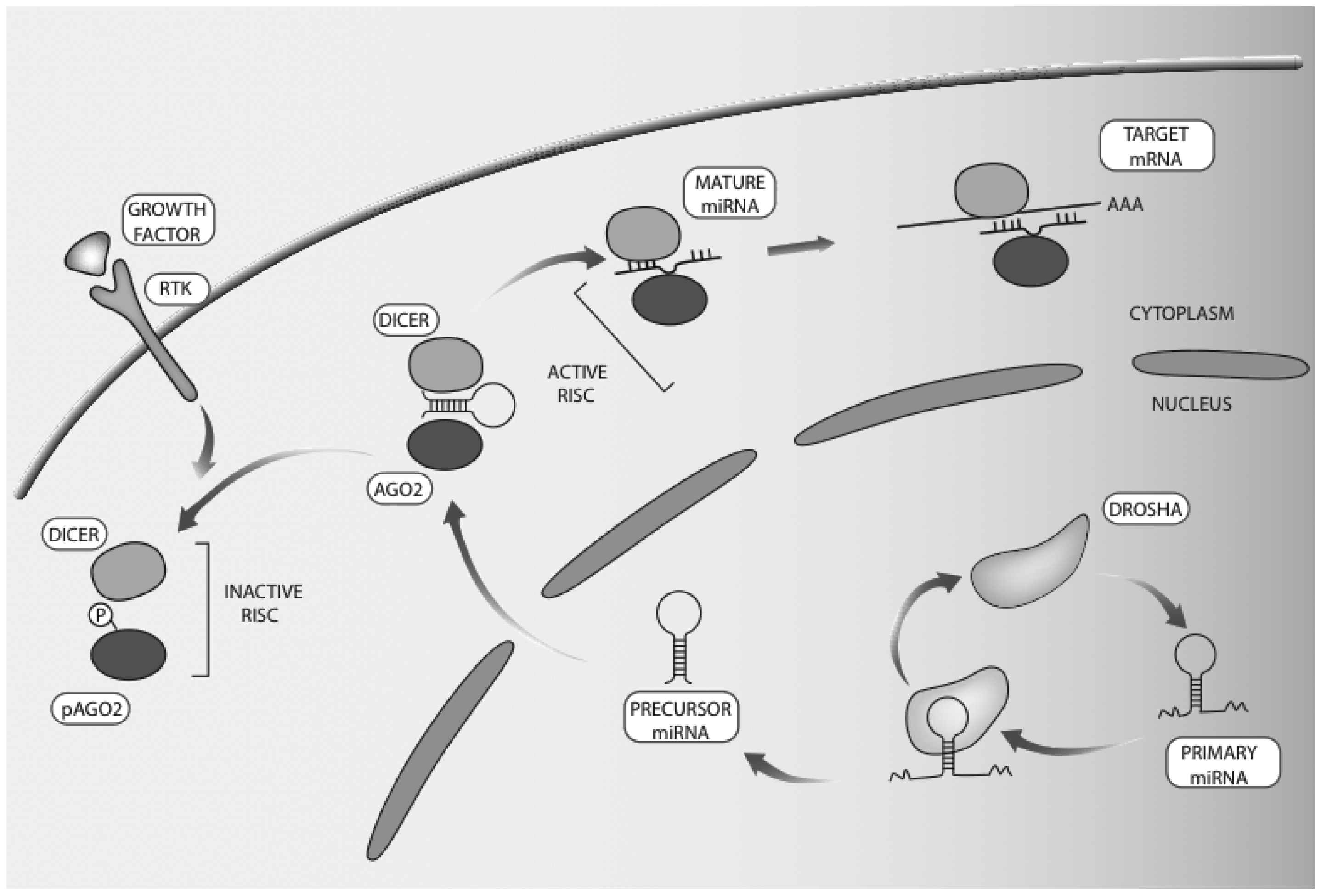
2. Occurrence and Biogenesis of microRNAs and Their Relevance to Cancer

According to the latest release of the miRBase database (release 21; June 2014), there are at least 2588 mature human microRNAs. miRNAs play profound roles in cancer progression, including metastasis. They can act both as oncogenes, namely, oncomiRs, and as tumor suppressor miRNAs. Changes in the abundance of specific miRNAs were demonstrated in many types of cancer, and their expression levels influence cell migration, invasion and proliferation [26]. Most of the miRNAs in cancer cells show down-regulated abundance compared to normal cells, however, several miRNAs are specifically up-regulated in cancer. In line with global alterations, it has been shown that malignant processes involve dysregulation or dysfunction of the miRNA biogenesis machinery due to mutations or epigenetic events (reviewed by [27] and by [28]). For example, expression of Drosha and/or Dicer is decreased in some tumor types, including neuroblastoma, liposarcoma, lung, breast, and ovarian cancers [29,30,31]. Growth factor signaling pathways, such as the epidermal growth factor receptor (EGFR) and the transforming growth factor beta (TGF-β) pathways, might affect general processing of miRNAs. EGFR restrains the maturation of specific tumor suppressor miRNAs, such as miR-31, -192, and miR-193a-5p, by phosphorylation of Argonaute 2 (AGO2) at Tyr393. This phosphorylation reduces the ability of Dicer to bind with AGO2, thereby inhibits processing of precursor miRNAs into mature miRNAs [32]. Under hypoxia, phosphorylation of Tyr393 by EGFR enhances cell survival and invasiveness and this was associated with poor prognosis of breast cancer patients [32]. TGF-β and bone morphogenic protein (BMP) signaling increases miR-21 abundance; specific SMAD signal transducers are recruited to the Drosha microprocessor complex and thus promote processing of primary miR-21 (pri-miR-21) into precursor miR-21 (pre-miR-21) [33]. Global effects of growth factors on miRNA biogenesis are associated in tumors with genomic rearrangements, which cause deletions or amplification of specific miRNAs loci. Conceivably, cancer cells make use of both growth factors and genetic aberrations to change miRNAs abundance, and consequently harness cellular machineries in favor of better adaptation to their changing environments.
Figure 2. Schematic representation of microRNA biosynthesis and regulation by RTK signaling. microRNA biogenesis starts with transcription of the respective gene by RNA polymerase II. The formed long primary microRNA (pri-miRNA) consists of a hairpin stem, terminal loop and two single stranded regions. The RNase III endonuclease called Drosha processes pri-miRNAs into 70-nucleotide imperfect stem loop structures (pre-miRNAs). The latter are exported to the cytoplasm, to undergo processing by another RNase III endonuclease, Dicer, which removes the loop and joins the two arms. The resulting RNA duplex of 19–24 nucleotides allows one strand to be loaded into the RISC, while the other strand undergoes degradation. Mature microRNAs lead to translational repression or to mRNA degradation. Note that the RISC includes members of the Argonaute family, such as AGO2. It has been reported that phosphorylation of AGO2, at tyrosine 393, by EGFR is enhanced under hypoxia [32]. This is associated with dissociation of the AGO2-Drosha complex and with inhibition of processing of precursor microRNA molecules.
Jcm 04 01578 g002

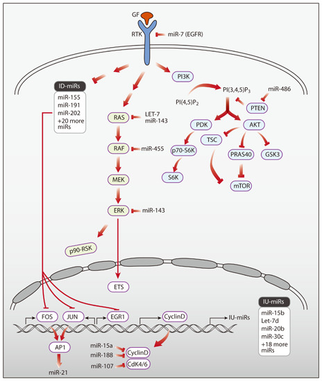
3. Networks of Growth Factors and microRNAs

3.1. Growth Factors Regulating miRNAs

Regulation of miRNA abundance might be induced, or otherwise influenced, by growth factors. Several studies analyzed changes in expression profiles of miRNAs following stimulation of cultured cells with specific growth factors. For instance, dynamic and coordinated changes in expression of groups of miRNAs were identified in normal mammary epithelial cells following stimulation with EGF. In less than 60 min post stimulation we observed both up- and down-regulation of distinct groups of miRNAs [34,35]. Interestingly the immediately down-regulated miRNAs we reported, a group consisting of 23 members, were over-represented among miRNAs that showed lower expression in breast cancer tumors compared to the surrounding normal tissue (peri-tumor) from the same patient [34]. Reciprocally, the up-regulated miRNAs were enriched among miRNAs with higher expression in the tumors [35]. Importantly, the mammary cells we tested, MCF10A, migrate in response to EGF stimulation [13,36]. Accordingly, we found that the migratory response of these cells is controlled by both up- and down-regulated miRNAs. For example, miR-15b, which was immediately up-regulated following EGF stimulation, significantly decelerated migration and invasion rates when silenced. In line with this observation, miR-15b expression was significantly higher in different breast cancer subtypes compared to control. MiR-15b’s novel target, metastasis suppressor 1 (MTSS1), a lipid-binder cytoskeletal protein, which is lost in some advanced tumors, was down-regulated following EGF treatment and mediated the effects on migration and invasion of normal and cancerous cells [35] (Figure 3A). Manipulation of the immediately down-regulated miRNAs following EGF stimulation also affected migration. Thus, silencing miR-191, which targets the immediate early gene called EGR1, elevated cell migration. Like miR-191, a significant number of the targets of immediately down-regulated miRNAs are IEGs, such as FOS and JUN. Under steady state, when EGF is not introduced to cells, the immediately down-regulated miRNAs (ID-miRs) inhibit the expression of the IEGs, some of which are proto-oncogenes. Correspondingly, upon EGF stimulation the expression levels of these miRNAs are decreased and a rapid up-regulation of the IEGs is achieved. For example, one of the ID-miRs, miR-155, directly targets FOS (Figure 3B). Interestingly, the oncogenic viral form of c-FOS, v-FOS, harbors a shorter 3′UTR than the c-FOS 3′UTR, which does not include miR-155’s target sequence. Hence, the transcript of v-FOS is not inhibited by miR-155, which allows v-FOS to exert its oncogenic ability [34]. In another comprehensive study, HeLa cells were stimulated with EGF for short times (15 min to 6 h) and miRNA expression levels were measured using microarray or deep-sequencing [37]. Dynamic changes in miRNA expression level were detected and the miRNA’s predicted targets were found to be involved in molecular functions that relate to EGF signaling, such as cellular development, proliferation, cell morphology, cell death, and cell-to-cell signaling and interaction [37].
Up regulation of three miRNAs, miR-31, miR-181b, and miR-222, was detected in oral cancer cells following treatment with EGF, and this was mediated by AKT and C/EBPβ signaling, at least in the case of miR-31 [38]. Increased expression of miR-31 was also observed in EGF-stimulated mammary cells [35]. MiR-31 directly targets synaptojanin 2 (SYNJ2), a lipid phosphatase transiently up-regulated following EGF treatment (Figure 3C). Hence, it is possible that miR-31 fine-tunes the expression of SYNJ2, meaning that it induces down-regulation of SYNJ2 back to baseline expression level. In patients with breast and brain cancer, SYNJ2’s high abundance was negatively correlated with miR-31 expression and associated with poor prognosis [39]. Congruently, forced expression of SYNJ2 enhanced tumor growth and metastasis in mice, and increased formation of invadopodia and lamellipodia, actin-filled cellular extensions involved in invasion and migration, respectively [39].
The delayed response to EGF stimulation (3–12 h post stimulation) involves miRNAs targeting both apoptotic and anti-apoptotic genes. Specifically, miR-134, miR-145, miR-146b, miR-432, and miR-494 had the largest number of apoptotic and anti-apoptotic targets, including targets that are part of the interferon pathway [40]. Other miRNAs that were identified as regulators of apoptosis and are induced by growth factor receptors, such as EGFR and MET, are miR-221/222 and miR-30b/30c. It was further suggested that in response to treatment of lung cancer cells with tyrosine kinase inhibitors these miRNAs repress the pro-apoptotic genes APAF1 and BIM, making cells less susceptible to apoptosis (reviewed by [41]). The miR-30 family has a tumor suppressive role. These family members were induced by SRC inhibitors and down-regulated by oncogenic growth factors signals such as EGF and the hepatocyte growth factor (HGF). Additionally, several of miR-30’s predicted targets, such as the MAPK-regulated transcription factor, ERG, are associated with epithelial-to-mesenchymal transition (EMT) [42]. In conclusion, several growth factor inducible miRNAs seem to act cooperatively to support survival, proliferation and motility, cellular functions vital for tumor progression.
Figure 3. Network modules incorporating microRNAs and growth factor signaling pathways. (A) A feed-forward loop (FFL), whereby EGFR signaling down-regulates the expression of MTSS1, an inhibitor of metastasis. Inhibition of MTSS1 is strengthened by the induction of its targeting IU-miR, miR-15b; (B) A feed-forward loop whereby EGFR signaling up-regulates expression of the transcription factor FOS and, in parallel, down-regulates an ID-miR, miR-155, which inhibits FOS; (C) An incoherent FFL, whereby up-regulation of SYNJ2, a lipid phosphatase gene, by EGFR signaling is fine-tuned through the induction of the delayed up-regulated microRNA (DU-miR), miR-31, which inhibits SYNJ2 expression; (D) Listed are miRNAs that directly target EGFR in different cancer cells. (ID-miR, immediately down-regulated miRNA; IU-miR, immediately up-regulated miRNA; DU-miR, delayed up-regulated miRNA).
Jcm 04 01578 g003

3.2. Specific microRNAs Regulating Growth Factor Signaling

The other side of the miRNAs-growth factor networks is miRNAs that regulate the expression of growth factors, growth factor receptors, and their intracellular effectors. Specifically, we focus here on miRNAs that regulate EGFR, the EGFR pathway, and EGFR’s family members. EGFR itself can be regulated by multiple miRNAs. MiR-7 was one of the first miRNA identified as directly regulating EGFR. In glioblastoma, lung and breast cancer cells miR-7 blocked EGFR expression by means of accelerating mRNA decay. Potentially, miR-7 induces tumor suppressive actions by regulating not only EGFR but also the downstream signaling pathway at multiple sites. For example, this miRNA can inhibit AKT and ERK1/2 in several human cancer cell lines and it might decrease invasiveness and arrest the cell cycle [43,44]. Similarly, miR-128 was among the first miRNAs identified as an upstream regulator of EGFR. Interestingly, miR-128 loss of heterezygosity was frequently detected in lung cancer samples, in correlation with patient survival following treatment with an EGFR-specific TKI [45]. Other miRNAs that directly target EGFR include miR-23b/27b [46], miR-133a [47,48], miR-133b [49], miR-146a [50], miR-146b-5p [51], miR-219-5p [52], miR-302b [53] and miR-608 [54] (Figure 3D).
As initially exemplified by miR-7, other miRNAs can also target more than one component of the EGFR pathway. These include miR-124, miR-147, and miR-193a-3p, which inhibit G1/S transition and cell proliferation by targeting EGFR-driven cell cycle proteins [55]. MiR-143 and miR-145 target KRAS, BRAF [56] and MEK2 [57] in colorectal cancer and also in other types of cancer such as prostate tumors [58]. MiR-27a (miR-27a-3p) and the complementary miR-27a* (miR-27a-5p), both targeting EGFR, were found to be significantly down-regulated in multiple head and neck squamous cell carcinoma cell lines. Interestingly, miR-27a* targets also AKT and mTOR (mammalian target of rapamycin) within the EGFR signaling pathway [59].
Other members of the EGFR/ERBB family are also regulated by miRNAs in cancer. Using miRNA gain-of-function screens and two HER2-amplified cell lines enabled identification of the following direct regulators of HER2: miR-552, miR-541, miR-193a-5p, miR-453, miR-134, miR-498, and miR-331-3p [60]. MiR-331-3p was found to target HER2 also in glioblastoma and prostate cancer cell lines [61,62]. In a similar way, miR-148b, miR-149, miR-326, and miR-520a-3p simultaneously down-regulated HER3/ERBB3 and components of the downstream signaling pathway in response to the direct ligand of HER3, neuregulin [63]. Interestingly, miR-125a and miR-125b target both HER2 and HER3 in breast cancer cells, and consequently inhibit phosphorylation of ERK and AKT [64]. miR-193a-3p directly targets HER4/ERBB4. Repression of HER4 by overexpression of miR-193a-3p resulted in decreased proliferation, migration, invasion and EMT, as well as increased apoptosis of lung cancer cells. Moreover, miR-193a-3p, which negatively regulates HER4 in xenograft tumor models, bears anti-tumor effects [65,66]. In esophageal squamous cell carcinoma, miR-302 targeted HER4, inhibited proliferation and invasion and induced apoptosis [67]. Taken together, it is conceivable that other subgroups of receptors for growth factors are regulated by multiple miRNAs, and the latter might coordinately control components of the downstream signaling pathway.

3.3. Feedback Loops Linking microRNAs and Growth Factors

The plot thickens. Bilateral regulation of growth factors and miRNAs in tumors generates high order complexity that is increasingly emerging now. Specifically, feedback regulatory loops in which a miRNA is both targeting a specific pathway and, at the same time, is regulated by the same pathway, likely confer module versatility and dynamicity. For example, several miRNAs that directly target EGFR are also regulated by EGFR signaling. Thus, miR-34a is up-regulated immediately following EGF stimulation [35], but it is also directly regulating EGFR. Possibly, through this complex regulation, miR-34a acts as a tumor suppressor in the development of chordoma [54]. As discussed above, miR-7 is a well-established regulator of EGFR, however it was also shown that miR-7 might be regulated by EGFR signaling: EGFR activation in lung cancer cells can stimulate miR-7 expression in an ERK-dependent manner, suggesting that EGFR induces miR-7 expression via the RAS-ERK pathway [68]. Feedback loops that involve specific miRNAs and different components of the EGFR pathway also exist: miR-143 and miR-145 regulate the EGFR pathway genes KRAS, BRAF, and MEK2 [56,57], but EGFR signals down-regulated these tumor suppressor miRNAs in a murine model of colon cancer [69]. In addition, in lung cancer cells, EGFR down-regulated miR-145 expression through ERK1/2 [70]. Other, less direct feedback loops, were also identified, in which EGFR regulated the expression of miRNAs that in turn targeted other partners of the same pathway. For example, miR-21 expression levels are regulated by EGFR via the activation of beta-catenin and AP-1 [71], and miR-21 is suppressed by the EGFR inhibitor, AG1478, suggesting that the EGFR can regulate miR-21 expression [72]. On the other hand, miR-21 regulates EGFR and AKT signaling through VHL/beta-catenin and the PPARα/AP-1axis [71]. These networks of miRNAs and growth factors have roles to play in cells motility, proliferation, and other processes that involved in cancer pathogenesis and metastasis. It is therefore important to resolve these networks in a global and systemic way.

4. Potential Clinical Applications of miRNAs Relevant to Growth Factors and Signal Transduction

Because growth factor signaling is pivotal to tumor progression and it is often targeted by anti-cancer drugs, major efforts are being made with the aim of better classifying malignancies and improving diagnosis and personalized therapy [73]. Since the first identification of miRNA dysregulation in cancer [74], profiling the abundance of miRNAs in tumor samples [75,76], as well as in patient fluids [77,78], is increasingly implicated as a tool enabling improved diagnosis, prognosis, and assessment of therapeutic responses. For example, in the pioneering case of let-7, reduced expression of this miRNA in human lung tumors is associated with shortened postoperative survival [79]. The use of let-7 and other miRNAs as biomarkers has been facilitated by their high stability in patient samples [77,80]. Furthermore, expression of miRNAs has been shown to be highly tissue specific [81], such that miRNA profiling might be able to infer developmental origins of specific tumors [76,82].
In the last few years, measuring global patterns of miRNAs is becoming an established method for classifying individual tumors of breast [83,84,85,86], lung [87,88], and other organs [89]. Some of these classifications will likely reach clinical application. For example, a microRNA-based test that identifies the primary origin of 42 different types of tumors (denoted miRview-mets2) has been established [90]. The panel involves testing 64 miRNAs, previously validated in 489 specimens, including 146 metastatic tumors from 42 tissues of origin. The panel is based on a tree-classifier, originally developed by Rosenfeld and co-workers, who profiled 205 primary versus 131 metastatic tumors from 22 different tumor origins [82]. Another comprehensive study by Nair and co-workers reported meta-analysis of 43 miRNA profiling studies across 20 types of malignancies [91]. These authors argue that stringent standardization must be introduced into the process of miRNA profiling. In addition, they found that for all classifier miRNAs in studies that evaluated overall survival across diverse malignancies, the miRNAs most frequently associated with poor outcome were let-7 (decreased expression in patients with cancer) and miR-21 (increased expression). In the context of growth factor signaling, relative abundance of subsets of EGF-regulated miRNAs in breast cancer models have been shown to correlate with the abundance of miRNAs in breast cancer patients [34,35]. Thus, miRNA classifiers, especially those based on molecular mechanisms of disease, will likely evolve into major diagnostic and prognostic tools of cancer pathologists.

5. MicroRNAs as Molecular Targets of Future Cancer Therapeutics

Apart from their increasing role in classification and prognosis of cancer, miRNAs are emerging as potential targets of novel drugs. Since miRNAs can function both as oncogenes and tumor suppressors, two complementary therapeutic approaches relate to miRNA-mediated therapy: silencing the action of a specific miRNA or re-introducing a specific miRNA into patients (reviewed in [92,93]). Initial efforts concentrated on silencing expression of specific miRNAs. For example, Krutzfeldt and co-workers conducted intravenous administration of “antagomirs” to mice. Injection of antagomirs resulted in significant reduction in abundance of several miRNAs in various tissues (i.e., miR-16, miR-122, miR-192, and miR-194) [94]. Since then, many delivery methods have been developed to systemically administer miRNA antisense oligonucleotides, which directly silence oncogenic miRNAs in tumors [95,96] or in the surrounding microenvironment [97]. Attenuation of miRNA action has also been achieved using miRNA sponges [98] and miR-masks [99], which contain sequences complementary to the miRNA target site or to the miRNA itself, respectively. On the other hand, replacement therapy focuses on re-expression of tumor suppressor miRNAs. Overcoming the loss of miRNA expression might be achieved either through introduction of synthetic miRNA mimics in the form of small double stranded and chemically modified oligonucleotides [100], or by using adenovirus-associated vectors (AAV), which do not stably integrate into the host genome [101].
Currently there are only three miRNA-targeting therapeutics in clinical trials, two of them in oncology. The first, miR-122, is an abundant liver-specific miRNA involved in the pathology of liver diseases, such as replication of hepatitis C virus (HCV) [102]. Since HCV infection depends on functional interactions between miR-122 and the HCV genome [103], miravirsen, a LNA-modified DNA phosphorothioate antisense oligonucleotide against miR-122, was developed as a potential drug [104]. Currently, miravirsen is applied in seven clinical trials, some are already in phase II. Another therapeutic endeavor employs MRX34, a liposome-formulated mimic of miR-34a, which is administered to patients with primary liver cancer or to those with liver metastasis from other cancers. Notably, miR-34a is embedded in the p53 transcriptional network [105]. Overexpression of miR-34a was found to inhibit tumor growth and to prolong survival of tumor-bearing mice [100,106,107]. A similar endeavor, which reached phase I trials, involves the tumor suppressor miR-16. Mice injected with miR-16 mimics showed dose-dependent inhibition of Malignant Pleural Mesothelioma (MPM) tumors [108]. This led to the development of TargomiRs, nanoparticles containing miR-16 mimics, which are administered to patients with MPM or with non-small cell lung cancer (NSCLC). Importantly, the nanoparticles are conjugated to anti-EGFR bi-specific antibodies that facilitate their targeted delivery to EGFR-expressing cells. Targeting microRNA-based therapeutics to cancer cells represents only one of many pharmacological challenges. These relate not only to drug efficacy, but also to potential toxicity due to the biology of miRNAs and their multiple targets.

6. MicroRNAs as Modulators of Patient Response to Drugs Targeting Growth Factor Signaling

Anti-cancer drugs able to intercept growth factor signaling currently outnumber other classes of therapies available to medical oncologists [109]. These drugs are effective on a broad range of carcinomas, and some drugs are active in more than one clinical indication, which is a rare situation in oncology. So far, only two classes of drugs have been approved: (i) monoclonal antibodies (mAbs), either naked or conjugated to a cytotoxic compound; and (ii) tyrosine kinase inhibitors (TKIs), which are either mono-specific or designed to inhibit several receptors. Along with weak efficacy, development of patient resistance to therapy hinders the effectiveness of both TKIs and mAbs. Mechanisms leading to resistance are only partially understood, and they include activation of surrogate pathways, acquired structural modifications of the drug target, and histological transformation, such as epithelial to mesenchymal transition (EMT) and small cell transformation [110]. Several studies point to potential roles of miRNAs in emergence of resistance to various cancer treatments, including chemotherapy [111], radiotherapy [112], and targeted therapy [113]. Below we discuss resistance to targeted therapy, as well as summarize experimental data relevant to clinical applications of mAbs against oncogenic receptors (Table 1) and small molecule TKIs (Table 2).
Overexpression of miR-7, a well-known regulator of EGFR expression [43], was shown to enhance the effect of an EGFR TKI, erlotinib, in a head and neck cancer model system [114]. Another receptor-targeting miRNA, miR-375, was shown to target the receptor for the insulin-like growth factor 1 (IGF1R) [115]. Importantly, miR-375 abundance negatively correlates to the expression of IGF1R in breast cancer specimens. Moreover, overexpression of miR-375 restored sensitivity to trastuzumab, an anti-HER2 mAb, and increased efficacy of trastuzumab in a xenograft model [115]. Sensitivity of breast cancer cells to trastuzumab was increased also by overexpression of miR-200c, a well-known miRNA regulating various cellular processes, including EMT [116]. Adam and co-workers reported that miR-200c is also associated with modulation of sensitivity of bladder carcinoma cell lines to an EGFR mAb, cetuximab [117]. Efficiency of cetuximab treatment was found to benefit also from concomitant overexpression of miR-146a: exposing hepatocellular carcinoma (HCC) cell lines to both cetuximab and miR-146a mimics elicited synergistic effects leading to increased apoptosis and decreased cell growth [118]. Like cetuximab, nimotuzumab binds to EGFR, and its inhibitory effects were enhanced by reducing abundance of miR-566 in glioblastoma cells [119]. The case of miR-221 is especially interesting as it was shown in two independent studies to facilitate the therapeutic effect of both trastuzumab [120] and gefitinib [121], in breast and lung cancer models, respectively. While miR-221 (along with miR-222) was shown to target APAF-1 in the lung model, its main target in the mammary model was the tumor suppressor PTEN. PTEN was also shown to be targeted by miR-21 [122]. This later report showed that overexpression of miR-21 decreases sensitivity of lung cells to gefitinib. Additionally, knock-down of miR-21 restored gefitinib sensitivity to a gefitinib-resistant lung cell line and caused a dramatic reduction in tumor size [122].
Interestingly, many studies relating to TKI-resistance focus on lung cancer models, and particularly on resistance to the EGFR-specific TKI called gefitinib. Accordingly, overexpression of miR-138-5p [123], miR-34a [124], miR-103 [121], or miR-203 [121] all resulted in increased sensitivity to gefitinib in lung cancer models, albeit by targeting different effectors. Conversely, in the case of miR-30b and miR-30c, inhibition of miRNA expression, rather than overexpression, increased drug sensitivity of gefitinib-resistant cells [121]. Likewise, overexpression of miR-203 was shown to enhance the effect of another EGFR-targeting TKI, CI-1033, and to reduce tumor size in a xenograft model of RAS-driven cells [125]. Collectively, the results we reviewed underscore potential roles for specific miRNAs in the acquisition of resistance to cancer drugs, and support the ability of yet other miRNAs to restore drug sensitivity. Therefore, targeting miRNAs in combination with conventional treatments may improve therapeutic efficacy of personalized cancer therapy.
Table 1. miRNAs modulating the efficacy of monoclonal antibodies targeting receptor tyrosine kinases.
miRNATarget Gene(s)EffectDrug & TumorReference
miR-566VHLKnockdown of miR-566 inhibited cell proliferation and invasion and led to cell cycle arrest in glioma cells. It further sensitized glioblastoma cells to NimotuzumabNimotuzumab (glioblastoma)[119]
miR-200cZNF217, ZEB1Overexpression of miR-200c increased sensitivity to trastuzumab and suppressed invasiveness of breast cancer cell linesTrastuzumab (breast)[116]
miR-375IGF1ROverexpression of miR-375 restored sensitivity to trastuzumab resistant cell lines and increased the efficacy of trastuzumab in a xenogarft model.Trastuzumab (breast)[115]
miR-221PTENOverexpression of miR-221 inhibited apoptosis, promoted metastasis and induced trastuzumab resistance in HER-2 positive breast cancer cells.Trastuzumab (breast)[120]
miR-200cERRFI-1Overexpression of miR-200c regains sensitivity of the resistant cell lines to cetuximab treatment resulting in reduced cell growth in vitroCetuximab (bladder)[117]
miR-146aEGFR signalingOverexpression of miR-146a suppressed cell growth and increased cellular apoptosis in HCC cell lines and displayed synergistic effects with cetuximabCetuximab (hepatocellular)[118]
miR-7EGFROverexpression of miR-7 enhanced the effect of erlotinib on growth inhibition of FaDu cellsErlotinib (head&neck)[114]
Table 2. miRNAs modulating efficacy of TKIs.
miRNATarget Gene(s)EffectDrug & TumorReference
miR-30b/30c and miR-221/222BIM, APAF-1 (respectively)Knockdown of miR-30b, -30c, -221 and -222 in gefitinib-resistant cells induces increased sensitivity gefitinibGefitinib (lung)[121]
miR-21PTENOverexpression of miR-21 decreased sensitivity of lung cells to gefitinib. Knock-down of miR-21 restored gefitinib sensitivity of the corresponding gefitinib-resistant cell line and caused a dramatic reduction in tumor sizeGefitinib (lung)[122]
miR-34aMETOverexpression of miR-34a in EGFR mutant NSCLC increased sensitivity to gefitinib, resulting in increased inhibition of cell growth and to induced apoptosis, which resulted in tumor regressionGefitinib (lung)[124]
miR-138-5pGPR124Overexpression of miR-138-5p in NSCLC cells increased sensitivity to gefitinib in vitroGefitinib (lung)[123]
miR-103 and miR-203PKC-e, SRC (respectively)Overexpression of miR-103 and miR-203 increased sensitivity to gefitinib in lung cells resistant to the drugGefitinib (lung)[121]
miR-203EREG, TGFA, API5, BIRC2, TRIAP1Overexpression of miR-203 synergistically enhanced the effect of CI-1033 on reduction of tumor size in a xenograft model of nude mice injected with Ras-activated cellsCI-1033 (prostate)[125]

7. Concluding Remarks

MicroRNAs can target growth factor pathways and, vice versa, growth factor pathways can regulate miRNA biogenesis. This bilateral crosstalk creates complex networks that are involved in multiple sub-programs of tumor progression, such as cell cycle regulation, EMT, and metastasis. Moreover, as we discussed herein, the complexity and robustness of these networks are enhanced by recurring feedback regulatory modules. As expected, miRNAs and growth factor signals are embedded in larger regulatory networks that control patient response to therapeutic interventions, such as monoclonal antibodies. It is therefore essential to resolve miRNA networks at high granularity and understand their functional logic. Thus, comprehensive mapping and understanding of the miRNA and growth factor alliance holds promise in terms of more effective cancer treatments that avoid emergence of patient resistance. Other future applications might include utilization of the miRNAs-growth factor networks as classifiers of cancer subtypes and markers for cancer diagnostics and prognosis.

Acknowledgments

Our team is located at the Marvin Tanner Laboratory for Cancer Research. We thank Orit Bechar for artwork. Our research is supported by the National Cancer Institute, the European Research Council, the Seventh Framework Program of the European Commission, the German-Israeli Project Cooperation (DIP), the Israel Cancer Research Fund and the Dr. Miriam and Sheldon G. Adelson Medical Research Foundation. Yosef YardenY.Y. is the incumbent of the Harold and Zelda Goldenberg Professorial Chair.

Conflicts of Interest

The authors declare no conflict of interest.

Author Contributions

Merav Kedmi, Aldema Sas-Chen and Yosef Yarden conceived the review, wrote the text and designed the figures.

References

  1. Solomon, E.; Borrow, J.; Goddard, A.D. Chromosome aberrations and cancer. Science 1991, 254, 1153–1160. [Google Scholar] [CrossRef] [PubMed]
  2. Fearon, E.R.; Vogelstein, B. A genetic model for colorectal tumorigenesis. Cell 1990, 61, 759–767. [Google Scholar] [CrossRef]
  3. Tomasetti, C.; Vogelstein, B. Cancer etiology. Variation in cancer risk among tissues can be explained by the number of stem cell divisions. Science 2015, 347, 78–81. [Google Scholar] [CrossRef] [PubMed]
  4. Chaffer, C.L.; Weinberg, R.A. How does multistep tumorigenesis really proceed? Cancer Discov. 2015, 5, 22–24. [Google Scholar] [CrossRef] [PubMed]
  5. Beerenwinkel, N.; Antal, T.; Dingli, D.; Traulsen, A.; Kinzler, K.W.; Velculescu, V.E.; Vogelstein, B.; Nowak, M.A. Genetic progression and the waiting time to cancer. PLoS Comput. Biol. 2007, 3, e225. [Google Scholar] [CrossRef] [PubMed]
  6. Witsch, E.; Sela, M.; Yarden, Y. Roles for growth factors in cancer progression. Physiology (Bethesda) 2010, 25, 85–101. [Google Scholar] [CrossRef] [PubMed]
  7. Futreal, P.A.; Coin, L.; Marshall, M.; Down, T.; Hubbard, T.; Wooster, R.; Rahman, N.; Stratton, M.R. A census of human cancer genes. Nat. Rev. Cancer 2004, 4, 177–183. [Google Scholar] [CrossRef] [PubMed]
  8. Sporn, M.B.; Todaro, G.J. Autocrine secretion and malignant transformation of cells. N. Engl. J. Med. 1980, 303, 878–880. [Google Scholar] [CrossRef] [PubMed]
  9. Greco, A.; Fusetti, L.; Villa, R.; Sozzi, G.; Minoletti, F.; Mauri, P.; Pierotti, M.A. Transforming activity of the chimeric sequence formed by the fusion of collagen gene col1a1 and the platelet derived growth factor b-chain gene in dermatofibrosarcoma protuberans. Oncogene 1998, 17, 1313–1319. [Google Scholar] [CrossRef] [PubMed]
  10. Shimizu, A.; O’Brien, K.P.; Sjoblom, T.; Pietras, K.; Buchdunger, E.; Collins, V.P.; Heldin, C.H.; Dumanski, J.P.; Ostman, A. The dermatofibrosarcoma protuberans-associated collagen type ialpha1/platelet-derived growth factor (pdgf) b-chain fusion gene generates a transforming protein that is processed to functional pdgf-bb. Cancer Res. 1999, 59, 3719–3723. [Google Scholar] [PubMed]
  11. Hynes, N.E.; Watson, C.J. Mammary gland growth factors: Roles in normal development and in cancer. Cold Spring Harb. Perspect. Biol. 2010, 2, a003186. [Google Scholar] [CrossRef] [PubMed]
  12. Kovacs, E.; Zorn, J.A.; Huang, Y.; Barros, T.; Kuriyan, J. A structural perspective on the regulation of the epidermal growth factor receptor. Annu. Rev. Biochem. 2015, 84, 739–764. [Google Scholar] [CrossRef] [PubMed]
  13. Tarcic, G.; Avraham, R.; Pines, G.; Amit, I.; Shay, T.; Lu, Y.; Zwang, Y.; Katz, M.; Ben-Chetrit, N.; Jacob-Hirsch, J.; et al. Egr1 and the erk-erf axis drive mammary cell migration in response to egf. FASEB J. 2012, 26, 1582–1592. [Google Scholar] [CrossRef] [PubMed]
  14. Huang, H.; Tindall, D.J. Dynamic foxo transcription factors. J. Cell Sci. 2007, 120, 2479–2487. [Google Scholar] [CrossRef] [PubMed]
  15. Amit, I.; Citri, A.; Shay, T.; Lu, Y.; Katz, M.; Zhang, F.; Tarcic, G.; Siwak, D.; Lahad, J.; Jacob-Hirsch, J.; et al. A module of negative feedback regulators defines growth factor signaling. Nat. Genet. 2007, 39, 503–512. [Google Scholar] [CrossRef] [PubMed]
  16. Tullai, J.W.; Schaffer, M.E.; Mullenbrock, S.; Sholder, G.; Kasif, S.; Cooper, G.M. Immediate-early and delayed primary response genes are distinct in function and genomic architecture. J. Biol. Chem. 2007, 282, 23981–23995. [Google Scholar] [CrossRef] [PubMed]
  17. Avraham, R.; Yarden, Y. Feedback regulation of egfr signalling: Decision making by early and delayed loops. Nat. Rev. Mol. Cell Biol. 2011, 12, 104–117. [Google Scholar] [CrossRef] [PubMed]
  18. Han, J.; Lee, Y.; Yeom, K.H.; Kim, Y.K.; Jin, H.; Kim, V.N. The drosha-dgcr8 complex in primary microrna processing. Genes Dev. 2004, 18, 3016–3027. [Google Scholar] [CrossRef] [PubMed]
  19. Grishok, A.; Pasquinelli, A.E.; Conte, D.; Li, N.; Parrish, S.; Ha, I.; Baillie, D.L.; Fire, A.; Ruvkun, G.; Mello, C.C. Genes and mechanisms related to rna interference regulate expression of the small temporal rnas that control c. Elegans developmental timing. Cell 2001, 106, 23–34. [Google Scholar] [CrossRef]
  20. Song, J.J.; Smith, S.K.; Hannon, G.J.; Joshua-Tor, L. Crystal structure of argonaute and its implications for risc slicer activity. Science 2004, 305, 1434–1437. [Google Scholar] [CrossRef] [PubMed]
  21. Chendrimada, T.P.; Finn, K.J.; Ji, X.; Baillat, D.; Gregory, R.I.; Liebhaber, S.A.; Pasquinelli, A.E.; Shiekhattar, R. Microrna silencing through risc recruitment of eif6. Nature 2007, 447, 823–828. [Google Scholar] [CrossRef] [PubMed]
  22. Tijsterman, M.; Plasterk, R.H. Dicers at risc; the mechanism of rnai. Cell 2004, 117, 1–3. [Google Scholar] [CrossRef]
  23. Lewis, B.P.; Burge, C.B.; Bartel, D.P. Conserved seed pairing, often flanked by adenosines, indicates that thousands of human genes are microrna targets. Cell 2005, 120, 15–20. [Google Scholar] [CrossRef] [PubMed]
  24. Zamore, P.D.; Haley, B. Ribo-gnome: The big world of small rnas. Science 2005, 309, 1519–1524. [Google Scholar] [CrossRef] [PubMed]
  25. Baek, D.; Villen, J.; Shin, C.; Camargo, F.D.; Gygi, S.P.; Bartel, D.P. The impact of micrornas on protein output. Nature 2008, 455, 64–71. [Google Scholar] [CrossRef] [PubMed]
  26. Croce, C.M.; Calin, G.A. Mirnas, cancer, and stem cell division. Cell 2005, 122, 6–7. [Google Scholar] [CrossRef] [PubMed]
  27. Lin, S.; Gregory, R.I. Microrna biogenesis pathways in cancer. Nat. Rev. Cancer 2015, 15, 321–333. [Google Scholar] [CrossRef] [PubMed]
  28. Hata, A.; Lieberman, J. Dysregulation of microrna biogenesis and gene silencing in cancer. Sci. Signal. 2015, 8, re3. [Google Scholar] [CrossRef] [PubMed]
  29. Avery-Kiejda, K.A.; Braye, S.G.; Forbes, J.F.; Scott, R.J. The expression of dicer and drosha in matched normal tissues, tumours and lymph node metastases in triple negative breast cancer. BMC Cancer 2014, 14, 253. [Google Scholar] [CrossRef] [PubMed]
  30. Poursadegh Zonouzi, A.A.; Nejatizadeh, A.; Rahmati-Yamchi, M.; Fardmanesh, H.; Shakerizadeh, S.; Poursadegh Zonouzi, A.; Nejati-Koshki, K.; Shekari, M. Dysregulated expression of dicer in invasive ductal breast carcinoma. Med. Oncol. 2015, 32, 643. [Google Scholar] [CrossRef] [PubMed]
  31. Vincenzi, B.; Iuliani, M.; Zoccoli, A.; Pantano, F.; Fioramonti, M.; de Lisi, D.; Frezza, A.M.; Rabitti, C.; Perrone, G.; Onetti Muda, A.; et al. Deregulation of dicer and mir-155 expression in liposarcoma. Oncotarget 2015, 6, 10586–10591. [Google Scholar] [PubMed]
  32. Shen, J.; Xia, W.; Khotskaya, Y.B.; Huo, L.; Nakanishi, K.; Lim, S.O.; Du, Y.; Wang, Y.; Chang, W.C.; Chen, C.H.; et al. Egfr modulates microrna maturation in response to hypoxia through phosphorylation of ago2. Nature 2013, 497, 383–387. [Google Scholar] [CrossRef] [PubMed]
  33. Davis, B.N.; Hilyard, A.C.; Lagna, G.; Hata, A. Smad proteins control drosha-mediated microrna maturation. Nature 2008, 454, 56–61. [Google Scholar] [CrossRef] [PubMed]
  34. Avraham, R.; Sas-Chen, A.; Manor, O.; Steinfeld, I.; Shalgi, R.; Tarcic, G.; Bossel, N.; Zeisel, A.; Amit, I.; Zwang, Y.; et al. Egf decreases the abundance of micrornas that restrain oncogenic transcription factors. Sci. Signal. 2010, 3, ra43. [Google Scholar] [CrossRef] [PubMed]
  35. Kedmi, M.; Ben-Chetrit, N.; Korner, C.; Mancini, M.; Ben-Moshe, N.B.; Lauriola, M.; Lavi, S.; Biagioni, F.; Carvalho, S.; Cohen-Dvashi, H.; et al. Egf induces micrornas that target suppressors of cell migration: Mir-15b targets mtss1 in breast cancer. Sci. Signal. 2015, 8, ra29. [Google Scholar] [CrossRef] [PubMed]
  36. Katz, M.; Amit, I.; Citri, A.; Shay, T.; Carvalho, S.; Lavi, S.; Milanezi, F.; Lyass, L.; Amariglio, N.; Jacob-Hirsch, J.; et al. A reciprocal tensin-3-cten switch mediates egf-driven mammary cell migration. Nat. Cell Biol. 2007, 9, 961–969. [Google Scholar] [CrossRef] [PubMed]
  37. Llorens, F.; Hummel, M.; Pantano, L.; Pastor, X.; Vivancos, A.; Castillo, E.; Mattlin, H.; Ferrer, A.; Ingham, M.; Noguera, M.; et al. Microarray and deep sequencing cross-platform analysis of the mirrnome and isomir variation in response to epidermal growth factor. BMC Genomics 2013, 14, 371. [Google Scholar] [CrossRef] [PubMed] [Green Version]
  38. Lu, W.C.; Kao, S.Y.; Yang, C.C.; Tu, H.F.; Wu, C.H.; Chang, K.W.; Lin, S.C. Egf up-regulates mir-31 through the c/ebpbeta signal cascade in oral carcinoma. PLoS ONE 2014, 9, e108049. [Google Scholar] [CrossRef] [PubMed]
  39. Ben-Chetrit, N.; Chetrit, D.; Russell, R.; Korner, C.; Mancini, M.; Abdul-Hai, A.; Itkin, T.; Carvalho, S.; Cohen-Dvashi, H.; Koestler, W.J.; et al. Synaptojanin 2 is a druggable mediator of metastasis and the gene is overexpressed and amplified in breast cancer. Sci. Signal. 2015, 8, ra7. [Google Scholar] [CrossRef] [PubMed]
  40. Alanazi, I.; Hoffmann, P.; Adelson, D.L. Micrornas are part of the regulatory network that controls egf induced apoptosis, including elements of the jak/stat pathway, in a431 cells. PLoS ONE 2015, 10, e0120337. [Google Scholar] [CrossRef] [PubMed]
  41. Gomez, G.G.; Volinia, S.; Croce, C.M.; Zanca, C.; Li, M.; Emnett, R.; Gutmann, D.H.; Brennan, C.W.; Furnari, F.B.; Cavenee, W.K. Suppression of microrna-9 by mutant egfr signaling upregulates foxp1 to enhance glioblastoma tumorigenicity. Cancer Res. 2014, 74, 1429–1439. [Google Scholar] [CrossRef] [PubMed]
  42. Kao, C.J.; Martiniez, A.; Shi, X.B.; Yang, J.; Evans, C.P.; Dobi, A.; deVere White, R.W.; Kung, H.J. Mir-30 as a tumor suppressor connects egf/src signal to erg and emt. Oncogene 2014, 33, 2495–2503. [Google Scholar] [CrossRef] [PubMed]
  43. Kefas, B.; Godlewski, J.; Comeau, L.; Li, Y.; Abounader, R.; Hawkinson, M.; Lee, J.; Fine, H.; Chiocca, E.A.; Lawler, S.; et al. Microrna-7 inhibits the epidermal growth factor receptor and the akt pathway and is down-regulated in glioblastoma. Cancer Res. 2008, 68, 3566–3572. [Google Scholar] [CrossRef] [PubMed]
  44. Webster, R.J.; Giles, K.M.; Price, K.J.; Zhang, P.M.; Mattick, J.S.; Leedman, P.J. Regulation of epidermal growth factor receptor signaling in human cancer cells by microrna-7. J. Biol. Chem. 2009, 284, 5731–5741. [Google Scholar] [CrossRef] [PubMed]
  45. Weiss, G.J.; Bemis, L.T.; Nakajima, E.; Sugita, M.; Birks, D.K.; Robinson, W.A.; Varella-Garcia, M.; Bunn, P.A., Jr.; Haney, J.; Helfrich, B.A.; et al. Egfr regulation by microrna in lung cancer: Correlation with clinical response and survival to gefitinib and egfr expression in cell lines. Ann. Oncol.: Off. J. Eur. Soc. Med. Oncol./ESMO 2008, 19, 1053–1059. [Google Scholar] [CrossRef] [PubMed]
  46. Chiyomaru, T.; Seki, N.; Inoguchi, S.; Ishihara, T.; Mataki, H.; Matsushita, R.; Goto, Y.; Nishikawa, R.; Tatarano, S.; Itesako, T.; et al. Dual regulation of receptor tyrosine kinase genes egfr and c-met by the tumor-suppressive microrna-23b/27b cluster in bladder cancer. Int. J. Oncol. 2015, 46, 487–496. [Google Scholar] [CrossRef] [PubMed]
  47. Wang, L.K.; Hsiao, T.H.; Hong, T.M.; Chen, H.Y.; Kao, S.H.; Wang, W.L.; Yu, S.L.; Lin, C.W.; Yang, P.C. Microrna-133a suppresses multiple oncogenic membrane receptors and cell invasion in non-small cell lung carcinoma. PLoS ONE 2014, 9, e96765. [Google Scholar] [CrossRef] [PubMed]
  48. Cui, W.; Zhang, S.; Shan, C.; Zhou, L.; Zhou, Z. Microrna-133a regulates the cell cycle and proliferation of breast cancer cells by targeting epidermal growth factor receptor through the egfr/akt signaling pathway. FEBS J. 2013, 280, 3962–3974. [Google Scholar] [CrossRef] [PubMed]
  49. Liu, L.; Shao, X.; Gao, W.; Zhang, Z.; Liu, P.; Wang, R.; Huang, P.; Yin, Y.; Shu, Y. Microrna-133b inhibits the growth of non-small-cell lung cancer by targeting the epidermal growth factor receptor. FEBS J. 2012, 279, 3800–3812. [Google Scholar] [CrossRef] [PubMed]
  50. Kumaraswamy, E.; Wendt, K.L.; Augustine, L.A.; Stecklein, S.R.; Sibala, E.C.; Li, D.; Gunewardena, S.; Jensen, R.A. Brca1 regulation of epidermal growth factor receptor (egfr) expression in human breast cancer cells involves microrna-146a and is critical for its tumor suppressor function. Oncogene 2014. [Google Scholar] [CrossRef] [PubMed]
  51. Katakowski, M.; Zheng, X.; Jiang, F.; Rogers, T.; Szalad, A.; Chopp, M. Mir-146b-5p suppresses egfr expression and reduces in vitro migration and invasion of glioma. Cancer Investig. 2010, 28, 1024–1030. [Google Scholar] [CrossRef] [PubMed]
  52. Rao, S.A.; Arimappamagan, A.; Pandey, P.; Santosh, V.; Hegde, A.S.; Chandramouli, B.A.; Somasundaram, K. Mir-219-5p inhibits receptor tyrosine kinase pathway by targeting egfr in glioblastoma. PLoS ONE 2013, 8, e63164. [Google Scholar] [CrossRef] [PubMed]
  53. Wang, L.; Yao, J.; Shi, X.; Hu, L.; Li, Z.; Song, T.; Huang, C. Microrna-302b suppresses cell proliferation by targeting egfr in human hepatocellular carcinoma smmc-7721 cells. BMC Cancer 2013, 13, 448. [Google Scholar] [CrossRef] [PubMed]
  54. Zhang, Y.; Schiff, D.; Park, D.; Abounader, R. Microrna-608 and microrna-34a regulate chordoma malignancy by targeting egfr, bcl-xl and met. PLoS ONE 2014, 9, e91546. [Google Scholar] [CrossRef] [PubMed]
  55. Uhlmann, S.; Mannsperger, H.; Zhang, J.D.; Horvat, E.A.; Schmidt, C.; Kublbeck, M.; Henjes, F.; Ward, A.; Tschulena, U.; Zweig, K.; et al. Global microrna level regulation of egfr-driven cell-cycle protein network in breast cancer. Mol. Syst. Biol. 2012, 8, 570. [Google Scholar] [CrossRef] [PubMed]
  56. Pagliuca, A.; Valvo, C.; Fabrizi, E.; di Martino, S.; Biffoni, M.; Runci, D.; Forte, S.; de Maria, R.; Ricci-Vitiani, L. Analysis of the combined action of mir-143 and mir-145 on oncogenic pathways in colorectal cancer cells reveals a coordinate program of gene repression. Oncogene 2013, 32, 4806–4813. [Google Scholar] [CrossRef] [PubMed]
  57. Pekow, J.R.; Dougherty, U.; Mustafi, R.; Zhu, H.; Kocherginsky, M.; Rubin, D.T.; Hanauer, S.B.; Hart, J.; Chang, E.B.; Fichera, A.; et al. Mir-143 and mir-145 are downregulated in ulcerative colitis: Putative regulators of inflammation and protooncogenes. Inflamm. Bowel Dis. 2012, 18, 94–100. [Google Scholar] [CrossRef] [PubMed]
  58. Xu, B.; Niu, X.; Zhang, X.; Tao, J.; Wu, D.; Wang, Z.; Li, P.; Zhang, W.; Wu, H.; Feng, N.; et al. Mir-143 decreases prostate cancer cells proliferation and migration and enhances their sensitivity to docetaxel through suppression of kras. Mol. Cell. Biochem. 2011, 350, 207–213. [Google Scholar] [CrossRef] [PubMed]
  59. Wu, X.; Bhayani, M.K.; Dodge, C.T.; Nicoloso, M.S.; Chen, Y.; Yan, X.; Adachi, M.; Thomas, L.; Galer, C.E.; Jiffar, T.; et al. Coordinated targeting of the egfr signaling axis by microrna-27a*. Oncotarget 2013, 4, 1388–1398. [Google Scholar] [PubMed]
  60. Leivonen, S.K.; Sahlberg, K.K.; Makela, R.; Due, E.U.; Kallioniemi, O.; Borresen-Dale, A.L.; Perala, M. High-throughput screens identify micrornas essential for her2 positive breast cancer cell growth. Mol. Oncol. 2014, 8, 93–104. [Google Scholar] [CrossRef] [PubMed]
  61. Giles, K.M.; Barker, A.; Zhang, P.M.; Epis, M.R.; Leedman, P.J. Microrna regulation of growth factor receptor signaling in human cancer cells. Methods Mol. Biol. 2011, 676, 147–163. [Google Scholar] [PubMed]
  62. Epis, M.R.; Giles, K.M.; Barker, A.; Kendrick, T.S.; Leedman, P.J. Mir-331-3p regulates erbb-2 expression and androgen receptor signaling in prostate cancer. J. Biol. Chem. 2009, 284, 24696–24704. [Google Scholar] [CrossRef] [PubMed]
  63. Bischoff, A.; Bayerlova, M.; Strotbek, M.; Schmid, S.; Beissbarth, T.; Olayioye, M.A. A global microrna screen identifies regulators of the erbb receptor signaling network. Cell Commun. Signal. 2015, 13, 5. [Google Scholar] [CrossRef] [PubMed]
  64. Scott, G.K.; Goga, A.; Bhaumik, D.; Berger, C.E.; Sullivan, C.S.; Benz, C.C. Coordinate suppression of erbb2 and erbb3 by enforced expression of micro-rna mir-125a or mir-125b. J. Biol. Chem. 2007, 282, 1479–1486. [Google Scholar] [CrossRef] [PubMed]
  65. Liang, H.; Liu, M.; Yan, X.; Zhou, Y.; Wang, W.; Wang, X.; Fu, Z.; Wang, N.; Zhang, S.; Wang, Y.; et al. Mir-193a-3p functions as a tumor suppressor in lung cancer by down-regulating erbb4. J. Biol. Chem. 2015, 290, 926–940. [Google Scholar] [CrossRef] [PubMed]
  66. Yu, T.; Li, J.; Yan, M.; Liu, L.; Lin, H.; Zhao, F.; Sun, L.; Zhang, Y.; Cui, Y.; Zhang, F.; et al. Microrna-193a-3p and -5p suppress the metastasis of human non-small-cell lung cancer by downregulating the erbb4/pik3r3/mtor/s6k2 signaling pathway. Oncogene 2015, 34, 413–423. [Google Scholar] [CrossRef] [PubMed]
  67. Zhang, M.; Yang, Q.; Zhang, L.; Zhou, S.; Ye, W.; Yao, Q.; Li, Z.; Huang, C.; Wen, Q.; Wang, J. Mir-302b is a potential molecular marker of esophageal squamous cell carcinoma and functions as a tumor suppressor by targeting erbb4. J. Exp. Clin. Cancer Res. 2014, 33, 10. [Google Scholar] [CrossRef] [PubMed]
  68. Cho, W.C.; Chow, A.S.; Au, J.S. Mir-145 inhibits cell proliferation of human lung adenocarcinoma by targeting egfr and nudt1. RNA Biol. 2011, 8, 125–131. [Google Scholar] [CrossRef] [PubMed]
  69. Zhu, H.; Dougherty, U.; Robinson, V.; Mustafi, R.; Pekow, J.; Kupfer, S.; Li, Y.C.; Hart, J.; Goss, K.; Fichera, A.; et al. Egfr signals downregulate tumor suppressors mir-143 and mir-145 in western diet-promoted murine colon cancer: Role of g1 regulators. Mol. Cancer Res. 2011, 9, 960–975. [Google Scholar] [CrossRef] [PubMed]
  70. Guo, Y.H.; Zhang, C.; Shi, J.; Xu, M.H.; Liu, F.; Yuan, H.H.; Wang, J.Y.; Jiang, B.; Gao, F.H. Abnormal activation of the egfr signaling pathway mediates the downregulation of mir145 through the erk1/2 in non-small cell lung cancer. Oncol. Rep. 2014, 31, 1940–1946. [Google Scholar] [PubMed]
  71. Zhang, K.L.; Han, L.; Chen, L.Y.; Shi, Z.D.; Yang, M.; Ren, Y.; Chen, L.C.; Zhang, J.X.; Pu, P.Y.; Kang, C.S. Blockage of a mir-21/egfr regulatory feedback loop augments anti-egfr therapy in glioblastomas. Cancer Lett. 2014, 342, 139–149. [Google Scholar] [CrossRef] [PubMed]
  72. Seike, M.; Goto, A.; Okano, T.; Bowman, E.D.; Schetter, A.J.; Horikawa, I.; Mathe, E.A.; Jen, J.; Yang, P.; Sugimura, H.; et al. Mir-21 is an egfr-regulated anti-apoptotic factor in lung cancer in never-smokers. Proc. Natl. Acad. Sci. USA 2009, 106, 12085–12090. [Google Scholar] [CrossRef] [PubMed]
  73. Perou, C.M.; Sorlie, T.; Eisen, M.B.; van de Rijn, M.; Jeffrey, S.S.; Rees, C.A.; Pollack, J.R.; Ross, D.T.; Johnsen, H.; Akslen, L.A.; et al. Molecular portraits of human breast tumours. Nature 2000, 406, 747–752. [Google Scholar] [CrossRef] [PubMed]
  74. Calin, G.A.; Dumitru, C.D.; Shimizu, M.; Bichi, R.; Zupo, S.; Noch, E.; Aldler, H.; Rattan, S.; Keating, M.; Rai, K.; et al. Frequent deletions and down-regulation of micro- rna genes mir15 and mir16 at 13q14 in chronic lymphocytic leukemia. Proc. Natl. Acad. Sci. USA 2002, 99, 15524–15529. [Google Scholar] [CrossRef] [PubMed]
  75. Volinia, S.; Calin, G.A.; Liu, C.G.; Ambs, S.; Cimmino, A.; Petrocca, F.; Visone, R.; Iorio, M.; Roldo, C.; Ferracin, M.; et al. A microrna expression signature of human solid tumors defines cancer gene targets. Proc. Natl. Acad. Sci. USA 2006, 103, 2257–2261. [Google Scholar] [CrossRef] [PubMed]
  76. Lu, J.; Getz, G.; Miska, E.A.; Alvarez-Saavedra, E.; Lamb, J.; Peck, D.; Sweet-Cordero, A.; Ebert, B.L.; Mak, R.H.; Ferrando, A.A.; et al. Microrna expression profiles classify human cancers. Nature 2005, 435, 834–838. [Google Scholar] [CrossRef] [PubMed]
  77. Mitchell, P.S.; Parkin, R.K.; Kroh, E.M.; Fritz, B.R.; Wyman, S.K.; Pogosova-Agadjanyan, E.L.; Peterson, A.; Noteboom, J.; O’Briant, K.C.; Allen, A.; et al. Circulating micrornas as stable blood-based markers for cancer detection. Proc. Natl. Acad. Sci. USA 2008, 105, 10513–10518. [Google Scholar] [CrossRef] [PubMed]
  78. Chen, X.; Ba, Y.; Ma, L.; Cai, X.; Yin, Y.; Wang, K.; Guo, J.; Zhang, Y.; Chen, J.; Guo, X.; et al. Characterization of micrornas in serum: A novel class of biomarkers for diagnosis of cancer and other diseases. Cell Res. 2008, 18, 997–1006. [Google Scholar] [CrossRef] [PubMed]
  79. Takamizawa, J.; Konishi, H.; Yanagisawa, K.; Tomida, S.; Osada, H.; Endoh, H.; Harano, T.; Yatabe, Y.; Nagino, M.; Nimura, Y.; et al. Reduced expression of the let-7 micrornas in human lung cancers in association with shortened postoperative survival. Cancer Res. 2004, 64, 3753–3756. [Google Scholar] [CrossRef] [PubMed]
  80. Nelson, P.T.; Baldwin, D.A.; Scearce, L.M.; Oberholtzer, J.C.; Tobias, J.W.; Mourelatos, Z. Microarray-based, high-throughput gene expression profiling of micrornas. Nat. Methods 2004, 1, 155–161. [Google Scholar] [CrossRef] [PubMed]
  81. Lagos-Quintana, M.; Rauhut, R.; Yalcin, A.; Meyer, J.; Lendeckel, W.; Tuschl, T. Identification of tissue-specific micrornas from mouse. Curr. Biol.: CB 2002, 12, 735–739. [Google Scholar] [CrossRef]
  82. Rosenfeld, N.; Aharonov, R.; Meiri, E.; Rosenwald, S.; Spector, Y.; Zepeniuk, M.; Benjamin, H.; Shabes, N.; Tabak, S.; Levy, A.; et al. Micrornas accurately identify cancer tissue origin. Nat. Biotechnol. 2008, 26, 462–469. [Google Scholar] [CrossRef] [PubMed]
  83. Dvinge, H.; Git, A.; Graf, S.; Salmon-Divon, M.; Curtis, C.; Sottoriva, A.; Zhao, Y.; Hirst, M.; Armisen, J.; Miska, E.A.; et al. The shaping and functional consequences of the microrna landscape in breast cancer. Nature 2013, 497, 378–382. [Google Scholar] [CrossRef] [PubMed]
  84. Blenkiron, C.; Goldstein, L.D.; Thorne, N.P.; Spiteri, I.; Chin, S.F.; Dunning, M.J.; Barbosa-Morais, N.L.; Teschendorff, A.E.; Green, A.R.; Ellis, I.O.; et al. Microrna expression profiling of human breast cancer identifies new markers of tumor subtype. Genome Biol. 2007, 8, R214. [Google Scholar] [CrossRef] [PubMed]
  85. Andorfer, C.A.; Necela, B.M.; Thompson, E.A.; Perez, E.A. Microrna signatures: Clinical biomarkers for the diagnosis and treatment of breast cancer. Trends Mol. Med. 2011, 17, 313–319. [Google Scholar] [CrossRef] [PubMed]
  86. Enerly, E.; Steinfeld, I.; Kleivi, K.; Leivonen, S.K.; Aure, M.R.; Russnes, H.G.; Ronneberg, J.A.; Johnsen, H.; Navon, R.; Rodland, E.; et al. Mirna-mrna integrated analysis reveals roles for mirnas in primary breast tumors. PLoS ONE 2011, 6, e16915. [Google Scholar] [CrossRef] [PubMed]
  87. Yanaihara, N.; Caplen, N.; Bowman, E.; Seike, M.; Kumamoto, K.; Yi, M.; Stephens, R.M.; Okamoto, A.; Yokota, J.; Tanaka, T.; et al. Unique microrna molecular profiles in lung cancer diagnosis and prognosis. Cancer Cell 2006, 9, 189–198. [Google Scholar] [CrossRef] [PubMed]
  88. Hu, Z.; Chen, X.; Zhao, Y.; Tian, T.; Jin, G.; Shu, Y.; Chen, Y.; Xu, L.; Zen, K.; Zhang, C.; et al. Serum microrna signatures identified in a genome-wide serum microrna expression profiling predict survival of non-small-cell lung cancer. J. Clin. Oncol.: Off. J. Am. Soc. Clin. Oncol. 2010, 28, 1721–1726. [Google Scholar] [CrossRef] [PubMed]
  89. Cho, W.C. Micrornas: Potential biomarkers for cancer diagnosis, prognosis and targets for therapy. Int. J. Biochem. Cell Biol. 2010, 42, 1273–1281. [Google Scholar] [CrossRef] [PubMed]
  90. Meiri, E.; Mueller, W.C.; Rosenwald, S.; Zepeniuk, M.; Klinke, E.; Edmonston, T.B.; Werner, M.; Lass, U.; Barshack, I.; Feinmesser, M.; et al. A second-generation microrna-based assay for diagnosing tumor tissue origin. Oncologist 2012, 17, 801–812. [Google Scholar] [CrossRef] [PubMed]
  91. Nair, V.S.; Maeda, L.S.; Ioannidis, J.P. Clinical outcome prediction by micrornas in human cancer: A systematic review. J. Natl. Cancer Inst. 2012, 104, 528–540. [Google Scholar] [CrossRef] [PubMed]
  92. Garzon, R.; Marcucci, G.; Croce, C.M. Targeting micrornas in cancer: Rationale, strategies and challenges. Nat. Rev. Drug Discov. 2010, 9, 775–789. [Google Scholar] [CrossRef] [PubMed]
  93. Shibata, C.; Otsuka, M.; Kishikawa, T.; Yoshikawa, T.; Ohno, M.; Takata, A.; Koike, K. Current status of mirna-targeting therapeutics and preclinical studies against gastroenterological carcinoma. Mol. Cell. Ther. 2013, 1, 5. [Google Scholar] [CrossRef] [PubMed]
  94. Krutzfeldt, J.; Rajewsky, N.; Braich, R.; Rajeev, K.G.; Tuschl, T.; Manoharan, M.; Stoffel, M. Silencing of micrornas in vivo with “antagomirs”. Nature 2005, 438, 685–689. [Google Scholar] [CrossRef] [PubMed]
  95. Elmen, J.; Lindow, M.; Schutz, S.; Lawrence, M.; Petri, A.; Obad, S.; Lindholm, M.; Hedtjarn, M.; Hansen, H.F.; Berger, U.; et al. Lna-mediated microrna silencing in non-human primates. Nature 2008, 452, 896–899. [Google Scholar] [CrossRef] [PubMed]
  96. Xie, J.; Ameres, S.L.; Friedline, R.; Hung, J.H.; Zhang, Y.; Xie, Q.; Zhong, L.; Su, Q.; He, R.; Li, M.; et al. Long-term, efficient inhibition of microrna function in mice using raav vectors. Nat. Methods 2012, 9, 403–409. [Google Scholar] [CrossRef] [PubMed]
  97. Cheng, C.J.; Bahal, R.; Babar, I.A.; Pincus, Z.; Barrera, F.; Liu, C.; Svoronos, A.; Braddock, D.T.; Glazer, P.M.; Engelman, D.M.; et al. Microrna silencing for cancer therapy targeted to the tumour microenvironment. Nature 2015, 518, 107–110. [Google Scholar] [CrossRef] [PubMed]
  98. Ebert, M.S.; Neilson, J.R.; Sharp, P.A. Microrna sponges: Competitive inhibitors of small rnas in mammalian cells. Nat. Methods 2007, 4, 721–726. [Google Scholar] [CrossRef] [PubMed]
  99. Choi, W.Y.; Giraldez, A.J.; Schier, A.F. Target protectors reveal dampening and balancing of nodal agonist and antagonist by mir-430. Science 2007, 318, 271–274. [Google Scholar] [CrossRef] [PubMed]
  100. Trang, P.; Wiggins, J.F.; Daige, C.L.; Cho, C.; Omotola, M.; Brown, D.; Weidhaas, J.B.; Bader, A.G.; Slack, F.J. Systemic delivery of tumor suppressor microrna mimics using a neutral lipid emulsion inhibits lung tumors in mice. Mol. Ther.: J. Am. Soc. Gene Ther. 2011, 19, 1116–1122. [Google Scholar] [CrossRef] [PubMed]
  101. Kota, J.; Chivukula, R.R.; O’Donnell, K.A.; Wentzel, E.A.; Montgomery, C.L.; Hwang, H.W.; Chang, T.C.; Vivekanandan, P.; Torbenson, M.; Clark, K.R.; et al. Therapeutic microrna delivery suppresses tumorigenesis in a murine liver cancer model. Cell 2009, 137, 1005–1017. [Google Scholar] [CrossRef] [PubMed]
  102. Zeisel, M.B.; Pfeffer, S.; Baumert, T.F. Mir-122 acts as a tumor suppressor in hepatocarcinogenesis in vivo. J. Hepatol. 2013, 58, 821–823. [Google Scholar] [CrossRef] [PubMed]
  103. Jopling, C.L.; Yi, M.; Lancaster, A.M.; Lemon, S.M.; Sarnow, P. Modulation of hepatitis c virus rna abundance by a liver-specific microrna. Science 2005, 309, 1577–1581. [Google Scholar] [CrossRef] [PubMed]
  104. Janssen, H.L.; Reesink, H.W.; Lawitz, E.J.; Zeuzem, S.; Rodriguez-Torres, M.; Patel, K.; van der Meer, A.J.; Patick, A.K.; Chen, A.; Zhou, Y.; et al. Treatment of hcv infection by targeting microrna. N. Engl. J. Med. 2013, 368, 1685–1694. [Google Scholar] [CrossRef] [PubMed]
  105. He, L.; He, X.; Lim, L.P.; de Stanchina, E.; Xuan, Z.; Liang, Y.; Xue, W.; Zender, L.; Magnus, J.; Ridzon, D.; et al. A microrna component of the p53 tumour suppressor network. Nature 2007, 447, 1130–1134. [Google Scholar] [CrossRef] [PubMed]
  106. Liu, C.; Kelnar, K.; Liu, B.; Chen, X.; Calhoun-Davis, T.; Li, H.; Patrawala, L.; Yan, H.; Jeter, C.; Honorio, S.; et al. The microrna mir-34a inhibits prostate cancer stem cells and metastasis by directly repressing cd44. Nat. Med. 2011, 17, 211–215. [Google Scholar] [CrossRef] [PubMed]
  107. Hu, Q.L.; Jiang, Q.Y.; Jin, X.; Shen, J.; Wang, K.; Li, Y.B.; Xu, F.J.; Tang, G.P.; Li, Z.H. Cationic microrna-delivering nanovectors with bifunctional peptides for efficient treatment of panc-1 xenograft model. Biomaterials 2013, 34, 2265–2276. [Google Scholar] [CrossRef] [PubMed]
  108. Reid, G.; Pel, M.E.; Kirschner, M.B.; Cheng, Y.Y.; Mugridge, N.; Weiss, J.; Williams, M.; Wright, C.; Edelman, J.J.; Vallely, M.P.; et al. Restoring expression of mir-16: A novel approach to therapy for malignant pleural mesothelioma. Ann. Oncol.: Off. J. Eur. Soc. Med. Oncol./ESMO 2013, 24, 3128–3135. [Google Scholar] [CrossRef] [PubMed]
  109. Yarden, Y.; Pines, G. The erbb network: At last, cancer therapy meets systems biology. Nat. Rev. Cancer 2012, 12, 553–563. [Google Scholar] [CrossRef] [PubMed]
  110. Lovly, C.M.; Shaw, A.T. Molecular pathways: Resistance to kinase inhibitors and implications for therapeutic strategies. Clin. Cancer Res.: Off. J. Am. Assoc. Cancer Res. 2014, 20, 2249–2256. [Google Scholar] [CrossRef] [PubMed]
  111. Donzelli, S.; Mori, F.; Biagioni, F.; Bellissimo, T.; Pulito, C.; Muti, P.; Strano, S.; Blandino, G. Micrornas: Short non-coding players in cancer chemoresistance. Mol. Cell. Ther. 2014, 2, 16. [Google Scholar] [CrossRef] [PubMed]
  112. Hummel, R.; Hussey, D.J.; Haier, J. Micrornas: Predictors and modifiers of chemo- and radiotherapy in different tumour types. Eur. J. Cancer 2010, 46, 298–311. [Google Scholar] [CrossRef] [PubMed]
  113. MacDonagh, L.; Gray, S.G.; Finn, S.P.; Cuffe, S.; O’Byrne, K.J.; Barr, M.P. The emerging role of micrornas in resistance to lung cancer treatments. Cancer Treat. Rev. 2015, 41, 160–169. [Google Scholar] [CrossRef] [PubMed]
  114. Kalinowski, F.C.; Giles, K.M.; Candy, P.A.; Ali, A.; Ganda, C.; Epis, M.R.; Webster, R.J.; Leedman, P.J. Regulation of epidermal growth factor receptor signaling and erlotinib sensitivity in head and neck cancer cells by mir-7. PLoS ONE 2012, 7, e47067. [Google Scholar] [CrossRef] [PubMed]
  115. Ye, X.M.; Zhu, H.Y.; Bai, W.D.; Wang, T.; Wang, L.; Chen, Y.; Yang, A.G.; Jia, L.T. Epigenetic silencing of mir-375 induces trastuzumab resistance in her2-positive breast cancer by targeting igf1r. BMC Cancer 2014, 14, 134. [Google Scholar] [CrossRef] [PubMed]
  116. Bai, W.D.; Ye, X.M.; Zhang, M.Y.; Zhu, H.Y.; Xi, W.J.; Huang, X.; Zhao, J.; Gu, B.; Zheng, G.X.; Yang, A.G.; et al. Mir-200c suppresses tgf-beta signaling and counteracts trastuzumab resistance and metastasis by targeting znf217 and zeb1 in breast cancer. Int. J. Cancer. J. Int. Cancer 2014, 135, 1356–1368. [Google Scholar] [CrossRef] [PubMed]
  117. Adam, L.; Zhong, M.; Choi, W.; Qi, W.; Nicoloso, M.; Arora, A.; Calin, G.; Wang, H.; Siefker-Radtke, A.; McConkey, D.; et al. Mir-200 expression regulates epithelial-to-mesenchymal transition in bladder cancer cells and reverses resistance to epidermal growth factor receptor therapy. Clin. Cancer Res.: Off. J. Am. Assoc. Cancer Res. 2009, 15, 5060–5072. [Google Scholar] [CrossRef] [PubMed]
  118. Huang, S.; He, R.; Rong, M.; Dang, Y.; Chen, G. Synergistic effect of mir-146a mimic and cetuximab on hepatocellular carcinoma cells. BioMed Res. Int. 2014, 2014, 384121. [Google Scholar] [CrossRef] [PubMed]
  119. Zhang, K.L.; Zhou, X.; Han, L.; Chen, L.Y.; Chen, L.C.; Shi, Z.D.; Yang, M.; Ren, Y.; Yang, J.X.; Frank, T.S.; et al. Microrna-566 activates egfr signaling and its inhibition sensitizes glioblastoma cells to nimotuzumab. Mol. Cancer 2014, 13, 63. [Google Scholar] [CrossRef] [PubMed]
  120. Ye, X.; Bai, W.; Zhu, H.; Zhang, X.; Chen, Y.; Wang, L.; Yang, A.; Zhao, J.; Jia, L. Mir-221 promotes trastuzumab-resistance and metastasis in her2-positive breast cancers by targeting pten. BMB Rep. 2014, 47, 268–273. [Google Scholar] [CrossRef] [PubMed]
  121. Garofalo, M.; Romano, G.; di Leva, G.; Nuovo, G.; Jeon, Y.J.; Ngankeu, A.; Sun, J.; Lovat, F.; Alder, H.; Condorelli, G.; et al. Egfr and met receptor tyrosine kinase-altered microrna expression induces tumorigenesis and gefitinib resistance in lung cancers. Nat. Med. 2012, 18, 74–82. [Google Scholar] [CrossRef] [PubMed]
  122. Shen, H.; Zhu, F.; Liu, J.; Xu, T.; Pei, D.; Wang, R.; Qian, Y.; Li, Q.; Wang, L.; Shi, Z.; et al. Alteration in mir-21/pten expression modulates gefitinib resistance in non-small cell lung cancer. PLoS ONE 2014, 9, e103305. [Google Scholar] [CrossRef] [PubMed]
  123. Gao, Y.; Fan, X.; Li, W.; Ping, W.; Deng, Y.; Fu, X. Mir-138-5p reverses gefitinib resistance in non-small cell lung cancer cells via negatively regulating g protein-coupled receptor 124. Biochem. Biophys. Res. Commun. 2014, 446, 179–186. [Google Scholar] [CrossRef] [PubMed]
  124. Zhou, J.Y.; Chen, X.; Zhao, J.; Bao, Z.; Chen, X.; Zhang, P.; Liu, Z.F.; Zhou, J.Y. Microrna-34a overcomes hgf-mediated gefitinib resistance in egfr mutant lung cancer cells partly by targeting met. Cancer Lett. 2014, 351, 265–271. [Google Scholar] [CrossRef] [PubMed]
  125. Siu, M.K.; Abou-Kheir, W.; Yin, J.J.; Chang, Y.S.; Barrett, B.; Suau, F.; Casey, O.; Chen, W.Y.; Fang, L.; Hynes, P.; et al. Loss of egfr signaling regulated mir-203 promotes prostate cancer bone metastasis and tyrosine kinase inhibitors resistance. Oncotarget 2014, 5, 3770–3784. [Google Scholar] [PubMed]

