Article

Consecutive Prostate Cancer Specimens Revealed Increased Aldo–Keto Reductase Family 1 Member C3 Expression with Progression to Castration-Resistant Prostate Cancer

1
Department of Urology, Kyoto University Graduate School of Medicine, Kyoto 606-8507, Japan
2
Department of Diagnostic Pathology, Kyoto University Hospital, Kyoto 606-8507, Japan
*
Author to whom correspondence should be addressed.
J. Clin. Med. 2019, 8(5), 601; https://doi.org/10.3390/jcm8050601
Submission received: 27 March 2019 / Revised: 23 April 2019 / Accepted: 26 April 2019 / Published: 1 May 2019

Abstract

:
Aldo-keto reductase family 1 member C3 (AKR1C3) is an enzyme in the steroidogenesis pathway, especially in formation of testosterone and dihydrotestosterone, and is believed to have a key role in promoting prostate cancer (PCa) progression, particularly in castration-resistant prostate cancer (CRPC). This study aims to compare the expression level of AKR1C3 between benign prostatic epithelium and cancer cells, and among hormone-naïve prostate cancer (HNPC) and CRPC from the same patients, to understand the role of AKR1C3 in PCa progression. Correlation of AKR1C3 immunohistochemical expression between benign and cancerous epithelia in 134 patient specimens was analyzed. Additionally, correlation between AKR1C3 expression and prostate-specific antigen (PSA) progression-free survival (PFS) after radical prostatectomy was analyzed. Furthermore, we evaluated the consecutive prostate samples derived from 11 patients both in the hormone-naïve and castration-resistant states. AKR1C3 immunostaining of cancer epithelium was significantly stronger than that of the benign epithelia in patients with localized HNPC (p < 0.0001). High AKR1C3 expression was an independent factor of poor PSA PFS (p = 0.032). Moreover, AKR1C3 immunostaining was significantly stronger in CRPC tissues than in HNPC tissues in the same patients (p = 0.0234). Our findings demonstrate that AKR1C3 is crucial in PCa progression.

1. Introduction

Prostate cancer (PCa) is one of the most commonly diagnosed malignancies and the second leading cause of cancer deaths in the United States [1]. In Japan, the mortality rate of PCa is the sixth among those of all male malignancies, although the estimated incidence rates have slightly declined, possibly due to reduced prostate-specific antigen (PSA) screening, the same as in the United States [2]. In the early 1940s, Huggins and Hodges demonstrated growth and survival of PCa to depend on androgens [3]. Therefore, androgen deprivation therapy (ADT) has been a standard clinical procedure for the control of PCa growth, with patients mostly responsive to ADT at the beginning of the therapy, which is also called hormone-naïve prostate cancer (HNPC); however, most of those patients relapse thereafter, developing castration-resistant prostate cancer (CRPC). ADT is required to treat advanced PCa and biochemical recurrent cases after curative radical treatment; however, despite the reduction of serum testosterone (T) to castration levels and an observed tumor response in 80%–90% of the patients, residual concentrations of intratumoral 5α-dihydrotestosterone (DHT) remain at 10%–40% of the pre-ADT levels in castration-resistant and hormone-naïve states [4,5]. The amount of residual androgens is substantial for triggering androgen receptor (AR) signaling, AR target gene expression, and cancer cell proliferation [6]. The de-novo pathway, which commences with cholesterol requiring multiple androgen synthetic enzymes, may be a result of the intratumoral androgen function; however, whether the complete repertoire of synthetic enzymes is required to generate androgens from cholesterol remains to be fully elucidated [7,8]. Circulating adrenal androgens, which are abundant in the form of dehydroepiandrosterone (DHEA) and with sulfated modification, dehydroepiandrosterone sulfate (DHEA-S), are other significant points of origin of the androgens. The acquired capacity of converting the adrenal androgens to more potent forms is a characteristic of CRPC. Therefore, abiraterone acetate, which is a potent CYP17A1 (17-hydroxylase/17, 20-lyase) inhibitor, is effective against CRPC, and has been recently approved for treating metastatic HNPC. Type 5 17α-hydroxysteroid dehydrogenase, in another name, aldo-keto reductase family 1 member C3 (AKR1C3) is a crucial enzyme in the steroidogenesis pathway. It catalyzes Δ4-androsetene-3, 17-dione to T, DHT to 5α-androstane-3α, 17β-diol (3α-diol), and 3α-diol to androsterone; thus, it plays an important role in the formation of T and DHT [9]. Additionally, AKR1C3 can also reduce the weak estrogen, estrone, to the potent estrogen, 17β-estradiol, which might induce local estrogen production, contributing to PCa occurrence [10,11]. Estrogen and estrogen receptor (ER) (ER alpha: ERα and ER beta: ERβ) axes play an important role in both prostate carcinogenesis and progression to CRPC [12,13]. Although PCa co-expresses classical ERs, ERα and ERβ, and also non-genomic receptor, GRP30, complex interactions between ERs and AR, and those among various ligands in PCa cells need further investigation [12,13]. Moreover, AKR1C3 is known as prostaglandin (PG) F synthase that catalyzes the conversion of PGD2 to 11-βPGF2α and PGF2α prostanoids, hence contributing to proliferation and radio-resistance in PCa cells [14,15]. All these issues imply that AKR1C3 could have a potential role in PCa biology. Several studies have demonstrated that AKR1C3 expression levels are elevated in PCa cells than in benign cells; moreover, it is highly expressed in the CRPC cell lines and human CRPC tissues rather than in the hormone-naïve ones [9,16,17,18,19,20,21,22]. Nevertheless, most reports have focused on the PCa tissues derived from different patients and compared the expression levels in normal/benign prostate hyperplasia tissues, localized cancer, and metastatic CRPC. This study aimed to compare the expression level of AKR1C3 between normal prostatic epithelium and cancer cells in the same patients. Moreover, we evaluated AKR1C3 expression and PSA progression-free survival after radical prostatectomy. We also investigated the expression level in hormone-naïve cancer and advanced CRPC in the same patients, to better understand the role of AKR1C3 in PCa progression.

2. Materials and Methods

2.1. Human Prostate Tissue Samples

All PCa patients included in this study were Japanese patients. Tissue-microarrays (TMAs) consisted of 175 radical prostatectomy (RP) specimens of patients with hormone-naïve PCa, who received RP between December 2004 and October 2012 at Kyoto University Hospital [23]. The TMA was developed with one core from each case. This study included 134 cases, which had both cancer and non-cancer tissues in each TMA core. We defined PSA failure as two consecutive measurements of PSA levels of ≥0.2 ng/mL, and the date of PSA failure as the time of the first measurement of PSA level of ≥0.2 ng/mL. When PSA levels after surgery did not decline below 0.2 ng/mL, we defined the date of PSA failure by the time of surgery. Consecutive prostate samples derived from 11 patients, both at hormone-naïve and castration-resistant states, were evaluated. HNPC specimens consisted of samples from needle biopsy or from transurethral resection of the prostate (TUR-P). CRPC specimens were collected from the TUR-P samples against urinary retention or gross hematuria, penectomy for pain control, and spinal laminectomy against spinal cord compression due to bone metastases.

2.2. Immunohistochemistry

Immunohistochemistry was carried out using anti-AKR1C3 antibody (at a dilution of 1:200; Abcam (ab49680, Abcam plc, Cambridge, UK)). As a positive control of anti-AKR1C3 antibody, we used surgical specimens of breast cancer (estrogen receptor (+) and progesterone receptor (+)) [24]. Immunohistostainings were performed using Ventana Discovery Ultra system (Roche diagnostics) as an automatic immunohistostaining apparatus. All specimens were evaluated by two urological pathologists (S.S. and T.Y.). AKR1C3 immunostaining of benign epithelium was relatively homogenous, whereas that of cancer epithelium was heterogeneous. Thus, the strongest immunostaining intensity of AKR1C3 was compared between benign epithelium and cancer cells at each spot. In order to compare AKR1C3 immunostaining in consecutive specimens of cancer cells in each individual, and to evaluate the correlation of AKR1C3 immunostaining of cancer cells with PSA progression-free survival (PFS) after RP, the pathologists evaluated each of the staining proportion and intensity, and the sum of these evaluation scores was considered as the total score (TS). “Proportion score (PS)” was evaluated according to the expression rate of stained tumor cells as: <1% (score 0), 1%–10% (score 1), 11%–33% (score 2), 34%–66% (score 3), and >67% (score 4). “Intensity score (IS)” was evaluated as none (score 0), weak (score 1), intermediate (score 2), and strong (score 3) in most immunostained cells. The Gleason score (GS) of hematoxylin and eosin staining was also evaluated by the urological pathologists.

2.3. Statistical Analysis

Results were analyzed with JMP13 software (SAS Institute Inc., Cary, NC, USA); p-values were calculated with the Kruskal–Wallis test, Pearson’s chi-squared test, and Wilcoxon signed-rank test. PSA PFS was estimated by Kaplan–Meier analysis and groups compared with the log-rank test. Cox proportional hazard analysis was used to examine the factors associated with PSA PFS. A p value less than 0.05 was considered to be statistically significant.

3. Results

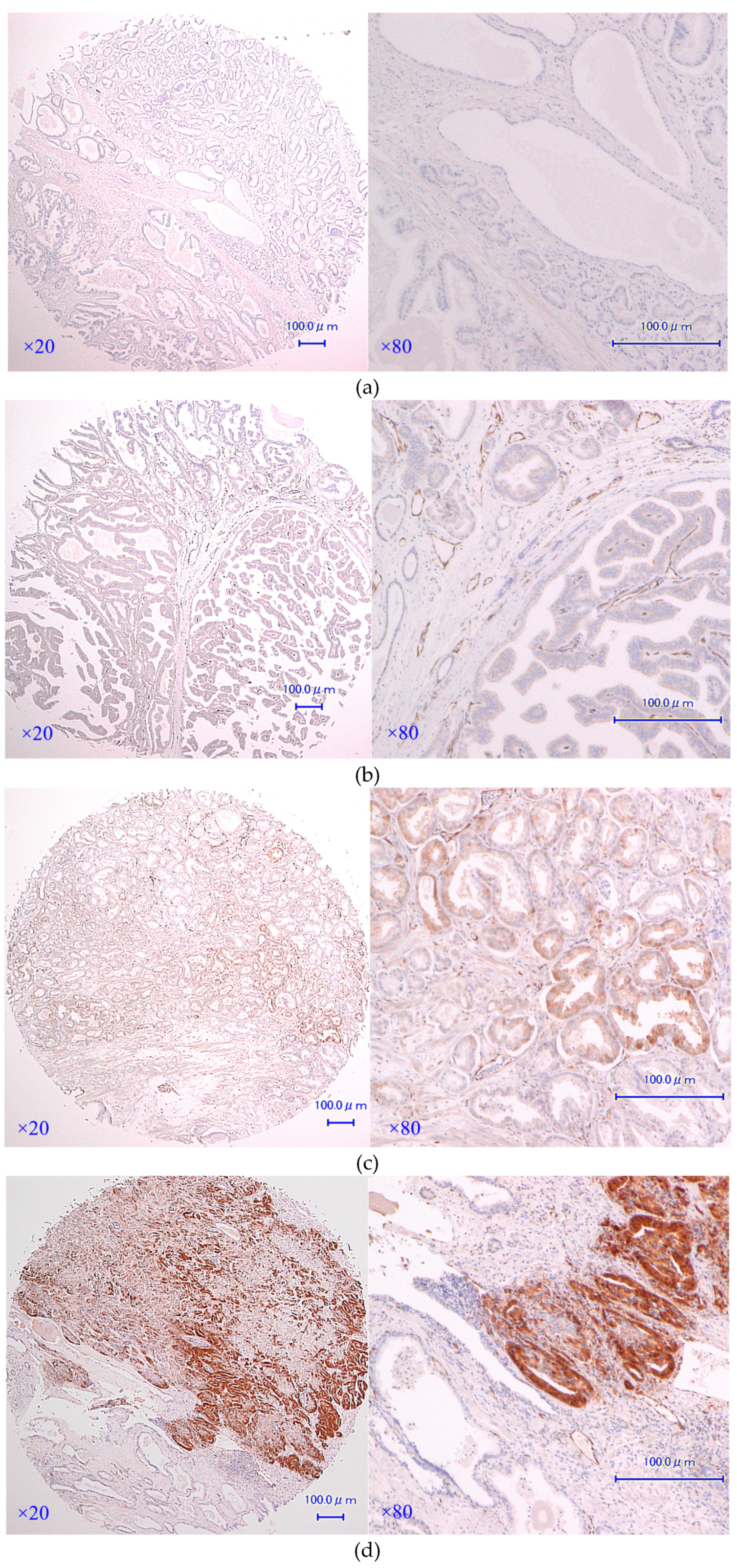
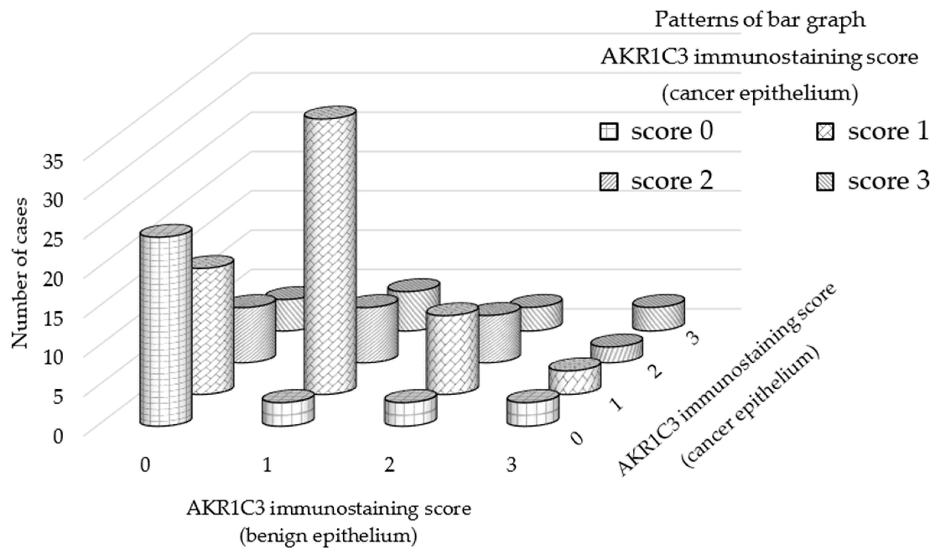
3.1. AKR1C3 Immunostaining of Cancer Epithelium Is Significantly Stronger than That of Benign Epithelia in Patients with Localized Hormone-Naïve Prostate Cancer

Clinical and pathological features are demonstrated and the results of statistical analysis of correlation between demographic features and AKR1C3 expression are presented as p-values in Table 1. Representative immunostaining of AKR1C3 is presented in Figure 1 (Figures S1–S4, Supplementary Materials). The distribution of AKR1C3 immunostaining scores are presented in Figure 2. AKR1C3 immunostaining was significantly stronger in cancer epithelia than in benign ones within the same spots (p < 0.0001). No correlation was observed between GS and AKR1C3 immunostaining in each spot (Table 1). These results suggested that AKR1C3 might play a role in PCa occurrence.

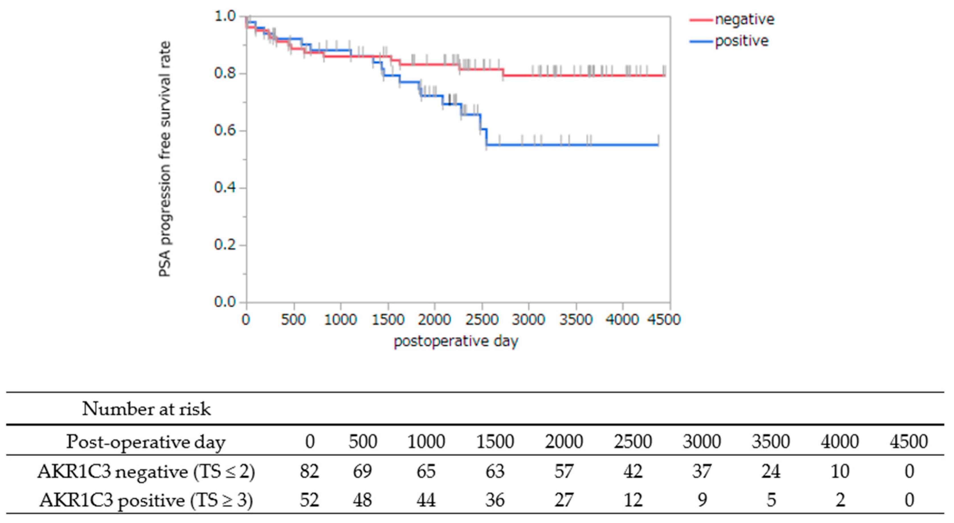
3.2. AKR1C3 Immunostaining of Cancer Cells Is Statistically Associated with PSA Progression-Free Survival after Radical Prostatectomy

The distribution of TS of AKR1C3 immunostaining in cancer cells from RP specimens is listed in Table 2. RP cases were dichotomized according to the median TS of the AKR1C3 immunostainings as: AKR1C3 TS ≤ 2 and AKR1C3 TS ≥ 3. AKR1C3 immunostainings and PSA PFS after RP were statistically correlated, and cases with a high AKR1C3 immunostaining TS had lower PSA PFS than those with a low AKR1C3 immunostaining TS (p = 0.042) (Figure 3). In order to evaluate prognostic factors for PSA PFS after RP, cox proportional hazards regression analysis was conducted with PSA at diagnosis, Gleason grade group, and AKR1C3 expression. AKR1C3 expression was an independent risk factor of PSA failure among our cohorts (p = 0.032, hazard ratio = 2.19) (Table 3). These results showed that AKR1C3 expression of cancer cells may be a prognostic marker of patients who received RP.

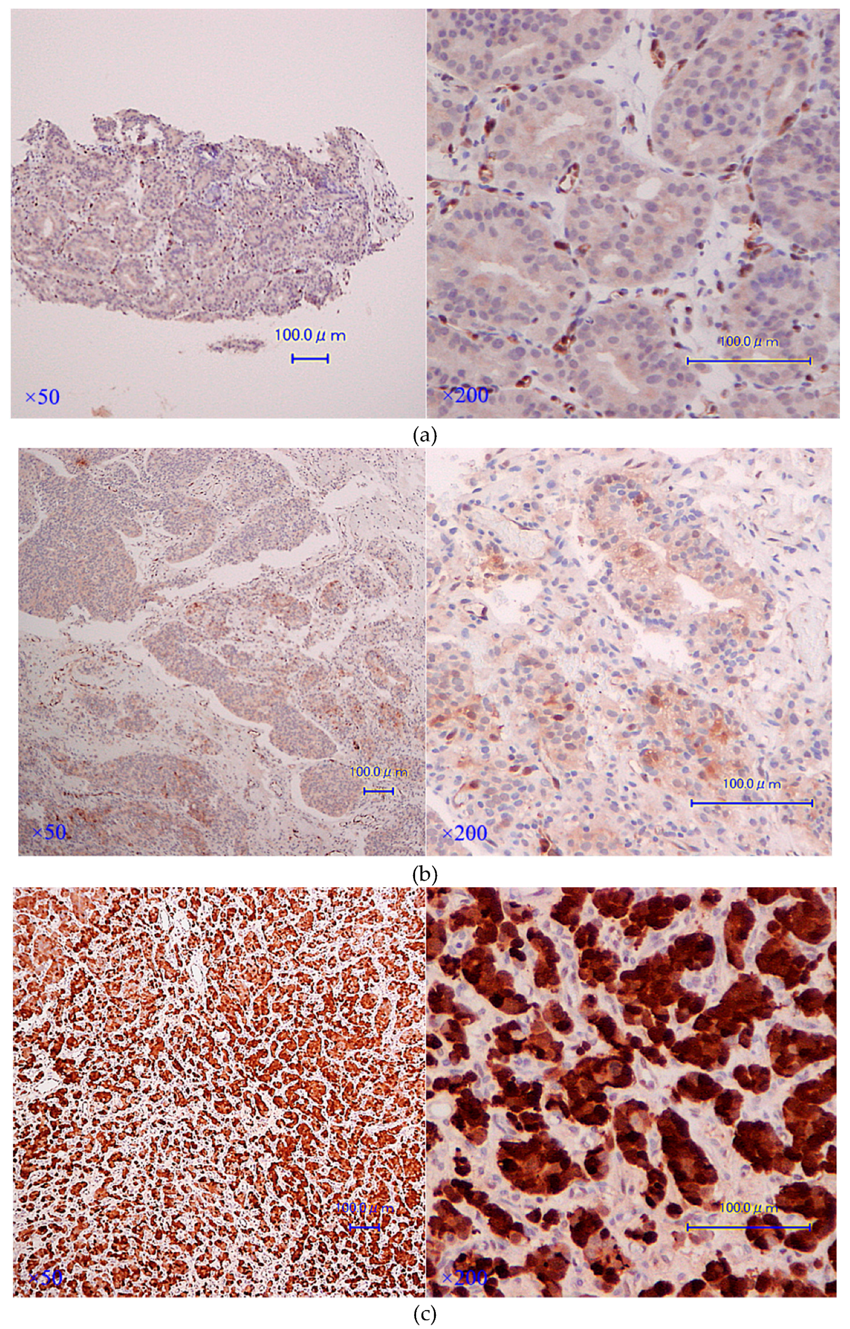
3.3. AKR1C3 Immunostaining Is Significantly Stronger in CRPC Tissues rather than in Hormone-Naïve Ones in the Same Cases

We obtained HNPC and CRPC tissues from the same patients in 11 cases; clinical and pathological characteristics are presented in Table 4. CRPC tissues revealed significantly stronger AKR1C3 immunostaining than hormone-naïve tissues in the same cases (p = 0.0234, Wilcoxon signed-rank test; Table 4). Interestingly, the longitudinal specimens at hormone-naïve, hormone-sensitive, and castration-resistant states were evaluated in one patient. Immunostainings of AKR1C3 are presented in Figure 4; AKR1C3 was gradually up-regulated with disease progression. These results implied that up-regulation of AKR1C3 might be required for the progression to CRPC in some cases, and it could be a therapeutic target for this complicated disease.

4. Discussion

Based on our immunohistochemical analysis of human prostate tissues, we confirmed that AKR1C3 might be crucial in PCa occurrence and progression. In particular, this is the first study to report that AKR1C3 immunostaining increases along the treatment course, that is, AKR1C3 expression elevates from the hormone-naïve status to the CRPC stage in the same patient. Moreover, our study is unique in showing that the PSA PFS rate of patients with high AKR1C3 expression in cancer cells derived from RP specimens was lower than that with low AKR1C3 expression. The majority of our CRPC cases received LHRH analog collectively with bicalutamide as a primary hormonal therapy. Nevertheless, we could not evaluate the correlation of pharmacological treatment with AKR1C3 immunostaining, since CRPC specimens were mostly obtained just during the time of transition to CRPC, before administration of docetaxel or androgen receptor-axis-targeted agents (ARATs), including enzalutamide and abiraterone. To the best of our knowledge, there is no report of AKR1C3 expression after ARAT treatment; however, AKR1C3 activation, both in vitro and in vivo, using PCa cell lines, has been shown as a factor of resistance against ARATs [25,26].
Lin et al. was the first to report a high-titer isoform-specific monoclonal antibody for AKR1C3 and demonstrated AKR1C3 expression in stromal cells, though only faintly in epithelial cells in normal prostate; however, in PCa cells, elevated expression was observed by immunostaining [23]. The same group also reported AKR1C3 to be positive in immunostaining, in 9 out of 11 PCa cases, and showed variation from strong to negative immunostaining within the same tumors, as in our study [26]. They also found no correlation in staining patterns between AR and AKR1C3 expression, consistent with our study (data not shown) [27]. Tian et al. examined the primary PCa biopsy specimens and showed that AKR1C3 expression by immunostaining gradually increases with an elevated GS in PCa epithelium [16]. In our cases, there was no correlation between GS and AKR1C3 immunostaining, which is incompatible with Tian’s results. This might be because our cases underwent RP and most of them had GSs of less than 8, while in Tian’s report, half of the PCa cases had GSs of 8 or higher. Stanbrough et al. revealed that AKR1C3 expression, as per immunohistochemistry, showed negative-to-heterogeneously weak staining in most primary PCa, but intermediate-to-strong AKR1C3 staining in CRPC specimens, which is in agreement with our results [22]. AKR1C3 expression analysis of cancer cells derived from RP showed immunostaining to be correlated with PSA PFS after RP. The result is reproducible even if we adopt IS of AKR1C3 as a representative of its expression (data not shown). In our knowledge, this is the first report of correlation of AKR1C3 expression with PSA PFS after RP. Additionally, AKR1C3 expression was an independent factor of poor PSA PFS when we analyzed the prognostic factors by multivariate analysis including the initial PSA level and Gleason grade group, which were previously considered to be significant predictors of recurrence-free survival after RP [28]. In future, we should analyze AKR1C3 expression and survival after RP in a much larger cohort to understand its role in clinical practice.
AKR1C3 is a multifunctional steroid-metabolizing enzyme that catalyzes androgen, estrogen, progesterone, and PG metabolism [29]. It reduces DHT to form 3α-diol, which is a neurosteroid that acts as a positive allosteric modulator of the gamma-aminobutyric acid type A receptor (GABAAR) [30]. The 3α-diol stimulates AR-negative PCa cells through GABAAR. Further, it up-regulates the epidermal growth factor (EGF) family members in AR-negative PCa cells and stimulates EGF receptor and Src. These results together suggest that AKR1C3 modulates intraprostatic neurosteroid that, in turn, activates AR-negative PCa progression. AKR1C3 is known to regulate its expression by ERG via direct binding to AKR1C3 gene [31]. Furthermore, ERG and AKR1C3 expression in human metastatic PCa tissues was revealed to positively correlate with each other by immunohistochemistry. AKR1C3 is known to regulate the stability of ubiquitin ligase Siah2, and thus enhance the Siah2-dependent regulation of AR activity via non-catalytic function [21]. Wang et al. reported that AKR1C3 could drive epithelial–mesenchymal transition (EMT) by activating the ERK signaling pathways and up-regulating transcription factors such as ZEB1, TWIST1, and SLUG, thereby facilitating PCa metastasis [19]. Therefore, AKR1C3 might be crucial in PCa progression.
There are several limitations in our study. The samples analyzed were relatively small, in particular, in consecutive PCa with CRPC progression. We were unaware of the role of AKR1C3 overexpression, in particular, in progression to CRPC, since we did not analyze the tissue concentrations of T and other androgens. Moreover, we did not know how AKR1C3 overexpression correlated in response to new ARATs, namely enzalutamide and abiraterone. Only three cases received ARATs before CRPC tissue extraction. Case 5 received abiraterone 3 months before laminectomy, case 7 was administered enzalutamide 8 months before TUR-P, and case 11 acquired enzalutamide 10 days before TUR-P.
In conclusion, expression of multifunctional AKR1C3, which is known to be the most up-regulated steroidogenic enzyme in patients with CRPC, might increase with the occurrence and longitudinal progression of the tumor in certain cases [17,32]. Targeting AKR1C3 might overcome the complicated disease, CRPC, and also control resistance against ARATs. We plan to further reveal the functions of AKR1C3 in PCa progression and discover potent and specific drugs to inhibit AKR1C3 function.

Supplementary Materials

The supplementary materials are available online at https://www.mdpi.com/2077-0383/8/5/601/s1.

Author Contributions

Y.M. and T.I. conceived the study. Y.M. and T.I. drafted the manuscript. Y.M. performed statistical analysis. Y.T. and S.S. performed pathological specimen evaluation and scoring. All the authors participated in patient enrolment and data collection, helped to draft the manuscript, and read and approved the final manuscript.

Funding

This study was supported by Grants-in-Aid for Scientific Research (project numbers: 15K10587 and 18H02936) from the Japan Society for the Promotion of Science.

Acknowledgments

We thank all members of Department of Urology and Diagnostic Pathology Kyoto University Graduate School of Medicine, for their devoted support and constructive advice.

Conflicts of Interest

The authors declare no conflicts of interest.

Abbreviations

AKR1C3Aldo-Keto Reductase Family 1 Member C3
PCaprostate cancer
CRPCcastration-resistant prostate cancer
HNPChormone-naïve prostate cancer
ADTandrogen deprivation therapy
Ttestosterone
DHT5α-dihydrotestosterone
ARandrogen receptor
DHEAdehydroepiandrosterone
ERestrogen receptor
PGprostaglandin
TMAtissue-microarray
RPradical prostatectomy
TUR-Ptransurethral resection of the prostate
PFSprogression-free survival
TStotal score
ISintensity score
PSproportion score
GSGleason score
CABcombined androgen blockade
DOCdocetaxel
Abiabiraterone
Enzenzalutamide
LHRHluteinizing hormone-releasing hormone
ARATsandrogen receptor-axis-targeted agents
GABAARgamma-aminobutyric acid type A receptor

References

  1. Siegel, R.L.; Miller, K.D.; Jemal, A. Cancer statistics, 2019. CA Cancer J. Clin. 2019, 69, 7–34. [Google Scholar] [CrossRef]
  2. Cancer Statistics in Japan-2017. Available online: https://ganjoho.jp/en/professional/statistics/brochure/2017_en.html (accessed on 1 April 2019).
  3. Huggins, C.; Hodges, C.V. Studies on Prostatic Cancer. I. The Effect of Castration, of Estrogen and of Androgen Injection on Serum Phosphatases in Metastatic Carcinoma of the Prostate. Cancer Res. 1941, 1, 293–297. [Google Scholar]
  4. Page, S.T.; Lin, D.W.; Mostaghel, E.A.; Hess, D.L.; True, L.D.; Amory, J.K.; Nelson, P.S.; Matsumoto, A.M.; Bremner, W.J. Persistent intraprostatic androgen concentrations after medical castration in healthy men. J. Clin. Endocrinol. MeTable 2006, 91, 3850–3856. [Google Scholar] [CrossRef]
  5. Nishiyama, T.; Hashimoto, Y.; Takahashi, K. The influence of androgen deprivation therapy on dihydrotestosterone levels in the prostatic tissue of patients with prostate cancer. Clin. Cancer Res. 2004, 10, 7121–7126. [Google Scholar] [CrossRef]
  6. Dai, C.; Heemers, H.; Sharifi, N. Androgen Signaling in Prostate Cancer. Cold Spring Harb. Perspect. Med. 2017, 7. [Google Scholar] [CrossRef] [PubMed]
  7. Locke, J.A.; Guns, E.S.; Lubik, A.A.; Adomat, H.H.; Hendy, S.C.; Wood, C.A.; Ettinger, S.L.; Gleave, M.E.; Nelson, C.C. Androgen levels increase by intratumoral de novo steroidogenesis during progression of castration-resistant prostate cancer. Cancer Res. 2008, 68, 6407–6415. [Google Scholar] [CrossRef]
  8. Hofland, J.; van Weerden, W.M.; Dits, N.F.; Steenbergen, J.; van Leenders, G.J.; Jenster, G.; Schroder, F.H.; de Jong, F.H. Evidence of limited contributions for intratumoral steroidogenesis in prostate cancer. Cancer Res. 2010, 70, 1256–1264. [Google Scholar] [CrossRef]
  9. Penning, T.M. Mechanisms of drug resistance that target the androgen axis in castration resistant prostate cancer (CRPC). J. Steroid Biochem. Mol. Biol. 2015, 153, 105–113. [Google Scholar] [CrossRef]
  10. Byrns, M.C.; Jin, Y.; Penning, T.M. Inhibitors of type 5 17beta-hydroxysteroid dehydrogenase (AKR1C3): Overview and structural insights. J. Steroid Biochem. Mol. Biol. 2011, 125, 95–104. [Google Scholar] [CrossRef]
  11. Ricke, W.A.; McPherson, S.J.; Bianco, J.J.; Cunha, G.R.; Wang, Y.; Risbridger, G.P. Prostatic hormonal carcinogenesis is mediated by in situ estrogen production and estrogen receptor alpha signaling. FASEB J. 2008, 22, 1512–1520. [Google Scholar] [CrossRef]
  12. Di Zazzo, E.; Galasso, G.; Giovannelli, P.; Di Donato, M.; Di Santi, A.; Cernera, G.; Rossi, V.; Abbondanza, C.; Moncharmont, B.; Sinisi, A.A.; et al. Prostate cancer stem cells: The role of androgen and estrogen receptors. Oncotarget 2016, 7, 193–208. [Google Scholar] [CrossRef] [PubMed]
  13. Di Zazzo, E.; Galasso, G.; Giovannelli, P.; Di Donato, M.; Castoria, G. Estrogens and Their Receptors in Prostate Cancer: Therapeutic Implications. Front. Oncol. 2018, 8, 2. [Google Scholar] [CrossRef]
  14. Komoto, J.; Yamada, T.; Watanabe, K.; Takusagawa, F. Crystal structure of human prostaglandin F synthase (AKR1C3). Biochemistry 2004, 43, 2188–2198. [Google Scholar] [CrossRef]
  15. Sun, S.Q.; Gu, X.; Gao, X.S.; Li, Y.; Yu, H.; Xiong, W.; Yu, H.; Wang, W.; Li, Y.; Teng, Y.; et al. Overexpression of AKR1C3 significantly enhances human prostate cancer cells resistance to radiation. Oncotarget 2016, 7, 48050–48058. [Google Scholar] [CrossRef] [Green Version]
  16. Tian, Y.; Zhao, L.; Zhang, H.; Liu, X.; Zhao, L.; Zhao, X.; Li, Y.; Li, J. AKR1C3 overexpression may serve as a promising biomarker for prostate cancer progression. Diagn. Pathol. 2014, 9, 42. [Google Scholar] [CrossRef]
  17. Hamid, A.R.; Pfeiffer, M.J.; Verhaegh, G.W.; Schaafsma, E.; Brandt, A.; Sweep, F.C.; Sedelaar, J.P.; Schalken, J.A. Aldo-keto reductase family 1 member C3 (AKR1C3) is a biomarker and therapeutic target for castration-resistant prostate cancer. Mol. Med. 2013, 18, 1449–1455. [Google Scholar] [CrossRef]
  18. Jernberg, E.; Thysell, E.; Bovinder Ylitalo, E.; Rudolfsson, S.; Crnalic, S.; Widmark, A.; Bergh, A.; Wikstrom, P. Characterization of prostate cancer bone metastases according to expression levels of steroidogenic enzymes and androgen receptor splice variants. PLoS ONE 2013, 8, e77407. [Google Scholar] [CrossRef]
  19. Wang, B.; Gu, Y.; Hui, K.; Huang, J.; Xu, S.; Wu, S.; Li, L.; Fan, J.; Wang, X.; Hsieh, J.T.; et al. AKR1C3, a crucial androgenic enzyme in prostate cancer, promotes epithelial-mesenchymal transition and metastasis through activating ERK signaling. Urol. Oncol. 2018, 36, 472.e11–472.e20. [Google Scholar] [CrossRef]
  20. Barnard, M.; Quanson, J.L.; Mostaghel, E.; Pretorius, E.; Snoep, J.L.; Storbeck, K.H. 11-Oxygenated androgen precursors are the preferred substrates for aldo-keto reductase 1C3 (AKR1C3): Implications for castration resistant prostate cancer. J. Steroid Biochem. Mol. Biol. 2018, 183, 192–201. [Google Scholar] [CrossRef]
  21. Fan, L.; Peng, G.; Hussain, A.; Fazli, L.; Guns, E.; Gleave, M.; Qi, J. The Steroidogenic Enzyme AKR1C3 Regulates Stability of the Ubiquitin Ligase Siah2 in Prostate Cancer Cells. J. Biol. Chem. 2015, 290, 20865–20879. [Google Scholar] [CrossRef]
  22. Stanbrough, M.; Bubley, G.J.; Ross, K.; Golub, T.R.; Rubin, M.A.; Penning, T.M.; Febbo, P.G.; Balk, S.P. Increased expression of genes converting adrenal androgens to testosterone in androgen-independent prostate cancer. Cancer Res. 2006, 66, 2815–2825. [Google Scholar] [CrossRef]
  23. Uegaki, M.; Kita, Y.; Shirakawa, R.; Teramoto, Y.; Kamiyama, Y.; Saito, R.; Yoshikawa, T.; Sakamoto, H.; Goto, T.; Akamatsu, S.; et al. Downregulation of RalGTPase-activating protein promotes invasion of prostatic epithelial cells and progression from intraepithelial neoplasia to cancer during prostate carcinogenesis. Carcinogenesis 2019, in press. [Google Scholar]
  24. Lin, H.K.; Steckelbroeck, S.; Fung, K.M.; Jones, A.N.; Penning, T.M. Characterization of a monoclonal antibody for human aldo-keto reductase AKR1C3 (type 2 3alpha-hydroxysteroid dehydrogenase/type 5 17beta-hydroxysteroid dehydrogenase); immunohistochemical detection in breast and prostate. Steroids 2004, 69, 795–801. [Google Scholar] [CrossRef]
  25. Liu, C.; Lou, W.; Zhu, Y.; Yang, J.C.; Nadiminty, N.; Gaikwad, N.W.; Evans, C.P.; Gao, A.C. Intracrine Androgens and AKR1C3 Activation Confer Resistance to Enzalutamide in Prostate Cancer. Cancer Res. 2015, 75, 1413–1422. [Google Scholar] [CrossRef]
  26. Liu, C.; Armstrong, C.M.; Lou, W.; Lombard, A.; Evans, C.P.; Gao, A.C. Inhibition of AKR1C3 Activation Overcomes Resistance to Abiraterone in Advanced Prostate Cancer. Mol. Cancer Ther. 2017, 16, 35–44. [Google Scholar] [CrossRef]
  27. Fung, K.M.; Samara, E.N.; Wong, C.; Metwalli, A.; Krlin, R.; Bane, B.; Liu, C.Z.; Yang, J.T.; Pitha, J.V.; Culkin, D.J.; et al. Increased expression of type 2 3alpha-hydroxysteroid dehydrogenase/type 5 17beta-hydroxysteroid dehydrogenase (AKR1C3) and its relationship with androgen receptor in prostate carcinoma. Endocr. Relat. Cancer 2006, 13, 169–180. [Google Scholar] [CrossRef]
  28. Nelson, C.P.; Dunn, R.L.; Wei, J.T.; Rubin, M.A.; Montie, J.E.; Sanda, M.G. Contemporary preoperative parameters predict cancer-free survival after radical prostatectomy: A tool to facilitate treatment decisions. Urol. Oncol. 2003, 21, 213–218. [Google Scholar] [CrossRef]
  29. Penning, T.M. Aldo-Keto Reductase Regulation by the Nrf2 System: Implications for Stress Response, Chemotherapy Drug Resistance, and Carcinogenesis. Chem. Res. Toxicol. 2017, 30, 162–176. [Google Scholar] [CrossRef]
  30. Xia, D.; Lai, D.V.; Wu, W.; Webb, Z.D.; Yang, Q.; Zhao, L.; Yu, Z.; Thorpe, J.E.; Disch, B.C.; Ihnat, M.A.; et al. Transition from androgenic to neurosteroidal action of 5alpha-androstane-3alpha, 17beta-diol through the type A gamma-aminobutyric acid receptor in prostate cancer progression. J. Steroid Biochem. Mol. Biol. 2018, 178, 89–98. [Google Scholar] [CrossRef]
  31. Powell, K.; Semaan, L.; Conley-LaComb, M.K.; Asangani, I.; Wu, Y.M.; Ginsburg, K.B.; Williams, J.; Squire, J.A.; Maddipati, K.R.; Cher, M.L.; et al. ERG/AKR1C3/AR Constitutes a Feed-Forward Loop for AR Signaling in Prostate Cancer Cells. Clin. Cancer Res. 2015, 21, 2569–2579. [Google Scholar] [CrossRef] [Green Version]
  32. Verma, K.; Gupta, N.; Zang, T.; Wangtrakluldee, P.; Srivastava, S.K.; Penning, T.M.; Trippier, P.C. AKR1C3 Inhibitor KV-37 Exhibits Antineoplastic Effects and Potentiates Enzalutamide in Combination Therapy in Prostate Adenocarcinoma Cells. Mol. Cancer Ther. 2018, 17, 1833–1845. [Google Scholar] [CrossRef]
Figure 1. Representative immunostainings of aldo-keto reductase family 1 member C3 (AKR1C3): (a) score 0 (none staining), (b) score 1 (weak staining), (c) score 2 (intermediate staining), and (d) score 3 (strong staining).
Jcm 08 00601 g001
Figure 2. Difference of AKR1C3 immunostaining score between benign and cancer epithelia in the same individuals. AKR1C3 immunostaining was significantly stronger in the cancer epithelia than in the benign ones at the same spots (p < 0.0001, Pearson’s chi-squared test).
Jcm 08 00601 g002
Figure 3. Kaplan–Meier survival curves revealed that the AKR1C3 positive group (TS ≥ 3) had a significantly lower PSA PFS rate than the negative group (TS ≤ 2) (p = 0.042, log-rank test).
Jcm 08 00601 g003
Figure 4. (ac) AKR1C3 immunostaining of HNPC, Hormone-sensitive prostate cancer (HSPC), and CRPC specimens in the same case (case 9). CAB (leuprolide acetate + bicalutamide) was initiated for case 9 after diagnosis (Figure 3a): (a) HNPC (biopsy) specimens at diagnosis, PSA: 29.9 ng/mL (normal reference range 0-4.0 ng/mL), AKR1C3: Proportion score (PS), 1; Intensity score (IS), 1; Total score (TS), 2; (b) HSPC specimens on day 45 after commencing CAB, PSA: 2.89 ng/mL, AKR1C3: PS, 2; IS, 2; TS, 4; (c) CRPC specimens, PSA: 46.74 ng/mL, AKR1C3: PS, 4; IS, 3; TS 7. After CAB initiation, transurethral lithotomy (TUL) and TUR-P were performed due to repeated urinary retention resulting from bladder stone (Figure 4b). After 1.5 years of bicalutamide, 2 months of flutamide, and 3 months of ethinylestradiol, together with continuous luteinizing hormone-releasing hormone (LHRH) agonist administration, TUR-P was performed due to urinary retention caused by enlargement of the local tumor (Figure 4c). The immunostaining results suggested increased expression of AKR1C3 in PCa tissues with disease progression.
Jcm 08 00601 g004
Table 1. Clinicopathological features of tissue-microarray (TMA) specimens with both benign epithelium and cancer cells in the same spot.
n = 134np Value
Age (mean ± SD)65.6 ± 6.310.6087
PSA, ng/mL (median)7.25 (IQR 5.40–9.88)0.9429
Pathological T stageT2a7N.A.
T2b1
T2c77
T3a37
T3b12
Grade group (pathological)1470.4119 ††
238
337
49
53
p-values indicate correlation of expression intensity with AKR1C3 total score; Kruskal–Wallis test, †† Pearson’s chi-squared test.
Table 2. AKR1C3) total score distribution of TMA specimens.
AKR1C3 ImmunostainingScoren = 134
AKR1C3 total score045
237
311
419
514
64
74
AKR1C3 total score (median)2 (IQR 0–4)
Table 3. Cox proportional hazard regression analysis of prostate-specific antigen progression free survival (PSA PFS) and clinical and pathological variables.
VariablesPSA PFS Rate
HR95% CIp Value †††
PSA level before RP1.121.05–1.180.0003
Grade group1.661.16–2.360.0053
AKR1C3 (TS)2.191.07–4.550.032
HR: hazard ratio, CI: confidence interval, and ††† Wald test.
Table 4. Clinicopathological features and results of AKR1C3 immunostaining total score in 11 cases, including both hormone-naïve and castration-resistant specimens.
CaseAge at DiagnosisClinical Stage at DiagnosisGleason Score at DiagnosisExcised CRPC OrganAge at ExcisionPSA at ExcisionDays from Diagnosis to CastrationTreatment until Excision of CRPC SpecimensAKR1C3 Immunostaining
Total Score
at HNPCat CRPC
Case 157cT3bN1M03 + 3Prostate6225786CAB + DOC67
Case 273cT3bN0M03 + 4Prostate8452860CAB47
Case 360cT3aN0M1c4 + 4Prostate65392603CAB + DOC44
Case 479cT3aN0M04 + 4Penis8216.11051CAB24
Case 568cT3aN0M1c4 + 4Thoracic vertebra7539.21702CAB + DOC + Abi05
Case 670cT3aN0M04 + 3Prostate78408.22112CAB66
Case 776cT4N1M1b4 + 5Prostate8514.072320CAB + Enz47
Case 878cT4N1M1b5 + 4Thoracic vertebra797.58321CAB77
Case 969cT4N0M04 + 5Prostate7146.74567CAB27
Case 1080cT3bN1M04 + 5Prostate826.21544CAB11
Case 1169cT4N0M1b4 + 5Prostate70127318CAB + Enz65
CAB: combined androgen blockade, DOC: docetaxel, Abi: abiraterone, and Enz: enzalutamide.

