Article

Emersion-Associated Responses of an Intertidal Coral and Its Suitability for Transplantation to Ecologically Engineer Seawalls

by
Clara Lei Xin Yong
1,†,
Rosa Celia Poquita-Du
1,*,†,
Danwei Huang
2 and
Peter Alan Todd
1,*
1
Experimental Marine Ecology Laboratory, Department of Biological Sciences, National University of Singapore, 16 Science Drive 4, S3 Level 2, Singapore 117558, Singapore
2
Department of Biological Sciences, Tropical Marine Science Institute and Centre for Nature-Based Climate Solutions, National University of Singapore, 16 Science Drive 4, Singapore 117558, Singapore
*
Authors to whom correspondence should be addressed.
These authors contributed equally for this study.
J. Mar. Sci. Eng. 2021, 9(10), 1096; https://doi.org/10.3390/jmse9101096
Submission received: 20 August 2021 / Revised: 22 September 2021 / Accepted: 1 October 2021 / Published: 7 October 2021
(This article belongs to the Special Issue Enhancing Marine Biodiversity of the Urban Coast)

Abstract

:
There is a growing interest in transplanting corals onto the intertidal section of artificial coastal defences (e.g., seawalls) as an ecological engineering strategy to enhance biodiversity on urban shores. However, this inevitably results in exposure to the harsh environmental conditions associated with emersion (aerial exposure). Although the effects of a multitude of environmental stressors on corals have been examined, their photophysiological and gene expression responses to emersion stress remain understudied, as does the among-genotype variation in these responses. In this study, we conducted an in situ experiment to test the effects of increased daily emersion duration on a locally common intertidal coral, Dipsastraea cf. lizardensis. Coral fragments (n = 3) from five genotypically distinct colonies were subjected to two treatments: (1) increased daily emersion duration (~4.5 h d−1) and, (2) control (~3 h d−1) for three consecutive days during spring low tide. We examined the post-experimental photophysiological responses and expression level of a stress-associated gene, Hsp16. Relative to the controls, coral fragments that were exposed to longer daily emersion duration displayed significantly reduced effective quantum yield, while endosymbiont density varied significantly among genotypes across the experimental conditions. We found no significant differences in chlorophyll a concentration and Hsp16 gene expression level, suggesting that changes in these processes may be gradual and the duration of treatment that the corals were subjected to is likely within their tolerance limits. Taken together, it appears that D. cf. lizardensis displays substantial capacity to cope with sup-optimal conditions associated with emersion which makes it a promising candidate for transplantation onto intertidal seawalls. However, within-species variation in their stress response indicates that not all genotypes respond similarly to emersion, and this should be taken into account when selecting donor colonies for transplantation.

1. Introduction

Almost three billion people live within 100 km from the coast [1]―a number expected to increase to six billion by 2025 [2]. This human pressure has led to massive shoreline changes, especially near coastal cities [3]. Natural intertidal habitats have been extensively replaced with hard amour such as seawalls to curb land erosion and combat threats arising from climate change (i.e., the impending rise of sea levels and increased frequency of floods and storms [4,5,6,7,8]). These artificial structures often support lower biodiversity and different community assemblages due to novel substrate materials, altered hydrodynamics, and reduced habitat complexity [5,7,9,10].
There is substantial potential to increase the ecological value to seawalls and other marine infrastructure (ocean sprawl) to mitigate the loss of coastal communities [11]. To achieve this, several studies have added enhancements such as crevices and pools to existing seawalls (see review in [5]) to create refuges for organisms and encourage resource partitioning [12,13] with the goal of boosting species richness [7,14]. In the tropics, some researchers have also attempted to increase biodiversity on seawalls by transplanting corals directly onto them [15,16]. As engineer species, scleractinian corals provide the reefs’ framework [17] and structural complexity which helps support additional species [18]. Although corals transplanted onto intertidal seawalls are exposed to environmental stressors associated with emersion, certain species are able to survive and provide ecosystem services, such as food for parrotfish [15].
The intertidal zone is known to be a harsh environment, with higher fluctuations in temperature, wave energy, salinity, and solar radiation [19] compared to the subtidal zone. Intertidal sessile organisms are frequently subjected to emersion due to the regular yet variable changes in tide levels [19], during which the intensity of these stressors is amplified along with additional factors such as desiccation [19,20]. Romaine et al. [21] observed reduced maximum gross photosynthesis and growth rates among aerially exposed corals, while Castrillón-Cifuentes et al. [19] reported reduced fecundity among corals in response to increased emersion duration. Corals found living in intertidal environments may already be at their physiological limit [22] and their capacity to cope with increased emersion duration is not fully understood.
Ng et al. [15] showed that some coral and sponge species survive better than others after being transplanted onto intertidal seawalls which were emersed for ~2 h daily for up to 3 consecutive days each month. However, little is known regarding among-genotype, phenotypically plastic, responses to emersion stress. To our knowledge, only a single study has demonstrated within-species variation in plastic responses of corals (i.e., Pocillopora acuta) to emersion stress (see [23]). Phenotypic plasticity, i.e., the capacity of an individual to change its behaviour, physiology, morphology and/or life history [24,25,26] in response to a change in environmental conditions [24], includes acclimatization responses. There are often different degrees of phenotypic plasticity among genotypically distinct coral colonies (or “genet”; see examples [27,28]). This variation can be illustrated by a reaction norm (see [29]), which illustrates the response of different genotypes to different environmental conditions. Most studies have tested morphologically and physiologically plastic responses of coral species to different types of stress, however, during the last decade, more have examined gene expression levels [30,31,32,33,34]. As sessile organisms, phenotypic plasticity can benefit corals as they are unable to escape their surrounding conditions [35,36,37,38]. Identifying responses to stress and genotypes that can tolerate emersion conditions can help inform coastal ecological engineering projects using coral transplantation.
By 2011, Singapore had already lost approximately 55% of its intertidal coral reef flats due to land reclamation [6] and installation of coastal armour [39]. This figure is expected to increase as more land reclamation projects are underway [6]. Along the southern coastline, corals have colonized areas of intertidal seawalls up to 0.8 m above chart datum (CD, lowest astronomical tide) [40,41], highlighting the potential to increase the biodiversity of seawalls through transplantation of these engineer species. This study aimed to examine the responses of five genotypes of a locally common intertidal coral, Dipsastraea cf. lizardensis, to increased emersion stress in the intertidal environment, with a view to identifying genotypes that would be suitable for transplantation onto seawalls. Specifically, we tested photophysiological performance, stress-associated gene regulation, and among-genotype variation in the level of plasticity. We hypothesized that: (1) Dipsastraea cf. lizardensis will exhibit lower photophysiological performance in response to increased emersion duration; (2) it will exhibit differential regulation of stress-associated genes in response to increased emersion duration; and (3) there exists variation in plasticity among genotypes.

2. Materials and Methods

2.1. Study Site, Species and Sample Collection

Singapore experiences semidiurnal tides with a tidal range of approximately 3 m. Low tides occur either early in the morning (between 04:00 and 10:00) or late in the afternoon (between 16:00 and 22:00; [42]) and intertidal corals can be emersed for these periods during low spring tides [15]. The study was conducted on the intertidal reef flat along the westward facing coastline of Pulau Hantu Besar (1°13′35.9″ N, 103°44′49.1″ E), an island off the south of Singapore’s main island (Figure 1).
Dipsastraea cf. lizardensis is a massive coral species commonly found in Singapore reefs, including intertidal areas, suggesting some capacity to withstand emersion stress. This intertidal form appears to be distinct, both genetically and morphologically, from the archetypical D. lizardensis (see phylogenetic analysis in Supplementary Materials). It is also present in the subtidal zone to about 5 m in depth [43]. There is only a single morphological form of D. cf. lizardensis in our collection, and it is superficially similar to the subtidal colonies observed in Huang et al. [44] and Chow et al. [43], although detailed morphological and morphometric analyses may uncover minute differences between them.
Out of six tagged colonies (at least 10 m apart along the intertidal zone), five colonies (G1–G5) were confirmed D. cf. lizardensis based on the mitochondrial intergenic region (see Supplementary Figure S1). Due to the nature of their growth form [45], massive corals are less susceptible to fragmentation by physical disturbances. Hence, the tagged colonies were unlikely to be clonemates. To verify their genetic distinction, we attempted to genotype the colonies using the commonly used nuclear marker, internal transcribed spacer 2 (ITS2) (see Supplementary Materials). Out of the five colonies, only gDNA samples from G1, G3 and G4 were successfully sequenced, and these were found to be genetically distinct. The average genetic identity match between the three genotypes was 97.87 ± 0.41%. (Supplementary Table S1). Based on the >10 m distance among colonies [46,47] and our molecular results, we assumed all five colonies represented distinct genotypes. Population genetics studies conducted to date in Singapore have not detected unambiguous clonality [48,49].
From each colony, six fragments (diameter: 50 mm) were obtained using a diamond core drill bit attached to a 36 V cordless drill, hammer and chisel (N = 30). To minimize within-colony variability, all coral fragments were taken from mid-level height of a colony. Fragments were immediately attached to polyvinyl chloride (PVC) mesh with epoxy putty and transplanted onto submerged holding platforms near the donor colonies. Coral fragments were allowed to acclimatize for two weeks to recover from the stress of being removed from their parent colony.

2.2. Experimental Setup

Coral fragments were subjected to two treatments: increased emersion duration (‘emersed’ hereafter) and a control. The different emersion durations were created by fabricating platforms of different heights. Increased emersion duration was achieved by placing fragments approximately 0.4 m above substratum and the control was placed at the height of the donor coral where the fragments were extracted (approximately 0.1 m above substratum). Emersed, control and holding platforms were made using PVC pipes with 2 × 2 cm square mesh cable-tied over the top to create a surface for the coral fragments. Control platforms were installed next to the coral colonies at the same height where the fragments were cored. Emersed platforms were installed on the upper part of the reef flat. Three fragment replicates from each genotype were haphazardly chosen, assigned and transplanted onto each emersed and control platform.
The experiment was carried out for three days between 31 August and 3 September 2019 during a spring tide period. At each platform, light level (in photosynthetically active radiation (PAR)) and temperature were measured using Odyssey Integrated PAR Sensor (Dataflow Systems, Christchurch, New Zealand) and HOBO Water Temp Pro v2 (Onset Computer Corporation, Bourne, MA, USA), respectively. Loggers were set to log every two minutes for the entire duration of the experiment. At the end of experiment, all coral fragments were collected. Each fragment (=sample) was halved with a sanitized chisel and hammer, of which one half was immediately preserved in RNAlater™ (Thermo Fisher Scientific, Massachusetts, USA) for differential gene expression analyses while the other half was wrapped in aluminium foil for photophysiological analyses. Only light data from the first day of treatment period was used due to logger failure.

2.3. RNA Extraction and Reverse Transcription (RT)

Total RNA for each coral sample (~150–200 mg of tissue) was extracted using the TRIzol (InvitrogenTM, Thermo Fisher Scientific, Waltham, MA, USA) following the manufacturer’s protocol and a few modifications following Barshis et al. [37]. The quality of extracted total RNA was examined via gel electrophoresis for presence of two ribosomal RNA bands. Quantification of RNA yield was performed using Qubit fluorometer with RNA Broad Range assay kit (ThermoFisher Scientific, Massachusetts, USA).
Complementary DNA (cDNA) was prepared for each sample from the amount of total RNA amounting to 1.0 μg using a one-step process with iScript™ gDNA Clear cDNA Synthesis Kit (Bio-Rad Laboratories, Inc., Hercules, CA, USA) according to manufacturer’s protocol.

2.4. Primer Design, Validation and Primer Efficiency Determination

Stress-associated genes that were known to be differentially expressed under heat and light stress from previous studies (e.g., [31,33,50]) were selected as genes of interest (GOIs) for this study (Table 1). Transcript sequences of genes predicted to be protein-coding from Coelastrea aspera, a closely-related species to D. lizardensis [44], were obtained from Reef Genomics database (http://reefgenomics.org/ (accessed on 4 October 2021); [51,52]) instead due to lack of transcriptome data for D. lizardensis. Transcript sequences were aligned against known protein sequences curated in the Uniprot Swiss-Prot protein database [53] using blastx in QIAGEN CLC Genomics Workbench (V10.0.1; Qiagen Bioinformatics GmbH, Hilden, Germany) with a threshold e-value of ≤1 × 10⁻⁵.. Sequences that matched to each GOI were collated to create consensus sequences for designing of primers using Primer-BLAST [54]. Selection of primer pairs was performed based on the following criteria: (i) a maximum temperature difference of one; (ii) GC content range of 50–60%; and (iii) sequence length of around 200 base pairs.
Specificity of the primer was validated by performing polymerase chain reaction (PCR; Labcycler Gradient and Thermoblock 96, Sensoquest, Göttingen, Germany) using the designed primers (Table 1) and prepared cDNA samples for each GOI. Each reaction consisted of 12.5 μL of GoTaq Green Master Mix (Promega, WI, USA), 1.0 μL of each forward and reverse primer (10 μM), 8.5 μL of water and 2.0 μL of diluted cDNA sample. PCR reaction steps were: (i) 94 °C for 2 min; (ii) 40 cycles of 94 °C for 45 s and 55–60 °C for 60 s; (iii) and 72 °C for 5 min. Successful amplifications of target genes were examined via gel electrophoresis.
Primer efficiency (E) was determined following Kenkel et al. [31] and Pfaffl [55]. A series of dilutions of cDNA samples, covering three orders of magnitude (50–0.0625 ng of equivalent RNA concentration) were prepared. Diluted cDNA samples were subsequently added to SsoAdvanced Universal Inhibitor-Tolerant SYBR Green Supermix (Bio-Rad Laboratories, Inc., California, USA) and primers to make up a qPCR reaction volume of 15.0 µL. Samples were amplified using qPCR (CFX96 Touch Real-Time PCR Detection System, Bio-Rad Laboratories, Inc., USA) following the thermocycling protocol: (i) 98 °C for 3 min; (ii) 40 cycles of 98 °C for 15 s and 60 °C for 45 s. Melt curve analysis was performed after qPCR amplification to ensure specific product was amplified. Cycle quantification (Cq) values were plotted against log2 cDNA and the regression slope was used to determine E via the expression E = 2−(1/slope). Primers with E ranging between 1.85 and 2.15 were selected for downstream gene expression level quantification via qPCR.

2.5. Quantification of Gene Expression

Amplifications of each GOI were performed following the qPCR steps established in Section 2.3. Three technical replicates of qPCR reactions (15 µL volume per reaction) were performed on each cDNA sample (50 ng µL−1 concentration) along with no template control and negative-RT control to ensure samples were void of contamination. Cycle of quantification (Cq) values for each sample were converted to molecule counts following the expression: Count = E(Cq1 − Cq), where Cq1 values are the number of PCR cycles needed for one target molecule, using MCMC.qpcr package for statistical analysis [56] on statistical software R (version 4.0.4; [57]).

2.6. Photophysiological Analyses (Effective Quantum Yield of Photosystem II; Endosymbiont Density, Chlorophyll a Concentration)

At the end of the experiment, effective quantum yield of Photosystem II (ΦPSII) (a parameter that can be measured using Diving-PAM fluorometer) for each fragment was determined using a pulse amplitude modulated (PAM) fluorometer (Diving-PAM-II underwater fluorometer, Walz, Effeltrich, Germany). ΦPSII is a widely used metric to assess the photosynthetic efficiency of corals, which is related to the organism’s ‘health’ [58]. Three random point measurements were taken at 5.0 mm distance from the surface of each fragment. Due to highly variable ambient light intensity in environment, ΦPSII values were normalized against light readings (in PAR) that were measured concurrently with ΦPSII using an external light sensor attached to the sample holder (DIVING-Universal Sample Holder, Walz, Germany) with the fluorometer’s fibre optic probe.
Algal endosymbiont density and chlorophyll (chl) a concentration were determined following Ben-Haim et al. [59] with a few modifications. To obtain the endosymbionts from coral fragments, coral tissue was extracted from the skeleton using an airbrush connected to a SCUBA tank. Each coral fragment was placed in an enclosed container with a punctured hole for the airbrush nozzle to fit through. Extracted tissue was collected by washing down tissue from the sides of the container using autoclaved seawater (ASW). Thereafter, the resultant slurry was filtered through a 100.0 µm followed by 50.0 µm plankton mesh to remove coral tissue and mucus. The resultant filtrate was centrifuged at 4000 rpm for 30 min at 20.0 °C and the supernatant was removed. The pellet was resuspended with ASW and sub-sampled (1 mL; five subsamples per fragment replicate) for enumeration of endosymbiont cells using Neubauer Improved Haemocytometer under a microscope.
To determine chl a concentration, the remaining endosymbiont cell suspension was centrifuged at 5000 rpm for 15 min at 4.0 °C and the supernatant was removed. The chl a pigment was extracted from endosymbionts by resuspending the pellet in 100% molecular grade acetone to a volume of 4.0 mL. Resuspended sample was incubated at 4.0 °C for 24 h, followed by centrifugation at 5000 rpm rpm for 10 min at 4.0 °C. Absorbance values of the supernatant were measured using the UV-Vis spectrophotometer (UV-1280, Shimadzu, Kyoto, Japan) at 630 nm, 663 nm, and 750 nm. Chl a content (in µg) was quantified following the equation, 11.43 × A630−0.64 × A663 [60]. The content was normalized against sample volume (after algal endosymbiont density sub-sampling) to obtain chl a concentration. Both measurements of endosymbiont density and chl a concentration were normalized to coral live tissue coverage obtained through images of coral fragments analysed with Coral Point Count with Excel extensions 4.1 (CPCe; [61]). Two of the coral fragments kept for photophysiological analyses were degraded after storage and hence excluded from the analyses for both endosymbiont density and chl a concentration.

2.7. Statistical Analyses

All analyses were performed using R (ver 4.0.4; [57]). To test whether photophysiological responses were influenced by genotype and treatments, the data was fitted into a Linear Mixed Effects (LME) model using ‘nlme’ package. Stepwise model simplification and selection of final model were based on Akaike Information Criterion. The data was later fitted into linear models due to the elimination of all random effects during stepwise simplification and observation of normality and non-heteroscedascity, which were checked using Shapiro–Wilk’s test [62] and standard residual diagnostic plots, respectively. Post-hoc analyses with Tukey’s adjustments were performed using emmeans package [63]. To test whether gene expression levels were influenced by genotype and treatments, the count data obtained from the MCMC.qpcr run was log-transformed and checked for normality and heteroscedascity before being fitted into an LME model followed by a linear model after elimination of random effects.

3. Results

At the end of the experiment, all coral fragments exposed to the different treatments were alive and did not show any signs of bleaching.
Corals that were transplanted to emersed and control platforms were exposed for approximately 4.5 h and 3.0 h of emersion, respectively. Emersion duration was estimated by identifying a sudden change in temperature around 06:00 and 10:00 (Supplementary Figure S2), which matched the receding and approaching tide level [42]. Increased emersion duration was accompanied by increased light intensity and temperature. Corals that were exposed to longer emersion duration experienced greater ranges of temperature (control: 26.39–31.79 °C; emersed: 26.82–34.83 °C) and light intensity (control: 0.0–440.88 µmol photons m−3 s−1; emersed: 0.0–703.60 µmol photons m−3 s−1).

3.1. Stress-Associated Gene Expression Responses

Hsp16, Adk and IκB were successfully amplified during primer validation using the designed primers, but not GFP. However, during the primer efficiency tests via qPCR, only Hsp16 was successfully amplified and showed an E value within the specified primer efficiency prerequisiteE value within the specified primer efficiency prerequisite (E = 1.99). Results showed no clear pattern for Hsp16 under increased emersion duration (Figure 2). There were no significant differences in Hsp16 expression levels among genotypes (F4,20 = 1.161; p > 0.05) and between treatments (F1,20 = 0.013; p > 0.05), with no significant influence of genotype on corals subjected to increased emersion (i.e., genotype × treatment, or G × E interaction; F4,20 = 0.212, p > 0.05).

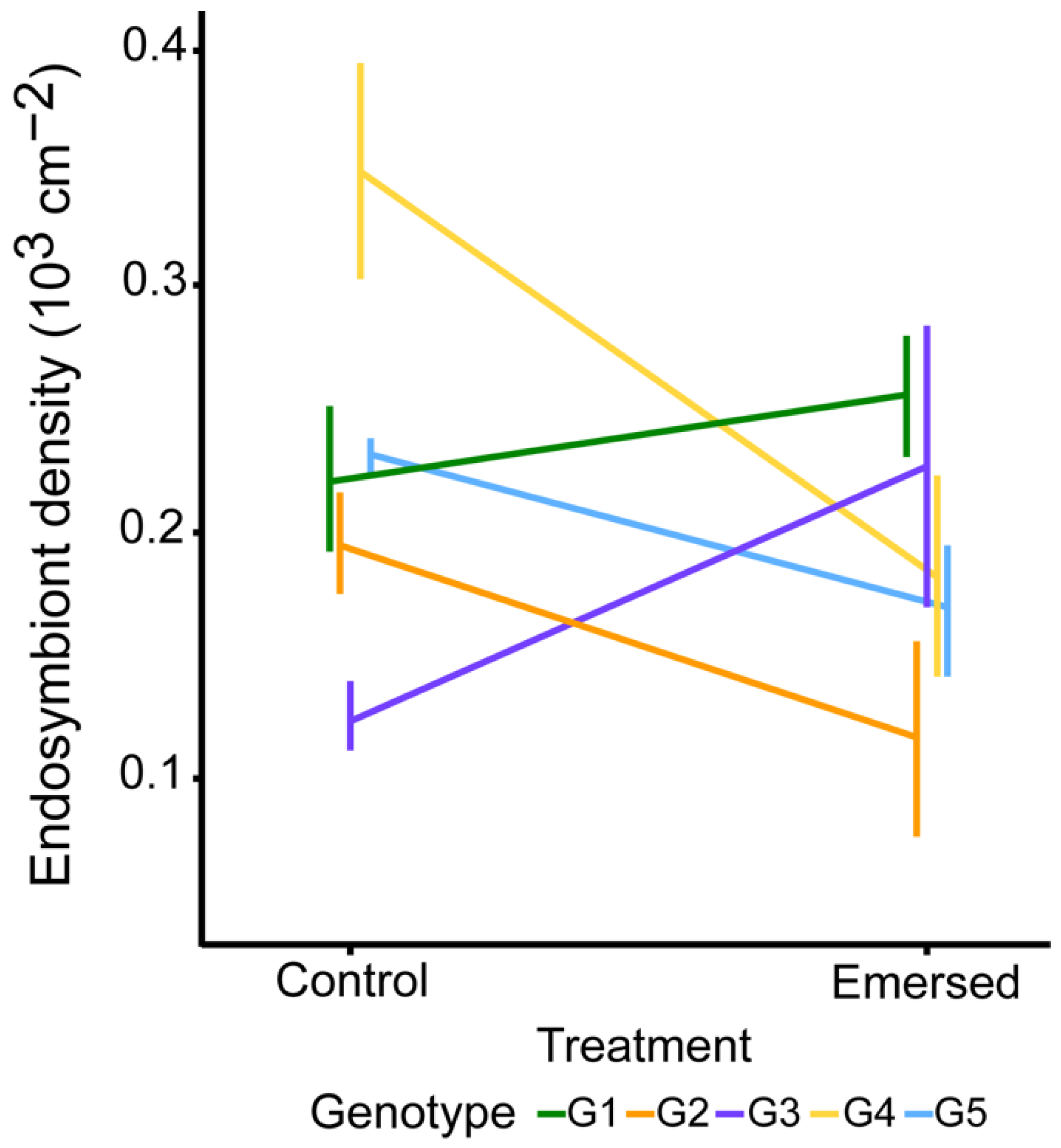
3.2. Photophysiological Responses (Effective Quantum Yield of Photosystem II (ΦPSII), Endosymbiont Density and chl a Concentration)

Overall, mean ΦPSII values were observed to decrease with increased emersion duration (Figure 3). ΦPSII was significantly different between treatments (F1,20 = 31.627; p < 0.01) and among genotypes (F4,20 = 4.167; p < 0.05) but there was no significant G × E interaction (F4,20 = 1.164; p > 0.05).
There was no distinguishable trend in algal endosymbiont density with increased emersion duration across genotype (Figure 4). Endosymbiont density was not significantly different among genotypes (F4,18 = 2.677; p > 0.05) and between treatments (F1,18 = 2.234; p > 0.05). However, changes in endosymbiont density for corals exposed to increased emersion duration relative to the control varied significantly among genotypes (F4,18 = 3.720; p < 0.05).
There was an overall slight decrease in chl a under increased emersion duration except for G2 (Figure 5). Two-way Analysis of Variance (ANOVA) analysis revealed that chl a concentration was not significantly different among genotypes (F4,18 = 0.951; p > 0.05) and between treatments (F1,18 = 1.418; p > 0.05) and there was no significant G × E interaction (F4,18 = 0.616; p > 0.05).

4. Discussion

With the growing interest in transplanting corals onto the intertidal seawalls as an ecological engineering strategy, it is critical to understand how different target species, or different genotypes within species, withstand exposure to the harsh environmental conditions associated with emersion. To date, only Pang et al. [23] has reported among-genotype variation of corals to emersion, using Pocillopora acuta as the model species, and demonstrated substantial intraspecific differences. Here, we examined a coral species more commonly found in the intertidal zone, Dipsastraea cf. lizardensis, and investigated among-genotype variation in photophysiological and gene expression level responses to increased emersion stress. Our results showed that algal endosymbiont density differed significantly among genotypes and treatments (i.e., emersed and control). The photosynthetic efficiency of coral fragments under increased emersion duration was significantly lower than controls. However, corals maintained similar Hsp16 expression levels and chl a concentration regardless of the treatment. Genotype × environment effects (crossing reaction norms) were detected for Hsp16 expression levels and photophysiological parameters. These findings indicate that D. cf. lizardensis is capable of acclimatizing to increased emersion by regulating their photophysiology, but the response can vary among genotypes.
Upregulation of heat-shock protein (hsp) genes in thermally-stressed corals has been reported in several studies (see [31,33,34,36,64,65]) and these responses are likely coping mechanisms of corals to elevated temperature. However, some research (see [37,50,66]) has also observed little to no differential expression of hsp genes, including D. cf. lizardensis in the current study. Bellantuono et al. [50] suggested that the absence of differential gene expression was because the genes that were upregulated returned to normal levels before the sampling timepoint. Barshis et al. [37], on the other hand, described two hypothetical physiological responses that could have taken place. Firstly, these genes could have been constantly expressed at higher levels as a pre-emptive response to frequently occurring stress. Secondly, the colonies could have already developed resilience against these stressors present in their natal site and hence displayed lower stress levels and changes in stress-associated gene expression levels. Barshis et al. [37] also noted that thermally resilient Acropora hyacinthus colonies have reduced transcription of small heat shock proteins including Hsp16.2 as compared to colonies that were more thermally sensitive. Although a comparison with a more thermally sensitive population was not conducted here, it may be that the coral has developed similar physiological responses against emersion stress, explaining the non-significant changes in Hsp16 expression levels.
We found a significant reduction in photosynthetic efficiency of D. cf. lizardensis in response to increased emersion (that included increased light and heat intensity) duration, corroborating the findings of Pang et al. [23]. Elevated light intensity and temperature can impose photoinhibition and damage on the photosynthetic apparatus of algal endosymbionts such as the Photosystem II [67,68]. When the rate of damage is faster than the rate of repair, reduction in photosynthetic efficiency occurs [69] and, for various coral species, increased temperature and/or light intensity has led to a decrease in photosynthetic efficiency [68,70]. Under intensive heat and/or light stress, corals may also undergo bleaching via reduction in endosymbiont density or pigment concentration [71,72]. Our results did not identify any emersion-driven change in chl a concentration and distinguishable trend in endosymbiont density, but the reaction norms suggested that the genotypes varied in the level of plasticity for endosymbiont density.
Variation in plastic responses among genotypes could be due to adaptations of a genotype to its natal site. Bowden-Kerby [73] and Howells et al. [38] reported that the genetic make-up of corals (i.e., genotype) influence their varying acclimatization capacities. These studies transplanted different genotypes of corals between contrasting environments (e.g., inshore and offshore habitats), and the results revealed that the acclimatization capacity of a genotype in a new environment may be limited or wide-ranging depending on their natal environment [38,73]. In the present study, the difference in depth between the shallowest and deepest colonies tested was 0.41 m and maximum temperature experienced by the two colonies during emersion differed by 2–4 °C (due to daily variation in tidal fluctuation). Thus, it is possible that the observed response variation among genotypes of D. cf. lizardensis here was influenced by the natal environment of each colony.
Is D. cf. lizardensis suitable for transplantation onto seawalls to increase biodiversity? Our results revealed that this species can cope with increased emersion by mainly regulating its photophysiology. Overall, D. cf. lizardensis displayed phenotypic plasticity in response to emersion but the varying levels exhibited among genotypes suggest a range of physiological capacity to cope with the stressors related to emersion. Although increased emersion stress caused a slight decrease in photosynthetic efficiency, no tissue bleaching was observed in all fragments. In addition, these fragments were subjected to an estimated 4.5 h of daily emersion, a longer duration compared to the corals that were transplanted onto the intertidal seawall by Ng et al. ([15]; approximately two hours over three consecutive days). Nevertheless, additional work is required to determine the acclimatization responses of D. cf. lizardensis after weeks and months of exposure to this new environment.
In general, D. cf. lizardensis appears to be a promising candidate for transplantation onto intertidal seawalls as an ecological engineering strategy. Where many previous studies have discussed “winners” and “losers” when comparing among-species capacity to survive challenging environments [74,75,76], our findings underscore the need to examine responses at a finer resolution (i.e., genotype-level). It is important to choose genotypes that are resistant to emersion stress as they may be more likely to survive the extreme conditions characteristic of intertidal seawalls. This genotype-level approach can be applied to other ecological engineering strategies, including coral restoration and seeding artificial reefs, to increase the survivability of transplants and enhance the effectiveness of these conservation efforts.

Supplementary Materials

The following are available online at https://www.mdpi.com/article/10.3390/jmse9101096/s1, Figure S1: Maximum likelihood tree of coral species under the family Merulinidae based on the mito-chondrial intergenic region (iGR), showing five tagged colonies (G1-G5, in red) clustered under Dipsastraea cf. lizardensis, Figure S2: Temperature (A), light intensity (B) and estimated emersion period pro-file, Table S1: Genetic identity matches between pairs of genotypes. References [77,78,79,80,81,82,83,84,85,86] are cited in the Supplementary Materials.

Author Contributions

Conceptualization, C.L.X.Y., R.C.P.-D. and P.A.T.; methodology, C.L.X.Y., R.C.P.-D., D.H. and P.A.T.; investigation, C.L.X.Y., R.C.P.-D. and D.H.; formal analysis, C.L.X.Y., R.C.P.-D.; data curation, C.L.X.Y., R.C.P.-D.; writing—original draft, C.L.X.Y., R.C.P.-D., D.H. and P.A.T.; writing—review and editing, C.L.X.Y., R.C.P.-D., D.H. and P.A.T.; supervision, R.C.P.-D., D.H. and P.A.T.; funding acquisition, P.A.T. All authors have read and agreed to the published version of the manuscript.

Funding

This research was funded by the National Research Foundation of Singapore under the Marine Science Research and Development Programme (MSRDP-P05).

Institutional Review Board Statement

Not applicable.

Informed Consent Statement

Not applicable.

Data Availability Statement

The data presented in this study are openly available in Zenodo at https://doi.org/10.5281/zenodo.5205263 (accessed on 7 October 2021).

Acknowledgments

We thank the lab members from the Experimental Marine Ecology Laboratory for their assistance in fieldwork. We also thank members from the Reef Ecology Laboratory for sharing their knowledge on the molecular techniques and statistical analyses. Lastly, we thank the undergraduate students Annie Chew, Jonas Khaw and Ng Yan Ze for their assistance with the laboratory work. All research was carried out with permission of the Singapore National Parks Board (Permit no. NP/RP17-099-1b).

Conflicts of Interest

The authors declare no conflict of interest.

References

  1. Burke, L.; Reytar, K.; Spalding, M.; Perry, A. Reefs at Risk Revisited; World Resources Institute: Washington, DC, USA, 2011. [Google Scholar]
  2. Creel, L. Ripple Effects: Population and Coastal Regions. Available online: https://pdf.usaid.gov/pdf_docs/Pnadd169.pdf (accessed on 7 October 2021).
  3. Todd, P.A.; Heery, E.C.; Loke, L.H.L.; Thurstan, R.H.; Kotze, D.J.; Swan, C. Towards an Urban Marine Ecology: Characterizing the Drivers, Patterns and Processes of Marine Ecosystems in Coastal Cities. Oikos 2019, 128, 1215–1242. [Google Scholar] [CrossRef]
  4. Charlier, R.H.; Chaineux, M.C.P.; Morcos, S. Panorama of the History of Coastal Protection. J. Coast. Res. 2005, 211, 79–111. [Google Scholar] [CrossRef]
  5. Firth, L.B.; Knights, A.M.; Bridger, D.; Evans, A.J.; Mieszkowska, N.; Moore, P.J.; O’Connor, N.E.; Sheeham, E.V.; Thompson, R.C.; Hawkins, S.J. Ocean Sprawl: Challenges and Opportunities for Biodiversity Management in a Changing World. In Oceanography and Marine Biology: An Annual Review; Hughes., R.N., Hughes, D.J., Smith, I.P., Dale, A.C., Eds.; CRC Press: Boca Raton, FL, USA, 2016; Volume 54, pp. 193–269. [Google Scholar] [CrossRef]
  6. Lai, S.; Loke, L.H.L.; Hilton, M.J.; Bouma, T.J.; Todd, P.A. The Effects of Urbanisation on Coastal Habitats and the Potential for Ecological Engineering: A Singapore Case Study. Ocean Coast. Manag. 2015, 103, 78–85. [Google Scholar] [CrossRef]
  7. Loke, L.H.L.; Bouma, T.J.; Todd, P.A. The Effects of Manipulating Microhabitat Size and Variability on Tropical Seawall Biodiversity: Field and Flume Experiments. J. Exp. Mar. Bio. Ecol. 2017, 492, 113–120. [Google Scholar] [CrossRef]
  8. Morris, R.L.; Heery, E.C.; Loke, L.H.; Lau, E.; Strain, E.; Airoldi, L.; Alexander, K.A.; Bishop, M.J.; Coleman, R.A.; Cordell, J.R.; et al. Design Options, Implementation Issues and Evaluating Success of Ecologically Engineered Shorelines. Oceanography and Marine Biology: An Annual Review; CRC Press: Boca Raton, FL, USA, 2019; Volume 57. [Google Scholar]
  9. Firth, L.B.; Thompson, R.C.; Bohn, K.; Abbiati, M.; Airoldi, L.; Bouma, T.J.; Bozzeda, F.; Ceccherelli, V.U.; Colangelo, M.A.; Evans, A.; et al. Between a Rock and a Hard Place: Environmental and Engineering Considerations When Designing Coastal Defence Structures. Coast. Eng. 2014, 87, 122–135. [Google Scholar] [CrossRef]
  10. Perkol-finkel, S.; Hadary, T.; Rella, A.; Shirazi, R.; Sella, I. Design of Coastal and Marine Infrastructure. Ecol. Eng. 2017, 740, 139981. [Google Scholar] [CrossRef]
  11. Chapman, M.G.; Underwood, A.J. Evaluation of Ecological Engineering of “Armoured” Shorelines to Improve Their Value as Habitat. J. Exp. Mar. Bio. Ecol. 2011, 400, 302–313. [Google Scholar] [CrossRef]
  12. Loke, L.H.L.; Todd, P.A. Structural Complexity and Component Type Increase Intertidal Biodiversity Independently of Area. Ecology 2016, 97, 383–393. [Google Scholar] [CrossRef] [PubMed]
  13. Moschella, P.S.; Abbiati, M.; Åberg, P.; Airoldi, L.; Anderson, J.M.; Bacchiocchi, F.; Bulleri, F.; Dinesen, G.E.; Frost, M.; Gacia, E.; et al. Low-Crested Coastal Defence Structures as Artificial Habitats for Marine Life: Using Ecological Criteria in Design. Coast. Eng. 2005, 52, 1053–1071. [Google Scholar] [CrossRef]
  14. Loke, L.H.L.; Heery, E.C.; Lai, S.; Bouma, T.J.; Todd, P.A. Area-Independent Effects of Water-Retaining Features on Intertidal Biodiversity on Eco-Engineered Seawalls in the Tropics. Front. Mar. Sci. 2019, 6, 16. [Google Scholar] [CrossRef]
  15. Ng, C.S.L.; Lim, S.C.; Ong, J.Y.; Teo, L.M.S.; Chou, L.M.; Chua, K.E.; Tan, K.S. Enhancing the Biodiversity of Coastal Defence Structures: Transplantation of Nursery-Reared Reef Biota onto Intertidal Seawalls. Ecol. Eng. 2015, 82, 480–486. [Google Scholar] [CrossRef]
  16. Toh, T.C.; Ng, C.S.; Loke, H.X.; Taira, D.; Toh, K.B.; Afiq-Rosli, L.; Du, R.C.; Cabaitan, P.; Sam, S.Q.; Kikuzawa, Y.P.; et al. A Cost-Effective Approach to Enhance Scleractinian Diversity on Artificial Shorelines. Ecol. Eng. 2017, 99, 349–357. [Google Scholar] [CrossRef]
  17. Wild, C.; Hoegh-Guldberg, O.; Naumann, M.S.; Colombo-Pallotta, M.F.; Ateweberhan, M.; Fitt, W.K.; Iglesias-Prieto, R.; Palmer, C.; Bythell, J.C.; Ortiz, J.C.; et al. Climate Change Impedes Scleractinian Corals as Primary Reef Ecosystem Engineers. Mar. Freshw. Res. 2011, 62, 205–215. [Google Scholar] [CrossRef]
  18. Nogueira, M.M.; Neves, E.; Johnsson, R. Effects of Habitat Structure on the Epifaunal Community in Mussismilia Corals: Does Coral Morphology Influence the Richness and Abundance of Associated Crustacean Fauna? Helgol. Mar. Res. 2015, 69, 221–229. [Google Scholar] [CrossRef]
  19. Castrillón-Cifuentes, A.L.; Lozano-Cortés, D.F.; Zapata, F.A. Effect of Short-Term Subaerial Exposure on the Cauliflower Coral, Pocillopora Damicornis, during a Simulated Extreme Low-Tide Event. Coral Reefs 2017, 36, 401–414. [Google Scholar] [CrossRef]
  20. Firth, L.B.; Schofield, M.; White, F.J.; Skov, M.W.; Hawkins, S.J. Biodiversity in Intertidal Rock Pools: Informing Engineering Criteria for Artificial Habitat Enhancement in the Built Environment. Mar. Environ. Res. 2014, 102, 122–130. [Google Scholar] [CrossRef]
  21. Romaine, S.; Tambutté, E.D.; Allemand, D.; Gattuso, J.-P. Photosynthesis, Respiration and Calcification of a Zooxanthellate Scleractinian Coral under Submerged and Exposed Conditions. Mar. Biol. 1997, 129, 175–182. [Google Scholar]
  22. Mieszkowska, N.; Firth, L.; Bentley, M. Impacts of Climate Change on Intertidal Habitats. MCCIP Sci. Rev. 2013, 2013, 180–192. [Google Scholar] [CrossRef]
  23. Pang, H.E.; Poquita-du, R.C.; Sanjeev, S.; Huang, D.; Todd, P.A. Among-Genotype Responses of the Coral Pocillopora Acuta to Emersion: Implications for the Ecological Engineering of Artificial Coastal Defences. Mar. Environ. Res. 2021, 168, 105312. [Google Scholar] [CrossRef]
  24. Bradshaw, A.D. Evolutionary Significance of Phenotypic Plasticity in Plants. In Advances in Genetics; Caspari, E.W., Thoday, J.M., Eds.; Academic Press: Cambridge, MA, USA, 1965; Volume 13, pp. 115–155. [Google Scholar] [CrossRef]
  25. Silvertown, J. Plant Phenotypic Plasticity and Non-Cognitive Behaviour. Trends Ecol. Evol. 1998, 13, 255–256. [Google Scholar] [CrossRef]
  26. Ghalambor, C.K.; McKay, J.K.; Carroll, S.P.; Reznick, D.N. Adaptive versus Non-Adaptive Phenotypic Plasticity and the Potential for Contemporary Adaptation in New Environments. Funct. Ecol. 2007, 21, 394–407. [Google Scholar] [CrossRef]
  27. Todd, P.A. Morphological Plasticity in Scleractinian Corals. Biol. Rev. 2008, 83, 315–337. [Google Scholar] [CrossRef]
  28. Lohr, K.E.; Patterson, J.T. Intraspecific Variation in Phenotype among Nursery-Reared Staghorn Coral Acropora Cervicornis (Lamarck, 1816). J. Exp. Mar. Bio. Ecol. 2017, 486, 87–92. [Google Scholar] [CrossRef]
  29. Stearns, S.C. The Evolutionary Significance of Phenotypic Plasticity: Phenotypic Sources of Variation among Organisms Can Be Described by Developmental Switches and Reaction Norms. Bioscience 1989, 39, 436–445. [Google Scholar] [CrossRef]
  30. Bay, L.K.; Ulstrup, K.E.; Nielsen, H.B.; Jarmer, H.; Goffard, N.; Willis, B.L.; Miller, D.J.; Van Oppen, M.J. Microarray Analysis Reveals Transcriptional Plasticity in the Reef Building Coral Acropora Millepora. Mol. Ecol. 2009, 18, 3062–3075. [Google Scholar] [CrossRef] [PubMed]
  31. Kenkel, C.D.; Aglyamova, G.; Alamaru, A.; Bhagooli, R.; Capper, R.; Cunning, R.; deVillers, A.; Haslun, J.A.; Hédouin, L.; Keshavmurthy, S.; et al. Development of Gene Expression Markers of Acute Heat-Light Stress in Reef-Building Corals of the Genus Porites. PLoS ONE 2011, 6, e26914. [Google Scholar] [CrossRef] [PubMed]
  32. Kenkel, C.D.; Matz, M.V. Gene Expression Plasticity as a Mechanism of Coral Adaptation to a Variable Environment. Nat. Ecol. Evol. 2016, 1, 14. [Google Scholar] [CrossRef] [PubMed]
  33. Poquita-Du, R.C.; Huang, D.; Chou, L.M.; Mrinalini; Todd, P.A. Short Term Exposure to Heat and Sediment Triggers Changes in Coral Gene Expression and Photo-Physiological Performance. Front. Mar. Sci. 2019, 6, 1227. [Google Scholar] [CrossRef]
  34. Poquita-Du, R.C.; Goh, Y.L.; Huang, D.; Chou, L.M.; Todd, P.A. Gene Expression and Photophysiological Changes in Pocillopora Acuta Coral Holobiont Following Heat Stress and Recovery. Microorganisms 2020, 8, 1227. [Google Scholar] [CrossRef]
  35. Kenkel, C.D.; Meyer, E.; Matz, M.V. Gene Expression under Chronic Heat Stress in Populations of the Mustard Hill Coral (Porites Astreoides) from Different Thermal Environments. Mol. Ecol. 2013, 22, 4322–4334. [Google Scholar] [CrossRef]
  36. Poli, D.; Fabbri, E.; Goffredo, S.; Airi, V.; Franzellitti, S. Physiological Plasticity Related to Zonation Affects hsp70 Expression in the Reef-Building Coral Pocillopora Verrucosa. PLoS ONE 2017, 12, e0171456. [Google Scholar] [CrossRef]
  37. Barshis, D.J.; Ladner, J.T.; Oliver, T.A.; Seneca, F.O.; Traylor-Knowles, N.; Palumbi, S.R. Genomic Basis for Coral Resilience to Climate Change. Proc. Natl. Acad. Sci. USA 2013, 110, 1387–1392. [Google Scholar] [CrossRef] [PubMed]
  38. Howells, E.J.; Berkelmans, R.; van Oppen, M.J.H.; Willis, B.L.; Bay, L.K. Historical Thermal Regimes Define Limits to Coral Acclimatization. Ecology 2013, 94, 1078–1088. [Google Scholar] [CrossRef] [PubMed]
  39. Chou, L.M. Marine Habitats in One of the World’s Busiest Harbours. In The Environment in Asia Pacific Harbours; Wolanski, E., Ed.; Springer: Dordrecht, the Netherlands, 2006. [Google Scholar] [CrossRef]
  40. Lee, Y.; Lam, S.Q.Y.; Yi, Q.; Teresa, L.; Tay, S.; Preslie, Y.; Koh, K.; Tan, S. Composition and Structure of Tropical Intertidal Hard Coral Communities on Natural and Man-Made Habitats. Coral Reefs 2021, 40, 685–700. [Google Scholar] [CrossRef]
  41. Ng, C.S.L.; Chen, D.; Chou, L.M. Hard Coral Assemblages on Seawalls in Singapore. In Contributions to Marine Science; Tan, K.S., Ed.; National University of Singapore: Singapore, 2012; pp. 75–79. [Google Scholar]
  42. Martime and Port Authority. Year 2019 Singapore Tide Tables; Hydrographic Division Maritime and Port Authority of Singapore: Singapore, 2019.
  43. Chow, G.S.E.; Chan, Y.K.S.; Jain, S.S.; Huang, D. Light Limitation Selects for Depth Generalists in Urbanised Reef Coral Communities. Mar. Environ. Res. 2019, 147, 101–112. [Google Scholar] [CrossRef] [PubMed]
  44. Huang, D.; Licuanan, W.Y.; Baird, A.H.; Fukami, H. Cleaning up the “Bigmessidae”: Molecular Phylogeny of Scleractinian Corals from Faviidae, Merulinidae, Pectiniidae and Trachyphylliidae. BMC Evol. Biol. 2011, 11, 37. [Google Scholar] [CrossRef] [PubMed]
  45. Riegl, B.; Riegl, A. How Episodic Coral Breakage Can Determine Community Structure: A South African Coral Reef Example. Mar. Ecol. 1996, 17, 399–410. [Google Scholar] [CrossRef]
  46. Foster, N.L.; Baums, I.B.; Mumby, P.J. Sexual vs. Asexual Reproduction in an Ecosystem Engineer: The Massive Coral Montastraea Annularis. J. Anim. Ecol. 2007, 76, 384–391. [Google Scholar] [CrossRef] [PubMed]
  47. Highsmith, R. Reproduction by Fragmentation in Corals. Mar. Ecol. Prog. Ser. 1982, 7, 207–226. [Google Scholar] [CrossRef]
  48. Feldman, B.; Afiq-Rosli, L.; Simon-Blecher, N.; Bollati, E.; Wainwright, B.J.; Bongaerts, P.; Huang, D.; Levy, O. Distinct Lineages and Population Genomic Structure of the Coral Pachyseris Speciosa in the Small Equatorial Reef System of Singapore. Coral Reefs 2021. [Google Scholar] [CrossRef]
  49. Afiq-Rosli, L.; Wainwright, B.J.; Gajanur, A.R.; Lee, A.C.; Ooi, S.K.; Chou, L.M.; Huang, D. Barriers and Corridors of Gene Flow in an Urbanized Tropical Reef System. Evol. Appl. 2021, 1–14. [Google Scholar] [CrossRef]
  50. Bellantuono, A.J.; Granados-Cifuentes, C.; Miller, D.J.; Hoegh-Guldberg, O.; Rodriguez-Lanetty, M. Coral Thermal Tolerance: Tuning Gene Expression to Resist Thermal Stress. PLoS ONE 2012, 7, e50685. [Google Scholar] [CrossRef] [PubMed]
  51. Voolstra, C.R.; Miller, D.J.; Ragan, M.A.; Hoffmann, A.A.; Hoegh-Guldberg, O.; Bourne, D.G.; Ball, E.E.; Ying, H.; Forêt, S.; Takahashi, S.; et al. The ReFuGe 2020 Consortium-Using “Omics” Approaches to Explore the Adaptability and Resilience of Coral Holobionts to Environmental Change. Front. Mar. Sci. 2015, 2, 68. [Google Scholar] [CrossRef]
  52. Liew, Y.J.; Aranda, M.; Voolstra, C.R. Reefgenomics.Org-a Repository for Marine Genomics Data. Database 2016, 2016, baw152. [Google Scholar] [CrossRef]
  53. Bairoch, A.A.R. The SWISS-PROT Protein Sequence Database and Its Supplement TrEMBL in 2000. Nucleic Acids Res. 2000, 28, 45–48. [Google Scholar] [CrossRef]
  54. Ye, J.; Coulouris, G.; Zaretskaya, I.; Cutcutache, I.; Rozen, S.; Madden, T.L. Primer-BLAST: A Tool to Design Target-Specific Primers for Polymerase Chain Reaction. BMC Bioinform. 2012, 13, 134. [Google Scholar] [CrossRef]
  55. Pfaffl, M.W. A New Mathematical Model for Relative Quantification in Real-Time RT-PCR. Nucleic Acids Res. 2001, 29, 45e. [Google Scholar] [CrossRef]
  56. Matz, M.V.; Wright, R.M.; Scott, J.G. No Control Genes Required: Bayesian Analysis of QRT-PCR Data. PLoS ONE 2013, 8, e71448. [Google Scholar] [CrossRef] [PubMed]
  57. R Core Team. R: A Language and Environment for Statistical Computing. R Found. Stat. Comput. 2020. [Google Scholar] [CrossRef]
  58. Ralph, P.J.; Gademann, R.; Larkum, A.W.D.; Schreiber, U. In Situ Underwater Measurements of Photosynthetic Activity of Coral Zooxanthellae and Other Reef-Dwelling Dinoflagellate Endosymbionts. Mar. Ecol. Prog. Ser. 1999, 180, 139–147. [Google Scholar] [CrossRef]
  59. Ben-Haim, Y.; Zicherman-Keren, M.; Rosenberg, E. Temperature-Regulated Bleaching and Lysis of the Coral Pocillopora Damicornis by the Novel Pathogen Vibrio Coralliilyticus. Appl. Environ. Microbiol. 2003, 69, 4236–4242. [Google Scholar] [CrossRef]
  60. Jeffrey, S.W.; Humphrey, G.F. New Spectrophotometric Equations for Determining Chlorophylls a, b, C1 and C2 in Higher Plants, Algae and Natural Phytoplankton. Biochem. Physiol. Pflanz. 1975, 167, 191–194. [Google Scholar] [CrossRef]
  61. Kohler, K.E.; Gill, S.M. Coral Point Count with Excel Extensions (CPCe): A Visual Basic Program for the Determination of Coral and Substrate Coverage Using Random Point Count Methodology. Comput. Geosci. 2006, 32, 1259–1269. [Google Scholar] [CrossRef]
  62. Zuur, A.F.; Ieno, E.N.; Walker, N.; Saveliev, A.A.; Smith, G.M. Mixed Effects Models and Extensions in Ecology with R, 1st ed.; Springer: New York, NY, USA, 2009. [Google Scholar] [CrossRef]
  63. Lenth, R. Emmeans: Estimated Marginal Means, Aka Least-Squares Means; R Package Version 1.6.0; 2019. Available online: https://github.com/rvlenth/emmeans (accessed on 7 October 2021).
  64. Leggat, W.; Seneca, F.; Wasmund, K.; Ukani, L.; Yellowlees, D.; Ainsworth, T.D. Differential Responses of the Coral Host and Their Algal Symbiont to Thermal Stress. PLoS ONE 2011, 6, e26687. [Google Scholar] [CrossRef] [PubMed]
  65. Seveso, D.; Montano, S.; Strona, G.; Orlandi, I.; Galli, P.; Vai, M. Hsp60 Expression Profiles in the Reef-Building Coral Seriatopora Caliendrum Subjected to Heat and Cold Shock Regimes. Mar. Environ. Res. 2016, 119, 118–123. [Google Scholar] [CrossRef]
  66. Voolstra, C.R.; Schnetzer, J.; Peshkin, L.; Randall, C.J.; Szmant, A.M.; Medina, M. Effects of Temperature on Gene Expression in Embryos of the Coral Montastraea Faveolata. BMC Genom. 2009, 10, 627. [Google Scholar] [CrossRef]
  67. Niyogi, K.K. Photoprotection Revisited: Genetic and Molecular Approaches. Annu. Rev. Plant Physiol. Plant Mol. Biol. 1999, 50, 333–359. [Google Scholar] [CrossRef]
  68. Warner, M.E.; Fitt, W.K.; Schmidt, G.W. The Effects of Elevated Temperature on the Photosynthetic Efficiency of Zooxanthellae in Hospite from Four Different Species of Reef Coral: A Novel Approach. Plant Cell Environ. 1996, 19, 291–299. [Google Scholar] [CrossRef]
  69. Roth, M.S. The Engine of the Reef: Photobiology of the Coral-Algal Symbiosis. Front. Microbiol. 2014, 5, 422. [Google Scholar] [CrossRef]
  70. Chow, A.M.; Ferrier-Pagès, C.; Khalouei, S.; Reynaud, S.; Brown, I.R. Increased Light Intensity Induces Heat Shock Protein Hsp60 in Coral Species. Cell Stress Chaperones 2009, 14, 469–476. [Google Scholar] [CrossRef]
  71. Ambarsari, I.; Brown, B.E.; Barlow, R.G.; Britton, G.; Cummings, D. Fluctuations in Algal Chlorophyll and Carotenoid Pigments during Solar Bleaching in the Coral Goniastrea Aspera at Phuket, Thailand. Mar. Ecol. Prog. Ser. 1997, 159, 303–307. [Google Scholar] [CrossRef]
  72. Venn, A.A.; Wilson, M.A.; Trapido-Rosenthal, H.G.; Keely, B.J.; Douglas, A.E. The Impact of Coral Bleaching on the Pigment Profile of the Symbiotic Alga, Symbiodinium. Plant Cell Environ. 2006, 29, 2133–2142. [Google Scholar] [CrossRef]
  73. Bowden-Kerby, A. Restoration of Threatened Acropora Cervicornis Corals: Intraspecific Variation as a Factor in Mortality, Growth, and Self-Attachment. In Proceedings of the 11th International Coral Reef Symposium, Lauderdale, FL, USA, 7–11 July 2008; pp. 1194–1198. [Google Scholar]
  74. Brown, B.E.; Dunne, R.P.; Phongsuwan, N.; Patchim, L.; Hawkridge, J.M. The Reef Coral Goniastrea Aspera: A “winner” Becomes a “Loser” during a Severe Bleaching Event in Thailand. Coral Reefs 2014, 33, 395–401. [Google Scholar] [CrossRef]
  75. Loya, Y.; Sakai, K.; Yamazato, K.; Nakano, Y.; Sambali, H.; Van Woesik, R. Coral Bleaching: The Winners and the Losers. Ecol. Lett. 2001, 4, 122–131. [Google Scholar] [CrossRef]
  76. Van Woesik, R.; Sakai, K.; Ganase, A.; Loya, Y. Revisiting the Winners and the Losers a Decade after Coral Bleaching. Mar. Ecol. Prog. Ser. 2011, 434, 67–76. [Google Scholar] [CrossRef]
  77. Fukami, H.; Budd, A.F.; Levitan, D.R.; Jara, J.; Kersanach, R.; Knowlton, N. Geographic differences in species boundaries among members of the Montastraea annularis complex based on molecular and morphological markers. Evolution 2004, 58, 324–337. [Google Scholar] [CrossRef] [PubMed]
  78. Huang, D.; Benzoni, F.; Arrigoni, R.; Baird, A.H.; Berumen, M.L.; Bouwmeester, J.; Chou, L.M.; Fukami, H.; Licuanan, W.Y.; Lovell, E.R.; et al. Towards a phylogenetic classification of reef corals: The Indo-Pacific genera Merulina, Goniastrea and Scapophyllia (Scleractinia, Merulinidae). Zool. Scr. 2014, 43, 531–548. [Google Scholar] [CrossRef]
  79. Maddison, W.P.; Maddison, D.R. Mesquite: A Modular System for Evolutionary Analysis, version 3.7. Available online: http://mesquiteproject.org (accessed on 5 October 2021).
  80. Katoh, K.; Standley, D.M. MAFFT multiple sequence alignment software version 7: Improvements in performance and usability. Mol. Biol. Evol. 2013, 30, 772–780. [Google Scholar] [CrossRef]
  81. Katoh, K.; Asimenos, G.; Toh, H. Multiple alignment of DNA sequences with MAFFT. Methods Mol. Biol. 2009, 537, 39–64. [Google Scholar] [CrossRef]
  82. Katoh, K.; Misawa, K.; Kuma, K.; Miyata, T. MAFFT: A novel method for rapid multiple sequence alignment based on fast Fourier transform. Nucleic Acids Res. 2002, 30, 3059–3066. [Google Scholar] [CrossRef]
  83. Stamatakis, A. (2006) RAxML-VI-HPC: Maximum likelihood-based phylogenetic analyses with thousands of taxa and mixed models. J. Bioinform. 2006, 22, 2688–2690. [Google Scholar] [CrossRef] [PubMed]
  84. Stamatakis, A.; Hoover, P.; Rougemont, J. A rapid bootstrap algorithm for the RAxML web servers. Syst. Biol. 2008, 57, 758–771. [Google Scholar] [CrossRef] [PubMed]
  85. Takabayashi, M.; Carter, D.; Ward, S.; Hoegh-Guldberg, O. Inter- and Intra-Specific Variability in Ribosomal DNA Sequence in the Internal Transcribed Spacer Region of Corals. In Proceedings of the Australian Coral Reef Society 75th Anniversary Conference, Heron Island, Australia, 2–6 October 1997; pp. 241–248. [Google Scholar]
  86. White, T.J.; Bruns, T.; Lee, S.; Taylor, J. Amplification and direct sequencing of fungal ribosomal RNA genes for phylogenetics. In PCR Protocols: A Guide to Methods and Applications; Academic Press: Cambridge, MA, USA, 1990; Volume 38, pp. 315–322. Available online: http://pdf.xuebalib.com:1262/3x0d5gC6z4eF.pdf (accessed on 7 October 2021).
Figure 1. Map of study site (black star), located at the westward facing coastline of Pulau Hantu Besar.
Jmse 09 01096 g001
Figure 2. Reaction norms of all genotypes showing changes in Hsp16 expression level following exposure to increased emersion duration.
Jmse 09 01096 g002
Figure 3. Reaction norms of all genotypes showing changes in ΦPSII following exposure to increased emersion duration.
Jmse 09 01096 g003
Figure 4. Reaction norms of all genotypes showing changes in algal endosymbiont density level following exposure to increased emersion duration.
Jmse 09 01096 g004
Figure 5. Reaction norms of all genotypes showing changes in chlorophyll a concentration following exposure to increased emersion duration.
Jmse 09 01096 g005
Table 1. List of stress-associated genes of interest and primer sequences used for expression level analyses.
Genes of InterestFunctional ProfileForward and Reverse Primer Sequence
Heat shock protein 16 (Hsp16)Prevents protein aggregation F: 5′–GTATTGCGCTGCCAAAGGAC–3′
R: 5′–TAGTTGGCAGTTTCACCCGC–3′
Adenosine kinase (Adk)Regulates metabolism and possibly growth F: 5′–ACCAGCGATTGGGCAATACA–3′
R: 5′–GCAGAGATGTCCAGGAGTGG–3′
Green fluorescent-like protein (GFP)Possibly photoprotective functionF: 5′–TTTGCATCGCCACAAACGAC–3′
R: 5′–CACATCGGTAATGGCCACCT–3′
NF-κB inhibitor (IκB)Regulates apoptosis process F: 5′–TCCGCAAACTCGTGTATCGT–3′
R: 5′–CCAGTCAAAAGCCTCCGAGT–3′
Publisher’s Note: MDPI stays neutral with regard to jurisdictional claims in published maps and institutional affiliations.

