Review

Utilizing Electrochemical-Based Sensing Approaches for the Detection of SARS-CoV-2 in Clinical Samples: A Review

by
Nor Syafirah Zambry
1,
Godwin Attah Obande
2,3,
Muhammad Fazli Khalid
1,
Yazmin Bustami
4,
Hairul Hisham Hamzah
5,
Mohd Syafiq Awang
6,
Ismail Aziah
1,* and
Asrulnizam Abd Manaf
6,*
1
Institute for Research in Molecular Medicine (INFORMM), Health Campus, Universiti Sains Malaysia, Kubang Kerian 16150, Kelantan, Malaysia
2
Department of Medical Microbiology and Parasitology, Universiti Sains Malaysia, Kubang Kerian 16150, Kelantan, Malaysia
3
Department of Microbiology, Faculty of Science, Federal University of Lafia, Lafia PMB 146, Nasarawa State, Nigeria
4
School of Biological Sciences, Universiti Sains Malaysia, Gelugor 11800, Pulau Pinang, Malaysia
5
School of Chemical Sciences, Universiti Sains Malaysia, Gelugor 11800, Pulau Pinang, Malaysia
6
Collaborative Microelectronic Design Excellence Centre (CEDEC), Sains@USM, Universiti Sains Malaysia, Bayan Lepas 11900, Pulau Pinang, Malaysia
*
Authors to whom correspondence should be addressed.
Biosensors 2022, 12(7), 473; https://doi.org/10.3390/bios12070473
Submission received: 22 May 2022 / Revised: 20 June 2022 / Accepted: 21 June 2022 / Published: 29 June 2022
(This article belongs to the Section Biosensor and Bioelectronic Devices)

Abstract

:
The development of precise and efficient diagnostic tools enables early treatment and proper isolation of infected individuals, hence limiting the spread of coronavirus disease 2019 (COVID-19). The standard diagnostic tests used by healthcare workers to diagnose severe acute respiratory syndrome coronavirus-2 (SARS-CoV-2) infection have some limitations, including longer detection time, the need for qualified individuals, and the use of sophisticated bench-top equipment, which limit their use for rapid SARS-CoV-2 assessment. Advances in sensor technology have renewed the interest in electrochemical biosensors miniaturization, which provide improved diagnostic qualities such as rapid response, simplicity of operation, portability, and readiness for on-site screening of infection. This review gives a condensed overview of the current electrochemical sensing platform strategies for SARS-CoV-2 detection in clinical samples. The fundamentals of fabricating electrochemical biosensors, such as the chosen electrode materials, electrochemical transducing techniques, and sensitive biorecognition molecules, are thoroughly discussed in this paper. Furthermore, we summarised electrochemical biosensors detection strategies and their analytical performance on diverse clinical samples, including saliva, blood, and nasopharyngeal swab. Finally, we address the employment of miniaturized electrochemical biosensors integrated with microfluidic technology in viral electrochemical biosensors, emphasizing its potential for on-site diagnostics applications.

1. Introduction

The novel coronavirus SARS-CoV-2 that caused COVID-19 disease was firstly discovered in Wuhan, Hubei Province, China, in December 2019. The World Health Organization (WHO) declared COVID-19 to be a pandemic due the capability of SARS-CoV-2 viral to rapidly spread worldwide [1]. The virus can be transmitted through respiratory droplets during coughing, talking, and sneezing, with the incubation time from 2 to 14 days [2,3]. People infected with this virus may present with very mild clinical symptoms, e.g., flu, headache, sore throat, cough, fever, and diarrhea, to life-threatening conditions such as multi-organ dysfunction and interstitial pneumonia that is possibly caused a thrombophilic vasculitis in the lung [4,5,6,7]. In most cases, SARS-CoV-2 patients demonstrate acute respiratory distress syndrome (ARDS). In this condition, individuals find it difficult to breathe since the oxygen level in blood keeps decreasing due to consumption by the virus during their replication [8]. The mortality rate of SARS-CoV-2 was varied depending on the geographic area [9]. Since their emergence, more than 20 million people worldwide have been infected and approximately more than 800,000 deaths were recorded [1]. The numbers kept elevated until presently, as no specific antiviral treatment is available for this virus [10]. Accordingly, the COVID-19 pandemic threatened the world by badly impacting the public health sector, which subsequently affected socio-economic aspects and national financial policies [11].
To overcome the current global pandemic, various diagnostic tests are developed to give extremely fast and accurate detection. The effective diagnostic systems enable the immediate isolation of individuals that present mild infection symptoms through a strict quarantine and thus interrupt the transmission chain of COVID-19 to the surrounding community [12,13]. Despite a high rate of disease spreading, the main challenge for the COVID-19 diagnostics lies in the asymptomatic individuals with SARS-CoV-2 infections. The asymptomatic individuals have higher chances to spread the virus efficiently, hence causing a predicament to control the outbreak of the disease [14]. Therefore, early diagnostic tests with high specificity and sensitivity, precise, and rapid are crucial for mass screening of SARS-CoV-2, to identify positive cases which enable contact tracing and containment. Such situations can curb the spreading and infection rate of the virus and thus provide ample time for developing vaccines or treatments to control this contagious virus [15,16]. Presently, numerous diagnostic tests are available for the early detection of virus infection. The diagnostic tests for SARS-CoV-2 mostly relied on detecting viral nucleic acid (DNA or RNA) and antigens or antibodies produced upon exposure to infection [17,18]. To date, healthcare workers have extensively used the quantitative real-time reverse transcription-polymerase chain reaction (real-time RT-qPCR), and enzyme-linked immunosorbent-assay (ELISA)-based testing to diagnose SARS-CoV-2 [19,20,21]. Although these methods provide high sensitivities and reliable results, they are not preferable for rapid on-site diagnosis. This is due to some restrictions such as tedious sample preparation, long detection process, the requirement of well-trained staff, and sophisticated instruments [18,22]. Therefore, the biosensor particularly an electrochemical biosensor is seen as a good alternative to the existing diagnostic tests since it quickly diagnoses viral diseases with high selectivity and sensitivity [23,24].
A biosensor is an analytical electronic device that composed of three associated elements: a biorecognition molecule; a transducer (an electronic part that transfers a biochemical signal from the interaction between analyte and biorecognition molecule into an electronic signal); and a processor (amplifies and shows the analytical response signal that can be quantifiable) [25,26]. Compared to the existing COVID-19 detection methods, biosensors enable selective and sensitive detection of a targeted analyte cost-effectively and rapidly. Biosensors can perform either semi-quantitative or quantitative real-time analyses of analytes without the need for sample preparation and reagents. More importantly, they have the potential to enable in situ analyses, which are crucial features for point-of-care (POC) diagnostic [27,28,29]. Aside from medical and POC applications, these innovative bioelectronic devices have been extensively used in food processing, food safety, environmental monitoring, drug recovery, forensics, and biomedical research [25,30,31]. There are various types of biosensors, and it is generally categorized based on the type of biorecognition molecules immobilized, and transducer used [32]. Biorecognition molecules used in the biosensors consist of antibodies, nucleic acids, enzymes, biomimetic materials, and whole cells. The signal recognition of biosensors is achieved through various types of transducers including electrochemical, piezoelectric, optical, and thermometric [25]. The electrochemical transducer is the most popular biosensor, and they are extensively used to detect high numbers of biomarkers and infectious diseases. The implementation of electrochemical biosensors for the quantitative or semi-quantitative analysis of respiratory viruses had been proven due to their intrinsic strength such as simplicity in design, flexibility, miniaturized instrumentation, fast detection time, low cost, and high sensitivity [33,34,35].
This review focuses on the use of electrochemical biosensors for the ultrasensitive detection of SARS-CoV-2. Here, we collated recent research papers that described numerous electrochemical biosensor designs for early detection of this infectious virus in a variety of clinical samples (serum, saliva, urine, and nasopharyngeal swab). Factors that determine the ideal fabrication of SARS-CoV-2 electrochemical biosensors such as the working electrode materials, biorecognition elements, and transducing techniques will be discussed. The addition of nanostructured materials (graphene derivates, carbon nanotubes, gold nanoparticles, etc.) as the surface modifier on the miniaturized electrochemical biosensor enhanced the sensitivity of the electrochemical biosensor. Further integration of the miniaturized electrochemical platform with microfluidic technology and smartphone enables rapid and on-site detection of SARS-CoV-2.

2. SARS-CoV-2 Diagnostic Tests Advantages and Challenges

Table 1 summarises the pros and cons of the existing diagnostic tests and electrochemical biosensors for SARS-CoV-2 detection. Formerly, a computerized tomography (CT) scan has been remarkably used as a reliable diagnostic tool for screening individuals infected with COVID-19. CT scan provides information on the various organs cross-sectional images, included blood vessels, soft tissues, bones, and inside the body. Such images can give detailed information on pathophysiology to diagnose and evaluate the severity of disease [36]. Chest CT scan is known as the primary diagnostic test for COVID-19 [37]. Chest CT scan images of a COVID-19 patient usually show bilateral, peripheral, patchy consolidation, and basal predominant ground-glass opacities (GGOs) with the sub-pleural distribution [38]. The continuous observation of chest CT scans could facilitate fast diagnosis, monitor disease progress, and determine the suitable treatment for COVID-19 patients. Nevertheless, the chest CT scan images cannot distinguish the SARS-CoV-2 from other acute respiratory diseases and often detect the patients at an advanced stage of infection [39]. Furthermore, the imaging features of a SARS-CoV-2 patient’s chest may vary according to the viral concentration and severity of infection after the appearance of severe symptoms. For example, during the early stage of infection (0–4 days), the CT scan images shows “halo” sign in the right upperlobe (25%). The ground-glass opacities, reticular pattern, crazy-paving pattern, subpleural lines, pleural thickening and fibrosis were commonly observed during the intermediate/progressive (5–9 days) and advanced (≥10 days) stages of the SARS-CoV-2 infection [40]. Other drawbacks of this method are the requirement of advanced, skilled personnel and expensive equipment [41]. Therefore, this method cannot be implemented in small rural towns that have limited technology.
The current standard diagnostic test for detecting SARS-CoV-2 depends on two strategies: detection of viral antigens and nucleic acid. Another method is the detection of specific antibodies produced against the virus by the patient’s immune response [17,42]. The real-time reverse transcription-polymerase chain reaction (real-time RT-PCR) is known as a standard gold method for diagnosing SARS-CoV-2 infection [43]. The nucleic acid-based method was acknowledged to rapidly detect the SARS-CoV using multiple primers and probe sets in distinct regions of the SARS-CoV genome. The current real-time RT-PCR assay has been established to detect the structural proteins and accessory genes (spike (S) protein, nucleocapsid (N), RNA-dependent RNA polymerase (RdRP), envelope (E), orORF1b, ORF8 genes), as these are specific biomarkers for SARS-CoV-2 [41,44]. The high selectivity of this technique distinguishes SARS-CoV-2 from other human and animal coronaviruses. To date, numerous quantitative RT-PCR (qRT-PCR) test kits are available in the market such as CDC 2019-Novel Coronavirus Real-Time RT-PCR Diagnostic Panel (Centers for Disease Control and Prevention, Atlanta, GA, USA), ExProbeTM SARS-CoV-2 Testing Kit (TBG Diagnostics Corp., New Taipei City, Taiwan), Xpert Xpress SARS-CoV-2 test (Cepheid, Sunnyvale, CA, USA), Abbott RealTime SARS-CoV-2 RT-PCR Kit (Abbott, Chicago, IL, USA), TaqPath COVID-19 Combo Kit (Thermo Fisher Scientific, Life Technologies Corporation, Carlsbad, CA, USA) and PerkinElmer® New Coronavirus Nucleic Acid Detection Kit (PerkinElmer, Inc., Austin, TX, USA) [43]. Despite these methods’ high selectivity and sensitivity, the assay requires a higher concentration of target molecules, lengthy processing time, sophisticated, expensive laboratory equipment, and skilled personnel [45,46]. To overcome these issues, isothermal amplification techniques such as nucleic acid sequence-based amplification (NASBA), loop-mediated isothermal amplification (LAMP), strand displacement amplification (SDA), nicking enzyme amplification reaction (NEAR), rolling circle amplification (RCA), and helicase dependent amplification (HDA) are developed for detection of SARS-CoV-2 infection [47]. Although the detection methods are highly sensitive, more specific, and reaction efficient, the current technology still faces some limitations [48,49]. For example, the ID NOW® instrument can provide a rapid detection time (within 13 min) [50]. However, the time taken, and labour needed for sample preparation, reagent loading, and the instrument’s set-up does not count. [47,51,52]. In addition, the technology used is costly and less attractive for mass screening applications.
Table 1. Summary of advantages and drawbacks of the existing diagnostic methods and electrochemical biosensors for SARS-CoV-2 detection.
Detection MethodTargetLaboratory or Point-of-Care (POC)QuantitativeAdvantagesCost of TestingDrawbacks
CT scanChestLaboratoryNoHigh sensitivityHighLack of specificity
Require sophisticated and expensive machines
Need trained personnel to interpret the results
Exposed to the radiation
RT-PCRNucleic acidLaboratorySemi-quantitativeHighly specific and sensitive
Suitable for early infection
Can detect the viral particles that cannot be cultured by cell culture method
HighRequire sample preparation and purification
Need specific reagents
Require sophisticated and expensive machines
Need skilled personnel
Chances of false results are higher for mixed infection cases
Longer analysis time (~50 min to 4 h)
Not suitable for mass population
Not suitable for large scale screening for multiple samples
ELISAAntigen
Antibody
LaboratorySemi-quantitativeSuitable for monitoring the immune response
Suitable for sero-surveillance
HighRequire sample preparation and purification
Low specificity
High risk of cross-reactivity
Longer analysis time (~2 to 5 h)
Not suitable for large scale screening for multiple samples
Electrochemical biosensorAny analyte depending on the biorecognition elementPOCYesRapid response time
(~10 s to 1 h)
Highly specific
No need complex reagents and sample preparation
Miniaturization capability
LowSample matrixes affect the sensitivity of assay
Low stability
CT: Computerized tomography, RT-PCR: Real-time polymerase chain reaction, ELISA: Enzyme-linked immunosorbent assay. Adapted from reference [43,53,54,55,56,57].
The most common antibody test used to diagnose SARS-CoV-2 infection is direct enzyme-linked immunosorbent assay (ELISA) [58]. In direct ELISA, the specific antigen (protein) is immobilised on a solid matrix (ELISA plates), then incubated with the primary antibody and followed by the addition of the substrates that can generate a color (e.g., horseradish peroxidase and alkaline phosphatase). The changes in color in the ELISA plates indicate the binding of a specific antigen to the antibody due to the enzymatic cleavage of a chromogenic substrate (Figure 1) [59,60]. ELISA provides an accurate result for infected patients and allows high throughput SARS-CoV-2 screening as it could process many samples simultaneously. The rapidness and sensitivity of this test allow the ELISA kits to be commercially available from various manufactures, namely Thermo Fisher Scientific, Abcam, and Merck [61]. Nonetheless, the assay is restricted to a single antigen per well and requires the complex production of antibodies [62,63]. For an asymptomatic person, ELISA might show an inaccurate result because the antibody only shows response after the 10th day of infections. Moreover, this assay might have a possibility of false-positive results due to the interferences with other proteins in serum samples of SARS-CoV-2 and other coronaviruses [42,64].
The limitations and problems related to the above-mentioned diagnostic tests encourage researchers nowadays to develop more robust and accurate detection systems for efficiently screening COVID-19 patients. The robust screening system enables effective isolation and prompt treatment to the patient, which subsequently breaks the COVID-19 transmission chain. To achieve the goal, the designed diagnostic assay must possess the following characteristics; the affordable sensing technologies (minimal cost and fewer demands for personnel and instrument), high sensitivity (ability to recognize infected individual, true positive rate), high specificity (ability to recognize non-infected individual, true negative rate), equipment-free (portable device), the faster response time (ideally in a minute or maximum is an hour), and deliverable (user-friendly). These characteristics are known as ASSURED criteria [47,65,66,67]. Based on these requirements, electrochemical biosensors are seen as suitable candidate with favorable characteristics.
The development of electrochemical biosensors has aroused the attention of most researchers nowadays due to their remarkable advantages such as high analytical sensitivity and selectivity, the simplicity in design, low cost of the device, miniaturization capacity, and inherent sustainability, due to the use of low reagents, and sample volumes, both in its development and application [68,69,70]. The features of electrochemical biosensors satisfy the industry requirement that emphasizes the rapidness of the method and the initial investment cost, technical support, and ease of handling [42,71,72]. Due to their significant properties, electrochemical biosensors have been established as a robust diagnostic device to test numerous clinical samples to detect infectious diseases caused by pathogenic viruses and bacteria [73]. Notably, the electrochemical biosensor has been emerging as an alternative tool for monitoring SARS-CoV-2 infection [74].
Despite having superior diagnostic features, the electrochemical biosensors platform faced several challenges such as the sample preparation procedure, stability and selectivity were dependent on the biorecognition molecules used. Moreover, the contamination of these biorecognition molecules might cause the performance of electrochemical biosensors to not be as effective [70]. Electrochemical viral infectious disease diagnosis also encountered difficulty in detecting pathogen directly from raw samples due to the presence of interferences such as vitamins and proteins that are commonly present in the bio-fluids. Thus, several separation steps are needed before the detection process, which subsequently prolongs the detection time [75]. The integration of the latest microfluidic technology with an electrochemical sensing platform could overcome this limitation as it facilitates the method for sample separation and electrochemical detection in one device [76]. Besides the biorecognition molecules, the electrochemical biosensor can achieve high selectivity and sensitivity due to easy modification of the electrode surface with nanomaterials in the lab scales [70]. However, the mass manufacturability aspect should be considered to produce a robust diagnostic tool for the end-users. The following section will discuss the ideal SARS-CoV-2 electrochemical biosensors system using the most robust electrode materials and specific biorecognition molecules that affect the sensitivity and selectivity of electrochemical detection.

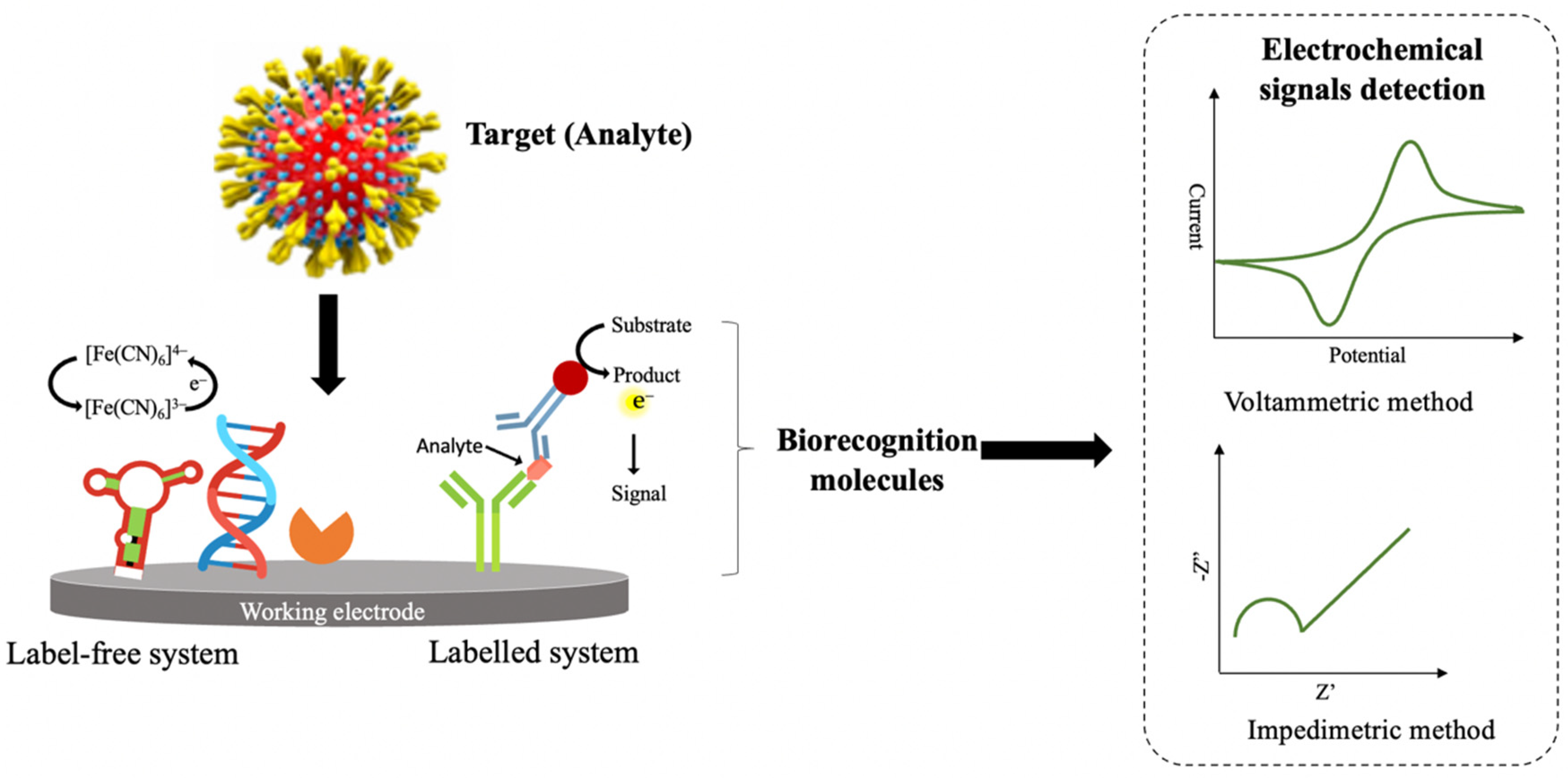
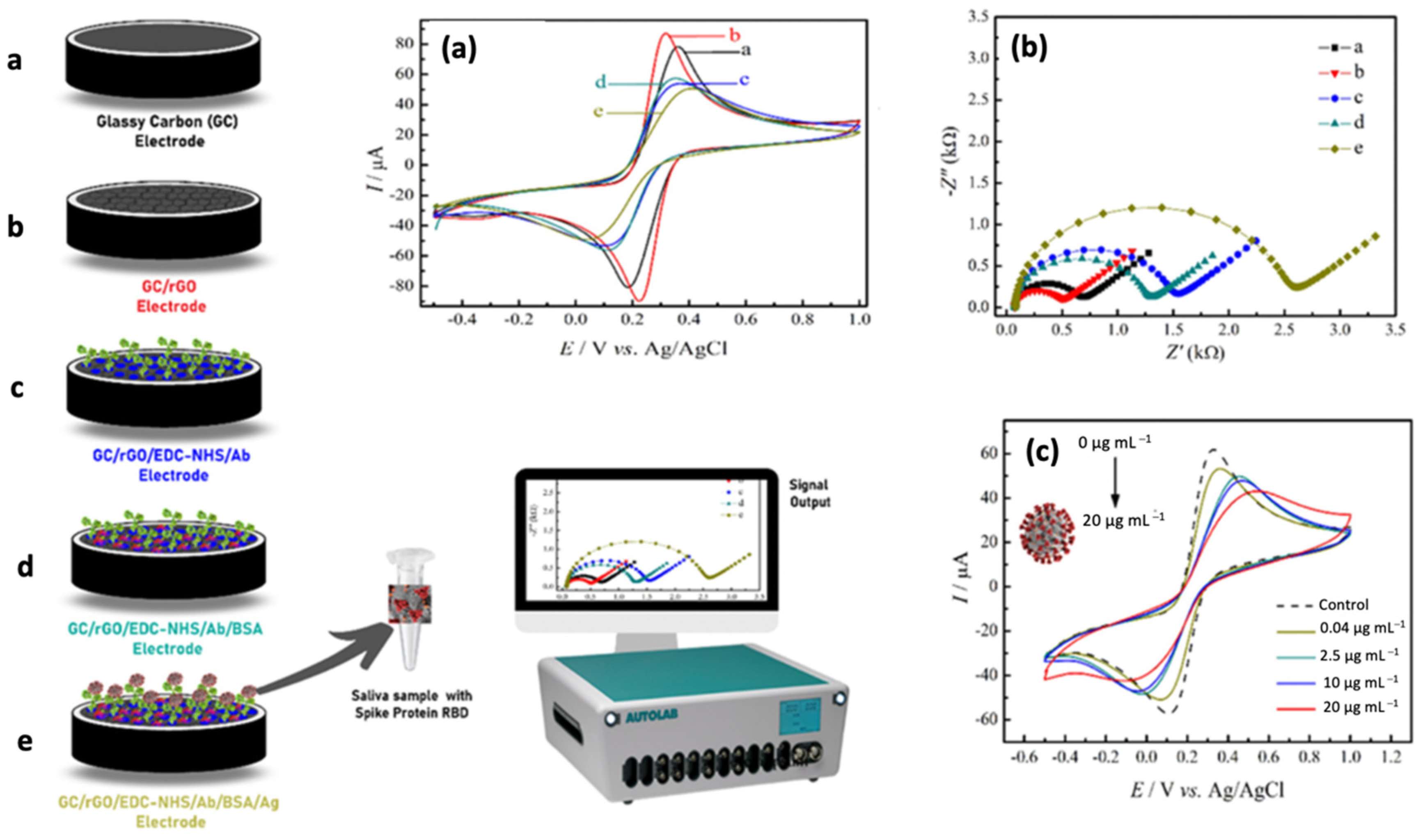
3. SARS-CoV-2 Electrochemical Biosensors

Electrochemical biosensors are known as the most promising and highly sensitive transduction systems for the early detection of SARS-CoV-2. This is due to its capability in detecting the target analyte at a very low concentration with low power consumption [77]. Generally, electrochemical biosensors are biosensing devices that transform the biochemical reactions between biorecognition molecule and target analyte into measurable signals via a current, voltage, or charge transfer resistance [78] (Figure 2). The generating signals are proportional to the concentration of target analytes in the biochemical reaction [28,79]. The utilization of electrochemical biosensors to detect a different group of viruses has been extensively reported in the literature. For instance, Anusha et al. [80] discussed numerous designs of electrochemical biosensing platforms and the function of biorecognition molecules in detecting the dengue virus. The recent advancement in developing robust electrochemical sensors strategies to detect the Zika virus has been extensively reviewed by Kaushik et al. [81]. Moreover, Rasouli et al. [77] highlight the progress of electrochemical DNA biosensors in monitoring the human papillomavirus virus. Due to its excellent performance in pathogenic virus detection, electrochemical biosensors are suggested as one of the promising diagnostic tests for the real-time observation of SARS-CoV-2. Many pieces of literature reported on the potential implementation of electrochemical biosensors for the detection of SARS-CoV-2. For example, Mahshid et al. [53] extensively reviewed the potential application of electrochemical biosensors for monitoring SARS-CoV-2. The most favourable electrochemical biosensors design was thoroughly discussed by Kotru et al. [82]. Indeed, Khan et al. [79] and Imran et al. [24] described the performance of different types of electrochemical biosensors for SARS-CoV-2 and other viral pathogens such as Ebola, influenza, Zika, and HIV.
In an electrochemical biosensors system, the target analyte can be detected either through a labeled system (indirect sensing) or a label-free system (direct sensing). The label-based electrochemical biosensor was constructed by adding a second probe or specific label (e.g., ferrocene, enzyme, metal nanoparticles, etc.) to the biorecognition element specific to the target analyte. In this sensing system, the concentration of the target analyte was indirectly measured based on generating signals from a specific label, and PBS was mostly used as an electrolyte solution for electrochemical measurement. Formerly, most of the highly sensitive electrochemical biosensors were fabricated based on a labelled system due to the potential for signal amplification and high resistance to non-specific binding as the detection of the analyte depends on two independent binding events. Nonetheless, the fabrication of this type of electrochemical biosensor is expensive because it requires two biorecognition systems, and multiple steps are needed during the surface functionalization of the working electrode. Furthermore, the additional preparation steps for complex labelling might prolong the time needed to construct the electrochemical biosensors. Moreover, electrochemical detection can only be carried out by highly trained personnel to ensure the quality of the analysis and interpretation of the results [83,84]. Hence, they are not preferable for real-time measurements.
Unlike label-based electrochemical biosensors, a label-free sensing system can minimize the preparation time and cost of analysis due to the elimination of complex labels and the requirement of only one biorecognition molecule. Such a sensing system can quantify the target analyte directly based on the electrical signals produced during the biochemical reaction without any labelling and chemical modification. The electrochemical detection in a label-free sensing system directly relies on the catalytic transfer of electrons between the surface of the working electrode and the active surface of a biorecognition molecule. The measurement of this sensing system often employs ferro/ferricyanide as a redox probe [22,85,86]. The capability of label-free electrochemical biosensors for rapid detection of target analyte enables them possible for real-time measurements and as a POC device [83,87]. Due to their significant advantages, label-free electrochemical biosensors have become a popular choice for electrochemical-based diagnostic of SARS-CoV-2 infection.
The fabrication of label-free electrochemical biosensors critically relies on several components such as the transducer (working electrode), biorecognition molecules, and electrochemical transducing techniques. These sensing platforms ensured more sensitive detection by fabricating the working electrode with materials that possess excellent electronic performance and applying the high specificity and affinity of biorecognition elements [88]. Table 2 demonstrates numerous types of electrochemical biosensors for detecting SARS-CoV-2 in clinical samples such as saliva, nasopharyngeal swab, serum, throat swab, urine, and feces. The limit of detections (LOD) was in the range from nanomolar to femtomolar using various fabrication strategies and electrochemical transducing techniques that will be discussed in the following subsections.

3.1. Transducer (Working Electrode) and Electrochemical Transducing Methods

The transducer or working electrode can be fabricated with semiconducting and conducting materials ranging from metals (e.g., platinum and gold) to non-metals (e.g., carbon) using diverse sizes of materials (from bulk to micro and nanostructured materials) [67,70,88]. Additionally, polymer electrodes and ceramic electrodes are often chosen in the fabrication of working electrodes of electrochemical biosensors due to their advantageous properties such as stability, biocompatibility, and tuneable electric conductivity [120]. The selection of working electrode materials will determine the performance of the electrochemical biosensor including the rate of heterogeneous electron transfer (affect the sensitivity and detection time), double-layer capacitance (affect the limit of detection), the character of the coupling chemistry required to attach the biorecognition molecules, and the propensity towards non-specific binding [67,88].
In electrochemical biosensors, the changes in electrical properties of the working electrode can be measured based on several transducing methods such as voltammetry (measure the current at a different range of potential), amperometric (measure the current at constant potential), potentiometric (measure the charge potential at fixed current), and conductometric (measure the conductivity of working electrode at varying of frequencies) depending on the detection principle and application of electrochemical biosensors. Among all the transducing techniques, the voltammetric techniques, namely cyclic voltammetry (CV), square wave voltammetry (SWV), and different pulse voltammetry (DPV), are often chosen in electrochemical diagnostics sensing due to the simplicity of the method and low-cost instrumentation (only a potentiostat is needed) [27,121,122,123]. Therefore, the detection times could be completed in a short period, and detection costs can significantly be reduced [124]. Moreover, these methods are significantly used in the fabrication of label-free electrochemical biosensors for viral detection [85]. The mode of applied potential differentiates among the CV, DPV and SWV techniques. In CV, the sample sweeps through varying potentials, whereas the potential of sample pulses from one potential to another in both DPV and SWV techniques [125].
CV technique provides information on the preliminary redox characteristics of the biorecognition elements or target analyte. Cyclic voltammograms can demonstrate the electrochemical characteristics and reversibility of the redox reaction (semi or quasi reversible, irreversible, completely reversible). Moreover, the CV technique is often used to measure the surface area of various materials that are functionalized onto the electrodes to observe the improvement of the electrochemical performance [75,126]. CV is commonly employed in diagnostics approaches to characterise target chemical substances and assess the electrochemical mechanism of chemical reactions. Nevertheless, CV is not recommended for quantitative detection of target chemical substances as it possess low resolution and sensitivity [107]. DPV and SWV are favored voltammetric methods used in electrochemical diagnostics due to their high resolution and sensitivity [75,107]. In DPV, the peak voltammogram was obtained through the measurement of differences between two current responses recorded in each pulse period [127]. Meanwhile, SWV voltammograms were obtained by plotting the differences between two current responses recorded in each pulse period against the staircase potential. The capability of DPV and SWV to reduce the effect of capacitive current and increase the signal-to-noise ratio by attenuating background current makes these voltammetry techniques important over other electrochemical techniques [127,128]. Due to these characteristics, DPV and SWV enable the detection of SARS-CoV-2 up to picomolar or femtomolar concentration [75].
Besides voltammetric techniques, the impedance technique, namely, electrochemical impedance spectroscopy (EIS), has been widely used in electrochemical diagnostics. Generally, it measured the impedance or capacitance of the electrochemical biosensors system by imposing a sine wave with an amplitude range from 5 to 10 mV. Through EIS, the interfacial characteristics (adsorption or desorption) between the electrode and electrolyte could be determined. Moreover, the EIS data can give information on the kinetic or mechanistic aspects and the electrochemical redox reaction rates [129]. Similar to CV, EIS measurement was also performed throughout every step of the electrochemical biosensor fabrication to follow the electrode surface modification. For example, Abrego-Martinez et al. [106] have performed the CV and EIS analyses in an electrolyte solution containing 5 mM [Fe(CN)6]3−/4− to evaluate the success of each fabrication step of aptasensor for monitoring the SARS-CoV-2 pseudovirus. The developed aptasensor could detect the spike protein of SARS-CoV-2 within 40 min of incubation time with a LOD of 66 pg/mL by EIS measurement.

3.2. Biorecognition Molecules Used for Fabrication of SARS-CoV-2 Electrochemical Biosensor

Until now, the immunoassay and nucleic-acid-based assay have been extensively utilized for clinical detection of SARS-CoV-2. Hence, the integration of these assays with an electrochemical sensing platform could overcome the existing limitations and drawbacks. This is due to the fact that electrochemical biosensors can provide more precise and sensitive results, as well as lower testing costs, user-friendly due to simple instrumentations, robust diagnosis, and rapid response time [26,130]. In the fabrication of electrochemical biosensors, DNA or RNA, aptamer, antibodies, and peptides are the common biorecognition elements used to detect various pathogens, including human coronaviruses [115,131]. Table 3 summarises the advantages and limitations of different biorecognition elements in the fabrication of electrochemical biosensors. Two types of electrochemical biosensors namely electrochemical immunosensor and electrochemical DNA sensors mainly utilized for SARS-CoV-2 detection are extensively discussed in the following section.

3.2.1. Electrochemical Immunosensor

Antibodies are recognized as the biorecognition molecules for electrochemical immunosensors. They are often chosen in most bioanalytical labs and commercial diagnostic kits to detect proteins, viruses and cancer cells because of their good affinity, high sensitivity, and specificity [132]. Among various types of antibodies, monoclonal antibodies (mAb), polyclonal antibodies (pAb) and antibody single-chain Fv (scFv) fragments are found as the most significant antibodies used to detect the respiratory virus infection. In particular, the reaction of mAb is highly specific compared with pAb as it can only bind with a single epitope. Thus, there are no chances of cross-reaction. Unlike mAb, pAb can recognize various epitopes on a single antigen [133]. Due to this characteristic, pAb is broadly applied in biosensors fabrication since its production cost is less expensive and can be mass-produced. The scFv fragments are preferably used for antigen capture compared with the whole antibody due to their smaller size and low variability [74,134]. A good affinity of antibodies towards antigens (can achieve nanomolar level) yielded numerous types of electrochemical biosensors. It can be in the form of enzyme-amplified ELISA to label-free formats [23]. Nevertheless, the antibody-based assay faced some limitations such as the production process being complex and expensive. Moreover, the addition of signals tags to the antibodies might disrupt their affinity [135].
In an electrochemical immunosensor, specific antibodies are immobilized onto the surface of the working electrode using several techniques such as direct absorption, magnetic beads (MBs) or self-assemble monolayer (SAM). They bind to specific target analytes (antigens) to generate electrical signals that can be measured using various transducing methods (Figure 3) [74,79]. Among the immobilization techniques of antibodies, SAMs of alkanethiols is mostly applied due to the capability to create strong covalent bonds on the surface of the working electrode in a simple way [136]. The generated signals in immunosensing assay are correlated to the reaction rate of antigen-antibody. Electrochemical immunosensors have been proven to detect most infectious diseases due to their excellent sensitivity, fast detection time, and the ability of miniaturization [132].
Accordingly, numerous immunosensors have been developed for the detection of various groups of viruses. The first immunosensor for detecting SARS-CoV had been developed by Ishikawa et al. [137] based on field-effect transistor (FET). In this study, the biochemical reaction between antibody and antigen was quantified based on the conductometric method. The virus antigen nucleocapsid (N) protein was used as a target analyte to fabricate this electrochemical biosensor. Antibody mimic proteins (AMPs) were used as an alternative for conventional antibodies due to their simple and low-cost production, smaller size (usually 2–5 nm, less than 10 kDa), and has stability in a broad range of pH and electrolyte concentrations. The surface of the working electrode was modified with an AMP capture agent namely fibronectin-based protein (Fn) to improve the selectivity of the designed sensor. The well-developed sensing platform could detect the N protein in 44 µM of bovine serum albumin (BSA) as low as 2 nM within 10 min.
Recently, Zaccariotto et al. [97], an impedimetric immunosensor was developed by immobilizing antibodies on modified glassy carbon for SARS-CoV-2 detection. Figure 4 illustrates the fabrication steps of the electrochemical immunosensor. The successful fabrication of the electrochemical immunosensor was evaluated using CV and EIS techniques. The modification of glassy carbon electrodes with reduced graphene oxide presents a low-cost diagnostic technology. Hence, this strategy can directly be implemented to single printed carbon electrodes to make the developed immunosensor become a POC device in the future. In this study, the fabricated impedimetric immunosensor was successfully detected the SARS-CoV-2 spike protein with LOD of 150 ng/mL in the range of linear concentration from 0.16 to 40 µg/mL. Besides an impedimetric method, the developed diagnostic platform can use a voltammetric technique such as SWV to detect this virus. The developed electrochemical immunosensor can detect this pathogenic virus at a concentration as low as 2.40 ng/mL using the SWV technique. The proposed immunosensor is a practical diagnostic test for screening SARS-CoV-2 infection as it demonstrated a good performance towards detecting the virus in the saliva samples. Hence, the detection process for the developed sensor is much simpler and rapid compared to other antibody-based antigen diagnostic methods.
In another study conducted by Liv [96], a novel electrochemical immunosensor was developed to detect the spike protein of the SARS-CoV-2 antigen in spiked saliva and oropharyngeal swab samples. This research group fabricated the sensor using a glassy carbon electrode modified with gold and capped with cysteamine and glutaraldehyde. The developed immunoassay platform employs the voltammetric method (CV and SWV) to detect the spike antibody in synthetic and real samples. The viral surface spike (S) protein was chosen as a biomarker for SARS-CoV-2 detection since it is a core transmembrane protein of the virus and highly immunogenic [138]. The fabricated electrochemical immunosensor can detect the target SARS-CoV-2 antigen protein with a LOD of 0.01 ag/mL in both synthetic media and clinical samples. This is the best reported LOD compared to another voltammetric immunoassay [102,113,118]. In addition, the developed electrochemical immunosensor demonstrated a simple sample preparation and shorter detection time (~35 min) compared to other established electrochemical methods in the literature [93,102,115]. The fabricated biosensor also showed high selectivity by the ability to distinguish the SARS-CoV-2 and MERS-CoV antigens. The accuracy of the developed immunosensor for detection of SARS-CoV-2 using saliva and oropharyngeal swab samples proved the successful fabrication of this immunosensor as most of the existing diagnostic tools used blood and serum samples that required a lengthy sample preparation process. However, further study is needed to integrate the ultrasensitive developed immunosensor into a ready-to-use commercial sensor and kit.
Accordingly, numerous electrochemical immunosensors have been developed for high sensitivity detection of different kinds of viruses. The first electrochemical immunosensor for the detection of SARS-CoV had been developed by Ishikawa et al., (2009) based on field-effect transistor (FET) [137]. Here, the change in conductance generated by the interaction of antibody-antigen can be measured and correlated to the concentration of the analyte. This FET-based electrochemical sensor used the virus antigen nucleocapsid (N) protein as a SARS biomarker. As an alternative for conventional antibodies, antibody mimic proteins (AMPs) were utilized as affinity binding agents due to their simple and low-cost production, smaller size (usually 2–5 nm, less than 10 kDa), and stability to a broad range of pH and electrolyte concentrations. The surface of working electrode was modified with a fibronection-based protein (Fn) as AMP capture agent to selectively bind the antigen N protein. To further improve the immobilization of the AMPs and transduced signal, the exposed gate region of the FET-based immunosensor was modified with In2O3 nanowires on a Si/SiO2 substrate. The well-developed sensing platform was able to detect the N protein at sub-nanomolar concentrations in a shorter time without the need for labelled reagents.
To date, a great number of electrochemical immunosensors have been reported for the detection of SARS-CoV-2 infection with good selectivity and LOD as presented in Table 2. In early 2020, Seo et al., (2020) have developed FET-based immunosensor for the detection of SARS-CoV-2 in clinical samples [91]. The surface of FET sensor was immobilized with a SARS-CoV-2 spike antibody through 1-pyrenebutyric acid N-hydroxysuccinimide ester (PBASE), an efficient interface coupling agent used as a probe linker. In this study, the viral surface spike (S) protein was chosen as a biomarker for virus detection because it is a major transmembrane protein of the virus and highly immunogenic. Moreover, the S protein exhibits diverse amino acid sequences among coronaviruses and thus enabling the specific detection of SARS-CoV-2 [138]. This FET immunosensor can detect the target SARS-CoV-2 antigen protein with LOD of 1 fg/mL in phosphate-buffered saline. In addition, the developed sensor demonstrated high sensitivity and selectivity by the ability to distinguish the SARS-CoV-2 antigen protein from those of MERS-CoV. The successful fabrication of this immunosensor enabling sensitive detection of the SARS-CoV-2 virus in clinical samples with LOD of 2.42 × 10² copies/mL without sample pre-treatment or any labelling.

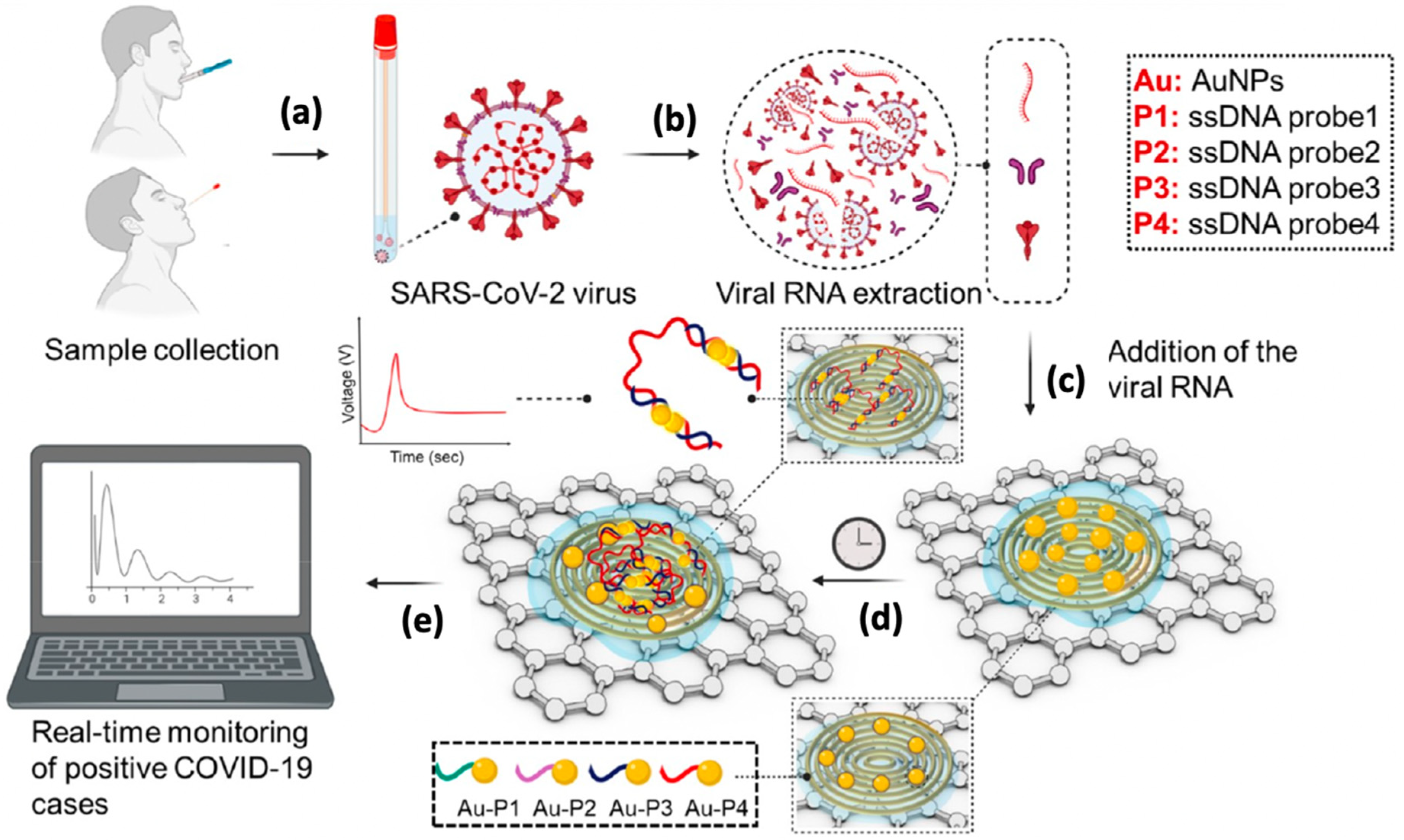
3.2.2. Electrochemical DNA Sensor

Viral nucleic acids are considered the most appropriate biorecognition elements for screening SARS-CoV-2 infection since the IgM and IgG immune responses are very low during the early stage of infection [23]. The common biorecognition elements that have been used in viral nucleic acid-based electrochemical sensors are ssDNA, RNA, peptide nucleic acid, and hairpin DNA [139]. Among them, ssDNA probes have been widely utilized in the construction of electrochemical-sensor-based nucleic acid which is known as electrochemical DNA sensor [74]. In an electrochemical DNA sensing, the ssDNA probe (15–30 kb) was immobilized onto the surface of the working electrode to recognize its complementary ssDNA target and produced a double-strand DNA (dsDNA) (Figure 5). The main interaction that occurred between the ssDNA probe and its complementary ssDNA target is called DNA hybridization. This hybridization reaction can be transferred into a quantifiable electrical signal via electrochemical techniques. The electrical signals were proportional to the concentration of viral nucleic acids [140,141]. Generally, the electrical signals are generated from the electron transfer of the redox-active probe with the electrode. In most electrochemical detection, [Fe(CN)6]3−/4− and [Ru(NH3)6]3+/2+ complexes re used as redox-active probes [142]. The immobilization of ssDNA onto the surface of the working electrode is a crucial step in fabricating an electrochemical DNA sensor because it will determine the excellent reactivity and orientation of the DNA probe to hybridize with its ssDNA target [143,144]. In an electrochemical DNA sensor, the ssDNA can be immobilized onto the surface of the working electrode through several techniques such as adsorption, covalent-bonding, and avidin-biotin interaction [145]. Among the techniques, chemisorption is widely applied for the covalent immobilization of thiol-modified DNA probe onto the gold electrode surface to form a self-assembly monolayer (SAM) due to the high-affinity interaction between the thiol group and gold electrode [146,147].
In early 2005, Abad-Valle et al. [148] developed the electrochemical DNA sensor to detect the SARS-CoV. This work designed the surface of the working electrode using 100 nm sputtered gold films. The gold surface was immobilized with a labelled thiolated DNA probe and hybridized with 30-mer complementary ssDNA that encodes a short lysine-rich region as target molecules. The electrochemical detection was measured indirectly using an alkaline-phosphatase-labelled streptavidin that converts a substrate into an electroactive product. The fabricated electrochemical DNA sensor successfully detects the SARS virus-specific sequence within 1 h with a LOD of 6 pM. Among many types of electrochemical DNA sensors, aptamers stand as the ideal probe on the sensing surface due to their extraordinary characteristics including high affinity and specificity to their target molecules, resistance to a wide range of temperatures, and can be easily modified by chemical groups for immobilization or labelling purposes [149]. Indeed, aptamers can be designed for a wide variety of targets, including antibodies, protein, enzymes, amino acids, growth factors, cancer biomarkers, toxins, metal ions and low molecular weight vitamins [150,151,152,153,154,155,156]. Owing to these properties, they exhibit various recognition mechanisms during electrochemical detection. In most electrochemical detections, their sensing mechanism relies on target-induced conformational changes that bring a redox reporter close to an electrode surface, triggering an increase in electrochemical signal [157]. The aptamer can be either artificial synthesis in vitro or biochemically synthesis via the systematic evolution of ligands by exponential enrichment (SELEX) process [131].
Unlike antibodies, aptamer offers enormous advantages such as small size, short synthesis time, cost-saving production, not involving animal production, and no batch-to-batch variation. More importantly, they possess lower detection limits (can reach the zeptomolar (zM) level) which are necessary for developing the high sensitivity electrochemical biosensor [131,158]. The utilization of aptamer as a recognition element for virus detection has been well described and reported in several pieces of literature. For instance, Bhardwaj et al. [159] fabricated the electrochemical sensor using ssDNA aptamers that target recombinant influenza A mini-hemagglutinin (mini-HA) protein and whole H1N1 viruses for detection of influenza H1N1 viruses. In this study, the selected aptamer was immobilized onto the surface of the glass working electrode that was chemically modified with indium tin oxide (ITO). The fabricated aptasensor demonstrates the high sensitivity and selectivity detection of the H1N1 virus with the LOD of 3.7 plaque-forming units (PFU)/mL. Besides using a single DNA aptamer as a recognition molecule, the aptamer target-antibody sandwich technique is another common sensing mechanism applied. The utilization of duplex recognition patterns in electrochemical detection would enhance accuracy and selectivity and give the lowest LOD of electrochemical biosensors [74].
Idili et al. [95] developed an electrochemical aptamer-based (EAB) biosensor for the detection of SARS-CoV-2 spike (S) protein in undiluted biological fluids (serum and artificial saliva). In this study, a redox reported-modified aptamer (Atto MB2 (methylene blue derivative)-aptamer) was covalently attached onto the surface of the gold working electrode. The binding of spike protein will trigger a change in the aptamer conformation, subsequently bringing the redox reporter closer to the gold electrode surface and generating the electrochemical signals. The SWV analysis was used to characterize the performance of the EAB biosensor in detecting different concentrations of spike protein in PBS buffer to mimic the condition of real clinical samples. The developed EAB biosensor can detect the SARS-CoV-2 RBD target as fast as within seconds (15 s), demonstrating its ability for POC devices. Moreover, this sensing platform showed a similar analytical performance to other diagnostic methods that use antibody and aptamer as they can detect the SARS-CoV-2 antigen down to picomolar levels in serum, buffer, and 50% artificial saliva.
Yousefi et al. [100] reported the first reagent-free electrochemical sensor using DNA aptamer-antibody conjugate as biorecognition elements to detect the SARS-CoV-2 infection (Figure 6). This research group successfully fabricated the electrochemical sensor that can directly detect the SARS-CoV-2 virus and its associated spike protein in processed and unprocessed patient saliva within 5 min, without any reagents. The designed sensor adopted a sensing mechanism that detects the potential triggered transport of a DNA-antibody conjugate. Here, an analyte-recognizing antibody was attached to a rigid, negative-charged linker composed of DNA and immobilized onto the surface of the gold working electrode. Ferrocene redox reporter molecule was attached to a thiolated DNA probe at 3′ to monitor the interaction on the surface working electrode. The electrochemical signals of the fabricated aptasensor were recorded using chronoamperometry (CA) using a potential window from 0 to +500 mV for 50 ms using Ag/AgCl as a reference electrode. Such electrochemical sensor demonstrates long-term stability as its performance was not much affected after 9 months of storage. Despite their benefits and versatility, aptamer also possesses some limitations that restrict their applications, such as being susceptible to nuclease degradation and not binding to the targets that lack functional groups [132].

4. The Advanced Electrochemical Sensing Technologies for Point-of-Care (POC) Detection of SARS-CoV-2

It is no doubt that the rise in SARS-CoV-2 outbreak cases demands rapid on-site and accurate diagnosis devices without the need for skilled technicians and sophisticated laboratories for high throughput sample screening [82,160]. In such situations, POC devices are urgently needed for the early detection of SARS-CoV-2 infection as this may be the best solution for now. Nowadays, the utilization of POC devices allows the diagnostic test to be performed near the patient site, providing faster results than conventional laboratory testing. Due to these features, they are significant in situations where rapid medical decisions need to be taken for example in emergency departments [161,162]. Notably, the use of POC diagnostic tools for early screening of viral infection in the population could potentially combat the spread of infection. Owing to the excellent diagnostic performance mentioned earlier, the electrochemical biosensors show potential and could be integrated into point-of-care testing (POCT) for SARS-CoV-2 diagnosis. The advanced design in electrochemical sensing platforms such as miniaturized sensors and microfluidic chips makes them a good alternative to the existing mainstream diagnostic COVID-19 methods [160]. As tabulated in Table 2, most of the developed SARS-CoV-2 electrochemical biosensors employed miniaturized electrode and microfluidic technologies for the ultrasensitive and on-site detection of this contagious virus that will be discussed further in the next section.

4.1. Nanomaterials as the Surface Modifier on the Miniaturized Electrochemical Sensor

In the current pandemic crisis, most sensor research moves towards developing nano-enabled miniaturized electrochemical biosensors to gain more accurate and fast detection of the SARS-CoV-2. The introduction of nanotechnology greatly improves the performance of SARS-CoV-2 electrochemical biosensors by enabling the detection at a very low level (picomolar (pM) to femtomolar (fM) level) and high selectivity toward virus protein [89]. Nanomaterials possess unique physical and chemical surface characteristics compared with the bulk material such as diffusivity, solubility, optical, thermodynamic, colour and magnetic features [163,164]. In biochemical sensing, nanomaterials exhibit functional electrical and mechanical characteristics that are responsible for enhancing the electrochemical, optical, and magnetic properties of biosensors [165,166]. Particularly, the addition of nanomaterials onto the surface of the working electrode can enlarge the biocompatible areas with the target analytes (e.g., enzymes, proteins, antibodies, DNA, and cells) which subsequently enhanced the sensor reactivity and sensitivity in an electrochemical sensing platform [165]. Moreover, the implementation of nanotechnology in viral electrochemical biosensors can reduce the detection time and experimental cost since a small volume of samples is needed for analysis [165,167].
Various types of nanomaterials have been used for fabricating the SARS-CoV-2 electrochemical biosensors such as gold nanoparticles (AuNPs), quantum dots (QDs), carbon nanotubes (CNTs), and graphene or graphene-derived nanomaterials (Figure 7). In an electro- chemical sensing platform, graphene is a good surface modifier to interface with diverse biomolecules and cells [167,168]. Meanwhile, graphene-derived nanomaterials namely graphene oxide (GO) and reduced graphene oxide (rGO) are an excellent choice for electrode surface modification because they possess good mechanical, chemical, electronic, and thermal characteristics. Sengupta and Hussain [169] had extensively reviewed the application of graphene-based electrochemical biosensors for rapid detection of SARS-CoV-2 and other pathogenic viruses.
Along with utilizing nanomaterials as the substrates for biorecognition element immobilization, the introduction of miniaturization technology makes the electrochemical biosensors suit for on-site diagnostics applications [160,170]. Due to the high demands for POCTs, miniaturized electrochemical sensors are broadly used to detect different target analytes including metal ions, small organic molecules, and biomolecules in diverse sectors such as food safety, environmental and healthcare [171]. Such sensor systems offer many benefits to electrochemical sensing platforms such as portability, simple sample preparation, ease to operate, good selectivity and sensitivity, rapid detection time, and low experimental cost [172,173]. Owing to their remarkable benefits, various designs of miniaturized electrochemical biosensors have been developed to detect SARS-CoV-2.
Last year, Alafeef et al. [108] research group developed the miniaturized electrochemical DNA sensor for quantitative measurement of SARS-CoV-2 in clinical samples. The electrochemical DNA sensor was fabricated using highly specific antisense thiolated ssDNA capped with gold nanoparticles (AuNPs) which target the viral nucleocapsid phosphoprotein (N gene) (Figure 8). The developed sensor can detect the SARS-CoV-2 RNA for only 5 min with a LOD of 6.9 copies/μL and a sensitivity of 231 copies/μL without any additional amplification technique. The obtained LOD in this study is comparable with other clinical approaches. Interestingly, the high sensitivity and specificity of this sensor manage to discriminate the positive COVID-19 samples from the negative ones with an accuracy of nearly 100%. Tripathy and Singh [66] proposed a miniaturized electrochemical sensor based on the DNA hybridization method for detecting SARS-CoV-2 as they successfully developed the label-free electrochemical DNA sensor for detection of dengue fever and breast/ovarian cancer in previous research [174,175]. The miniaturized device was fabricated on oxidized silicon substrates, using standard CMOS fabrication process flow and electrodeposition techniques. Such sensors utilized gold nanoparticles (AuNP) as the transducing element. In this sensing approach, a thiolated ssDNA probe which is complementary to SARS-CoV-2 RNA or its corresponding cDNA was immobilized on the surface of AuNP via gold-thiol self-assembly. Although this label-free miniaturized sensor could potentially become a POC biosensing device, additional steps such as the extraction of target DNA/RNA from the infected host and the following sample preparations are needed before the electrochemical detection. Such tedious sample preparation steps could affect the diagnostic accuracy and prolong the time required for analysis.
A further advance in the electrochemical sensing platform allows Mahari et al. [89] to develop an in-house built single printed carbon electrode (SPCE) called eCovSens. They fabricated the sensor by drop-casting the gold nanoparticles (AuNPs) onto the surface of the fluorine-doped tin oxide electrode (FTO) electrode before being allowed for immobilization with antibody probe. The addition of AuNPs on the surface of the FTO electrode will amplify the electrical signals of the fabricated sensor. Then, an nCOVID-19 monoclonal antibody probe was immobilized onto FTO/AuNPs electrode to recognize the spike antigen of nCOVID-19. This surface modification and antibody immobilization strategies provide rapid detection (10–30 s) with LOD of 90 fM using saliva samples. Furthermore, the eCovSens is an ultrasensitive sensor as they require only 20 μL sample volume compared to the other electrochemical biosensors that require at least 100 μL sample volume. Therefore, eCovSens could be a promising POC device as it is portable and stable until one month of storage.
Miripour et al. [116] presented a portable disposable sensor with an automatic electrochemical readout for real-time monitoring the reactive oxygen species (ROS) levels in the sputum of COVID 19 patients. Multi-Wall Carbon Nanotubes (MWCNTs) were selected as a nanomaterial to functionalized on the surface of the working electrode because they have good mechanical and electrical characteristics [176]. In this sensing system, the reaction between ROS molecules and the surface of the MWCNTs electrode generates an electrical signal measured via cyclic voltammetry. Such electrical signals correlate with the viral load in the sputum of COVID 19 samples. The responses of this disposable sensor were comparable with the CT scan results of the COVID-19 patient. In addition, the electrochemical ROS sensor could potentially use for rapid screening of patients that require prompt medical examination since it can give results less than 30 s.
In recent work, Hashemi et al. [107] established the electrochemical nanosensor for screening SARS-CoV-2 glycoprotein in biological samples. They fabricated the nanosensor using a layer of GO coated with gold nanostars (AuNS). The addition of these excellent biochemical sensing compounds on the surface of the working electrode allows this nanosensor to demonstrate a good LOD (1.68 × 10⁻²² μg/mL) and sensitivity (0.0048 μAμg/mL/cm). More importantly, the developed nanosensor can detect the trace of viruses in less than 1 min without the requirement of any biological marker and sample pre-treatment. In addition, the fabricated nanosensor exhibits high sensitivity (95%) for the detection of unknown clinical SARS-CoV-2 samples. Such good performances promote the developed nanosensor as a rapid diagnostic test for the detection of viral disease.
Besides electrochemical sensing strategies, data transmission is another crucial aspect that needs to be ascertained for rapid deliverable results to final-user and at-home diagnosis [177]. Following the previous miniaturized sensing strategies, Chandra [178] had proposed the miniaturized label-free electrochemical sensor integrated with smartphones for rapid screening of SARS-CoV-2 (Figure 9). A smartphone-based “cloud” directory was suggested to provide real-time surveillance of this pathogenic virus through geo-tagging. Through this system, the sensors were capable of tracing the disease spreading around the world and enabling the form of a library of data and details required for future preparation to manage and control such pandemics. A similar strategy has been reported by Balaji et al. [114], where they developed an electrochemical biosensor-based smartphone for assessing COVID-19 patients. Herein, the electrochemical signals generated from the detection of RNA COVID-19 were transferred to the portable smartphone for displaying the results. The obtained results are stored in a database using the Internet of Things (IoT). Through an IoT system, the database can be shared with the concerned department to monitor the patients to avoid contact with other people and health surveillance of the infected patients. Accordingly, the risk of disease spreading can be reduced and expedited the best therapy for patients. In another study, Zhao et al. [115] developed the super sandwich-type electrochemical biosensor that can be adapted to a smartphone to detect SARS-CoV-2 RNA. They implemented a ‘plug and play method’ to produce a portable sensor that can easily be accessible by users to assess the diagnosis results conveniently. Interestingly, the fabricated portable electrochemical smartphone does not require further nucleic acid amplification to detect SARS-CoV-2, making them more convenient as a POC device. Moreover, Torrente-Rodríguez et al. [118] introduced the advanced wireless telemedicine-based electrochemical platform, SARS-CoV-2 RapidPlex, to screen COVID-19. Such sensing systems possess multiplexed platforms that provide information on the level of immune response (IgG and IgM), viral infection rate (nucleocapsid protein), and severity of disease (C-reactive protein) [179,180]. The amperometric data from the multiplexed sensor are recorded by custom PCB-based wireless potentiostat. The data are wirelessly transferred to a user smartphone via Bluetooth. This sensor can be used for at-home diagnosis since it is simple to operate and can give ultrasensitive detection of SARS-CoV-2.

4.2. Microfluidic Chip

The integration of microfluidic technology with the miniaturized electrochemical sensing platform can further accelerate the performance of virus detection. This device is a so-called microfluidic chip or lab-on-chip (LoC). Microfluidic technology revolutionizes the method for sampling, sample separation, mixing, chemical reaction, and electrochemical detection in one device. Such technology is beneficial for real-time detection, enabling multiplexing, and assembling multiple microfluidic components [76,181]. The use of microfluidic technology in an electrochemical sensing platform can significantly minimize the volume of samples as it can process a small volume of fluids by using tiny channels with dimensions at the microscale, usually tens to hundreds of micrometers [182]. Such advanced features allow the microfluidic electrochemical devices to emerge as great alternatives to conventional diagnostic methods due to their huge benefits such as fast detection times, better process control, automation, portability, reduced waste generation, low experimental cost and superior detection limit and sensitivity [183,184]. In addition, the microfluidic electrochemical devices can offer a high quality of pathogenic viral assessment since their physical and chemical environments can be precisely controlled [185]. The advancement in microfluidic technologies allows the integration of smart solutions such as the Internet of Medical Things (IoMT), e-health, artificial intelligence (AI), and machine learning to develop innovative healthcare technologies [186,187]. The microfluidic devices can be fabricated using various material bases such as polytetrafluoroethylene (PTFE), polydimethylsiloxane (PDMS), polycarbonate, silicon, glass, quartz, polymethyl methacrylate (PMMA), paper, hydrogel, three-dimensional (3D) printing and thermoset materials that subsequently offer diversity in their development [76,181,188].
To date, the application of microfluidic devices for SARS-CoV-2 detection are extensively reported in the literature. For instance, a 3D-nanoprinted COVID-19 microfluidic chip (3DcC) was presented by Ali et al. [103] (Figure 10). The integration of the PDMS microfluidic channel with a 3D electrochemical sensor allows the utilization of a small volume of antibodies (30 µL) and antigen (20 µL) to detect SARS-CoV-2. Such features enable the 3DcC device to detect SARS-CoV-2 in seconds with LOD of 2.8 × 10⁻¹⁵ for spike S1 protein and 16.9 × 10⁻¹⁵ M for its receptor-binding-domain (RBD) using a portable smartphone-based potentiostat. Moreover, Li et al. [105] developed a microfluidic paper-based analytical device (μPADs) to detect the IgG antibodies of SARS-CoV-2 in serum samples. In this research, only 3 μL of spiked human samples and antibodies are needed for the detection process completed within 30 min. A similar approach was applied by Yakoh et al. [104] to monitor the level of IgG and IgM of SARS-CoV-2. They fabricated the microfluidic chips using paper because it is cheaper, has a natural abundance and can be discarded by incineration after use. This is highly preferable for infectious disease diagnosing as the test kits need to be disposed of after testing. The electrochemical paper-based device, COVID-19 ePAD, can detect the targeted antibodies in serum of infected patients with 90% specificity and 100% sensitivity within 30 min.

5. Conclusions

The existence of the COVID-19 pandemic encourages many researchers around the world to develop robust and accurate diagnostic tests for screening of SARS-CoV-2 to give immediate therapeutics and effective isolation of infected individuals. Up to now, the primary clinical diagnostic tests for COVID-19 have relied on the detection of viral nucleic acids, antigens, and antibodies specific for SARS-CoV-2. Due to the limitations of currently available diagnostics kits, electrochemical biosensors may be an excellent diagnostic tool for screening SARS-CoV-2 infection, as they meet the ASSURED criteria. The present review discusses current electrochemical sensing platform strategies for detecting SARS-CoV-2, including the use of the most sensitive biorecognition molecules, electrode surface modification with nanomaterials in conjunction with the miniaturized electrode and microfluidic technologies that could provide high-quality SARS-CoV-2 assessment. The majority of studies on electrochemical biosensors have used antibodies rather than nucleic acid as the biorecognition molecule since this method allows for easier sample preparation while maintaining good selectivity and sensitivity results. The antibodies were mainly immobilized on the surface of the working electrode, which had been modified using graphene-derived nanomaterials such as GO and rGo. The developed electrochemical biosensors can achieve the LOD as low as 2 copies/mL in clinical samples in less than 1 min of detection time without sample pre-treatment or labelling with any biorecognition molecule. The integration of the most sensitive electrochemical sensing platforms with smartphones enables the end-user to receive results quickly. The development of robust electrochemical biosensors for SARS-CoV-2 detection could be used to combat future pandemics. Although electrochemical biosensors have been proven for diagnosing a variety of SARS-CoV-2 clinical samples, they face a few limitations when moving from laboratory to on-site detection. Numerous issues, including the stability and reproducibility of the electrochemical biosensors, sample preparation processes, device engineering scale-up, and commercialization of electrochemical biosensors as a diagnostic device need to be further investigated. This aims to facilitate the electrochemical biosensors as a precise platform for rapid, simple, robust, and portable sensors for COVID-19 infection mass screening.

Author Contributions

Conceptualization, A.A.M., I.A. and H.H.H.; formal analysis, N.S.Z. and G.A.O.; investigation, N.S.Z., M.S.A. and G.A.O.; resources, N.S.Z., M.S.A., H.H.H., Y.B. and M.F.K.; data curation, N.S.Z., Y.B. and H.H.H.; writing—original draft preparation, N.S.Z.; writing—review and editing, A.A.M., Y.B., H.H.H. and I.A.; visualization, M.S.A., Y.B. and H.H.H.; supervision, A.A.M., Y.B., H.H.H. and I.A.; project administration, I.A.; funding acquisition, A.A.M. and I.A. All authors have read and agreed to the published version of the manuscript.

Funding

This research was funded by the Higher Institution Centre of Excellence (HICoE) Grant from the Ministry of Higher Education, Malaysia (311/CIPPM/4401005), Top-Down Research University Grant, Universiti Sains Malaysia (1001/PCEDEC/870050), and Research University (RUI) COVID-19 Grant, Universiti Sains Malaysia (1001/CIPPM/8012383).

Institutional Review Board Statement

Not applicable.

Informed Consent Statement

Not applicable.

Data Availability Statement

Not applicable.

Conflicts of Interest

The authors declare no conflict of interest.

References

  1. Nicola, M.; Alsafi, Z.; Sohrabi, C.; Kerwan, A.; Al-Jabir, A.; Iosifidis, C.; Agha, M.; Agha, R. The Socio-Economic Implications of the Coronavirus Pandemic (COVID-19): A Review. Int. J. Surg. 2020, 78, 185–193. [Google Scholar] [CrossRef] [PubMed]
  2. Singhal, T. A Review of Coronavirus Disease-2019 (COVID-19). Indian J. Pediatr. 2020, 87, 281–286. [Google Scholar] [CrossRef] [Green Version]
  3. Naserghandi, A.; Allameh, S.F.; Saffarpour, R. All about COVID-19 in Brief. New Microbes New Infect. 2020, 35, 100678. [Google Scholar] [CrossRef] [PubMed]
  4. Boraschi, P. COVID-19 Pulmonary Involvement: Is Really an Interstitial Pneumonia? Acad. Radiol. 2020, 27, 900. [Google Scholar] [CrossRef] [PubMed]
  5. Huang, J.C.; Chang, Y.-F.; Chen, K.-H.; Su, L.-C.; Lee, C.-W.; Chen, C.-C.; Chen, Y.-M.A.; Chou, C. Detection of Severe Acute Respiratory Syndrome (SARS) Coronavirus Nucleocapsid Protein in Human Serum Using a Localized Surface Plasmon Coupled Fluorescence Fiber-Optic Biosensor. Biosens. Bioelectron. 2009, 25, 320–325. [Google Scholar] [CrossRef]
  6. Chen, N.; Zhou, M.; Dong, X.; Qu, J.; Gong, F.; Han, Y.; Qiu, Y.; Wang, J.; Liu, Y.; Wei, Y. Epidemiological and Clinical Characteristics of 99 Cases of 2019 Novel Coronavirus Pneumonia in Wuhan, China: A Descriptive Study. Lancet 2020, 395, 507–513. [Google Scholar] [CrossRef] [Green Version]
  7. Huang, C.; Wang, Y.; Li, X.; Ren, L.; Zhao, J.; Hu, Y.; Zhang, L.; Fan, G.; Xu, J.; Gu, X. Clinical Features of Patients Infected with 2019 Novel Coronavirus in Wuhan, China. Lancet 2020, 395, 497–506. [Google Scholar] [CrossRef] [Green Version]
  8. Wadman, M.; Couzin-Frankel, J.; Kaiser, J.; Matacic, C. A Rampage through the Body 2020. Science 2020, 368, 356–360. [Google Scholar] [CrossRef]
  9. Pachetti, M.; Marini, B.; Benedetti, F.; Giudici, F.; Mauro, E.; Storici, P.; Masciovecchio, C.; Angeletti, S.; Ciccozzi, M.; Gallo, R.C. Emerging SARS-CoV-2 Mutation Hot Spots Include a Novel RNA-Dependent-RNA Polymerase Variant. J. Transl. Med. 2020, 18, 179. [Google Scholar] [CrossRef] [Green Version]
  10. Tang, J.W.; Tambyah, P.A.; Hui, D.S.C. Emergence of a Novel Coronavirus Causing Respiratory Illness from Wuhan, China. J. Infect. 2020, 80, 350. [Google Scholar] [CrossRef]
  11. Kelly-Cirino, C.D.; Nkengasong, J.; Kettler, H.; Tongio, I.; Gay-Andrieu, F.; Escadafal, C.; Piot, P.; Peeling, R.W.; Gadde, R.; Boehme, C. Importance of Diagnostics in Epidemic and Pandemic Preparedness. BMJ Glob. Health 2019, 4, e001179. [Google Scholar] [CrossRef] [PubMed]
  12. Signorelli, C.; Scognamiglio, T.; Odone, A. COVID-19 in Italy: Impact of Containment Measures and Prevalence Estimates of Infection in the General Population. Acta Biol. Med. Atenei Parm. 2020, 91, 175. [Google Scholar]
  13. Chathappady House, N.N.; Palissery, S.; Sebastian, H. Corona Viruses: A Review on SARS, MERS and COVID-19. Microbiol. Insights 2021, 14, 11786361211002480. [Google Scholar] [CrossRef] [PubMed]
  14. Long, Q.X.; Tang, X.J.; Shi, Q.L.; Li, Q.; Deng, H.J.; Yuan, J.; Hu, J.L.; Xu, W.; Zhang, Y.; Lv, F.J.; et al. Clinical and Immunological Assessment of Asymptomatic SARS-CoV-2 Infections. Nat. Med. 2020, 26, 1200–1204. [Google Scholar] [CrossRef]
  15. Bedford, J.; Enria, D.; Giesecke, J.; Heymann, D.L.; Ihekweazu, C.; Kobinger, G.; Lane, H.C.; Memish, Z.; Oh, M.; Schuchat, A. COVID-19: Towards Controlling of a Pandemic. Lancet 2020, 395, 1015–1018. [Google Scholar] [CrossRef]
  16. Perkins, M.D.; Dye, C.; Balasegaram, M.; Bréchot, C.; Mombouli, J.-V.; Røttingen, J.-A.; Tanner, M.; Boehme, C.C. Diagnostic Preparedness for Infectious Disease Outbreaks. Lancet 2017, 390, 2211–2214. [Google Scholar] [CrossRef] [Green Version]
  17. Ozer, T.; Geiss, B.J.; Henry, C.S. Chemical and Biological Sensors for Viral Detection. J. Electrochem. Soc. 2019, 167, 037523. [Google Scholar] [CrossRef] [Green Version]
  18. Sheikhzadeh, E.; Eissa, S.; Ismail, A.; Zourob, M. Diagnostic Techniques for COVID-19 and New Developments. Talanta 2020, 220, 121392. [Google Scholar] [CrossRef]
  19. Lan, L.; Xu, D.; Ye, G.; Xia, C.; Wang, S.; Li, Y.; Xu, H. Positive RT-PCR Test Results in Patients Recovered from COVID-19. JAMA 2020, 323, 1502–1503. [Google Scholar] [CrossRef] [Green Version]
  20. Carter, L.J.; Garner, L.V.; Smoot, J.W.; Li, Y.; Zhou, Q.; Saveson, C.J.; Sasso, J.M.; Gregg, A.C.; Soares, D.J.; Beskid, T.R. Assay Techniques and Test Development for COVID-19 Diagnosis 2020. ACS Cent. Sci. 2020, 6, 591–605. [Google Scholar] [CrossRef]
  21. Bramhachari, P.V.; Sheela, G.M.; Prathyusha, A.; Madhavi, M.; Kumar, K.S.; Reddy, N.N.R.; Berde, C.P. Advanced Immunotechnological Methods for Detection and Diagnosis of Viral Infections: Current Applications and Future Challenges. In Dynamics of Immune Activation in Viral Diseases; Springer Nature: Singapore, 2020; pp. 261–275. [Google Scholar]
  22. Antiochia, R. Developments in Biosensors for CoV Detection and Future Trends. Biosens. Bioelectron. 2021, 173, 112777. [Google Scholar] [CrossRef] [PubMed]
  23. Kudr, J.; Michalek, P.; Ilieva, L.; Adam, V.; Zitka, O. COVID-19: A Challenge for Electrochemical Biosensors. Trends Anal. Chem. 2021, 136, 116192. [Google Scholar] [CrossRef] [PubMed]
  24. Imran, S.; Ahmadi, S.; Kerman, K. Electrochemical Biosensors for the Detection of SARS-CoV-2 and Other Viruses. Micromachines 2021, 12, 174. [Google Scholar] [CrossRef] [PubMed]
  25. Bhalla, N.; Jolly, P.; Formisano, N.; Estrela, P. Introduction to Biosensors. Essays Biochem. 2016, 60, 1–8. [Google Scholar] [CrossRef] [PubMed] [Green Version]
  26. Huang, Y.; Xu, J.; Liu, J.; Wang, X.; Chen, B. Disease-Related Detection with Electrochemical Biosensors: A Review. Sensors 2017, 17, 2375. [Google Scholar] [CrossRef]
  27. Kaya, H.O.; Cetin, A.E.; Azimzadeh, M.; Topkaya, S.N. Pathogen Detection with Electrochemical Biosensors: Advantages, Challenges and Future Perspectives. J. Electroanal. Chem. 2021, 882, 114989. [Google Scholar] [CrossRef]
  28. Cesewski, E.; Johnson, B.N. Electrochemical Biosensors for Pathogen Detection. Biosens. Bioelectron. 2020, 159, 112214. [Google Scholar] [CrossRef]
  29. Bukkitgar, S.D.; Shetti, N.P.; Aminabhavi, T.M. Electrochemical Investigations for COVID-19 Detection-A Comparison with Other Viral Detection Methods. Chem. Eng. J. 2020, 420 Pt 2, 127575. [Google Scholar] [CrossRef]
  30. Mortari, A.; Lorenzelli, L. Recent Sensing Technologies for Pathogen Detection in Milk: A Review. Biosens. Bioelectron. 2014, 60, 8–21. [Google Scholar] [CrossRef]
  31. Bahadır, E.B.; Sezgintürk, M.K. Applications of Electrochemical Immunosensors for Early Clinical Diagnostics. Talanta 2015, 132, 162–174. [Google Scholar] [CrossRef]
  32. Xu, L.; Shoaei, N.; Jahanpeyma, F.; Zhao, J.; Azimzadeh, M.; Al, K.T. Optical, Electrochemical and Electrical (Nano) Biosensors for Detection of Exosomes: A Comprehensive Overview. Biosens. Bioelectron. 2020, 161, 112222. [Google Scholar] [CrossRef] [PubMed]
  33. Topkaya, S.N.; Azimzadeh, M. Biosensors of in Vitro Detection of Cancer and Bacterial Cells. In Nanobiosensors for Personalized and Onsite Biomedical Diagnosis; Chandra, P., Ed.; Institution of Engineering and Technology: Lucknow, India, 2016; pp. 73–94. [Google Scholar]
  34. Amiri, M.; Bezaatpour, A.; Jafari, H.; Boukherroub, R.; Szunerits, S. Electrochemical Methodologies for the Detection of Pathogens. ACS Sens. 2018, 3, 1069–1086. [Google Scholar] [CrossRef] [PubMed]
  35. McEachern, F.; Harvey, E.; Merle, G. Emerging Technologies for the Electrochemical Detection of Bacteria. Biotechnol. J. 2020, 15, 2000140. [Google Scholar] [CrossRef]
  36. Yu, M.; Xu, D.; Lan, L.; Tu, M.; Liao, R.; Cai, S.; Cao, Y.; Xu, L.; Liao, M.; Zhang, X. Thin-Section Chest CT Imaging of COVID-19 Pneumonia: A Comparison between Patients with Mild and Severe Disease. Radiol. Cardiothorac. Imaging 2020, 2, e200126. [Google Scholar] [CrossRef] [Green Version]
  37. Zhao, W.; Zhong, Z.; Xie, X.; Yu, Q.; Liu, J. Relation between Chest CT Findings and Clinical Conditions of Coronavirus Disease (COVID-19) Pneumonia: A Multicenter Study. AJR Am. J. Roentgenol. 2020, 214, 1072–1077. [Google Scholar] [CrossRef] [PubMed]
  38. Parekh, M.; Donuru, A.; Balasubramanya, R.; Kapur, S. Review of the Chest CT Differential Diagnosis of Ground-Glass Opacities in the COVID Era. Radiology 2020, 297, E289–E302. [Google Scholar] [CrossRef] [PubMed]
  39. Xie, X.; Zhong, Z.; Zhao, W.; Zheng, C.; Wang, F.; Liu, J. Chest CT for Typical Coronavirus Disease 2019 (COVID-19) Pneumonia: Relationship to Negative RT-PCR Testing. Radiology 2020, 296, E41–E45. [Google Scholar] [CrossRef] [Green Version]
  40. Soriano Aguadero, I.; Ezponda Casajús, A.; Mendoza Ferradas, F.; Igual Rouilleault, A.; Paternain Nuin, A.; Pueyo Villoslada, J.; Bastarrika, G. Chest Computed Tomography Findings in Different Phases of SARS-CoV-2 Infection. Radiologia 2021, 63, 218–227. [Google Scholar] [CrossRef]
  41. Aziz, A.; Asif, M.; Ashraf, G.; Farooq, U.; Yang, Q.; Wang, S. Trends in Biosensing Platforms for SARS-CoV-2 Detection: A Critical Appraisal against Standard Detection Tools. Curr. Opin. Colloid Interface Sci. 2021, 52, 101418. [Google Scholar] [CrossRef]
  42. Ashraf, G.; Aziz, A.; Qaisrani, R.N.; Chen, W.; Asif, M. Detecting and Inactivating Severe Acute Respiratory Syndrome Coronavirus-2 under the Auspices of Electrochemistry. Curr. Res. Chem. Biol. 2021, 1, 100001. [Google Scholar] [CrossRef]
  43. Mobed, A.; Shafigh, E.S. Biosensors Promising Bio-Device for Pandemic Screening “COVID-19 ”. Microchem. J. 2021, 164, 106094. [Google Scholar] [CrossRef] [PubMed]
  44. Corman, V.M.; Landt, O.; Kaiser, M.; Molenkamp, R.; Meijer, A.; Chu, D.K.W.; Bleicker, T.; Brünink, S.; Schneider, J.; Schmidt, M.L. Detection of 2019 Novel Coronavirus (2019-NCoV) by Real-Time RT-PCR. Eurosurveillance 2020, 25, 2000045. [Google Scholar] [CrossRef] [Green Version]
  45. Zeng, L.; Li, Y.; Liu, J.; Guo, L.; Wang, Z.; Xu, X.; Song, S.; Hao, C.; Liu, L.; Xin, M. Rapid, Ultrasensitive and Highly Specific Biosensor for the Diagnosis of SARS-CoV-2 in Clinical Blood Samples. Mater. Chem. Front. 2020, 4, 2000–2005. [Google Scholar] [CrossRef]
  46. Udugama, B.; Kadhiresan, P.; Kozlowski, H.N.; Malekjahani, A.; Osborne, M.; Li, V.Y.C.; Chen, H.; Mubareka, S.; Gubbay, J.B.; Chan, W.C.W. Diagnosing COVID-19: The Disease and Tools for Detection. ACS Nano 2020, 14, 3822–3835. [Google Scholar] [CrossRef] [PubMed] [Green Version]
  47. Shen, Y.; Anwar, T.B.; Mulchandani, A. Current Status, Advances, Challenges and Perspectives on Biosensors for COVID-19 Diagnosis in Resource-Limited Settings. Sens. Actuators Rep. 2021, 3, 100025. [Google Scholar] [CrossRef]
  48. Zhang, Y.; Odiwuor, N.; Xiong, J.; Sun, L.; Nyaruaba, R.O.; Wei, H.; Tanner, N.A. Rapid Molecular Detection of SARS-CoV-2 (COVID-19) Virus RNA Using Colorimetric LAMP. medRxiv 2020. medRxiv:2020.02.26.20028373. [Google Scholar]
  49. Ghodake, G.S.; Shinde, S.K.; Kadam, A.A.; Saratale, R.G.; Saratale, G.D.; Syed, A.; Elgorban, A.M.; Marraiki, N.; Kim, D.-Y. Biological Characteristics and Biomarkers of Novel SARS-CoV-2 Facilitated Rapid Development and Implementation of Diagnostic Tools and Surveillance Measures. Biosens. Bioelectron. 2021, 177, 112969. [Google Scholar] [CrossRef]
  50. Rapid Diagnostics. Abbott Point of Care Testing. Available online: https://www.globalpointofcare.abbott/en/index.html (accessed on 14 June 2021).
  51. Zhen, W.; Smith, E.; Manji, R.; Schron, D.; Berry, G.J. Clinical Evaluation of Three Sample-to-Answer Platforms for Detection of SARS-CoV-2. J. Clin. Microbiol. 2020, 58, e00783-20. [Google Scholar] [CrossRef] [Green Version]
  52. Basu, A.; Zinger, T.; Inglima, K.; Woo, K.M.; Atie, O.; Yurasits, L.; See, B.; Aguero-Rosenfeld, M.E. Performance of Abbott Id Now COVID-19 Rapid Nucleic Acid Amplification Test Using Nasopharyngeal Swabs Transported in Viral Transport Media and Dry Nasal Swabs in a New York City Academic Institution. J. Clin. Microbiol. 2020, 58, e01136-20. [Google Scholar] [CrossRef]
  53. Mahshid, S.S.; Flynn, S.E.; Mahshid, S. The Potential Application of Electrochemical Biosensors in the COVID-19 Pandemic: A Perspective on the Rapid Diagnostics of SARS-CoV-2. Biosens. Bioelectron. 2021, 176, 112905. [Google Scholar] [CrossRef]
  54. Parihar, A.; Ranjan, P.; Sanghi, S.K.; Srivastava, A.K.; Khan, R. Point-of-Care Biosensor-Based Diagnosis of COVID-19 Holds Promise to Combat Current and Future Pandemics. ACS Appl. Biol. Mater. 2020, 3, 7326–7343. [Google Scholar] [CrossRef] [PubMed]
  55. Rasmi, Y.; Li, X.; Khan, J.; Ozer, T.; Choi, J.R. Emerging Point-of-Care Biosensors for Rapid Diagnosis of COVID-19: Current Progress, Challenges, and Future Prospects. Anal. Bioanal. Chem. 2021, 413, 4137–4159. [Google Scholar] [CrossRef] [PubMed]
  56. Ghaffari, A.; Meurant, R.; Ardakani, A. COVID-19 Serological Tests: How Well Do They Actually Perform? Diagnostics 2020, 10, 453. [Google Scholar] [CrossRef]
  57. Kwee, T.C.; Kwee, R.M. Chest CT in COVID-19: What the Radiologist Needs to Know. RadioGraphics 2020, 40, 1848–1865. [Google Scholar] [CrossRef] [PubMed]
  58. Nadoushan, M.J.; Ahmadi, S.; Nadoushan, P.J. Serology Testing for SARS-CoV-2: Benefits and Challenges. Iran. J. Pathol. 2020, 15, 154. [Google Scholar] [CrossRef] [PubMed]
  59. Alhajj, M.; Farhana, A. Enzyme Linked Immunosorbent Assay. In StatPearls [Internet]; StatPearls Publishing: Tampa, FL, USA, 2022. Available online: https://www.ncbi.nlm.nih.gov/books/NBK555922/ (accessed on 21 May 2022).
  60. Lin, A.V. Direct ELISA. In ELISA; Springer: Berlin/Heidelberg, Germany, 2015; pp. 61–67. [Google Scholar]
  61. Garg, M.; Sharma, A.L.; Singh, S. Advancement in Biosensors for Inflammatory Biomarkers of SARS-CoV-2 during 2019–2020. Biosens. Bioelectron. 2020, 171, 112703. [Google Scholar] [CrossRef]
  62. Infantino, M.; Damiani, A.; Gobbi, F.L.; Grossi, V.; Lari, B.; Macchia, D.; Casprini, P.; Veneziani, F.; Villalta, D.; Bizzaro, N. Serological Assays for SARS-CoV-2 Infectious Disease: Benefits, Limitations and Perspectives. Isr. Med. Assoc. J. 2020, 22, 203–210. [Google Scholar]
  63. Younes, N.; Al-Sadeq, D.W.; Al-Jighefee, H.; Younes, S.; Al-Jamal, O.; Daas, H.I.; Yassine, H.; Nasrallah, G.K. Challenges in Laboratory Diagnosis of the Novel Coronavirus SARS-CoV-2. Viruses 2020, 12, 582. [Google Scholar] [CrossRef]
  64. Sidiq, Z.; Hanif, M.; KumarDwivedi, K.; Chopra, K.K. Benefits and Limitations of Serological Assays in COVID-19 Infection. Indian J. Tuberc. 2020, 67, S163–S166. [Google Scholar] [CrossRef]
  65. Kosack, C.S.; Page, A.-L.; Klatser, P.R. A Guide to Aid the Selection of Diagnostic Tests. Bull. World Health Organ. 2017, 95, 639. [Google Scholar] [CrossRef]
  66. Tripathy, S.; Singh, S.G. Label-Free Electrochemical Detection of DNA Hybridization: A Method for COVID-19 Diagnosis. Trans. Indian Natl. Acad. Eng. 2020, 5, 205–209. [Google Scholar] [CrossRef]
  67. De Eguilaz, M.R.; Cumba, L.R.; Forster, R.J. Electrochemical Detection of Viruses and Antibodies: A Mini Review. Electrochem. Commun. 2020, 116, 106762. [Google Scholar] [CrossRef] [PubMed]
  68. Ricci, F.; Adornetto, G.; Palleschi, G. A Review of Experimental Aspects of Electrochemical Immunosensors. Electrochim. Acta 2012, 84, 74–83. [Google Scholar] [CrossRef]
  69. Bukkitgar, S.D.; Shetti, N.P. Electrochemical Behavior of an Anticancer Drug 5-Fluorouracil at Methylene Blue Modified Carbon Paste Electrode. Mater. Sci. Eng. C 2016, 65, 262–268. [Google Scholar] [CrossRef] [PubMed]
  70. Bukkitgar, S.D.; Shetti, N.P.; Reddy, K.R.; Saleh, T.A.; Aminabhavi, T.M. Ultrasonication and Electrochemically-Assisted Synthesis of Reduced Graphene Oxide Nanosheets for Electrochemical Sensor Applications. FlatChem 2020, 23, 100183. [Google Scholar] [CrossRef]
  71. Soler, M.; Huertas, C.S.; Lechuga, L.M. Label-Free Plasmonic Biosensors for Point-of-Care Diagnostics: A Review. Expert Rev. Mol. Diagn. 2019, 19, 71–81. [Google Scholar] [CrossRef]
  72. Zheng, J.; Shi, H.; Wang, M.; Duan, C.; Huang, Y.; Li, C.; Xiang, Y.; Li, G. Homogenous Electrochemical Method for Ultrasensitive Detection of Tumor Cells Designed by Introduction of Poly (A) Tails onto Cell Membranes. Anal. Chem. 2019, 92, 2194–2200. [Google Scholar] [CrossRef]
  73. Silva, N.F.D.; Magalhães, J.M.C.S.; Freire, C.; Delerue-Matos, C. Electrochemical Biosensors for Salmonella: State of the Art and Challenges in Food Safety Assessment. Biosens. Bioelectron. 2018, 99, 667–682. [Google Scholar] [CrossRef] [Green Version]
  74. Zhao, Z.; Huang, C.; Huang, Z.; Lin, F.; He, Q.; Tao, D.; Jaffrezic-Renault, N.; Guo, Z. Advancements in Electrochemical Biosensing for Respiratory Virus Detection: A Review. Trends Anal. Chem. 2021, 139, 116253. [Google Scholar] [CrossRef]
  75. Goud, K.Y.; Reddy, K.K.; Khorshed, A.; Kumar, V.S.; Mishra, R.K.; Oraby, M.; Ibrahim, A.H.; Kim, H.; Gobi, K.V. Electrochemical Diagnostics of Infectious Viral Diseases: Trends and Challenges. Biosens. Bioelectron. 2021, 180, 113112. [Google Scholar] [CrossRef]
  76. Halldorsson, S.; Lucumi, E.; Gómez-Sjöberg, R.; Fleming, R.M.T. Advantages and Challenges of Microfluidic Cell Culture in Polydimethylsiloxane Devices. Biosens. Bioelectron. 2015, 63, 218–231. [Google Scholar] [CrossRef] [PubMed] [Green Version]
  77. Rasouli, E.; Shahnavaz, Z.; Basirun, W.J.; Rezayi, M.; Avan, A.; Ghayour-Mobarhan, M.; Khandanlou, R.; Johan, M.R. Advancements in Electrochemical DNA Sensor for Detection of Human Papilloma Virus-A Review. Anal. Biochem. 2018, 556, 136–144. [Google Scholar] [CrossRef] [PubMed]
  78. Taha, B.A.; al Mashhadany, Y.; Hafiz Mokhtar, M.H.; Dzulkefly Bin Zan, M.S.; Arsad, N. An Analysis Review of Detection Coronavirus Disease 2019 (COVID-19) Based on Biosensor Application. Sensors 2020, 20, 6764. [Google Scholar] [CrossRef]
  79. Khan, M.Z.H.; Hasan, M.R.; Hossain, S.I.; Ahommed, M.S.; Daizy, M. Ultrasensitive Detection of Pathogenic Viruses with Electrochemical Biosensor: State of the Art. Biosens. Bioelectron. 2020, 166, 112431. [Google Scholar] [CrossRef] [PubMed]
  80. Anusha, J.R.; Kim, B.C.; Yu, K.-H.; Raj, C.J. Electrochemical Biosensing of Mosquito-Borne Viral Disease, Dengue: A Review. Biosens. Bioelectron. 2019, 142, 111511. [Google Scholar] [CrossRef]
  81. Kaushik, A.; Tiwari, S.; Jayant, R.D.; Vashist, A.; Nikkhah-Moshaie, R.; El-Hage, N.; Nair, M. Electrochemical Biosensors for Early Stage Zika Diagnostics. Trends Biotechnol. 2017, 35, 308–317. [Google Scholar] [CrossRef] [PubMed] [Green Version]
  82. Kotru, S.; Klimuntowski, M.; Ridha, H.; Uddin, Z.; Askhar, A.A.; Singh, G.; Howlader, M.M.R. Electrochemical Sensing: A Prognostic Tool in the Fight against COVID-19. Trends Anal. Chem. 2021, 136, 116198. [Google Scholar] [CrossRef]
  83. Zaccari, I.; Davies, A.G.; Walti, C.; Laurenson, S.X. Label-Free Electrochemical Biosensors for Clinical Diagnostic. In Proceedings of the 2014 Cairo International Biomedical Engineering Conference (CIBEC), Giza, Egypt, 11–13 December 2014; IEEE: Piscataway, NJ, USA, 2014; pp. 15–18. [Google Scholar]
  84. Koyappayil, A.; Lee, M.-H. Ultrasensitive Materials for Electrochemical Biosensor Labels. Sensors 2021, 21, 89. [Google Scholar] [CrossRef]
  85. Khristunova, E.; Dorozhko, E.; Korotkova, E.; Kratochvil, B.; Vyskocil, V.; Barek, J. Label-Free Electrochemical Biosensors for the Determination of Flaviviruses: Dengue, Zika, and Japanese Encephalitis. Sensors 2020, 20, 4600. [Google Scholar] [CrossRef]
  86. Andryukov, B.G.; Besednova, N.N.; Romashko, R.V.; Zaporozhets, T.S.; Efimov, T.A. Label-Free Biosensors for Laboratory-Based Diagnostics of Infections: Current Achievements and New Trends. Biosensors 2020, 10, 11. [Google Scholar] [CrossRef] [Green Version]
  87. Chen, L.C.; Wang, E.; Tai, C.S.; Chiu, Y.C.; Li, C.W.; Lin, Y.R.; Lee, T.H.; Huang, C.W.; Chen, J.C.; Chen, W.L. Improving the Reproducibility, Accuracy, and Stability of an Electrochemical Biosensor Platform for Point-of-Care Use. Biosens. Bioelectron. 2020, 155, 112111. [Google Scholar] [CrossRef] [PubMed]
  88. Blair, E.O.; Corrigan, D.K. A Review of Microfabricated Electrochemical Biosensors for DNA Detection. Biosens. Bioelectron. 2019, 134, 57–67. [Google Scholar] [CrossRef] [PubMed] [Green Version]
  89. Mahari, S.; Roberts, A.; Shahdeo, D.; Gandhi, S. ECovSens-Ultrasensitive Novel in-House Built Printed Circuit Board Based Electrochemical Device for Rapid Detection of NCovid-19 Antigen, a Spike Protein Domain 1 of SARS-CoV-2. bioRxiv 2020. bioRxiv:2020.04.24.059204. [Google Scholar]
  90. Beduk, T.; Beduk, D.; de Oliveira Filho, J.I.; Zihnioglu, F.; Cicek, C.; Sertoz, R.; Arda, B.; Goksel, T.; Turhan, K.; Salama, K.N. Rapid Point-of-Care COVID-19 Diagnosis with a Gold-Nanoarchitecture-Assisted Laser-Scribed Graphene Biosensor. Anal. Chem. 2021, 93, 8585–8594. [Google Scholar] [CrossRef]
  91. Seo, G.; Lee, G.; Kim, M.J.; Baek, S.-H.; Choi, M.; Ku, K.B.; Lee, C.-S.; Jun, S.; Park, D.; Kim, H.G. Rapid Detection of COVID-19 Causative Virus (SARS-CoV-2) in Human Nasopharyngeal Swab Specimens Using Field-Effect Transistor-Based Biosensor. ACS Nano 2020, 14, 5135–5142. [Google Scholar] [CrossRef] [Green Version]
  92. Rahmati, Z.; Roushani, M.; Hosseini, H.; Choobin, H. Electrochemical Immunosensor with Cu 2 O Nanocube Coating for Detection of SARS-CoV-2 Spike Protein. Microchim. Acta 2021, 188, 105. [Google Scholar] [CrossRef]
  93. Mojsoska, B.; Larsen, S.; Olsen, D.A.; Madsen, J.S.; Brandslund, I.; Alatraktchi, F.A. Rapid SARS-CoV-2 Detection Using Electrochemical Immunosensor. Sensors 2021, 21, 390. [Google Scholar] [CrossRef] [PubMed]
  94. De Lima, L.F.; Ferreira, A.L.; Torres, M.D.T.; de Araujo, W.R.; de la Fuente-Nunez, C. Minute-Scale Detection of SARS-CoV-2 Using a Low-Cost Biosensor Composed of Pencil Graphite Electrodes. Proc. Natl. Acad. Sci. USA 2021, 118, e2106724118. [Google Scholar] [CrossRef]
  95. Idili, A.; Parolo, C.; Alvarez-Diduk, R.; Merkoçi, A. Rapid and Efficient Detection of the SARS-CoV-2 Spike Protein Using an Electrochemical Aptamer-Based Sensor. ACS Sens. 2021, 6, 3093–3101. [Google Scholar] [CrossRef]
  96. Liv, L. Electrochemical Immunosensor Platform Based on Gold-Clusters, Cysteamine and Glutaraldehyde Modified Electrode for Diagnosing COVID-19. Microchem. J. 2021, 168, 106445. [Google Scholar] [CrossRef]
  97. Zaccariotto, G.C.; Silva, M.K.L.; Rocha, G.S.; Cesarino, I. A Novel Method for the Detection of SARS-CoV-2 Based on Graphene-Impedimetric Immunosensor. Materials 2021, 14, 4230. [Google Scholar] [CrossRef] [PubMed]
  98. Torres, M.D.T.; de Araujo, W.R.; de Lima, L.F.; Ferreira, A.L.; de la Fuente-Nunez, C. Low-Cost Biosensor for Rapid Detection of SARS-CoV-2 at the Point of Care. Matter 2021, 4, 2403–2416. [Google Scholar] [CrossRef] [PubMed]
  99. Ehsan, M.A.; Khan, S.A.; Rehman, A. Screen-Printed Graphene/Carbon Electrodes on Paper Substrates as Impedance Sensors for Detection of Coronavirus in Nasopharyngeal Fluid Samples. Diagnostics 2021, 11, 1030. [Google Scholar] [CrossRef] [PubMed]
  100. Yousefi, H.; Mahmud, A.; Chang, D.; Das, J.; Gomis, S.; Chen, J.B.; Wang, H.; Been, T.; Yip, L.; Coomes, E. Detection of SARS-CoV-2 Viral Particles Using Direct, Reagent-Free Electrochemical Sensing. J. Am. Chem. Soc. 2021, 143, 1722–1727. [Google Scholar] [CrossRef]
  101. Rashed, M.Z.; Kopechek, J.A.; Priddy, M.C.; Hamorsky, K.T.; Palmer, K.E.; Mittal, N.; Valdez, J.; Flynn, J.; Williams, S.J. Rapid Detection of SARS-CoV-2 Antibodies Using Electrochemical Impedance-Based Detector. Biosens. Bioelectron. 2021, 171, 112709. [Google Scholar] [CrossRef]
  102. Vadlamani, B.S.; Uppal, T.; Verma, S.C.; Misra, M. Functionalized TiO2 Nanotube-Based Electrochemical Biosensor for Rapid Detection of SARS-CoV-2. Sensors 2020, 20, 5871. [Google Scholar] [CrossRef]
  103. Ali, M.A.; Hu, C.; Jahan, S.; Yuan, B.; Saleh, M.S.; Ju, E.; Gao, S.; Panat, R. Sensing of COVID-19 Antibodies in Seconds via Aerosol Jet Nanoprinted Reduced-Graphene-Oxide-Coated 3D Electrodes. Adv. Mater. 2021, 33, 2006647. [Google Scholar] [CrossRef]
  104. Yakoh, A.; Pimpitak, U.; Rengpipat, S.; Hirankarn, N.; Chailapakul, O.; Chaiyo, S. Based Electrochemical Biosensor for Diagnosing COVID-19: Detection of SARS-CoV-2 Antibodies and Antigen. Biosens. Bioelectron. 2021, 176, 112912. [Google Scholar] [CrossRef]
  105. Li, X.; Qin, Z.; Fu, H.; Li, T.; Peng, R.; Li, Z.; Rini, J.M.; Liu, X. Enhancing the Performance of Paper-Based Electrochemical Impedance Spectroscopy Nanobiosensors: An Experimental Approach. Biosens. Bioelectron. 2021, 177, 112672. [Google Scholar] [CrossRef]
  106. Abrego-Martinez, J.C.; Jafari, M.; Chergui, S.; Pavel, C.; Che, D.; Siaj, M. Aptamer-Based Electrochemical Biosensor for Rapid Detection of SARS-CoV-2: Nanoscale Electrode-Aptamer-SARS-CoV-2 Imaging by Photo-Induced Force Microscopy. Biosens. Bioelectron. 2022, 195, 113595. [Google Scholar] [CrossRef]
  107. Hashemi, S.A.; Behbahan, N.G.G.; Bahrani, S.; Mousavi, S.M.; Gholami, A.; Ramakrishna, S.; Firoozsani, M.; Moghadami, M.; Lankarani, K.B.; Omidifar, N. Ultra-Sensitive Viral Glycoprotein Detection NanoSystem toward Accurate Tracing SARS-CoV-2 in Biological/Non-Biological Media. Biosens. Bioelectron. 2021, 171, 112731. [Google Scholar] [CrossRef] [PubMed]
  108. Alafeef, M.; Dighe, K.; Moitra, P.; Pan, D. Rapid, Ultrasensitive, and Quantitative Detection of SARS-CoV-2 Using Antisense Oligonucleotides Directed Electrochemical Biosensor Chip. ACS Nano 2020, 14, 17028–17045. [Google Scholar] [CrossRef] [PubMed]
  109. Eissa, S.; Zourob, M. Development of a Low-Cost Cotton-Tipped Electrochemical Immunosensor for the Detection of SARS-CoV-2. Anal. Chem. 2020, 93, 1826–1833. [Google Scholar] [CrossRef] [PubMed]
  110. Avelino, K.Y.P.S.; dos Santos, G.S.; Frías, I.A.M.; da Silva Júnior, A.G.; Pereira, M.C.; Pitta, M.G.R.; de Araújo, B.C.; Errachid, A.; Oliveira, M.D.L.; Andrade, C.A.S. Nanostructured Sensor Platform Based on Organic Polymer Conjugated to Metallic Nanoparticle for the Impedimetric Detection of SARS-CoV-2 at Various Stages of Viral Infection. J. Pharm. Biomed. Anal. 2021, 206, 114392. [Google Scholar] [CrossRef] [PubMed]
  111. Kumar, M.S.; Nandeshwar, R.; Lad, S.B.; Megha, K.; Mangat, M.; Butterworth, A.; Knapp, C.W.; Knapp, M.; Hoskisson, P.A.; Corrigan, D.K. Electrochemical Sensing of SARS-CoV-2 Amplicons with PCB Electrodes. Sens. Actuators B Chem. 2021, 343, 130169. [Google Scholar] [CrossRef]
  112. Chaibun, T.; Puenpa, J.; Ngamdee, T.; Boonapatcharoen, N.; Athamanolap, P.; O’Mullane, A.P.; Vongpunsawad, S.; Poovorawan, Y.; Lee, S.Y.; Lertanantawong, B. Rapid Electrochemical Detection of Coronavirus SARS-CoV-2. Nat. Commun. 2021, 12, 802. [Google Scholar] [CrossRef]
  113. Fabiani, L.; Saroglia, M.; Galatà, G.; de Santis, R.; Fillo, S.; Luca, V.; Faggioni, G.; D’Amore, N.; Regalbuto, E.; Salvatori, P. Magnetic Beads Combined with Carbon Black-Based Screen-Printed Electrodes for COVID-19: A Reliable and Miniaturized Electrochemical Immunosensor for SARS-CoV-2 Detection in Saliva. Biosens. Bioelectron. 2021, 171, 112686. [Google Scholar] [CrossRef]
  114. Balaji, A.; Maurya, S.; Kumar, S. Smartphone-Based Electrochemical Sensor for Assessing COVID-19 Infected Patients. Int. J. Pervasive Comput. Commun. 2020; ahead of print. [Google Scholar]
  115. Zhao, H.; Liu, F.; Xie, W.; Zhou, T.-C.; OuYang, J.; Jin, L.; Li, H.; Zhao, C.-Y.; Zhang, L.; Wei, J.; et al. Ultrasensitive Supersandwich-Type Electrochemical Sensor for SARS-CoV-2 from the Infected COVID-19 Patients Using a Smartphone. Sens. Actuators B Chem. 2021, 327, 128899. [Google Scholar] [CrossRef]
  116. Miripour, Z.S.; Sarrami-Forooshani, R.; Sanati, H.; Makarem, J.; Taheri, M.S.; Shojaeian, F.; Eskafi, A.H.; Abbasvandi, F.; Namdar, N.; Ghafari, H. Real-Time Diagnosis of Reactive Oxygen Species (ROS) in Fresh Sputum by Electrochemical Tracing; Correlation between COVID-19 and Viral-Induced ROS in Lung/Respiratory Epithelium during This Pandemic. Biosens. Bioelectron. 2020, 165, 112435. [Google Scholar] [CrossRef]
  117. Guo, K.; Wustoni, S.; Koklu, A.; Díaz-Galicia, E.; Moser, M.; Hama, A.; Alqahtani, A.A.; Nazir Ahmad, A.; Alhamlan, F.S.; McCulloch, I. A Nanobody-Functionalized Organic Electrochemical Transistor for the Rapid Detection of SARS-CoV-2 or MERS Antigens at the Physical Limit. medRxiv 2020. medRxiv:2020.11.12.20228874. [Google Scholar]
  118. Torrente-Rodríguez, R.M.; Lukas, H.; Tu, J.; Min, J.; Yang, Y.; Xu, C.; Rossiter, H.B.; Gao, W. SARS-CoV-2 RapidPlex: A Graphene-Based Multiplexed Telemedicine Platform for Rapid and Low-Cost COVID-19 Diagnosis and Monitoring. Matter 2020, 3, 1981–1998. [Google Scholar] [CrossRef] [PubMed]
  119. Peng, Y.; Pan, Y.; Sun, Z.; Li, J.; Yi, Y.; Yang, J.; Li, G. An Electrochemical Biosensor for Sensitive Analysis of the SARS-CoV-2 RNA. Biosens. Bioelectron. 2021, 186, 113309. [Google Scholar] [CrossRef] [PubMed]
  120. Shetti, N.P.; Malode, S.J.; Bukkitgar, S.D.; Bagihalli, G.B.; Kulkarni, R.M.; Pujari, S.B.; Reddy, K.R. Electro-Oxidation and Determination of Nimesulide at Nanosilica Modified Sensor. Mater. Sci. Energy Technol. 2019, 2, 396–400. [Google Scholar] [CrossRef]
  121. Rahman, M.; Li, X.-B.; Lopa, N.S.; Ahn, S.J.; Lee, J.-J. Electrochemical DNA Hybridization Sensors Based on Conducting Polymers. Sensors 2015, 15, 3801–3829. [Google Scholar] [CrossRef] [Green Version]
  122. Chaubey, A.; Malhotra, B. Mediated Biosensors. Biosens. Bioelectron. 2002, 17, 441–456. [Google Scholar] [CrossRef]
  123. Silva, M.M.S.; Dias, A.; Cordeiro, M.T.; Marques, E., Jr.; Goulart, M.O.F.; Dutra, R.F. A Thiophene-Modified Screen Printed Electrode for Detection of Dengue Virus NS1 Protein. Talanta 2014, 128, 505–510. [Google Scholar] [CrossRef]
  124. Leva-Bueno, J.; Peyman, S.A.; Millner, P.A. A Review on Impedimetric Immunosensors for Pathogen and Biomarker Detection. Med. Microbiol. Immunol. 2020, 209, 343–362. [Google Scholar] [CrossRef] [Green Version]
  125. Chen, A.; Shah, B. Electrochemical Sensing and Biosensing Based on Square Wave Voltammetry. Anal. Methods 2013, 5, 2158–2173. [Google Scholar] [CrossRef]
  126. Elgrishi, N.; Rountree, K.J.; McCarthy, B.D.; Rountree, E.S.; Eisenhart, T.T.; Dempsey, J.L. A Practical Beginner’s Guide to Cyclic Voltammetry. J. Chem. Educ. 2018, 95, 197–206. [Google Scholar] [CrossRef]
  127. Aqmar, N.Z.N.; Abdullah, W.F.H.; Zain, Z.M.; Rani, S. Embedded 32-Bit Differential Pulse Voltammetry (DPV) Technique for 3-Electrode Cell Sensing. IOP Conf. Ser. Mater. Sci. Eng. 2018, 340, 012016. [Google Scholar]
  128. Cobb, S.J.; Macpherson, J.V. Enhancing Square Wave Voltammetry Measurements via Electrochemical Analysis of the Non-Faradaic Potential Window. Anal. Chem. 2019, 91, 7935–7942. [Google Scholar] [CrossRef] [PubMed]
  129. Reddy, K.K.; Bandal, H.; Satyanarayana, M.; Goud, K.Y.; Gobi, K.V.; Jayaramudu, T.; Amalraj, J.; Kim, H. Recent Trends in Electrochemical Sensors for Vital Biomedical Markers Using Hybrid Nanostructured Materials. Adv. Sci. 2020, 7, 1902980. [Google Scholar] [CrossRef] [PubMed]
  130. Turner, A.P.F. Biosensors: Sense and Sensibility. Chem. Soc. Rev. 2013, 42, 3184–3196. [Google Scholar] [CrossRef] [PubMed] [Green Version]
  131. Orooji, Y.; Sohrabi, H.; Hemmat, N.; Oroojalian, F.; Baradaran, B.; Mokhtarzadeh, A.; Mohaghegh, M.; Karimi-Maleh, H. An Overview on SARS-CoV-2 (COVID-19) and Other Human Coronaviruses and Their Detection Capability via Amplification Assay, Chemical Sensing, Biosensing, Immunosensing, and Clinical Assays. Nano-Micro Lett. 2021, 13, 18. [Google Scholar] [CrossRef]
  132. Chen, W.; Yuan, Y.; Jiang, X. Antibody and Antibody Fragments for Cancer Immunotherapy. J. Control. Release 2020, 328, 395–406. [Google Scholar] [CrossRef]
  133. Tzouvadaki, I.; Zapatero-Rodríguez, J.; Naus, S.; de Micheli, G.; O’Kennedy, R.; Carrara, S. Memristive Biosensors Based on Full-Size Antibodies and Antibody Fragments. Sens. Actuators B Chem. 2019, 286, 346–352. [Google Scholar] [CrossRef]
  134. Tsumoto, K.; Shinoki, K.; Kondo, H.; Uchikawa, M.; Juji, T.; Kumagai, I. Highly Efficient Recovery of Functional Single-Chain Fv Fragments from Inclusion Bodies Overexpressed in Escherichia Coli by Controlled Introduction of Oxidizing Reagent—Application to a Human Single-Chain Fv Fragment. J. Immunol. Methods 1998, 219, 119–129. [Google Scholar] [CrossRef]
  135. Tao, X.; Wang, X.; Liu, B.; Liu, J. Conjugation of Antibodies and Aptamers on Nanozymes for Developing Biosensors. Biosens. Bioelectron. 2020, 168, 112537. [Google Scholar] [CrossRef]
  136. Haji-Hashemi, H.; Norouzi, P.; Safarnejad, M.R.; Ganjali, M.R. Label-Free Electrochemical Immunosensor for Direct Detection of Citrus Tristeza Virus Using Modified Gold Electrode. Sens. Actuators B Chem. 2017, 244, 211–216. [Google Scholar] [CrossRef]
  137. Ishikawa, F.N.; Chang, H.-K.; Curreli, M.; Liao, H.-I.; Olson, C.A.; Chen, P.-C.; Zhang, R.; Roberts, R.W.; Sun, R.; Cote, R.J. Label-Free, Electrical Detection of the SARS Virus N-Protein with Nanowire Biosensors Utilizing Antibody Mimics as Capture Probes. ACS Nano 2009, 3, 1219–1224. [Google Scholar] [CrossRef] [Green Version]
  138. Lu, R.; Zhao, X.; Li, J.; Niu, P.; Yang, B.; Wu, H.; Wang, W.; Song, H.; Huang, B.; Zhu, N. Genomic Characterisation and Epidemiology of 2019 Novel Coronavirus: Implications for Virus Origins and Receptor Binding. Lancet 2020, 395, 565–574. [Google Scholar] [CrossRef] [Green Version]
  139. Astill, J.; Dara, R.A.; Fraser, E.D.G.; Sharif, S. Detecting and Predicting Emerging Disease in Poultry with the Implementation of New Technologies and Big Data: A Focus on Avian Influenza Virus. Front. Vet. Sci. 2018, 5, 263. [Google Scholar] [CrossRef] [PubMed]
  140. Sun, W.; Qin, P.; Gao, H.; Li, G.; Jiao, K. Electrochemical DNA Biosensor Based on Chitosan/Nano-V2O5/MWCNTs Composite Film Modified Carbon Ionic Liquid Electrode and Its Application to the LAMP Product of Yersinia Enterocolitica Gene Sequence. Biosens. Bioelectron. 2010, 25, 1264–1270. [Google Scholar] [CrossRef] [PubMed]
  141. Wang, K.; Lei, Y.; Zhong, G.-X.; Zheng, Y.-J.; Sun, Z.-L.; Peng, H.-P.; Chen, W.; Liu, A.-L.; Chen, Y.-Z.; Lin, X.-H. Dual-Probe Electrochemical DNA Biosensor Based on the “Y” Junction Structure and Restriction Endonuclease Assisted Cyclic Enzymatic Amplification for Detection of Double-Strand DNA of PML/RARα Related Fusion Gene. Biosens. Bioelectron. 2015, 71, 463–469. [Google Scholar] [CrossRef]
  142. Saylan, Y.; Erdem, Ö.; Ünal, S.; Denizli, A. An Alternative Medical Diagnosis Method: Biosensors for Virus Detection. Biosensors 2019, 9, 65. [Google Scholar] [CrossRef] [Green Version]
  143. Xu, B.; Zheng, D.; Qiu, W.; Gao, F.; Jiang, S.; Wang, Q. An Ultrasensitive DNA Biosensor Based on Covalent Immobilization of Probe DNA on Fern Leaf-like α-Fe2O3 and Chitosan Hybrid Film Using Terephthalaldehyde as Arm-Linker. Biosens. Bioelectron. 2015, 72, 175–181. [Google Scholar] [CrossRef]
  144. Ulianas, A.; Heng, L.Y.; Hanifah, S.A.; Ling, T.L. An Electrochemical DNA Microbiosensor Based on Succinimide-Modified Acrylic Microspheres. Sensors 2012, 12, 5445–5460. [Google Scholar] [CrossRef] [Green Version]
  145. Rashid, J.I.A.; Yusof, N.A. The Strategies of DNA Immobilization and Hybridization Detection Mechanism in the Construction of Electrochemical DNA Sensor: A Review. Sens. Bio-Sens. Res. 2017, 16, 19–31. [Google Scholar] [CrossRef]
  146. Benvidi, A.; Firouzabadi, A.D.; Tezerjani, M.D.; Moshtaghiun, S.M.; Mazloum-Ardakani, M.; Ansarin, A. A Highly Sensitive and Selective Electrochemical DNA Biosensor to Diagnose Breast Cancer. J. Electroanal. Chem. 2015, 750, 57–64. [Google Scholar] [CrossRef]
  147. Wang, J.; Shi, A.; Fang, X.; Han, X.; Zhang, Y. Ultrasensitive Electrochemical Supersandwich DNA Biosensor Using a Glassy Carbon Electrode Modified with Gold Particle-Decorated Sheets of Graphene Oxide. Microchim. Acta 2014, 181, 935–940. [Google Scholar] [CrossRef]
  148. Abad-Valle, P.; Fernández-Abedul, M.T.; Costa-García, A. Genosensor on Gold Films with Enzymatic Electrochemical Detection of a SARS Virus Sequence. Biosens. Bioelectron. 2005, 20, 2251–2260. [Google Scholar] [CrossRef]
  149. Fu, X.; Peng, F.; Lee, J.; Yang, Q.; Zhang, F.; Xiong, M.; Kong, G.; Meng, H.; Ke, G.; Zhang, X.-B. Aptamer-Functionalized DNA Nanostructures for Biological Applications. In DNA Nanotechnology; Springer: Cham, Switzerland, 2020; pp. 301–343. [Google Scholar]
  150. Arshavsky-Graham, S.; Urmann, K.; Salama, R.; Massad-Ivanir, N.; Walter, J.-G.; Scheper, T.; Segal, E. Aptamers vs. Antibodies as Capture Probes in Optical Porous Silicon Biosensors. Analyst 2020, 145, 4991–5003. [Google Scholar] [CrossRef] [PubMed]
  151. Eivazzadeh-Keihan, R.; Pashazadeh-Panahi, P.; Baradaran, B.; Maleki, A.; Hejazi, M.; Mokhtarzadeh, A.; de la Guardia, M. Recent Advances on Nanomaterial Based Electrochemical and Optical Aptasensors for Detection of Cancer Biomarkers. Trends Anal. Chem. 2018, 100, 103–115. [Google Scholar] [CrossRef]
  152. Hasanzadeh, M.; Zargami, A.; Baghban, H.N.; Mokhtarzadeh, A.; Shadjou, N.; Mahboob, S. Aptamer-Based Assay for Monitoring Genetic Disorder Phenylketonuria (PKU). Int. J. Biol. Macromol. 2018, 116, 735–743. [Google Scholar] [CrossRef] [PubMed]
  153. Tian, F.; Zhou, J.; Fu, R.; Cui, Y.; Zhao, Q.; Jiao, B.; He, Y. Multicolor Colorimetric Detection of Ochratoxin A via Structure-Switching Aptamer and Enzyme-Induced Metallization of Gold Nanorods. Food Chem. 2020, 320, 126607. [Google Scholar] [CrossRef]
  154. Wadhwa, S.; John, A.T.; Nagabooshanam, S.; Mathur, A.; Narang, J. Graphene Quantum Dot-Gold Hybrid Nanoparticles Integrated Aptasensor for Ultra-Sensitive Detection of Vitamin D3 towards Point-of-Care Application. Appl. Surf. Sci. 2020, 521, 146427. [Google Scholar] [CrossRef]
  155. Wang, L.; Peng, X.; Fu, H.; Huang, C.; Li, Y.; Liu, Z. Recent Advances in the Development of Electrochemical Aptasensors for Detection of Heavy Metals in Food. Biosens. Bioelectron. 2020, 147, 111777. [Google Scholar] [CrossRef]
  156. Xie, S.; Du, Y.; Zhang, Y.; Wang, Z.; Zhang, D.; He, L.; Qiu, L.; Jiang, J.; Tan, W. Aptamer-Based Optical Manipulation of Protein Subcellular Localization in Cells. Nat. Commun. 2020, 11, 1347. [Google Scholar] [CrossRef]
  157. Xiao, Y.; Lubin, A.A.; Heeger, A.J.; Plaxco, K.W. Label-free Electronic Detection of Thrombin in Blood Serum by Using an Aptamer-based Sensor. Angew. Chem. 2005, 117, 5592–5595. [Google Scholar] [CrossRef]
  158. Abd-Ellatief, R.; Abd-Ellatief, M.R. Electrochemical Aptasensors: Current Status and Future Perspectives. Diagnostics 2021, 11, 104. [Google Scholar]
  159. Bhardwaj, J.; Chaudhary, N.; Kim, H.; Jang, J. Subtyping of Influenza A H1N1 Virus Using a Label-Free Electrochemical Biosensor Based on the DNA Aptamer Targeting the Stem Region of HA Protein. Anal. Chim. Acta 2019, 1064, 94–103. [Google Scholar] [CrossRef] [PubMed]
  160. Kaushik, A.K.; Dhau, J.S.; Gohel, H.; Mishra, Y.K.; Kateb, B.; Kim, N.-Y.; Goswami, D.Y. Electrochemical SARS-CoV-2 Sensing at Point-of-Care and Artificial Intelligence for Intelligent COVID-19 Management. ACS Appl. Biol. Mater. 2020, 3, 7306–7325. [Google Scholar] [CrossRef] [PubMed]
  161. Sachdeva, S.; Davis, R.W.; Saha, A.K. Microfluidic Point-of-Care Testing: Commercial Landscape and Future Directions. Front. Bioeng. Biotechnol. 2021, 8, 1537. [Google Scholar] [CrossRef] [PubMed]
  162. Choi, J.R. Development of Point-of-Care Biosensors for COVID-19. Front. Chem. 2020, 8, 517. [Google Scholar] [CrossRef]
  163. Jeevanandam, J.; Barhoum, A.; Chan, Y.S.; Dufresne, A.; Danquah, M.K. Review on Nanoparticles and Nanostructured Materials: History, Sources, Toxicity and Regulations. Beilstein J. Nanotechnol. 2018, 9, 1050–1074. [Google Scholar] [CrossRef] [Green Version]
  164. Singh, T.; Shukla, S.; Kumar, P.; Wahla, V.; Bajpai, V.K. Application of Nanotechnology in Food Science: Perception and Overview. Front. Microbiol. 2017, 8, 2517. [Google Scholar] [CrossRef]
  165. Mokhtarzadeh, A.; Eivazzadeh-Keihan, R.; Pashazadeh, P.; Hejazi, M.; Gharaatifar, N.; Hasanzadeh, M.; Baradaran, B.; de la Guardia, M. Nanomaterial-Based Biosensors for Detection of Pathogenic Virus. Trends Anal. Chem. 2017, 97, 445–457. [Google Scholar] [CrossRef]
  166. Li, Z.; Fu, Y.; Liao, M.; Li, Y. Biosensing Methods for the Detection of Highly Pathogenic Avian Influenza H5N1 and H7N9 Viruses. Anal. Methods 2017, 9, 5238–5248. [Google Scholar] [CrossRef]
  167. Gowri, A.; Kumar, A.; Anand, S. Recent Advances in Nanomaterials Based Biosensors for Point of Care (PoC) Diagnosis of COVID-19-A Minireview. Trends Anal. Chem. 2021, 137, 116205. [Google Scholar] [CrossRef]
  168. Khristunova, E.; Barek, J.; Kratochvil, B.; Korotkova, E.; Dorozhko, E.; Vyskocil, V. Electrochemical Immunoassay for the Detection of Antibodies to Tick-Borne Encephalitis Virus by Using Various Types of Bioconjugates Based on Silver Nanoparticles. Bioelectrochemistry 2020, 135, 107576. [Google Scholar] [CrossRef]
  169. Sengupta, J.; Hussain, C.M. Graphene-Based Field-Effect Transistor Biosensors for the Rapid Detection and Analysis of Viruses: A Perspective in View of COVID-19. Carbon Trends 2020, 2, 100011. [Google Scholar] [CrossRef]
  170. Kaushik, A.; Vasudev, A.; Arya, S.K.; Pasha, S.K.; Bhansali, S. Recent Advances in Cortisol Sensing Technologies for Point-of-Care Application. Biosens. Bioelectron. 2014, 53, 499–512. [Google Scholar] [CrossRef] [PubMed]
  171. Zhang, W.; Wang, R.; Luo, F.; Wang, P.; Lin, Z. Miniaturized Electrochemical Sensors and Their Point-of-Care Applications. Chin. Chem. Lett. 2020, 31, 589–600. [Google Scholar] [CrossRef]
  172. Wu, Z.; Xu, N.; Li, W.; Lin, J.-M. A Membrane Separation Technique for Optimizing Sample Preparation of MALDI-TOF MS Detection. Chin. Chem. Lett. 2019, 30, 95–98. [Google Scholar] [CrossRef]
  173. Radi, A.-E. Electrochemical Aptamer-Based Biosensors: Recent Advances and Perspectives. Int. J. Electrochem. Sci. 2011, 2011, 863196. [Google Scholar] [CrossRef] [Green Version]
  174. Tripathy, S.; Vanjari, S.R.K.; Singh, V.; Swaminathan, S.; Singh, S.G. Electrospun Manganese (III) Oxide Nanofiber Based Electrochemical DNA-Nanobiosensor for Zeptomolar Detection of Dengue Consensus Primer. Biosens. Bioelectron. 2017, 90, 378–387. [Google Scholar] [CrossRef]
  175. Tripathy, S.; Gangwar, R.; Supraja, P.; Rao, A.N.; Vanjari, S.R.K.; Singh, S.G. Graphene Doped Mn2O3 Nanofibers as a Facile Electroanalytical DNA Point Mutation Detection Platform for Early Diagnosis of Breast/Ovarian Cancer. Electroanalysis 2018, 30, 2110–2120. [Google Scholar] [CrossRef]
  176. Ahn, K.; Kim, S.-M.; Yu, I.J. Multi-Walled Carbon Nanotube (MWCNT) Dispersion and Aerosolization with Hot Water Atomization without Addition of Any Surfactant. Saf. Health Work 2011, 2, 65–69. [Google Scholar] [CrossRef] [Green Version]
  177. Song, Q.; Sun, X.; Dai, Z.; Gao, Y.; Gong, X.; Zhou, B.; Wu, J.; Wen, W. Point-of-Care Testing Detection Methods for COVID-19. Lab Chip 2021, 21, 1634–1660. [Google Scholar] [CrossRef]
  178. Chandra, P. Miniaturized Label-Free Smartphone Assisted Electrochemical Sensing Approach for Personalized COVID-19 Diagnosis. Sens. Int. 2020, 1, 100019. [Google Scholar] [CrossRef]
  179. Braun, V.; Blackmore, J.; Cleveland, R.O.; Butler, C.R. Transcranial Ultrasound Stimulation in Humans Is Associated with an Auditory Confound That Can Be Effectively Masked. Brain Stimul. 2020, 13, 1527–1534. [Google Scholar] [CrossRef] [PubMed]
  180. Zhou, B.; She, J.; Wang, Y.; Ma, X. Utility of Ferritin, Procalcitonin, and C-Reactive Protein in Severe Patients with 2019 Novel Coronavirus Disease. preprint 2020. [Google Scholar] [CrossRef] [Green Version]
  181. Dai, L.; Zhao, X.; Guo, J.; Feng, S.; Fu, Y.; Kang, Y.; Guo, J. Microfluidics-Based Microwave Sensor. Sens. Actuators A Phys. 2020, 309, 111910. [Google Scholar] [CrossRef]
  182. Liao, Z.; Wang, J.; Zhang, P.; Zhang, Y.; Miao, Y.; Gao, S.; Deng, Y.; Geng, L. Recent Advances in Microfluidic Chip Integrated Electronic Biosensors for Multiplexed Detection. Biosens. Bioelectron. 2018, 121, 272–280. [Google Scholar] [CrossRef]
  183. Yi-Qiang, F.; Hong-Liang, W.; Ke-Xin, G.; Jing-Ji, L.I.U.; Dong-Ping, C.; Zhang, Y.-J. Applications of Modular Microfluidics Technology. Chin. J. Anal. Chem. 2018, 46, 1863–1871. [Google Scholar]
  184. Dutse, S.W.; Yusof, N.A. Microfluidics-Based Lab-on-Chip Systems in DNA-Based Biosensing: An Overview. Sensors 2011, 11, 5754–5768. [Google Scholar] [CrossRef]
  185. Simpson, C.; Lee, S.S.; Lee, C.-S.; Yamauchi, Y. Microfluidics: An Untapped Resource in Viral Diagnostics and Viral Cell Biology. Curr. Clin. Microbiol. Rep. 2018, 5, 245–251. [Google Scholar] [CrossRef] [Green Version]
  186. Mujawar, M.A.; Gohel, H.; Bhardwaj, S.K.; Srinivasan, S.; Hickman, N.; Kaushik, A. Nano-Enabled Biosensing Systems for Intelligent Healthcare: Towards COVID-19 Management. Mater. Today Chem. 2020, 17, 100306. [Google Scholar] [CrossRef]
  187. Kaushik, A. Manipulative Magnetic Nanomedicine: The Future of COVID-19 Pandemic/Endemic Therapy. Expert. Opin. Drug Deliv. 2021, 18, 531–534. [Google Scholar] [CrossRef]
  188. Kecili, S.; Tekin, H.C. Adhesive Bonding Strategies to Fabricate High-Strength and Transparent 3D Printed Microfluidic Device. Biomicrofluidics 2020, 14, 24113. [Google Scholar] [CrossRef] [Green Version]
Figure 1. Schematic illustration of direct enzyme-linked immunosorbent assay (ELISA) that generates a color signal when an antibody binds to a specific antigen (protein).
Biosensors 12 00473 g001
Figure 2. Schematic illustration of electrochemical biosensors platform based on label-free and labelled systems with various types of biorecognition molecules and electrochemical transducing techniques for the detection of SARS-CoV-2 in clinical samples. Adapted with permission from ref. [79]. Copyright 2020 Elsevier.
Biosensors 12 00473 g002
Figure 3. The schematic illustration for general fabrication of electrochemical immunosensor based on label-free and labelled systems (e.g., sandwich-type immunosensor) using gold electrode substrates.
Biosensors 12 00473 g003
Figure 4. Schematic representation of fabrication steps for a label free impedimetric immunosensor for detection of SARS-CoV-2 in a saliva sample. (a) CV and (b) EIS measurements for each fabrication step in 0.2 mol/L PBS, pH 7.4, 0.1 mol/L KCl containing 5.0 mmol/L of [Fe(CN6)]3−/4− for the working electrodes. (c) CV measurement of the immunosensor after the incubation with different antigen concentrations. Reproduced with permission from [97]. Copyright 2021 Multidisciplinary Digital Publishing Institute (MDPI).
Biosensors 12 00473 g004
Figure 5. The schematic illustration for general fabrication of label-free and labelled electrochemical DNA sensors based on gold electrode substrates via self-assembly monolayer technique (thiol chemistry).
Biosensors 12 00473 g005
Figure 6. The fabrication of electrochemical sensors based on a labelled system using DNA aptamer-antibody conjugate as recognition elements for detection of SARS-CoV-2 virus. (a) Detection of SARS-CoV-2 viral particles by the fabricated sensor coated with gold on the electrode surface. (b) The design of the sensor consists of an analyte-specific antibody tethered to a linker composed of dsDNA that also includes the redox probe ferrocene. (c) The changes in electrical properties that occurred on the electrode sensor surface. (df) The peak chronoamperometric current of fabricated biosensor after exposure to target. The figure has been reproduced with permission from [100]. Copyright 2021 American Chemical Society (ACS).
Biosensors 12 00473 g006
Figure 7. The surface modification of miniaturized electrochemical sensors with nanomaterials such as (a) graphene, (b) gold nanoparticle (AuNPs), and (c) cobalt-functionalized TiO2 nanotubes (Co-TNTs) together with its electrochemical measurements for rapid detection of SARS-CoV-2. (a) has been reproduced from [93] and (c) from [102] with permission from the Multidisciplinary Digital Publishing Institute (MDPI). (b) has been reproduced with permission from [108]. Copyright 2022 American Chemical Society (ACS).
Biosensors 12 00473 g007
Figure 8. Schematic representation of the principal detection of label-free paper-based electrochemical DNA biosensors for SARS-CoV-2 detection in nasal swabs or saliva of the patients. (a) Step 1: Samples will be collected from the nasal swab or saliva of the infected individuals. (b) Step 2: The viral RNA of SARS-CoV-2 will be extracted from samples. (c) Step 3: The extracted RNA samples will be dropped onto the paper-based electrochemical DNA biosensor and (d) incubated for 5 min. (e) Step 4: The electrochemical measurement will be performed using a potentiostat. The figure has been reproduced with permission from [108]. Copyright 2022 American Chemical Society (ACS).
Biosensors 12 00473 g008
Figure 9. The miniaturized label-free electrochemical sensor is integrated with smartphone-based “cloud” directory for the real-time surveillance of COVID-19 through geo-tagging. This figure has been adapted with permission from ref. [178]. Copyright 2020 Elsevier.
Biosensors 12 00473 g009
Figure 10. (a) A 3D-nanoprinted COVID-19 microfluidic chip (3DcC) that fabricated using PDMS. (b) The detection of 3DcC at different concentrations of SARS-CoV-2 antibodies in PBS solution using the electrical impedance spectroscopy (EIS) method. This figure has been reproduced with permission from [103]. Copyright 2022 John Wiley and Sons.
Biosensors 12 00473 g010
Table 2. Several reported electrochemical biosensors for SARS-CoV-2.
Target
Analyte
Recognition ElementElectrode ModificationPlatform TechnologyNameSample TypeIntegration with SmartphoneElectrochemical Detection MethodResponse TimeLimit of DetectionReference
Spike proteinMonoclonal antibodyFluorine doped tin oxide electrode with gold nanoparticlesScreen-printed carbon electrodeeCovSensSalivaNoDPV10–30 s90 fM[89]
AntibodyLaser-scribed graphene electrode combined with three-dimensional gold nanostructuresMiniaturise laser-scribed graphene electrode-SerumYesDPV1 h2.9 ng/mL[90]
AntibodyGrapheneGraphene-field effect transistorCOVID-19 field effect transistor sensorNasopharyngealNo->1 min1 fg/mL in antigen protein
1.6 × 101 pfu/mL in culture medium
2.42 × 10² copies/mL in clinical samples
[91]
IgG antibodyCu2O nanocubes Staphylococcal protein AScreen-printed carbon electrodeNanobiodeviceSaliva and artificial nasopharyngealNoCV, EIS20 min0.04 fg/mL[92]
Monoclonal antibodyGrapheneScreen printed electrode--NoCV, EIS45 min260 nM[93]
Human angiotensin-converting enzymeGold nanoparticlesGraphite printed electrodeLow-cost Electrochemical Advanced Diagnostic (LEAD)Saliva, nasopharyngeal swabNoSWV6.5 min229 fg/mL[94]
DNA AptamerGold electrode Electrochemical-aptamer-based (EAB) sensorSerum and artificial salivaNoSWV15 s-[95]
AntibodyGlassy carbon electrode-gold cluster--Saliva and oropharyngeal swabNoCV, SWV~35 min0.01 ag/mL[96]
Monoclonal antibodyGlassy carbon electrode-reduced graphene oxide--SalivaNoCV, EIS, SWV-150 ng/mL[97]
Angiotensin-converting enzyme-2 (ACE2)Carbon electrode-Nafion permeable membraneScreen-printed electrodeRAPID 1.0Saliva and Nasopharyngeal/oropharyngeal swabYesCV, EIS4 min1.16 PFU/mL[98]
IgG antibodyGraphene electrodeScreen-printed electrode (cellulose paper substrate) Nasopharyngeal swabYesCV, EIS-0.25 fg/mL[99]
Spike protein and viral particlesDNA-spike antibody conjugateElectrode-tethered sensors--SalivaNoChronoamperometry (CA)5 min-[100]
Spike protein and receptor-binding domainMonoclonal antibodyACEA Bioscience’s 96-well platform integrated with sensing electrode-SerumNoEIS<5 min--[101]
-Cobalt-functionalized titanium dioxide nanotubesCustom-cobalt-titanium dioxide nanotubes packaged printed circuit board setup--NoAmperometry30 s~0.7 nM[102]
Antibodies3D nanoprinting of electrodes coated
by reduced-graphene oxide
Microfluidic chip3D-printed COVID-19 test chip (3DcC)-YesEIS~ 11.5 s2.8 fM for S protein
16.9 fM for RBD
[103]
AntibodiesGraphene oxideFolding paper-based electrochemical sensorCOVID-19 ePADSerumYesSWV30 min0.11 ng/mL[104]
IgG antibodyZinc oxide nanowiresMicrofluidic paper-based analytical devices (μPADs)-SerumNoEIS15 min-[105]
ssDNA aptamerScreen-printed carbon electrodes-gold nanoparticlesScreen-printed electrode--NoEIS40 min66 pg/mL[106]
S1 and S2 glycoproteins-Graphene oxide and gold nanostarsScreen-printed electrode-Blood, saliva and nasopharyngeal swabNoCV, DPV1 min1.68 × 10−22 µg/mL[107]
Nucleocapsid phosphoproteinssDNAGold nanoparticle and graphene nanoplateletsPaper-based electrochemical platform-Nasopharyngeal and salivaNoCV<5 min6.9 copies/μL[108]
AntibodyCarbon nanofiberScreen-printed carbon electrode coating with absorbing cotton paddingCotton-tipped electrochemical immunosensorNasopharyngeal swabYesSWV~20 min0.8 pg/mL[109]
ssDNAIndium doped tin oxide-polypyrrole-gold nanoparticlesScreen-printed indium doped tin oxide electrode-Nasopharyngeal swabNoCV, EIS15 min258.01 copies/µL[110]
Nucleocapsid gene amplicons-Gold electrodePrinted circuit-board-based lab-on-chips--NoCV, DPV-10 pg/μL (approximately 1.7 fM[111]
Nucleocapsid and spike proteinOne-step sandwich hybridization of isothermal rolling circle amplification amplicons-Screen-printed carbon electrode-Nasopharyngeal swab sampleNoDPV30 min
<2 h from RNA extraction to the detection step
1 copy/µL of N and S gene[112]
AntibodyMagnetic bead-based immunosensor
combined with carbon black nanomaterial
Screen-printed electrode-SalivaNoDPV30 min19 ng/mL
for S protein
8 ng/mL
for N protein
[113]
RNA-Gold--NasopharyngealYes-70-80 sAccuracy of 81%[114]
Replicase complex (ORF1ab)p-sulfocalix[8]arene functionalized grapheneScreen-printed carbon electrode-Throat swab,
urine, feces, serum, saliva
YesDPV<10 s200 copies/mL[115]
Reactive oxygen species-Multi-wall carbon nanotubes decorated electrodePortable automatic electrochemical readout board and a sensing disposable sensorCOVID-19 associated ROS diagnosis (CRD)SputumNoCV<30 sAccuracy: 97%
Sensitivity: 97%
[116]
Recombinant protein with anti-GFP nanobodyNanobodiesGold organic transistorsNanobody-organic electrochemical transistors (OECT)
disposable platform
-Nasopharyngeal swab and salivaNoCV, EIS10 min
<15 min from sample to result
1.2 × 10⁻²¹ M in
saliva
1.8 × 10⁻²⁰ M in
buffer
[117]
Antigen nucleocapsid protein, IgM and IgG antibodies, inflammatory biomarker C-reactive proteinCapture antigens and antibodiesLaser-engraved grapheneMultiplexed telemedicine platform system with a graphene sensor array connected to a printed circuit board for signal processing and wireless communicationSARS-CoV-2 RapidPlexSerum and salivaYesDPV, open-circuit potential-electrochemical impedance spectroscopy (OCP-EIS)~1 min-[118]
ORF1ab fragmentCatalytic
hairpin assembly and terminal
deoxynucleotidyl transferase mediated-DNA polymerization
Gold electrode--Serum and salivaNoEIS, DPV-26 fM[119]
CV-Cyclic voltammetry; DPV-Differential pulse voltammetry; SWV-Square wave voltammetry; EIS-Electrochemical impedance spectroscopy.
Table 3. Advantages and drawbacks of common biorecognition elements applied in the fabrication of electrochemical biosensors for SARS-CoV-2 detection.
Type of Electrochemical
Biosensors
Biorecognition
Elements
Binding InteractionAdvantagesDrawbacks
Nucleic acid-basedssDNA/RNADNA-DNA, DNA-RNADetection of ssDNA PCR products, simple to produce, stable, very specific, ability to miniaturize, easy to implementRestricted for gene sequence detection, strict to hybridization conditions and expensive
AptamerAptamer-binding protein
Aptamer-DNA
Aptamer-antibody
Small size, low-cost, stable, simple to produce, high affinity and selectivity, wide variety of targetsStrict to hybridization conditions, long-term SELEX process and may require additional complex steps
ImmunosensorMonoclonal antibodies
(mAb)
Non-covalent interaction between antibody-antigen/proteinMore specific than pAb, low chances of cross-reactionHigh cost, unstable (very sensitive to environmental conditions) and complex production
Polyclonal antibodies
(pAb)
Low production cost, various epitopes and mass-produceUnstable (very sensitive to environmental conditions) and high chances of cross-reaction
Antibody single chain
Fv fragments (scFv)
Small size compared with the whole antibody and low variabilityLonger time to produce, lower affinities compared with whole antibodies and not applicable for small molecules
Adapted from reference [74,79].
Publisher’s Note: MDPI stays neutral with regard to jurisdictional claims in published maps and institutional affiliations.

