Advancements in Brain Research: The In Vivo/In Vitro Electrochemical Detection of Neurochemicals

1
Key Laboratory of Theoretical Organic Chemistry and Functional Molecule of Ministry of Education, School of Chemistry and Chemical Engineering, Hunan University of Science and Technology, Xiangtan 411201, China
2
Department of Chemistry, Institute for Advanced Studies in Basic Sciences (IASBS), Prof. Sobouti Boulevard, P.O. Box 45195-1159, Zanjan 45137-66731, Iran
3
Department of Chemistry and Molecular Biology, University of Gothenburg, 405 30 Gothenburg, Sweden
*
Authors to whom correspondence should be addressed.
Biosensors 2024, 14(3), 125; https://doi.org/10.3390/bios14030125
Submission received: 19 January 2024
/
Revised: 12 February 2024
/
Accepted: 20 February 2024
/
Published: 26 February 2024
(This article belongs to the Special Issue Trends in Development of Biosensors for Disease Diagnosis, Treatment and Management—2nd Edition)
Abstract
:Neurochemicals, crucial for nervous system function, influence vital bodily processes and their fluctuations are linked to neurodegenerative diseases and mental health conditions. Monitoring these compounds is pivotal, yet the intricate nature of the central nervous system poses challenges. Researchers have devised methods, notably electrochemical sensing with micro-nanoscale electrodes, offering high-resolution monitoring despite low concentrations and rapid changes. Implantable sensors enable precise detection in brain tissues with minimal damage, while microdialysis-coupled platforms allow in vivo sampling and subsequent in vitro analysis, addressing the selectivity issues seen in other methods. While lacking temporal resolution, techniques like HPLC and CE complement electrochemical sensing’s selectivity, particularly for structurally similar neurochemicals. This review covers essential neurochemicals and explores miniaturized electrochemical sensors for brain analysis, emphasizing microdialysis integration. It discusses the pros and cons of these techniques, forecasting electrochemical sensing’s future in neuroscience research. Overall, this comprehensive review outlines the evolution, strengths, and potential applications of electrochemical sensing in the study of neurochemicals, offering insights into future advancements in the field.
1. Introduction
Brain neurochemicals constitute a range of crucial chemicals for the central nervous system’s function, collectively encoding brain activities in physiological and pathological processes [1,2]. These neurochemicals intricately regulate the nervous system, ensuring its proper function and significantly contributing to signaling, learning, motor control, and even treating neurological diseases. The evolution of advanced in vitro neurochemical detection technologies has revolutionized our comprehension of neurochemical action mechanisms. However, these assays pose challenges, like potential neurochemical degradation and limitations in understanding real-time dynamic bioprocesses. Consequently, quantitatively monitoring brain neurochemicals in vivo holds profound significance in understanding human cognitive brain function.
The complexity of the brain’s environment and the fluctuating nature of neurochemicals during different physiological and pathological processes set stringent requirements for analytical methods in in vivo cerebral neurochemical monitoring. Within this domain, two major categories—implanted microsensors and microdialysis sampling—have demonstrated robust application prospects [3]. These technologies have rapidly advanced in recent years, fortifying in vivo neurochemical analysis. The popularity of microelectrodes in neuroscience surged after Gilbert Ning Ling successfully developed glass microelectrodes with apertures smaller than 1 micron. These microelectrodes, due to their minute size, allow brain implantation with reduced damage while providing excellent temporal and spatial resolutions [4,5]. In the late 1960s, Ralph Adams [6] and colleagues studied the electrochemical behavior of various biogenic amines and implanted a carbon paste electrode in an anesthetized rat’s brain, marking the initial attempt to monitor neurochemicals using conventional voltammetric techniques. Despite initially recording electrical signals likely from ascorbic acid instead of dopamine, this work crucially demonstrated that neurochemicals could diffuse onto electrode surfaces, inspiring researchers to explore microelectrodes for in vivo neurochemical measurements. Then, fast scan cyclic voltammetry (FSCV), introduced by Wightman in 1981, offered groundbreaking insights into neurochemical analysis, enabling ultra-high temporal and spatial resolution on-site signal output [7].
Moreover, various electrochemical techniques, including differential pulse voltammetry, amperometry, among others, have been employed for the in vivo monitoring of chemical neuro-substances [8,9,10,11,12,13], laying a solid foundation for microelectrode applications in neurochemistry. Initially focused on electroactive molecules, like ascorbic acid and 5-hydroxytryptamine, microelectrode-based research encountered challenges with substances like catecholamines due to an overlapping electrochemical redox potential. However, recent advancements in microelectrodes designed to recognize molecules via enzymes, aptamers, or electrochemical probes have significantly overcome these hurdles, and these electrodes have been used for monitoring non-electroactive molecules. For example, Gerhard’s team, in 2001, successfully monitored non-electrochemically active glutamate molecules in the rat prefrontal cortex using glutamate oxidase through a self-referencing microelectrode array (MEA) platform [14].
Despite many advantages, microelectrodes are susceptible to the biological contamination of the tissue microenvironment and struggle with quantitatively determining neurochemicals at basal levels [15,16]. Nonetheless, these limitations have not notably diminished the prominence of microsensors compared to other analytical techniques [17,18,19,20,21,22].
In parallel, microdialysis, introduced in the late 1950s, transformed our understanding of in vivo neurochemicals by measuring endogenous compound concentrations in animal brains [17,18]. This technology, often combined with instruments like high-performance liquid chromatography (HPLC) or capillary electrophoresis (CE), enhances selectivity and time resolution. Continuous efforts in coupling analytical methods with microdialysis have substantially improved its time resolution to seconds, challenging the initial notion of a poor time resolution associated with microdialysis [22,23,24,25]. Recent advancements, especially coupling microdialysis with biosensors [26,27,28], have revolutionized its capabilities. This combination leads to a sensitive analysis, low detection limit, and prevents analyte degradation. For example, in 2001, M. M. Rhemrev-Boom’s group utilized mobile biosensors for direct-coupled continuous low-flow microdialysis, showcasing enhanced analyte selectivity for glucose and lactic acid [29].
In summary, the review aims to analyze the suitability and advantages of implanted microsensors and microdialysis for the in vivo analysis of neurochemicals in brain samples. This review offers readers a comprehensive understanding to effectively choose analytical methods, fostering advancements in neurotransmitter analysis that benefit downstream research, disease diagnosis, drug discovery, neurochemicals, associated diseases, and treatment.
2. Types of Neurochemicals and Associated Diseases
The brain, composed of billions of neurons and neuroglia cells, hosts a diverse array of neurochemicals that continuously interact, forming a dynamic neural network responsible for regulating consciousness and behavior. These neurochemicals fall into two main classifications: neurotransmitters and neuromodulators. Neurotransmitters act as messengers, facilitating information transfer between synapses through direct electrical contact and converting action potentials into chemical signals. In contrast, neuromodulators often regulate neurotransmission and some bioprocesses. Neurochemicals encompass small ions, gases, reactive oxygen species (ROS), energy suppliers, peptides, and bioactive macromolecules, collectively governing vesicles, neurons, and circuits. They play specific roles in regulating daily physiological behavior, and imbalances in neurochemicals can lead to diseases, disrupting organism functions (Figure 1).
Neurotransmitters, like dopamine (DA), serotonin (5-HT), epinephrine (E), norepinephrine (NE), glutamate, and acetylcholine, amplify, transmit, and convert signals within cells, influencing mood regulation, cognition, memory formation, learning, and motor control. Catecholamine neurotransmitters, including DA and E, are extensively investigated, with alterations in their concentrations linked to neurodegenerative and psychiatric disorders [30,31]. Oscillating NE concentrations correlate with disorders like Parkinson’s disease and attention deficit hyperactivity disorder [32]. Reduced levels of 5-hydroxytryptophan are associated with depression, insomnia, and endocrine disruptions [33,34]. A decline in acetylcholine is linked to age-related memory loss and Alzheimer’s disease. Glutamate (Glu) contributes to neuronal excitation, with excessive levels leading to excitotoxicity and damaging and killing nerve cells [35]. Gamma-aminobutyric acid (GABA), a primary inhibitory neurochemical, plays a pivotal role in overall neuronal function. Fluctuations in Glu and GABA levels serve as markers for various neurological and psychiatric disorders [36,37,38].
In what follows, we review neuromodulators and their roles in our body. Ascorbic acid (AA) and extracellular adenosine triphosphate (ATP) are recognized neuromodulators and play critical roles in physiological and pathological processes. The antioxidant properties of AA scavenge reactive oxygen species, reducing oxidative stress and exerting neuroprotective effects. Variations in AA levels indicate neurodegenerative diseases, and its deficiency may lead to limb weakness, depression, bone pain, and osteoporosis. Dysregulated AA concentrations trigger diseases like ischemic stroke, auditory dysfunction, and olfactory dysfunction [39,40,41,42]. ATP, the universal energy currency, regulates physiological processes through purinergic receptor activation. Abnormal ATP levels are linked to immune function impairment in diseases like rheumatoid arthritis and AIDS.
Various ions (Ca2+, Cu2+, Na+, H+, and Cl−) play vital roles as neuromodulators in cellular signaling, molecular structure formation, and (co)enzyme activation. Metal ions chelate with biomolecules, enhancing activity and stability or act as redox centers, impacting biological processes. Dysregulated ion levels correlate with neurodegenerative diseases, cancer, and diabetes. Anions like chloride and bicarbonate regulate cell volume, membrane potential, and vesicle pH, with alterations linked to diseases such as cystic fibrosis and myasthenia gravis [43,44,45,46]. Among them, H+ ion balance is crucial for monitoring the body’s acid–base balance, with deviations triggering conditions like epilepsy, ischemia, and psychiatric disorders [47,48,49].
Neurochemicals encompass soluble gases, like nitric oxide (NO), hydrogen sulfide (H2S), and carbon monoxide (CO), known as gas transmitters. These lipophilic, soluble molecules readily cross cell membranes and are synthesized only as needed. Gas transmitters impact neuropsychiatric disorders, like anxiety. For example, nitric oxide enhances long-term synaptic transmission, influencing learning and memory. Hydrogen sulfide (H2S) regulates gamma-aminobutyric acid B receptor receptors, pH balance, and calcium homeostasis and provides neuroprotection against oxidative stress [50,51,52,53,54]. Abnormal concentrations of CO are associated with inflammation, liver disease, diabetes, and cancer [55,56,57,58,59].
Reactive oxygen species (ROS), generated from oxygen-containing molecules, play crucial roles in physiological and pathological processes. ROS accumulation causes oxidative stress, inflammation, and cellular damage, leading to necrosis and cancer.
Glucose and lactic acid serve as primary energy sources for the brain. A dysfunctional glucose metabolism correlates with neuropathologies like ischemic brain injury and neurodegenerative disorders. Lactate contributes to brain energy metabolism, regulating microcirculation and neuronal excitability and offering neuroprotection [60,61].
Another group of neuromodulators is neuropeptides, categorized as hypothalamic and pituitary, impacting synaptic transmission, neuronal inhibition, cognitive impairment, and stress response [62,63,64]. For example, the neuropeptide γ is associated with inhibiting the transmission of excitatory amino acids and reducing neuronal excitation. Endorphins affect anxiety and pain perception.
The last group of neuromodulators comprises metabolites. Generally, cell metabolism produces metabolites, like lipids, vitamins, antibiotics, toxins, and hormones, impacting neuromodulation. An abnormal brain metabolism often coexists with psychiatric disorders, like major depressive disorder (MDD) [65].
As previously mentioned, neurochemical imbalances can cause extensive harm to organisms. Analyzing their concentrations and monitoring their interactions under pathophysiological conditions at the molecular level aids in understanding brain function, guiding diagnosis, and the treatment of neurological diseases. Among all analytical techniques, electrochemical analytical methods stand out due to their speed, sensitivity, cost effectiveness, and ability to enable online detection. These methods rely on the direct redox of electrochemically active analytes on electrode surfaces or specific recognition units for indirect detection. However, the complex in vivo environment often requires an indirect detection strategy for some electrically active molecules to prevent sensor toxicity (Figure 2).
3. In Vivo Electrochemical Measurements of Neurochemicals in the Brain Tissue
Two main types of analytical methods are commonly utilized to electrochemically detect dynamic changes in neurochemicals in vivo (Figure 3). The first method involves implantable in vivo electrochemical (bio)sensing, where miniature electrochemical sensors are directly implanted into brain regions to record real-time dynamic changes in neurochemical levels within the central nervous system (CNS). The second method involves in vivo sampling-based neurochemical analysis, typically encompassing the electroanalysis of neurochemicals sampled in vivo from brain regions in vitro. This process relies on in vivo microdialysis combined with electrochemical measurements [25,66,67], alongside sample separation and offline assays. These techniques allow for multiple neurochemical analyses to be conducted simultaneously. Alternatively, selective online assays enable the continuous monitoring of one or more neurochemicals. In this section, we discuss the research progress in detecting neurochemicals in the brain tissue using two primary techniques: implantable microelectrodes and microdialysis (Figure 3).
3.1. Implantable Electrochemical Biosensors to Monitor Neurochemicals in the Brain Tissue
Implantable electrochemical biosensors are considered highly promising and effective technologies for real-time analyte monitoring due to their excellent spatial and temporal resolution as well as adaptable electrode interfaces. The commonly used electrode materials are divided into carbon and metal. Due to the complexity of the in vivo environment, carbon-based materials are widely used for the detection of neurochemicals in vivo because of their inertness. Common carbon electrodes include glass carbon electrodes (GCEs), carbon paste electrodes (CPEs), and screen-printed carbon electrodes. However, the most popular electrodes for neurochemical measurements are carbon fiber microelectrodes (CFMEs) because of their excellent biocompatibility, small size, and good electron transfer of neurotransmitters. In order to reduce tissue damage and inflammatory reactions during implantation, the size of the electrodes is crucial and can be regulated down to the micrometer and even nanometer levels [68,69]. Generally, electrode geometries are limited to discs, cylinders, or cones. Advances in nanolithography and 3D printing allow electrodes to have customizable geometries as well as optimized chemical and surface structures, which greatly improve the performance of the electrodes. Additionally, the advancement in microelectrode arrays facilitates the placement of multiple micrometer-sized electrodes on the same device, enabling the simultaneous detection of specific neurochemical molecules across various brain regions.
Electrochemically active substances can undergo direct redox reactions at the electrode interface, generating electrical signals for direct monitoring. However, non-electrochemically active neurochemicals require specific recognition units to capture and then convert chemical signals into electrical signals for indirect detection, because the redox potentials of some electrically active neurochemicals overlap extensively in vivo, and indirect monitoring methods are also available to improve selectivity. The in vivo neural environment is complex, featuring low analyte concentrations and thousands of potential interfering compounds, making sensitivity and selectivity crucial challenges in designing effective electrochemical sensors. In this way, advancements in various nanomaterials and recognition units have been instrumental in enhancing sensitivity and selectivity for detecting various neurochemicals.
3.1.1. Non-Electroactive Neurochemicals
The detection of neurochemicals typically involves directly measuring the corresponding current generated by the target species on the electrode surface, providing a quantitative assessment of dynamic chemical changes. Electrochemically active neurochemicals can be reoxidized directly on the electrode surface. However, the detection of non-electroactive neurochemicals (e.g., choline, acetylcholine, and glutamate) necessitates modified sensors. These sensors require a recognition unit capable of interacting with the target and producing an electroactive molecule, such as H2O2, or transferring the signal to the electrochemical process, like an intermediate [70]. This capability is achieved through enzymes [71,72], aptamers, and electrochemical probes to complete the capture of the target analyte signal (refer to Table 1). Next, we present a summary of these strategies.
● Enzymes intrinsically possess catalytic activity and exhibit specific recognition toward analytes. Enzyme-based biosensors usually rely on a medium that facilitates electron transfer (MET), generating an electrical signal by oxidizing or reducing electrically active substances on the electrode surface. The resulting current is directly proportional to the analyte concentration, enabling the detection of the target analyte. Examples include choline oxidase and glutamate oxidase, known for their simplicity, stability, and high sensitivity. O. Frey’s group [86] used a semipermeable m-phenylenediamine layer to significantly enhance sensor immunity to interference. They developed a micro-biosensor for the simultaneous detection of glutamate and choline in the rat brain using in silico process technology, opening new directions for detecting these substances within physiologically relevant concentration ranges. For reliable in vivo detection, Gerhard’s group [14] created a self-referencing microelectrode array (MEA) platform. By introducing a self-reference electrode and subtracting its current signal as background noise, interference from other species was eliminated. MEAs have been extensively used to measure rapid changes in glutamate levels in anesthetized and awake animals [14,87,88,89,90,91,92,93,94,95], lactate and glucose levels, and for the real-time monitoring of choline, acetylcholine (ACh) [96], and oxygen [97] in vivo.
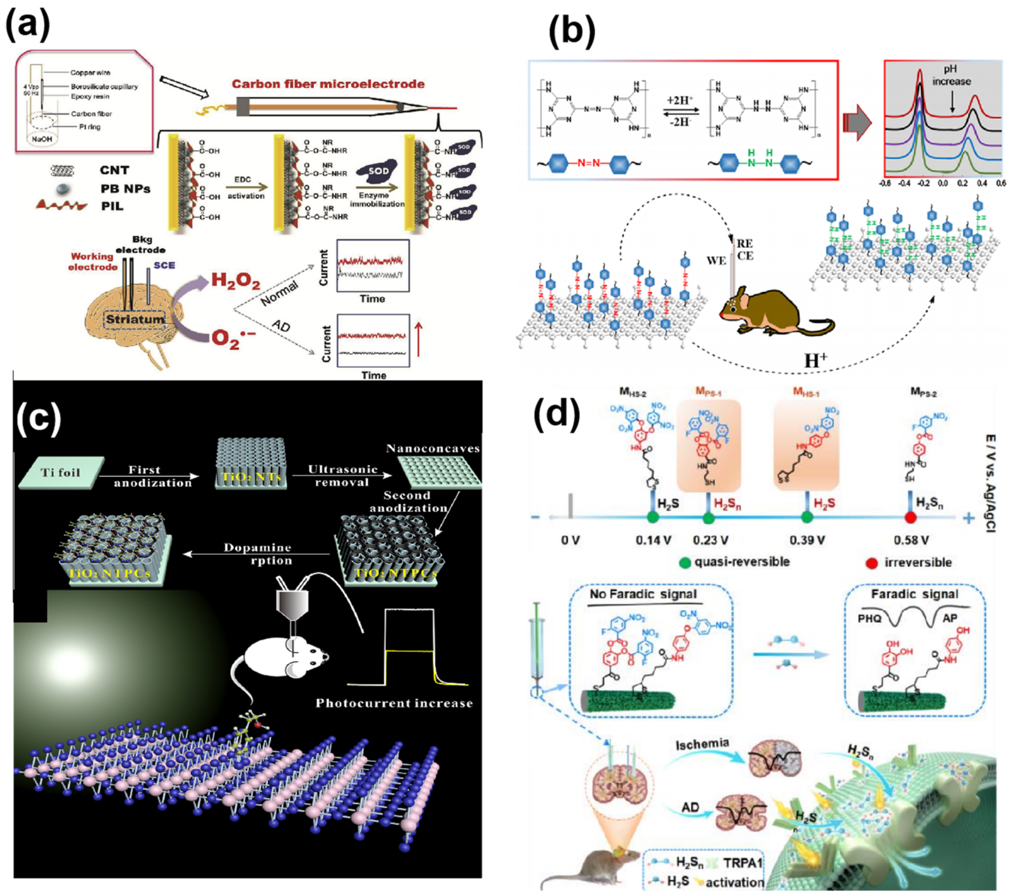
The ideal enzyme-based sensor capable of direct electron transfer (DET) from the redox-active center to the electrode surface belongs to the DET enzyme sensors category. DET enzyme sensors remain unaffected by changes in ambient oxygen concentration or additional mediators, making them adaptable to complex in vivo environments, ideal for neurochemical detection. Yu’s group [98] contributed significantly to ROS detection using functionalized ionic liquid polymers (PILs) coated on Prussian blue nanoparticles and carbon nanotubes (CNTs), enhancing sensor sites for SOD to improve sensitivity and stability to low O2•− concentrations (Figure 4a). Hence, the sensor effectively tracked changes in O2•− levels under normal and pathological conditions in the living brain system. Overall, enzymes play a crucial role in in vivo neurochemical detection owing to their unique selectivity and rapid kinetics. The development of various enzymes has expanded the scope of detectable analytes, and the emergence of synthetic nano-enzymes [99] holds promise for the future of biosensors.
● Aptamers, single-stranded DNA or RNA molecules from randomly sequenced nucleic acid pools, have gained attention for their high affinity and specificity to the target. Compared to enzymes, they offer convenient synthesis, design flexibility, and chemical stability. Aptamer-based electrochemical sensors undergo conformational changes when the aptamer on the electrode surface combines with the ligand, triggering electron transfer between the electrode and the modified REDOX group on the aptamer [100]. Currently, aptamers are used to detect neurochemicals, like DA [101], 5-HT [102], and ATP [103]. Aptamers possess a good biological compatibility. Due to a complex screening process, only specific neurochemicals have aptamer recognition units. In 2005, Yi Xiao et al. [104] pioneered an electrochemical aptamer sensor, sparking research interest. Subsequently, various aptamer electrochemical sensors have been developed, mostly for blood analyte detection [105,106]. However, few electrochemical aptamers have been applied successfully to brain tissue detection. Cui’s group [107] developed an electrochemical aptamer-based in vivo cocaine sensor on a silica-based neurorecording probe platform capable of measuring cocaine directly from discrete brain locations using square wave voltammetry, capturing real-time cocaine transient events in multiple brain regions over the entire pharmacokinetic time course.
Despite the many advantages of aptamers, several challenges persist, including limited measurement duration and obtaining high-performance aptamers for new targets. New aptamers need adaptation to sensing platforms, potentially bottlenecking sensor development for novel targets. Specificity remains a challenge; aptamers often bind chemical groups present in multiple targets, leading to cross-reactivity during detection [108,109]. This presents challenges for in vivo monitoring. The potential duration of in vivo aptamer-based measurements remains uncertain; prolonged in vivo measurements risk signal loss and electrode contamination, mitigated by surface coating or increasing single-layer packaging density to reduce protein contamination [110,111,112]. Despite being in its infancy, aptamer-based in vivo detection offers excellent time resolution, miniaturization, and other advantages, holding potential for in situ detection platforms.
● Recognition Elements for Ions play a crucial role in maintaining the central nervous system’s normal functioning. For instance, calcium ions act as second messengers in neurotransmitter regulation, while dysregulated Cl− levels and brain pH disorders are associated with various neurological disorders. Efforts have been made to detect ions accurately in vivo. To enhance in vivo detection accuracy, a dual-channel recognition strategy using ratiometric microelectrodes was proposed for real-time monitoring. Fan Zhao et al. [113] achieved dual-channel recognition by modifying recognition elements on different microelectrodes, enabling the real-time in situ monitoring of pH in the rat brain, minimizing brain damage and inaccuracies between the electrodes. Gu’s group [76] constructed a ratiometric microsensor for pH monitoring, utilizing electrochemically oxidized graphene oxide (EOGO) to generate a built-in correction signal and a poly(melamine) (PMel) film as a pH-selective recognition membrane (Figure 4b). PMel, a highly pH-sensitive conductive polymer, facilitated the successful real-time monitoring of rat brain pH post-whole-brain ischemia/reperfusion events, confirming the robustness of the proposed ratiometric electrochemical microsensor platform.
● Molecularly Imprinted Polymers (MIPs), formed by a template molecule’s size, shape, and functional groups, draw inspiration from the specific binding of antigens and antibodies—the ‘lock and key’ mechanism. These polymers, like a template, can absorb specific targets that fit the fabricated template in terms of shape, size, and chemical function. Due to their distinct structure and specific recognition ability or high selectivity, MIPs find extensive use, particularly in molecularly imprinted electrochemical sensors (MIPESs) [114]. Mosbach and Haupt [115] pioneered the integration of electrochemical sensors with MIPs, introducing MIPESs in 1999. Since then, MIPESs have detected organic compounds, heavy metal ions, emerging pollutants, and in vitro biomolecules. In 2012 [116], the first in vivo microsensor based on a MIP detected dopamine in the rat brain. Subsequently, focusing on neurochemical detection in the body, it has been employed for selective DA detection (Figure 4c) [117], as well as norepinephrine (NE) [118] and epinephrine (EP) [119].
● Electrochemical Probes: While in vitro environments can be corrected, the complexity of in vivo environments poses a risk to the selectivity of in vivo assays. To address this issue, electrochemical probes based on a dual recognition strategy were proposed for in vivo measurements, enhancing assay selectivity through the synergistic chemical recognition of specific ligands and the redox activity of chemicals. Recent studies suggest that biological activities initially attributed to hydrogen sulfide may actually be mediated by H2Sn, which converts endogenous hydrogen sulfide to hydrogen polysulfide in the presence of ROS. H2Sn is hypothesized to have a stronger oxidative capacity and reactivity than hydrogen sulfide, potentially being the true regulator in cellular signaling [120,121]. The further development of sensors that selectively detect multiple neurochemicals simultaneously is required for an in-depth understanding of in vivo molecular mechanisms. Despite the direct oxidation of H2S on the electrode surface, the slow oxidation of other electroactive biomolecules and the organism’s internal complexity cause significant interference. Tian’s group [9] designed two electrochemical probes, 3,4-bis((2-fluoro-5-nitrobenzoyl)oxy)-benzoic acid and N-(4-(2,5-dinitrophenoxy) phenyl)-5-(1, 2-dithiolan-3-yl)pentanamide, specifically recognizing H2Sn and hydrogen sulfide, respectively. Co-assembling these probes on a mesoporous gold membrane yielded a microsensor that responded well to both hydrogen sulfide (0.2–50 µM) and H2Sn (0.2 to 40 µM) (Figure 4d). This study contributed to understanding the molecular mechanisms of H2Sn and hydrogen sulfide and emphasized the significance of simultaneous detection for elucidating their pathophysiological roles in the brain.
● Electrochemical Microarray Detection, an electrochemical microarray detection platform, offers a promising approach to simultaneously sense multiple ions in the body under specific physiological or pathological conditions. Tian’s group [81] developed an electrochemical physiological microarray (ECPM) to quantify K+, Ca2+, and Na+ concentrations and pH using an open-circuit potentiostatic method. Different ion-recognizing elements designed for simultaneous electrical signal recording without cross-talk were employed. An internal reference electrode coated with a polyvinyl chloride membrane avoided the complex brain environment interference, ensuring the accuracy and selectivity of the developed ECPM. It provides new paths for the real-time monitoring of K+, Ca2+, and Na+ ions’ dynamics and quantitative concentrations, establishing the correlation between electrical and chemical signals.
Figure 4.
Implantable electrochemical microsensors for in vivo analysis. (a) Schematic diagram of the fabrication of an enzyme-based electrochemical microsensor for the determination of superoxide anion. (The black line represents the background current, which increases with the presence of superoxide radicals, as shown by the red line). Reprinted from Ref. [98] with permission. Copyright 2019 Elsevier B.V. (b) Diagram of an electrochemical microsensor strategy for rat brain pH measurement. (Different colored DPV curves represent tests at different pH, and the pH decreases from the top down). Reprinted from Ref. [76] with permission. Copyright 2021 American Chemical Society. (c) Molecularly imprinted polymer-based PEC-sensing platform for DA detection. Reprinted from Ref. [117] with permission. Copyright 2016 Elsevier B.V. (d) The working principle of the sensing strategy and specific probes for the simultaneous detection of H2S and H2Sn in the living mouse brain. Reprinted from Ref. [9] with permission. Copyright 1999–2023 John Wiley & Sons, Inc.
3.1.2. Electroactive Neurochemicals
Electrochemical sensing for detecting neurochemicals typically involves the direct oxidation or reduction of the target species on the electrode surface, generating corresponding electric currents that quantify dynamic chemical changes. Electrochemically active neurochemicals undergo direct oxidation or reduction on the electrode surface, producing detectable electrical signals. However, challenges arise due to the presence of multiple redox substances and the overlapping peak potentials of their reactions, hindering selective monitoring.
Nevertheless, functional brain regulatory networks enable electrochemical monitoring. Techniques, such as cyclic voltammetry (CV), chronoamperometry (CA), and potentiometric pulse (DPV), are commonly employed. CV is the recording of current–potential curves by controlling the electrode potential at different rates over time with one or more repeated scans in a triangular waveform. The basic principle is that a pulsed voltage with a triangular waveform is applied to the closed loop formed by the working electrode and the counter electrode to change the potential at the working electrode/electrolyte interface at a certain rate, forcing the active material at the working electrode to undergo an oxidation/reduction reaction. CA monitors the gain or loss of electrons in the presence of a fixed potential. In addition to a three-electrode setup consisting of a working electrode, a reference electrode, and a counter electrode, it can also be performed using a two-electrode setup with the reference and counter short-circuited together. The differential current of DPV is a symmetrical volt–ampere peak whose intensity is proportional to the concentration of the analyte. Due to the high Faraday to charging current ratio of the DPV, the direct detection of the base analyte at low concentrations can be achieved. Fast scan cyclic voltammetry (FSCV) stands out due to its scanning potentials and high scan rate, enabling the detection of small changes in neurochemical concentrations and differentiation based on unique redox potentials. In their study, Butcher et al. [122] utilized carbon fiber microelectrodes to monitor electroactive neurochemicals, such as dopamine, epinephrine, norepinephrine, and 5-hydroxytryptophan, enhancing selectivity and temporal resolution.
Despite advancements like waveform alterations enhancing sensitivity in rapid CV [123,124,125], varying chemical basal concentrations pose detection challenges. To achieve sensitivity and selectivity in in vivo electrochemical sensors, the modulation of specific electrochemical processes at the electrode/brain interface is essential. Nanomaterial advancements, encompassing carbon nanotubes, graphene, MOFs (metal–organic frameworks), metals, and metal oxides, bolster electron transfer kinetics. This enhancement elevates sensitivity, diminishes activation overpotential, and minimizes interference from unwanted substances. Additionally, recognition units, such as enzymes, aptamers, and electrochemical probes, play crucial roles in the construction of electroactive neurochemical sensors.
Among these electrode modifiers, metallic nanomaterials exhibit exceptional properties in amplifying electrical signals. Guoyue Shi et al. [126] pioneered the development of a glass-sealed Au nanoelectrode with cluster-like gold nanostructures modified with Nafion, marking the first instance of improving sensitivity and selectivity for the in vivo detection of dopamine (DA). This electrochemical biosensor offers several advantages, including easy insertion, low tissue interference, good biocompatibility, and strong analytical performance. It provides an effective means to monitor brain DA.
In addition to metallic nanomaterials, certain carbon-based nanomaterials possess remarkable electrochemical catalytic properties. Graphene, renowned for its high conductivity, mechanical strength, expansive surface area, and catalytic characteristics [127,128], is extensively used as an electrode in efficient electrochemical sensors. Its wide potential window and rapid electron transfer rate lead to a minimal charge transfer resistance and heightened electrochemical activity. The hydrophobic and p-p interactions between dopamine and graphene enable the distinct separation of the oxidation potentials of dopamine and uric acid. Chen’s group [129] significantly increased the electrode surface area and enhanced the electrocatalytic performance of platinum wires by employing gold nanoparticles (AuNPs) and reduced graphene oxide (rGO)-modified platinum wire microelectrodes on AuNPs/rGO composites, achieving the successful detection of dopamine in the rat brain striatum. Single- and multi-walled carbon nanotubes (SWCNTs and MWCNTs, respectively) were utilized to augment the electroactive/adsorption sites, ensuring a higher sensitivity, and electrocatalytic/defect-enriched sites, ensuring a higher selectivity in monitoring neurochemicals.
In 2019 [130], Taylor et al. enhanced the sensitivity to dopamine by modifying the coating on carbon fiber surfaces, introducing a highly sensitive and selective carbon fiber electrode (CFE). PEDOT finds wide utility in neutral physiological environment analysis for detecting electrophysiological signals and neurotransmission due to its strong adhesion and porous structure. PEDOT/CNT-functionalized sensors have been extensively applied in in vivo DA detection. Cai’s group [131] utilized SWCNTs/PEDOT: PSS-modified microelectrode arrays, creating a four-stalk implantable microelectrode array (MEA) for simultaneously and in real-time detecting bimodal signals—electrophysiological signals and dopamine (DA) concentration—in the rat striatum. This setup effectively monitored the dynamic changes in striatal bimodal signals during isoflurane anesthesia.
To improve the selectivity, some recognition units, such as enzymes, aptamers, and electrochemical probes, have been developed for the detection of electroactive neurochemicals. Mao’s group [132] introduced a novel interface functionalization strategy involving the assembly of aptamer–cholesterol amphiphiles (aptCAs) on alkyl chain-functionalized carbon fiber electrodes (CFEs). This approach proved more effective than pre-treating the electrode surface with a positively charged coating, enabling the efficient immobilization of aptamers on the CFE surface via electrostatic interaction. The resulting modified electrode exhibited increased stability in physiological fluids and demonstrated versatility across various oligonucleotide sequences. Upon implantation into the nucleus ambiguus (NAc) and medial forebrain bundle (MFB) regions of rats, this modified electrode successfully monitored dopamine (DA) dynamics in the living rat brain using amperometry to track changes in DA levels during electrical stimulation. Mao’s group also proposed an electrochemical coupling strategy, covalently placing a specific catechol on the carbon fiber surface, initiating an initial electrochemical coupling on the CFE. This process resulted in a thin layer of quinone intermediates that rapidly bound to thiol-containing oligonucleotides at controlled potentials. This innovative strategy not only simplified and enhanced the efficiency of carbon surface modification but also significantly improved sensitivity and stability in dopamine sensing. It established a robust system for continuously detecting dopamine dynamics in the living animal brain [133].
Based on the reaction of electroactive DBPs with hydrogen peroxide to generate electroactive phenols, a series of phenyl borate derivatives (DBPs) were designed as electrochemical probe molecules. Through analyzing the performance of the synthesized probes, we found that the o-Cl-DBPs exhibited optimal performance. Ratiometric electrodes were successfully constructed using graphene oxide as the internal reference for detection in the three brain regions of Parkinson’s rats. These ratiometric electrodes better mitigated errors arising from differences between in vitro and in vivo environments. Notably, due to graphene’s favorable electrocatalytic effect on ascorbic acid, we unexpectedly discovered the regulation of the balance between the antioxidant AA and hydrogen peroxide in the brain [134].
Researchers have demonstrated a strong interest in other electroactive neurochemicals, developing multiple strategies for in vivo in situ real-time monitoring, as summarized in Table 2.
3.2. Microdialysis Coupled with Electrochemical Detection
Microdialysis serves as a pivotal analytical technique for sampling in neurochemical analyses. Especially beneficial for in vivo applications, microdialysis minimizes damage to the brain tissue and exhibits extensive applicability in various extracellular fluid analyses, encompassing heart, fat, liver, and brain tissues [148]. Originating in 1966, the technique relies on analyte diffusion across a porous membrane. Bito’s group [18,149] accessed a dog’s cortical layer using a sterile dialysis capsule, allowing the collection of analytes into a brine stream, subsequently detectable in the flowing salt species. Notably, microdialysis often integrates with chromatography or electrophoresis systems for sample separation. When combined with electrochemical detection systems, it forms a separation-based sensor capable of achieving the highly selective, near real-time monitoring of multiple analytes. However, microdialysis monitoring relying on separation systems requires sample pretreatment, risking sample degradation, causing delays, and limiting spatial resolution due to separation properties. The coupling of microdialysis with biosensors, driven by advancements in electrochemical biosensors, enables the direct analysis of biological samples from living bodies. While less selective than separation-based microdialysis monitoring, this approach’s simplicity negates the need for additional pretreatment, avoids potential analyte degradation, and allows the monitoring of biological events over extended periods, such as 2 h [150].
3.2.1. Detection Techniques with Separation Means
Microdialysis can be paired with separation devices, like high-performance liquid chromatography (HPLC) and capillary electrophoresis (CE), along with associated detectors, to detect samples in dialysate. Continuous efforts have substantially improved microdialysis coupled with separation systems, achieving temporal resolutions in seconds, overturning the prior notion of a poor temporal resolution. As a result, it has also been favored by researchers, with a variety of microdialysis-coupled separation systems emerging as detection strategies that can be applied to the detection of different neurochemicals (Table 3).
● Microdialysis–capillary electrophoresis/microchip electrophoresis (MD–CE/ME): MD–CE/ME encounters a challenge due to the high-volume injection requirements (1–10 µL) of conventional separation techniques, leading to a compromised time resolution—a consistent limitation in microdialysis systems. However, the emergence of capillary electrophoresis has significantly alleviated this issue with its high separation efficiency and shorter separation times [151,152,153,154,155]. As early as the 1990s [156,157], the coupling of MD with CE enabled the efficient analysis of polar and charged compounds in biological samples. Initially, the integration of MD and CE was often paired with laser-induced fluorescence (LIF) detection, facilitating the in vivo detection of various neurochemicals, like norepinephrine, glutamate, and aspartate [155,158].
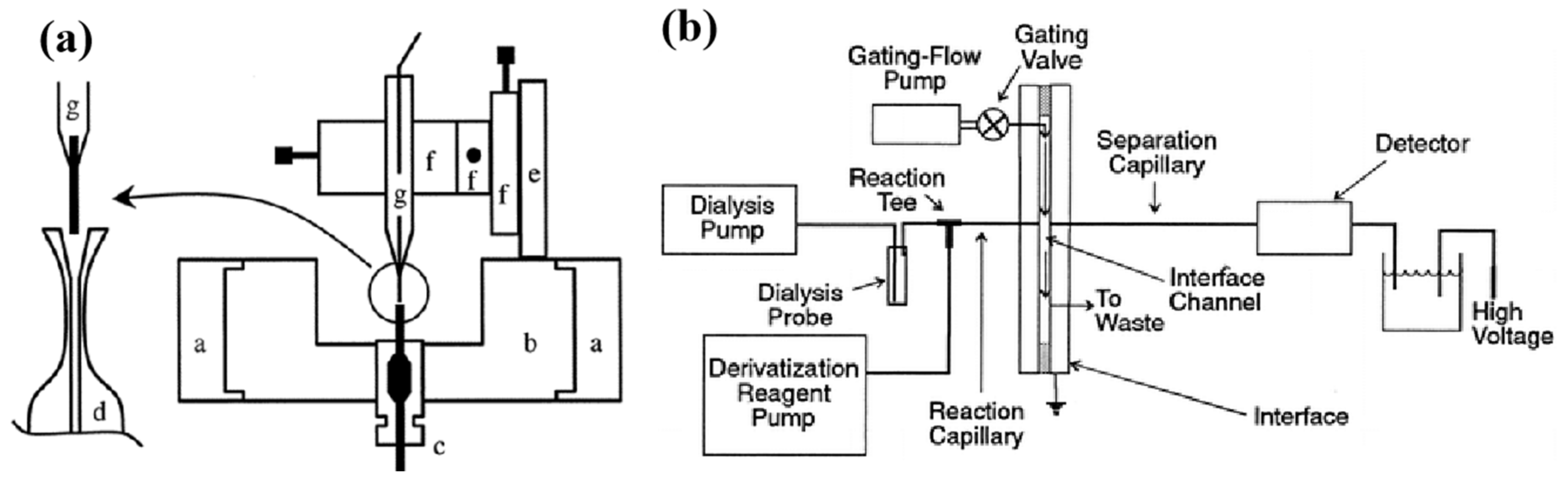
Despite CE-LIF detection enhancing the potential for the high temporal resolution monitoring of neurochemicals in microdialysis fluids, it necessitates derivatization to introduce a fluorophore into compounds, limiting its applicability to all metabolites and reducing selectivity for biomolecules. However, capillary electrophoresis (CE) combined with electrochemical detection has emerged as a robust analytical tool [159,160,161]. Thomas J. O’Shea et al. [162] demonstrated the continuous monitoring of amino acids in the brain by integrating CE with electrochemical detection using microdialysis sampling. This setup was showcased by observing “K+-induced stimulation of excitatory amino acid release.” Qian’s group [21] developed an integrated end column decoupler with conductive discs in a fused silica capillary wall, providing a low detection limit. This CE-EC system effectively determined dopamine in 1 min brain microdialysis fluid samples from anesthetized rats (Figure 5a).
On the other hand, microchip electrophoresis (ME) not only facilitates rapid MD-LC separation and demands minimal sample volumes but also enables the continuous, simultaneous monitoring of multiple analytes using online separation sensors. When combined with continuous microdialysis sampling, microchip electrophoretic separation-based sensors provide near real-time dynamic information on sample chemical composition. EC detection in ME was first described in 1998 [163] and offers unique advantages [164] over the initially used laser-induced fluorescence techniques [165,166,167]. It allows the direct detection of small, naturally electrically active biomolecules using an amperometric method. Additionally, the integration of microchip electrophoresis separation with the electrochemical system eliminates the need for bulky optical detection instruments, enhancing convenience.
Fabricating MD-ME-EC systems faces challenges in integrating working electrodes into the device. Rachel A. Saylor et al. [168] employed a PDMS/glass hybrid device fabrication process, enabling the successful integration of a high ionic-strength, pressure-driven MD stream with ME and CE on carbon electrodes. This integration allowed the near real-time in vivo monitoring of catecholamines in the rat brain. To enhance electrode alignment reproducibility with the separation channel, Shamal M. Gunawardhana et al. [169] utilized a thermally cracked photoresist film working electrode and a poly (Dimethylsiloxane) microchip with a flow-gated sample injection interface. This setup coupled MD for the online monitoring of levodopa conversion to dopamine and monitoring dopamine release in anesthetized rats after high K+ stimulation.
Flow-gated capillary electrophoresis combines conventional CE and microchip CE, using a quartz capillary as a separation channel and adopting a fast flow-gated injection technique primarily from microchip CE. This method utilizes silica capillaries as separation tubes while employing a single-cross microchip configuration for rapid flow-gated injection [20]. As early as 1996 [170], Kennedy’s group investigated capillary electrophoresis coupled with microdialysis using a flow-gated interface for the in vivo quantitative monitoring of compounds like aspartic acid and glutamate in the rat caudate nucleus (Figure 5b). This strategy led to an enhanced separation efficiency and rapid sampling despite trace sample amounts. Subsequently, to further enhance flow-gated CE in vivo monitoring, Bowser and Kennedy [171] reported an improved separation efficiency using smaller diameter capillaries (10 µm), higher electric field strength (2000 V/cm), and sheath-flow cuvettes. Additionally, Kennedy and Bowser [172] utilized derivatization with NDA and NBD-F to enhance sensitivity in detecting primary amines for rat brain neurotransmitter detection.
Figure 5.
(a) Schematic of the etched end-column decoupler and the rotatable detection cell for CE-EC. (a, stationary support collar; b, rotatable detection cell; c, butt connector; d, capillary; e, micropositioner mount; f, three translation stages of the micropositioner; g, carbon fiber microelectrode). Reprinted from Ref. [21] with permission. Copyright 1999 American Chemical Society. (b) Block diagram of the microdialysis/capillary zone electrophoresis-LIF system with online derivatization. Reprinted from Ref. [170] with permission. Copyright 1996 American Chemical Society.
● Microdialysis–liquid chromatography (MD–LC): MD–LC is a prevalent method for analyzing microdialysis samples, often employing liquid chromatography (LC) coupled with electrochemical, fluorescence, mass spectrometry (MS), or absorbance detection. While capillary electrophoresis boasts a high temporal resolution, HPLC has emerged as the preferred method for studying low concentrations of neurochemicals, like dopamine and 5-HT, due to its reliability and reproducibility. HPLC can provide both base concentrations and dynamic changes. However, conventional liquid chromatography necessitates sufficient dialysate, leading to longer processing times, inadequate temporal resolution, and significant delays between the sample collection and clinical response. Conversely, nano (capillary or microporous)-based LC methods coupled with fluorescence or electrochemical detection offer the selective analysis of neurochemicals in microvolume samples. Previous efforts to enhance the speed of HPLC techniques include using high pressure to increase the linear velocity of the mobile phase, elevating column temperature, and utilizing shorter columns filled with shorter particles [173,174,175]. The temporal resolution of measurements at HPLC not only depends on the sample stream flow rate but also on the sample volume. Capillary columns possess significantly smaller peak volumes compared to the standard or microbore (1 mm diameter) HPLC columns, minimizing the sample volume dilution and greatly improving the temporal resolution. Weber’s group integrated these modifications to determine serotonin (5-HT) levels in the central nervous system of awake animals using capillary LC and electrochemical detection [176]. To further expedite 5-HT chromatographic determinations and enhance the temporal resolution, Weber’s group explored on-column pre-concentration as part of optimization, highlighting the column diameter’s importance in achieving high speed and optimal sensitivity conditions. Smaller particle diameters and higher temperatures were found to yield faster optimal separations and a greater sensitivity. Under ideal conditions, the separation time for 5-HT was reduced to approximately 22.7 s. Online analysis feasibility was demonstrated by simulating repeated injections to achieve a time resolution of 36 s [22].
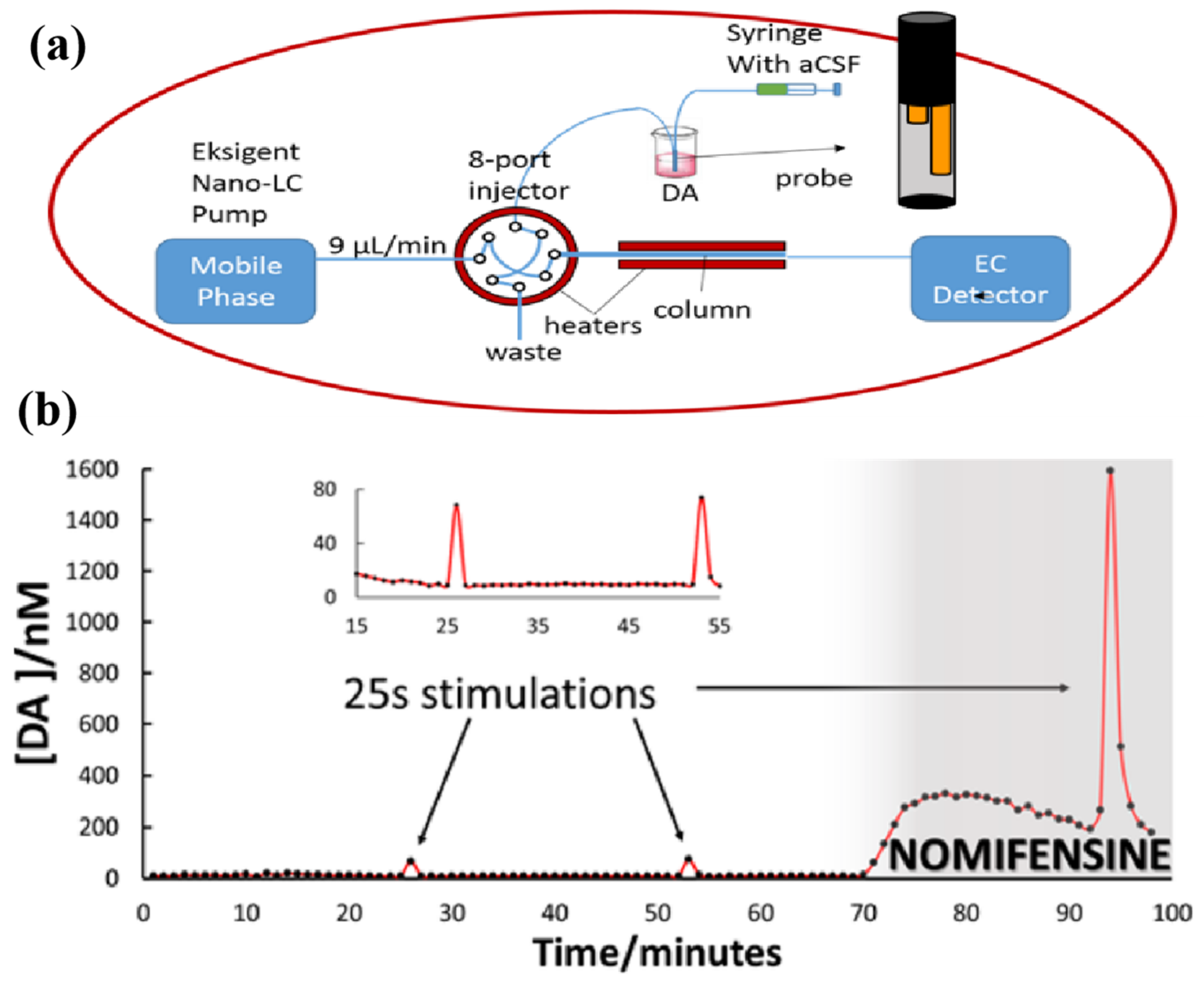
In another work, the online monitoring of 5-HT in vivo was accomplished by Andrews’ group, employing this rapid microdialysis method in awake animals over extended periods, achieving a temporal resolution of 3 min [177]. This represented a significant advancement. Weber’s group [178] optimized the pH, buffer composition, and surfactant concentration to eliminate interference with dopamine peaks. Employing the system to monitor electrically evoked DA transients, we achieved a true temporal resolution of less than 1 min. The evoked transients were observable in single, 1 min dialysate samples (Figure 6). To ensure robust in vivo online measurements, Weber’s group introduced a six-port recirculating syringe between the syringe pump and the microdialysis probe, utilizing capillary tubing (75 μm inner diameter, 70 cm long) at the inlet and outlet of the probe to minimize solute dispersion. Sampling and analysis were optimized to monitor basal 5-HT concentrations in the striatum of freely moving rats and subsequent responses to 1 min time-resolved stimuli [19]. In another work, Mao Lanqun’s group examined the spontaneous firing rate (SFR) and neurochemical kinetics of the auditory cortex in rats following sodium salicylate injection. They utilized in vivo microdialysis in combination with an online electrochemical system (OECS) and HPLC, revealing significantly elevated levels of glutamate and ascorbic acid [179].

Table 3.
In vivo detection of neurochemicals in the brain tissue by microdialysis coupled with electrochemical sensing based on separation systems.
Table 3.
In vivo detection of neurochemicals in the brain tissue by microdialysis coupled with electrochemical sensing based on separation systems.
| Neurochemical | Technique | Sampling Site | Temporal Resolution | LOD | Ref. |
|---|---|---|---|---|---|
| DA | MD-LC | Striatum | 1 min | - | [180] |
| DA | MD-HPLC | Nucleus accumbens septi | 1 min | - | [181] |
| DA | MD-HPLC | Striatum | 2 min | - | [182,183] |
| DA | MD-HPLC | Striatum, Cortex | 2 min | - | [184] |
| DA | MD-HPLC | Striatum | 1 min | 40 nM | [185] |
| MD-CapUHPLC | Striatum | 1 min | 0.15 nM | [178] | |
| DA | MD-ME | Striatum | 65 | 200 μM | [168] |
| DA | MD-ME | Striatum | 100 s | 1 μM | [169] |
| GABA | MD-HPLC | - | 1 min | - | [186] |
| Glu | MD-CE | Striatum | 25 s | - | [187] |
| Glu | MD-HPLC | Hippocampus | 1 min | - | [186] |
| Glu and AA | MD-HPLC | Auditory cortex | 0.1/1 μM | [179] | |
| Neuroactive amines and AAs | MD-CapLC | Striatum | 10 s | 0.09−0.35 nM | [188] |
| 5-HIAA | MD-CapLC | Hippocampus | 2 min | 3 nM | [189] |
| 5-HT | MD-CapLC | Hippocampus | 1 min | 56 μM | [189] |
| 5-HT | MD-HPLC | Hippocampus | 90 s | - | [186] |
| 5-HT | MD-HPLC | Striatum, Cortex | 2 min | - | [184] |
| 5-HT | MD-HPLC | Hippocampus, Striatum | 3 min | 0.8 fM | [177] |
| 5-HT | MD-CapUHPLC | Hippocampus | 1 min | 70 pM | [176] |
| 5-HT | MD-CapUHPLC | Striatum | 36 s | 0.3 nM | [22] |
| 5-HT | MD-CapUHPLC | Striatum | 30 s | 160 pM | [19] |
| NA, DA, and 5-HT | MD-UHPLC | Prefrontal cortex | 12 min | 32/42/83 pM | [190] |
| NA, DA, and 5-HT | MD-CapUHPLC | Striatum, Amygdala, Hippocampus | 21 min | 0.75/0.75/1.5 nM | [191] |
| NA, DA, and 5-HT | MD-UHPLC | Hippocampus | 8 min | 83/58/60 pM | [192] |
| Monoamines | MD-UHPLC | Hippocampus, Prefrontal cortex, Striatum | 20 min | 100 pM | [193] |
| Ach | MD-HPLC | Striatum | 7 min | 20 fmol | [194] |
| Ach | MD-HPLC | Hippocampus | 6 min | 10 fmol | [195] |
LC: liquid chromatography; HPLC: high-performance liquid chromatography; CapUHPLC: capillary ultra-high-performance liquid chromatography; ME: microchip electrophoresis; CE: capillary electrophoresis; CapLC: capillary liquid chromatography; UHPLC: ultra-high-performance liquid chromatography.
3.2.2. Detection Techniques with Biosensors
The use of biosensors offers a viable alternative to separation devices, which often necessitate bulky hardware and intricate operational mechanisms. Coupling biosensors with microdialysis introduces near real-time capabilities and shorter analysis times. This method involves fewer technical requirements as it avoids the need for sample collection and separation. While the use of biosensors has significantly improved the drawback of low temporal resolution, other challenges have emerged, including issues related to stability, reproducibility, etc. [196]
An ideal biosensor should have the capability to continuously and reliably monitor analytes in complex bodily fluids from living organisms over an extended period. Typically, a biosensor comprises one or more recognition elements designed to identify target molecules [197,198]. This process generates a chemical signal captured by the sensor component and translated into an optical or electrochemical signal (Table 4).
The utilization of enzymes stands as a common strategy in biosensors. In 2005, Mao’s group [25] summarized the principles, development, and notable applications of early enzyme-based continuous online monitoring combined with microdialysis sampling and biosensors. Notably, their work involved the development of enzyme-based biosensors integrated with microdialysis, employing thionine and xanthine oxidase (XOD) as low-potential mediators and oxidases [23]. This demonstrated that the use of low-potential mediators for electron transfer in oxidases offers a novel approach for developing oxidase-based biosensors, both theoretically and technologically simple.
Table 4.
In vivo detection of neurochemicals in the brain tissue using microdialysis coupled with electrochemical biosensors.
Table 4.
In vivo detection of neurochemicals in the brain tissue using microdialysis coupled with electrochemical biosensors.
| Neurochemical | Technique | Sampling Site | Temporal Resolution | LOD | Ref. |
|---|---|---|---|---|---|
| DA | MD-Biosensors | Striatum | 4 min | 0.31 nM | [199] |
| DA | MD-Biosensors | Striatum | 10 min | - | [200] |
| Glucose and lactate | MD-Biosensors | Cortex | 30 s | - | [201,202,203] |
| Glucose and lactate | MD-Biosensors | Cortex | 1 min | - | [204] |
| Glucose and lactate | MD-Biosensors | Striatum | - | 2.39/2.52 µM | [205] |
| Lactate | MD-Biosensors | Striatum | 1 min | - | [206] |
| Glucose | MD-Biosensors | Cortex | 2 min | 50 nM | [207] |
| Glucose | MD-Biosensors | Auditory cortex | - | 10 µM | [208] |
| Glucose | MD-Biosensors | Striatum | - | 1.8 µM | [209] |
| Glucose | MD-Biosensors | Striatum | - | 0.28 µM | [210] |
| Glucose | MD-Biosensors | Striatum | - | 3.33 µM | [211] |
| Ach | MD-Biosensors | Striatum | - | 1 µM | [24] |
| Hypoxanthine | MD-Biosensors | Striatum | - | 0.40 µM | [23] |
| Cu2+ | MD-Biosensors | Striatum | - | 13 nM | [212] |
| ATP | MD-Biosensors | Cortex | - | 50 pM | [213] |
| ATP | MD-Biosensors | Cortex | 0.5 s | 0.1 fmol | [214] |
| H2O2 | MD-Biosensors | Cortex, Striatum, Hippocampus | - | 1 µM | [79] |
DA: dopamin; Glu: glutamate; Ach: acetylcholine; ATP: adenosine triphosphate; MD: microdialysis.
The combination of microdialysis enables the highly selective detection of endogenous species within the brain system. Their research showcased an online electrochemical system (OECS) for the selective and continuous measurement of acetylcholine (ACh) [24], achieved by efficiently integrating in vivo microdialysis, a multienzymatic microreactor, and an electrochemical detector.
In a separate study, Yanyan Yu et al. [215] synthesized a new room-temperature ionic liquid, [C3(OH)2mim][BF4], with two hydroxyl functionalities in the imidazolium core. This introduction of functional groups significantly enhanced the stabilization of Au/Pt alloys and facilitated the formation of well-dispersed small metal nanoparticles, exhibiting a good catalytic activity against hydrogen peroxide. Using glutamate oxidase as a biorecognition element, the continuous detection of glutamate was achieved in the rat striatum, observing normal levels and changes in concentration following various stimuli (Figure 7a).
To further enhance in vivo sensing accuracy and selectivity, a dual recognition unit strategy (DRUS) was proposed. Yanyan Yu et al. [214] constructed a DRUS ATP biosensor with high selectivity and sensitivity. Utilizing aptamer-to-base and polyimidazole-to-phosphate recognition abilities, the biosensor exhibited an ultra-high sensitivity to subinosinazole levels of ATP-LOD and a remarkable selectivity against interfering ADP and AMP sensing in the extracellular ATP microdialysate sampled from the brain system.
Ratiometric electrochemical sensors (RECSs) have shown improved reproducibility, stability, and reliability in correcting errors during in vivo sensing, making them ideally suited for repetitive in vivo analyses [9,212,216]. Building upon this concept, Gu’s group constructed RECSs by modifying graphene oxide on electrodes via electrodeposition. Subsequently, methylene blue was adsorbed onto the graphene oxide using electrostatic attraction, serving as an effective internal reference ratio. The introduction of graphene sp3 C-C defects via subsequent electroreduction facilitated the electrochemical oxidation of AA at low potentials, ensuring a high selectivity in the brain system against potential interferences. One point that is worth noting is that this voltammetric RECS to accomplish in vivo/online repetitive measurements included a time interval of more than 1 min, which is capable of tracking the physiological process happening over several minutes [217] (Figure 7b).
Figure 7.
Several microdialysis-based sampling combined with electrochemical biosensors for the in vivo analysis of neurotransmitters. (a) Schematic presentation of the online microdialysis system with GlutaOx-modified biosensors for the continuous monitoring of glutamate. Reprinted from Ref. [215] with permission. Copyright 2023 Elsevier B.V. (b) Construction of a RECS for the selective determination of AA in cerebral microdialysis fluids and changes in AA concentration under whole-brain ischemia–reperfusion. Reprinted from Ref. [217] with permission. Copyright 2020 American Chemical Society.

All in all, the integration of microdialysis with electrochemical systems offers advantages, such as simple equipment, low cost, and high sensitivity. Initially, the coupling of microdialysis with separation systems and integrated electrochemical detection was primarily used for detecting electroactive neurochemicals. As the coupling extended to biosensors, designing recognition units on the sensor surface allowed the detection of not only electroactive neurological substances but also non-electrochemically active neurochemicals. However, the sometimes slow electrochemical reaction rates and coupled biocatalytic processes limit the temporal resolution. Spectroscopic methods, such as laser-induced fluorescence (LIF) and visible light absorption, have fast reaction kinetics. The application of microdialysis in combination with spectroscopic techniques has been shown to provide a high temporal resolution for the detection of neurochemicals. Additionally, integrating microdialysis with mass spectrometry serves as a powerful tool for analyzing biologically active molecules. This integration provides sequence specificity and enhanced mass sensitivity, enabling detection across a broad range of analytes. These assays significantly compensate for the limitations of integrated microdialysis and electrochemical detection platforms. As this review primarily focused on the application of electrochemical sensing to the brain tissue, we thus omitting detailed descriptions of other assays.
3.3. Emerging Techniques
In recent years, a liquid/liquid interface microsensor (LLIM) using a nano-micropipette emerged, utilizing electrochemical techniques to monitor the charge transfer from the aqueous phase to the organic phase of an analyte. This innovation allows the monitoring of neurochemicals in the living brain, even those lacking redox activity. Electrochemistry at the liquid/liquid interface, or the interface between two immiscible electrolyte solutions, has advanced the direct analysis of ions by leveraging the difference in electrolytic energy between the ionic solvents of neighboring phases. Adjusting the pipette tip to the micrometer scale enhanced the spatial resolution of in vivo analyses. In recent decades, electrochemical sensing at liquid/liquid interfaces has garnered attention for analyzing non-electrochemically active substances, like neurochemicals, amino acids, peptides, and proteins. For example, choline (Ch) exhibits specific ion transfer potential and a distinct ion transfer current signal. Zhang’s group [218] employed 1,2-dichloroethane as the organic phase and choline-containing rat cerebrospinal fluid as the aqueous phase, utilizing the disparity in solvation energies between the liquid phases. This approach demonstrated a good linearity and selectivity in responding to Ch, achieving a detection limit of 0.37 μM (Figure 8). Also, nanotechnology and liquid–liquid interfacial sensing have combined to enable neuronal monitoring for neurochemicals. Novel sensing principles based on nanochannels, particularly ionic current rectification (ICR) technology, have been developed. Lanqun’s group explored ICR at the micrometer scale [219], unveiling a new strategy using the microscale to selectively sense ATP in the brain system. They employed polyimidazole [220] to modify the inner wall surface of the microtubule, offering a good linearity for ATP within the 5–100 nM concentration range through differential binding between the positively charged polyimidazole and negatively charged ATP aptamers.
In another study, field-effect transistor (FET) technology, highly sensitive and selective in biosensing, allows miniaturization and has gained prominence for real-time, high-throughput, and high-sensitivity assays. Among biosensing platforms, FET biosensors excel in enzyme-modified biosensors due to their precise analyte detection, small size, integrated compactness, and potentially cost-effective quality. These sensors detect hydrogen ions (H+), byproducts produced by enzymes and analytes, proportionate to the analyte concentration. Graphene, with its unique structural geometry, excellent electrical properties, biocompatibility, and sensitivity to surface charge alterations, serves as an active channel for immobilizing enzyme molecules in FET biosensors. Hwang et al. [221] developed a reduced graphene oxide-based enzyme-modified FET (RGO-EnFET) to study acetylcholinesterase enzyme kinetics and the impact of acetylcholinesterase inhibitors on AD therapy. Additionally, Fenoy’s group [222] proposed modifying the FET graphene channel with a copolymer poly(3-aminobenzylaminobenzidine-co-phenylene amine) (PABA) film, enhancing the electrostatic charge and creating a non-denaturing environment for enzyme immobilization. This improved the FETs’ pH sensitivity, enabling real-time detection of acetylcholinesterase within the 5–1000 μM range for acetylcholine sensing.
FET technology has also integrated aptamers. Andrews’ group [223] obtained DA- and 5-HT-specific aptamers through SELEX screening, constructing flexible aptamer field-effect transistor sensors for DA and 5-HT detection. These sensors successfully detected target analytes in artificial cerebrospinal fluid and mouse brain slices.
The quality of behavioral correlates of neurochemical measurements can be improved as new wireless data transmission systems are able to perform the tests without tethering the animal [224]. As physics, neurophysiology, chemistry, and other disciplines continue to advance, microelectronic devices for wireless neurochemical sensing have been developed to detect neurochemicals in non-tethered animals. In addition, wireless transmission systems can also be used in human medicine. Roham et al. [225] proposed an integrated chip for wireless neurochemical measurements that provides both amperometry and fast scanning cyclic voltammetry. With the continued efforts of researchers, wireless transmission system test instruments are slowly becoming portable and wearable. Tonello et al. [226] have developed a low-cost, high-sensitivity, portable point-of-care (PoC) detection system based on a screen-printed electrochemical sensor that used a proprietary antibody to detect unfolded p53, enabling the detection of this biomarker in Alzheimer’s patients.
In recent years, Drakakis’ group [227] has developed a battery-powered potentiostat and wireless data transmission system that includes integrated biosensors and a microfluidic system for microdialysis. It can detect glucose and lactate in the brain of brain-injured patients using amperometry and potassium ions using potentiometry.
4. In Vitro Electrochemical Measurements of Biomolecules in the Brain Tissue
Both the in vivo and in vitro assays of neurochemicals play crucial roles in exploring brain science. Conducting detections on brain slices in vitro helps to avoid interference from the in vivo environment. Isolated brain slice cultures serve as common analytical models for studying neurophysiology, frequently employed for monitoring neurochemicals in the brain tissue in vitro. Moreover, multiple slices from the same brain can be obtained and measured, allowing access to deeper tissue regions. The experimental setup is typically more convenient with brain slices. However, brain-sectioning experiments come with limitations. The tissues undergo mechanical trauma during sectioning, raising concerns about whether measurements are taken from healthy or injured regions. Moreover, phenomena like Donnan swelling, due to the exposure of intracellular charged molecules during sectioning, are likely to alter the tissue’s mechanical properties.
In vivo assays are more time-sensitive compared to in vitro assays, enabling the real-time tracking of dynamic changes in target analyte concentrations throughout the organism. Yet, the complexity of the in vivo environment poses additional detection challenges. Both in vitro and in vivo assays possess strengths and weaknesses, continuously evolving and progressing in parallel, complementing each other with their respective strengths. In this regard, Wu’s group [228] enhanced electrode biocompatibility and stability by modifying the CFE with chitosan (CS) membranes, brain cell membranes, and the aptamer cholesterol amphiphile (DNA-Cho). Their electrode demonstrated a high sensitivity, specificity, and stability in detecting DA and was utilized to detect potassium-ion-induced DA release in brain slices and PC12 cells, unveiling the specific process of DA release inhibition by lipopolysaccharide (Figure 9). This robust electrode modification strategy facilitates the in vivo monitoring of DA sensing activities in complex environments. The development of microarray probes (MEAs) has offered new directions for researchers. Hossain et al. [229] designed and validated a platinum (Pt) microelectrode array-based GABA probe, utilized for in vitro measurements in brain slices. The probe includes two microbial sensors, with the concentration of GABA corrected by the difference in oxidation currents of hydrogen peroxide generated during measurements by the two microbial sensors. This approach minimizes the impact of the complex brain tissue environment. To enhance enzyme stability, Asri’s group [230] developed an enzyme-coating method that significantly optimized the mechanical stability, enabling its use in in vitro slices for selective acetylcholine detection. However, compared to in vivo monitoring, in vitro assays do not fully reflect the overall physiological trends in organisms, leading researchers to prefer in situ real-time monitoring in vivo.
5. Conclusions and Outlook
The accurate measurement of fast neurochemicals within the central nervous system remains pivotal in neuroscience, offering a comprehensive understanding of the molecular mechanisms driving physiological and pathological brain processes. Electrochemical sensing stands as a potent tool for monitoring small biomolecules in the brain tissue. Implantable electrochemical sensors and microdialysis represent two complementary categories in electrochemical sensing. Implantable sensors, providing high temporal and spatial resolution, enable the real-time monitoring of neurochemical signals in specific brain regions. Their sub-second temporal resolution allows for the study of rapid events associated with specific neurophysiological activities, eliminating isolation processes.
Nanomaterial-based electrochemical sensors, like graphene, carbon nanotubes, molecularly imprinted polymers, metal–organic frameworks, and metal nanoparticles, showcase robustness, selectivity, sensitivity, precision, and accuracy in quantifying neurochemicals. These sensors excel in measuring target molecules amidst complex physiological environments. Further advancements in recognition units (e.g., enzymes, aptamers, and molecularly imprinted polymers) have enhanced selectivity, improving the reliability of in vivo measurements. However, challenges, such as foreign body reaction from implantable electrodes, electrode contamination, and protein adsorption, still impede the stability and accuracy of long-term monitoring.
The development of flexible and degradable materials holds promise for sustained long-term bioanalytical tracking. Microdialysis, a potent sampling technique, facilitates the continuous monitoring of biomolecule concentrations in vivo and in vitro. Its broad sampling range encompasses diverse small molecules, like neurochemicals, amino acids, and neuropeptides, enabling simultaneous detection in microdialysates.
Though microdialysis suffers from a poor spatial and temporal resolution, its coupling with separation techniques, like high-performance liquid chromatography and capillary electrophoresis, has alleviated this drawback. Integrating microdialysis with electrochemical biosensors circumvents the need for sample separation. However, biosensors often rely on specific enzymes or aptamers, limiting target molecule detection. Moreover, costly microdialysis probes and the susceptibility of dialysis membranes to clogging pose challenges for reuse.
Continuous advancements in microdialysis probes, refining capillary column diameters, and optimizing biosensor surface construction strategies have propelled microdialysis in neurochemistry. The current neurochemical detection focuses on in vivo measurements in freely moving or conscious mice/rats, aligning studies closer to natural conditions. Emerging technologies, like ionic current rectification and field-effect transistors, open new frontiers and wireless data transmission systems for in vivo electrochemical sensing. Future research directions may focus more on the real-time in situ measurement, visualization, and simultaneous detection of multiple neurochemicals without interference. Advanced clinical neurotransmitter measurements are anticipated to develop, enriching neuroscience research, with electrochemical neurotransmission analysis poised as a potent tool in disease diagnosis and treatment.
Author Contributions
Conceptualization, Y.Z.; methodology, S.C.; investigation, X.X. and Y.Z.; data curation, X.X. and Y.Z.; writing—original draft preparation, X.X.; writing—review and editing, H.G. and A.H.; supervision, S.C., H.G. and A.H. All authors have read and agreed to the published version of the manuscript.
Funding
This research was funded by the Hunan Provincial Natural Science Foundation of China (Project No. 2021JJ30238, 2022JJ90045).
Conflicts of Interest
The authors declare no conflicts of interest.
References
- Teleanu, R.I.; Niculescu, A.G.; Roza, E.; Vladâcenco, O.; Grumezescu, A.M.; Teleanu, D.M. Neurotransmitters—Key Factors in Neurological and Neurodegenerative Disorders of the Central Nervous System. Int. J. Mol. Sci. 2022, 23, 5954. [Google Scholar] [CrossRef]
- Zhang, X.; Hatamie, A.; Ewing, A.G. Nanoelectrochemical analysis inside a single living cell. Curr. Opin. Electrochem. 2020, 22, 94–101. [Google Scholar] [CrossRef]
- Song, Q.; Li, Q.; Yan, J.; Song, Y. Echem methods and electrode types of the current in vivo electrochemical sensing. RSC Adv. 2022, 12, 17715–17739. [Google Scholar] [CrossRef]
- Hatamie, A.; He, X.; Zhang, X.-W.; Oomen, P.E.; Ewing, A.G. Advances in nano/microscale electrochemical sensors and biosensors for analysis of single vesicles, a key nanoscale organelle in cellular communication. Biosens. Bioelectron. 2023, 220, 114899. [Google Scholar] [CrossRef]
- Hatami, A.; Zhang, X.W.; Pieter, E.O.; Andrew, G.E. Nanoscale Electrochemical Sensors for Intracellular Measurements at the Single Cell. In Handbook of Nanobioelectrochemistry: Application in Devices and Biomolecular Sensing; Springer Nature: Singapore, 2023; pp. 131–152. [Google Scholar]
- Kissinger, P.T.; Hart, J.B.; Adams, R.N. Voltammetry in brain tissue—A new neurophysiological measurement. Brain Res. 1973, 55, 209–213. [Google Scholar] [CrossRef]
- Shin, M.; Venton, B.J. Fast-Scan Cyclic Voltammetry (FSCV) Reveals Behaviorally Evoked Dopamine Release by Sugar Feeding in the Adult Drosophila Mushroom Body. Angew. Chem. Int. Ed. 2022, 61, e202207399. [Google Scholar] [CrossRef] [PubMed]
- Cao, Q.; Shin, M.; Lavrik, N.V.; Venton, B.J. 3D-Printed Carbon Nanoelectrodes for In Vivo Neurotransmitter Sensing. Nano Lett. 2020, 20, 6831–6836. [Google Scholar] [CrossRef]
- Dong, H.; Zhou, Q.; Zhang, L.; Tian, Y. Rational Design of Specific Recognition Molecules for Simultaneously Monitoring of Endogenous Polysulfide and Hydrogen Sulfide in the Mouse Brain. Angew. Chem. Int. Ed. 2019, 58, 13948–13953. [Google Scholar] [CrossRef] [PubMed]
- Liang, H.; Zhu, M.; Ye, H.; Zeng, C.; Wang, S.; Niu, Y. Carbon fiber microelectrode array loaded with the diazonium salt-single-walled carbon nanotubes composites for the simultaneous monitoring of dopamine and serotonin in vivo. Anal. Chim. Acta 2021, 1186, 339086. [Google Scholar] [CrossRef]
- Ganesana, M.; Trikantzopoulos, E.; Maniar, Y.; Lee, S.T.; Venton, B.J. Development of a novel micro biosensor for in vivo monitoring of glutamate release in the brain. Biosens. Bioelectron. 2019, 130, 103–109. [Google Scholar] [CrossRef] [PubMed]
- Wei, H.; Wu, F.; Li, L.; Yang, X.; Xu, C.; Yu, P.; Ma, F.; Mao, L. Natural Leukocyte Membrane-Masked Microelectrodes with an Enhanced Antifouling Ability and Biocompatibility for In Vivo Electrochemical Sensing. Anal. Chem. 2020, 92, 11374–11379. [Google Scholar] [CrossRef]
- Hu, K.; Le Vo, K.L.; Hatamie, A.; Ewing, A.G. Quantifying Intracellular Single Vesicular Catecholamine Concentration with Open Carbon Nanopipettes to Unveil the Effect of L-DOPA on Vesicular Structure. Angew. Chem. Int. Ed. 2022, 61, e202113406. [Google Scholar] [CrossRef]
- Burmeister, J.J.; Gerhardt, G.A. Self-Referencing Ceramic-Based Multisite Microelectrodes for the Detection and Elimination of Interferences from the Measurement of l-Glutamate and Other Analytes. Anal. Chem. 2001, 73, 1037–1042. [Google Scholar] [CrossRef] [PubMed]
- Xu, C.; Li, G.; Gan, L.; Yuan, B. In Situ Electrochemical Formation of Oxo-Functionalized Graphene on Glassy Carbon Electrode with Chemical Fouling Recovery and Antibiofouling Properties for Electrochemical Sensing of Reduced Glutathione. Antioxidants 2023, 12, 8. [Google Scholar] [CrossRef] [PubMed]
- Feng, T.; Ji, W.; Tang, Q.; Wei, H.; Zhang, S.; Mao, J.; Zhang, Y.; Mao, L.; Zhang, M. Low-Fouling Nanoporous Conductive Polymer-Coated Microelectrode for In Vivo Monitoring of Dopamine in the Rat Brain. Anal. Chem. 2019, 91, 10786–10791. [Google Scholar] [CrossRef]
- Kalant, H. A microdialysis procedure for extraction and isolation of corticosteroids from peripheral blood plasma. Biochem. J. 1958, 69, 99–103. [Google Scholar] [CrossRef]
- Bito, L.; Davson, H.; Levin, E.; Murray, M.; Snider, N. The Concentrations of Free Amino acids and Other Electrolytes in Cerebrospinal Fluid, In Vivo Dialysate of Brain, and Blood Plasma of the DOG. J. Neurochem. 1966, 13, 1057–1067. [Google Scholar] [CrossRef]
- Zhang, J.; Jaquins-Gerstl, A.; Nesbitt, K.M.; Rutan, S.C.; Michael, A.C.; Weber, S.G. In Vivo Monitoring of Serotonin in the Striatum of Freely Moving Rats with One Minute Temporal Resolution by Online Microdialysis–Capillary High-Performance Liquid Chromatography at Elevated Temperature and Pressure. Anal. Chem. 2013, 85, 9889–9897. [Google Scholar] [CrossRef] [PubMed]
- Gong, M.; Zhang, N.; Maddukuri, N. Flow-gated capillary electrophoresis: A powerful technique for rapid and efficient chemical separation. Anal. Methods 2018, 10, 3131–3143. [Google Scholar] [CrossRef]
- Qian, J.; Wu, Y.; Yang, H.; Michael, A.C. An Integrated Decoupler for Capillary Electrophoresis with Electrochemical Detection: Application to Analysis of Brain Microdialysate. Anal. Chem. 1999, 71, 4486–4492. [Google Scholar] [CrossRef]
- Zhang, J.; Liu, Y.; Jaquins-Gerstl, A.; Shu, Z.; Michael, A.C.; Weber, S.G. Optimization for speed and sensitivity in capillary high performance liquid chromatography. The importance of column diameter in online monitoring of serotonin by microdialysis. J. Chromatogr. A 2012, 1251, 54–62. [Google Scholar] [CrossRef] [PubMed]
- König, M.; Thinnes, A.; Klein, J. Microdialysis and its use in behavioural studies: Focus on acetylcholine. J. Neurosci. Methods 2018, 300, 206–215. [Google Scholar] [CrossRef]
- Kho, C.M.; Enche Ab Rahim, S.K.; Ahmad, Z.A.; Abdullah, N.S. A Review on Microdialysis Calibration Methods: The Theory and Current Related Efforts. Mol. Neurobiol. 2016, 54, 3506–3527. [Google Scholar] [CrossRef]
- Saylor, R.A.; Lunte, S.M. A review of microdialysis coupled to microchip electrophoresis for monitoring biological events. J. Chromatogr. A 2015, 1382, 48–64. [Google Scholar] [CrossRef]
- Zhang, Z.; Hao, J.; Xiao, T.; Yu, P.; Mao, L. Online electrochemical systems for continuous neurochemical measurements with low-potential mediator-based electrochemical biosensors as selective detectors. Analyst 2015, 140, 5039–5047. [Google Scholar] [CrossRef]
- Lin, Y.; Yu, P.; Mao, L. A multi-enzyme microreactor-based online electrochemical system for selective and continuous monitoring of acetylcholine. Analyst 2015, 140, 3781–3787. [Google Scholar] [CrossRef]
- Zhang, M.; Mao, L. Enzyme-based amperometric biosensors for continuous and on-line monitoring of cerebral extracellular microdialysate. FBL 2005, 10, 345–352. [Google Scholar] [CrossRef] [PubMed]
- Bobin, S.; Popot, M.A.; Bonnaire, Y.; Tabet, J.C. Approach to the determination of insulin-like-growth-factor-I (IGF-I) concentration in plasma by high-performance liquid chromatography-ion trap mass spectrometry: Use of a deconvolution algorithm for the quantification of multiprotonated molecules in electrospray ionization. Analyst 2001, 126, 1996–2001. [Google Scholar]
- Dagher, A.; Robbins, T.W. Personality, addiction, dopamine: Insights from Parkinson’s disease. Neuron 2009, 61, 502–510. [Google Scholar] [CrossRef]
- Volkow, N.D.; Fowler, J.S.; Wang, G.J.; Swanson, J.M. Dopamine in drug abuse and addiction: Results from imaging studies and treatment implications. Mol. Psychiatry 2004, 9, 557–569. [Google Scholar] [CrossRef]
- Brennan, A.R.; Arnsten, A.F.T. Neuronal mechanisms underlying attention deficit hyperactivity disorder—The influence of arousal on prefrontal cortical function. Ann. N. Y. Acad. Sci. 2008, 1129, 236–245. [Google Scholar] [CrossRef]
- Kurian, M.A.; Gissen, P.; Smith, M.; Heales, S.J.R.; Clayton, P.T. The monoamine neurotransmitter disorders: An expanding range of neurological syndromes. Lancet Neurol. 2011, 10, 721–733. [Google Scholar] [CrossRef]
- Marecos, C.; Ng, J.; Kurian, M.A. What is new for monoamine neurotransmitter disorders? J. Inherit. Metab. Dis. 2014, 37, 619–626. [Google Scholar] [CrossRef]
- Hamdan, S.K.; Mohd Zain, A. In vivo Electrochemical Biosensor for Brain Glutamate Detection: A Mini Review. Malays. J. Med. Sci. 2014, 21, 12–26. [Google Scholar] [PubMed]
- Lau, A.; Tymianski, M. Glutamate receptors, neurotoxicity and neurodegeneration. Pflügers Arch. Eur. J. Physiol. 2010, 460, 525–542. [Google Scholar] [CrossRef] [PubMed]
- Martin, W.R.W. MR Spectroscopy in Neurodegenerative Disease. Mol. Imaging Biol. 2007, 9, 196–203. [Google Scholar] [CrossRef] [PubMed]
- Stone, J.M.; Morrison, P.D.; Pilowsky, L.S. Review: Glutamate and dopamine dysregulation in schizophrenia—A synthesis and selective review. J. Psychopharmacol. 2007, 21, 440–452. [Google Scholar] [CrossRef] [PubMed]
- Rice, M.E. Ascorbate regulation and its neuroprotective role in the brain. Trends Neurosci. 2000, 23, 209–216. [Google Scholar] [CrossRef] [PubMed]
- Liu, K.; Lin, Y.; Xiang, L.; Yu, P.; Su, L.; Mao, L. Comparative study of change in extracellular ascorbic acid in different brain ischemia/reperfusion models with in vivo microdialysis combined with on-line electrochemical detection. Neurochem. Int. 2008, 52, 1247–1255. [Google Scholar] [CrossRef] [PubMed]
- Zhang, N.; Liu, J.-x.; Ma, F.-r.; Yu, L.-s.; Lin, Y.-q.; Liu, K.; Mao, L.-q. Change of extracellular ascorbic acid in the brain cortex following ice water vestibular stimulation: An on-line electrochemical detection coupled with in vivo microdialysis sampling for guinea pigs. Chin. Med. J. 2008, 121, 1120–1125. [Google Scholar] [CrossRef]
- Li, L.; Zhang, Y.; Hao, J.; Liu, J.; Yu, P.; Ma, F.; Mao, L. Online electrochemical system as an in vivo method to study dynamic changes of ascorbate in rat brain during 3-methylindole-induced olfactory dysfunction. Analyst 2016, 141, 2199–2207. [Google Scholar] [CrossRef]
- Fouani, L.; Menezes, S.V.; Paulson, M.; Richardson, D.R.; Kovacevic, Z. Metals and metastasis: Exploiting the role of metals in cancer metastasis to develop novel anti-metastatic agents. Pharmacol. Res. 2017, 115, 275–287. [Google Scholar] [CrossRef]
- Jentsch, T.J.; Stein, V.; Weinreich, F.; Zdebik, A.A. Molecular Structure and Physiological Function of Chloride Channels. Physiol. Rev. 2002, 82, 503–568. [Google Scholar] [CrossRef]
- Graefe, A.; Stanca, S.E.; Nietzsche, S.; Kubicova, L.; Beckert, R.; Biskup, C.; Mohr, G.J. Development and Critical Evaluation of Fluorescent Chloride Nanosensors. Anal. Chem. 2008, 80, 6526–6531. [Google Scholar] [CrossRef]
- Ashton, T.D.; Jolliffe, K.A.; Pfeffer, F.M. Luminescent probes for the bioimaging of small anionic species in vitro and in vivo. Chem. Soc. Rev. 2015, 44, 4547–4595. [Google Scholar] [CrossRef] [PubMed]
- Zhao, H.; Carney, K.E.; Falgoust, L.; Pan, J.W.; Sun, D.; Zhang, Z. Emerging roles of Na+/H+ exchangers in epilepsy and developmental brain disorders. Prog. Neurobiol. 2016, 138–140, 19–35. [Google Scholar] [CrossRef] [PubMed]
- Rossi, D.J.; Brady, J.D.; Mohr, C. Astrocyte metabolism and signaling during brain ischemia. Nat. Neurosci. 2007, 10, 1377–1386. [Google Scholar] [CrossRef] [PubMed]
- Vollmer, L.L.; Strawn, J.R.; Sah, R. Acid–base dysregulation and chemosensory mechanisms in panic disorder: A translational update. Transl. Psychiatry 2015, 5, e572. [Google Scholar] [CrossRef] [PubMed]
- Takei, Y.; Ando, H.; Tsutsui, K. Chapter 103—Gasotransmitter Family. In Handbook of Hormones; Academic Press: San Diego, CA, USA, 2016; pp. 601–602. [Google Scholar]
- Pałasz, A.; Menezes, I.C.; Worthington, J.J. The role of brain gaseous neurotransmitters in anxiety. Pharmacol. Rep. 2021, 73, 357–371. [Google Scholar] [CrossRef] [PubMed]
- Han, Y.; Qin, J.; Chang, X.; Yang, Z.; Bu, D.; Du, J. Modulating effect of hydrogen sulfide on gamma-aminobutyric acid B receptor in recurrent febrile seizures in rats. Neurosci. Res. 2005, 53, 216–219. [Google Scholar] [CrossRef] [PubMed]
- Garcia-Bereguiain, M.A.; Samhan-Arias, A.K.; Martin-Romero, F.J.; Gutierrez-Merino, C. Hydrogen Sulfide Raises Cytosolic Calcium in Neurons Through Activation of L-Type Ca2+ Channels. Antioxid. Redox Signal. 2008, 10, 31–42. [Google Scholar] [CrossRef]
- Schreier, S.M.; Muellner, M.K.; Steinkellner, H.; Hermann, M.; Esterbauer, H.; Exner, M.; Gmeiner, B.M.K.; Kapiotis, S.; Laggner, H. Hydrogen Sulfide Scavenges the Cytotoxic Lipid Oxidation Product 4-HNE. Neurotox. Res. 2010, 17, 249–256. [Google Scholar] [CrossRef]
- Xie, C.; Luo, K.; Tan, L.; Yang, Q.; Zhao, X.; Zhou, L. A Review for In Vitro and In Vivo Detection and Imaging of Gaseous Signal Molecule Carbon Monoxide by Fluorescent Probes. Molecules 2022, 27, 8842. [Google Scholar] [CrossRef]
- Li, W.; Li, R.; Chen, R.; Liang, X.; Song, W.; Lin, W. Activatable Photoacoustic Probe for In Situ Imaging of Endogenous Carbon Monoxide in the Murine Inflammation Model. Anal. Chem. 2021, 93, 8978–8985. [Google Scholar] [CrossRef]
- Fu, G.-Q.; Xia, Y.-S.; Jiang, W.-L.; Wang, W.-X.; Tan, Z.-K.; Guo, K.-Y.; Mao, G.-J.; Li, C.-Y. A novel precipitating-fluorochrome-based fluorescent probe for monitoring carbon monoxide during drug-induced liver injury. Talanta 2022, 243, 123398. [Google Scholar] [CrossRef]
- Zhang, C.; Peng, S.-Y.; Hong, S.; Chen, Q.-W.; Zeng, X.; Rong, L.; Zhong, Z.-L.; Zhang, X.-Z. Biomimetic carbon monoxide nanogenerator ameliorates streptozotocin induced type 1 diabetes in mice. Biomaterials 2020, 245, 119986. [Google Scholar] [CrossRef] [PubMed]
- Yue, L.; Tang, Y.; Huang, H.; Song, W.; Lin, W. A fluorogenic probe for detecting CO with the potential integration of diagnosis and therapy (IDT) for cancer. Sens. Actuators B Chem. 2021, 344, 130245. [Google Scholar] [CrossRef]
- Zhang, S.; Lachance, B.B.; Mattson, M.P.; Jia, X. Glucose metabolic crosstalk and regulation in brain function and diseases. Prog. Neurobiol. 2021, 204, 102089. [Google Scholar] [CrossRef]
- Hollyer, T.R.; Bordoni, L.; Kousholt, B.S.; van Luijk, J.; Ritskes-Hoitinga, M.; Østergaard, L. The evidence for the physiological effects of lactate on the cerebral microcirculation: A systematic review. J. Neurochem. 2019, 148, 712–730. [Google Scholar] [CrossRef]
- Xapelli, S.; Agasse, F.; Ferreira, R.; Silva, P.A.; Malva, O.J. Neuropeptide Y as an Endogenous Antiepileptic, Neuroprotective and Pro-Neurogenic Peptide. Recent Pat. CNS Drug Discov. 2006, 1, 315–324. [Google Scholar] [CrossRef]
- Kalra, S.P.; Dube, M.G.; Pu, S.; Xu, B.; Horvath, T.L.; Kalra, P.S. Interacting Appetite-Regulating Pathways in the Hypothalamic Regulation of Body Weight. Endocr. Rev. 1999, 20, 68–100. [Google Scholar]
- O’Loughlin, E.K.; Pakan, J.M.P.; McDermott, K.W.; Yilmazer-Hanke, D. Expression of neuropeptide Y1 receptors in the amygdala and hippocampus and anxiety-like behavior associated with Ammon’s horn sclerosis following intrahippocampal kainate injection in C57BL/6J mice. Epilepsy Behav. 2014, 37, 175–183. [Google Scholar] [CrossRef]
- Loewi, O. Über humorale übertragbarkeit der Herznervenwirkung. Pflüger’s Arch. Gesamte Physiol. Menschen Tiere 1921, 189, 239–242. [Google Scholar] [CrossRef]
- Watson, C.J.; Venton, B.J.; Kennedy, R.T. In Vivo Measurements of Neurotransmitters by Microdialysis Sampling. Anal. Chem. 2006, 78, 1391–1399. [Google Scholar] [CrossRef]
- Khan, A.S.; Michael, A.C. Invasive consequences of using micro-electrodes and microdialysis probes in the brain. TrAC Trends Anal. Chem. 2003, 22, 503–508. [Google Scholar] [CrossRef]
- Khoshnevisan, K.; Maleki, H.; Honarvarfard, E.; Baharifar, H.; Gholami, M.; Faridbod, F.; Larijani, B.; Faridi Majidi, R.; Khorramizadeh, M.R. Nanomaterial based electrochemical sensing of the biomarker serotonin: A comprehensive review. Microchim. Acta 2019, 186, 49. [Google Scholar] [CrossRef]
- Tiwari, J.N.; Vij, V.; Kemp, K.C.; Kim, K.S. Engineered Carbon-Nanomaterial-Based Electrochemical Sensors for Biomolecules. ACS Nano 2016, 10, 46–80. [Google Scholar] [CrossRef] [PubMed]
- Shleev, S.; Tkac, J.; Christenson, A.; Ruzgas, T.; Yaropolov, A.I.; Whittaker, J.W.; Gorton, L. Direct electron transfer between copper-containing proteins and electrodes. Biosens. Bioelectron. 2005, 20, 2517–2554. [Google Scholar] [CrossRef] [PubMed]
- Tian, Y.; Mao, L.; Okajima, T.; Ohsaka, T. Superoxide Dismutase-Based Third-Generation Biosensor for Superoxide Anion. Anal. Chem. 2002, 74, 2428–2434. [Google Scholar] [CrossRef] [PubMed]
- Tian, Y.; Mao, L.; Okajima, T.; Ohsaka, T. Electrochemistry and Electrocatalytic Activities of Superoxide Dismutases at Gold Electrodes Modified with a Self-Assembled Monolayer. Anal. Chem. 2004, 76, 4162–4168. [Google Scholar] [CrossRef] [PubMed]
- Zhang, X.; Song, C.; Yang, K.; Hong, W.; Lu, Y.; Yu, P.; Mao, L. Photoinduced Regeneration of an Aptamer-Based Electrochemical Sensor for Sensitively Detecting Adenosine Triphosphate. Anal. Chem. 2018, 90, 4968–4971. [Google Scholar] [CrossRef] [PubMed]
- Li, C.; Zhuo, Y.; Xiao, X.; Li, S.; Han, K.; Lu, M.; Zhang, J.; Chen, S.; Gu, H. Facile Electrochemical Microbiosensor Based on In Situ Self-Assembly of Ag Nanoparticles Coated on Ti3C2Tx for In Vivo Measurements of Chloride Ions in the PD Mouse Brain. Anal. Chem. 2021, 93, 7647–7656. [Google Scholar] [CrossRef] [PubMed]
- Xiao, X.; Li, C.; Liu, Y.; Feng, Y.; Han, K.; Xiang, H.; Shi, G.; Gu, H. A ratiometric electrochemical microsensor for monitoring chloride ions in vivo. Analyst 2021, 146, 6202–6210. [Google Scholar] [CrossRef] [PubMed]
- Zhang, Z.; Li, M.; Zuo, Y.; Chen, S.; Zhuo, Y.; Lu, M.; Shi, G.; Gu, H. In Vivo Monitoring of pH in Subacute PD Mouse Brains with a Ratiometric Electrochemical Microsensor Based on Poly(melamine) Films. ACS Sens. 2022, 7, 235–244. [Google Scholar] [CrossRef] [PubMed]
- Dong, H.; Zhao, L.; Zhou, Y.; Wei, X.; Xu, C.; Zhang, Y.; Xu, M. Novel Self-Calibrating Amperometric and Ratiometric Electrochemical Nanotip Microsensor for pH Measurement in Rat Brain. Anal. Chem. 2021, 93, 13815–13822. [Google Scholar] [CrossRef] [PubMed]
- Liu, L.; Zhao, F.; Liu, W.; Zhu, T.; Zhang, J.Z.H.; Chen, C.; Dai, Z.; Peng, H.; Huang, J.-L.; Hu, Q.; et al. An Electrochemical Biosensor with Dual Signal Outputs: Toward Simultaneous Quantification of pH and O2 in the Brain upon Ischemia and in a Tumor during Cancer Starvation Therapy. Angew. Chem. Int. Ed. 2017, 56, 10471–10475. [Google Scholar] [CrossRef] [PubMed]
- Li, S.; Tian, Y. An Electrochemical Biosensor with Dual Signal Outputs for Ratiometric Monitoring the Levels of H2O2 and pH in the Microdialysates from a Rat Brain. Electroanalysis 2018, 30, 1047–1053. [Google Scholar] [CrossRef]
- Moon, J.; Ha, Y.; Kim, M.; Sim, J.; Lee, Y.; Suh, M. Dual Electrochemical Microsensor for Real-Time Simultaneous Monitoring of Nitric Oxide and Potassium Ion Changes in a Rat Brain during Spontaneous Neocortical Epileptic Seizure. Anal. Chem. 2016, 88, 8942–8948. [Google Scholar] [CrossRef]
- Zhao, F.; Liu, Y.; Dong, H.; Feng, S.; Shi, G.; Lin, L.; Tian, Y. An Electrochemophysiological Microarray for Real-Time Monitoring and Quantification of Multiple Ions in the Brain of a Freely Moving Rat. Angew. Chem. Int. Ed. 2020, 59, 10426–10430. [Google Scholar] [CrossRef]
- Huang, S.; Zhang, L.; Dai, L.; Wang, Y.; Tian, Y. Nonenzymatic Electrochemical Sensor with Ratiometric Signal Output for Selective Determination of Superoxide Anion in Rat Brain. Anal. Chem. 2021, 93, 5570–5576. [Google Scholar] [CrossRef]
- Li, R.; Guo, D.; Ye, J.; Zhang, M. Stabilization of Prussian blue with polyaniline and carbon nanotubes in neutral media for in vivo determination of glucose in rat brains. Analyst 2015, 140, 3746–3752. [Google Scholar] [CrossRef]
- Forderhase, A.G.; Styers, H.C.; Lee, C.A.; Sombers, L.A. Simultaneous voltammetric detection of glucose and lactate fluctuations in rat striatum evoked by electrical stimulation of the midbrain. Anal. Bioanal. Chem. 2020, 412, 6611–6624. [Google Scholar] [CrossRef]
- Weltin, A.; Kieninger, J.; Enderle, B.; Gellner, A.-K.; Fritsch, B.; Urban, G.A. Polymer-based, flexible glutamate and lactate microsensors for in vivo applications. Biosens. Bioelectron. 2014, 61, 192–199. [Google Scholar] [CrossRef]
- Frey, O.; Holtzman, T.; McNamara, R.M.; Theobald, D.E.H.; van der Wal, P.D.; de Rooij, N.F.; Dalley, J.W.; Koudelka-Hep, M. Enzyme-based choline and l-glutamate biosensor electrodes on silicon microprobe arrays. Biosens. Bioelectron. 2010, 26, 477–484. [Google Scholar] [CrossRef]
- Burmeister, J.J.; Davis, V.A.; Quintero, J.E.; Pomerleau, F.; Huettl, P.; Gerhardt, G.A. Glutaraldehyde Cross-Linked Glutamate Oxidase Coated Microelectrode Arrays: Selectivity and Resting Levels of Glutamate in the CNS. ACS Chem. Neurosci. 2013, 4, 721–728. [Google Scholar] [CrossRef]
- Burmeister, J.J.; Pomerleau, F.; Palmer, M.; Day, B.K.; Huettl, P.; Gerhardt, G.A. Improved ceramic-based multisite microelectrode for rapid measurements of l-glutamate in the CNS. J. Neurosci. Methods 2002, 119, 163–171. [Google Scholar] [CrossRef] [PubMed]
- Day, B.K.; Pomerleau, F.; Burmeister, J.J.; Huettl, P.; Gerhardt, G.A. Microelectrode array studies of basal and potassium-evoked release of l-glutamate in the anesthetized rat brain. J. Neurochem. 2006, 96, 1626–1635. [Google Scholar] [CrossRef]
- Hascup, K.N.; Hascup, E.R.; Pomerleau, F.; Huettl, P.; Gerhardt, G.A. Second-by-second measures of L-glutamate in the prefrontal cortex and striatum of freely moving mice. J. Pharmacol. Exp. Ther. 2008, 324, 725. [Google Scholar] [CrossRef] [PubMed]
- McLamore, E.S.; Mohanty, S.; Shi, J.; Claussen, J.; Jedlicka, S.S.; Rickus, J.L.; Porterfield, D.M. A self-referencing glutamate biosensor for measuring real time neuronal glutamate flux. J. Neurosci. Methods 2010, 189, 14–22. [Google Scholar] [CrossRef] [PubMed]
- Rutherford, E.C.; Pomerleau, F.; Huettl, P.; Strömberg, I.; Gerhardt, G.A. Chronic second-by-second measures of l-glutamate in the central nervous system of freely moving rats. J. Neurochem. 2007, 102, 712–722. [Google Scholar] [CrossRef] [PubMed]
- Stephens, M.L.; Pomerleau, F.; Huettl, P.; Gerhardt, G.A.; Zhang, Z. Real-time glutamate measurements in the putamen of awake rhesus monkeys using an enzyme-based human microelectrode array prototype. J. Neurosci. Methods 2010, 185, 264–272. [Google Scholar] [CrossRef] [PubMed]
- Konradsson-Geuken, Å.; Gash, C.R.; Alexander, K.; Pomerleau, F.; Huettl, P.; Gerhardt, G.A.; Bruno, J.P. Second-by-second analysis of alpha 7 nicotine receptor regulation of glutamate release in the prefrontal cortex of awake rats. Synapse 2009, 63, 1069–1082. [Google Scholar] [CrossRef]
- Burmeister, J.J.; Palmer, M.; Gerhardt, G.A. l-lactate measures in brain tissue with ceramic-based multisite microelectrodes. Biosens. Bioelectron. 2005, 20, 1772–1779. [Google Scholar] [CrossRef]
- Burmeister, J.J.; Pomerleau, F.; Huettl, P.; Gash, C.R.; Werner, C.E.; Bruno, J.P.; Gerhardt, G.A. Ceramic-based multisite microelectrode arrays for simultaneous measures of choline and acetylcholine in CNS. Biosens. Bioelectron. 2008, 23, 1382–1389. [Google Scholar] [CrossRef]
- Ledo, A.; Lourenço, C.F.; Laranjinha, J.; Brett, C.M.A.; Gerhardt, G.A.; Barbosa, R.M. Ceramic-Based Multisite Platinum Microelectrode Arrays: Morphological Characteristics and Electrochemical Performance for Extracellular Oxygen Measurements in Brain Tissue. Anal. Chem. 2017, 89, 1674–1683. [Google Scholar] [CrossRef] [PubMed]
- Peng, Q.; Yan, X.; Shi, X.; Ou, S.; Gu, H.; Yin, X.; Shi, G.; Yu, Y. In vivo monitoring of superoxide anion from Alzheimer’s rat brains with functionalized ionic liquid polymer decorated microsensor. Biosens. Bioelectron. 2019, 144, 111665. [Google Scholar] [CrossRef]
- Wang, Q.; Wei, H.; Zhang, Z.; Wang, E.; Dong, S. Nanozyme: An emerging alternative to natural enzyme for biosensing and immunoassay. TrAC Trends Anal. Chem. 2018, 105, 218–224. [Google Scholar] [CrossRef]
- Lubin, A.A.; Plaxco, K.W. Folding-Based Electrochemical Biosensors: The Case for Responsive Nucleic Acid Architectures. Acc. Chem. Res. 2010, 43, 496–505. [Google Scholar] [CrossRef]
- Farjami, E.; Campos, R.; Nielsen, J.S.; Gothelf, K.V.; Kjems, J.; Ferapontova, E.E. RNA Aptamer-Based Electrochemical Biosensor for Selective and Label-Free Analysis of Dopamine. Anal. Chem. 2013, 85, 121–128. [Google Scholar] [CrossRef]
- Chávez, J.L.; Hagen, J.A.; Kelley-Loughnane, N. Fast and Selective Plasmonic Serotonin Detection with Aptamer-Gold Nanoparticle Conjugates. Sensors 2017, 17, 681. [Google Scholar] [CrossRef]
- Santos-Cancel, M.; Simpson, L.W.; Leach, J.B.; White, R.J. Direct, Real-Time Detection of Adenosine Triphosphate Release from Astrocytes in Three-Dimensional Culture Using an Integrated Electrochemical Aptamer-Based Sensor. ACS Chem. Neurosci. 2019, 10, 2070–2079. [Google Scholar] [CrossRef]
- Xiao, Y.; Piorek, B.D.; Plaxco, K.W.; Heeger, A.J. A Reagentless Signal-On Architecture for Electronic, Aptamer-Based Sensors via Target-Induced Strand Displacement. J. Am. Chem. Soc. 2005, 127, 17990–17991. [Google Scholar] [CrossRef]
- Li, H.; Arroyo-Currás, N.; Kang, D.; Ricci, F.; Plaxco, K.W. Dual-Reporter Drift Correction to Enhance the Performance of Electrochemical Aptamer-Based Sensors in Whole Blood. J. Am. Chem. Soc. 2016, 138, 15809–15812. [Google Scholar] [CrossRef]
- Swensen, J.S.; Xiao, Y.; Ferguson, B.S.; Lubin, A.A.; Lai, R.Y.; Heeger, A.J.; Plaxco, K.W.; Soh, H.T. Continuous, Real-Time Monitoring of Cocaine in Undiluted Blood Serum via a Microfluidic, Electrochemical Aptamer-Based Sensor. J. Am. Chem. Soc. 2009, 131, 4262–4266. [Google Scholar] [CrossRef] [PubMed]
- Taylor, I.M.; Du, Z.; Bigelow, E.T.; Eles, J.R.; Horner, A.R.; Catt, K.A.; Weber, S.G.; Jamieson, B.G.; Cui, X.T. Aptamer-functionalized neural recording electrodes for the direct measurement of cocaine in vivo. J. Mater. Chem. B 2017, 5, 2445–2458. [Google Scholar] [CrossRef]
- Stoltenburg, R.; Reinemann, C.; Strehlitz, B. SELEX—A (r)evolutionary method to generate high-affinity nucleic acid ligands. Biomol. Eng. 2007, 24, 381–403. [Google Scholar] [CrossRef] [PubMed]
- Shaver, A.; Kundu, N.; Young, B.E.; Vieira, P.A.; Sczepanski, J.T.; Arroyo-Currás, N. Nuclease Hydrolysis Does Not Drive the Rapid Signaling Decay of DNA Aptamer-Based Electrochemical Sensors in Biological Fluids. Langmuir 2021, 37, 5213–5221. [Google Scholar] [CrossRef]
- Leung, K.K.; Downs, A.M.; Ortega, G.; Kurnik, M.; Plaxco, K.W. Elucidating the Mechanisms Underlying the Signal Drift of Electrochemical Aptamer-Based Sensors in Whole Blood. ACS Sens. 2021, 6, 3340–3347. [Google Scholar] [CrossRef]
- Arroyo-Currás, N.; Somerson, J.; Vieira, P.A.; Ploense, K.L.; Kippin, T.E.; Plaxco, K.W. Real-time measurement of small molecules directly in awake, ambulatory animals. Proc. Natl. Acad. Sci. USA 2017, 114, 645–650. [Google Scholar] [CrossRef] [PubMed]
- Li, H.; Dauphin-Ducharme, P.; Arroyo-Currás, N.; Tran, C.H.; Vieira, P.A.; Li, S.; Shin, C.; Somerson, J.; Kippin, T.E.; Plaxco, K.W. A Biomimetic Phosphatidylcholine-Terminated Monolayer Greatly Improves the In Vivo Performance of Electrochemical Aptamer-Based Sensors. Angew. Chem. Int. Ed. 2017, 56, 7492–7495. [Google Scholar] [CrossRef]
- Zhao, F.; Zhang, L.; Zhu, A.; Shi, G.; Tian, Y. In vivo monitoring of local pH values in a live rat brain based on the design of a specific electroactive molecule for H+. Chem. Commun. 2016, 52, 3717–3720. [Google Scholar] [CrossRef]
- Cui, B.; Liu, P.; Liu, X.; Liu, S.; Zhang, Z. Molecularly imprinted polymers for electrochemical detection and analysis: Progress and perspectives. J. Mater. Res. Technol. 2020, 9, 12568–12584. [Google Scholar] [CrossRef]
- Kröger, S.; Turner, A.P.F.; Mosbach, K.; Haupt, K. Imprinted Polymer-Based Sensor System for Herbicides Using Differential-Pulse Voltammetry on Screen-Printed Electrodes. Anal. Chem. 1999, 71, 3698–3702. [Google Scholar] [CrossRef]
- Tsai, T.-C.; Han, H.-Z.; Cheng, C.-C.; Chen, L.-C.; Chang, H.-C.; Chen, J.-J.J. Modification of platinum microelectrode with molecularly imprinted over-oxidized polypyrrole for dopamine measurement in rat striatum. Sens. Actuators B Chem. 2012, 171–172, 93–101. [Google Scholar] [CrossRef]
- Xin, Y.; Li, Z.; Wu, W.; Fu, B.; Wu, H.; Zhang, Z. Recognition unit-free and self-cleaning photoelectrochemical sensing platform on TiO2 nanotube photonic crystals for sensitive and selective detection of dopamine release from mouse brain. Biosens. Bioelectron. 2017, 87, 396–403. [Google Scholar] [CrossRef]
- Si, B.; Song, E. Molecularly imprinted polymers for the selective detection of multi-analyte neurotransmitters. Microelectron. Eng. 2018, 187–188, 58–65. [Google Scholar] [CrossRef]
- Li, Y.; Song, H.; Zhang, L.; Zuo, P.; Ye, B.-c.; Yao, J.; Chen, W. Supportless electrochemical sensor based on molecularly imprinted polymer modified nanoporous microrod for determination of dopamine at trace level. Biosens. Bioelectron. 2016, 78, 308–314. [Google Scholar] [CrossRef] [PubMed]
- Yadav, P.K.; Martinov, M.; Vitvitsky, V.; Seravalli, J.; Wedmann, R.; Filipovic, M.R.; Banerjee, R. Biosynthesis and Reactivity of Cysteine Persulfides in Signaling. J. Am. Chem. Soc. 2016, 138, 289–299. [Google Scholar] [CrossRef] [PubMed]
- Tan, B.H.; Wong, P.T.H.; Bian, J.-S. Hydrogen sulfide: A novel signaling molecule in the central nervous system. Neurochem. Int. 2010, 56, 3–10. [Google Scholar] [CrossRef] [PubMed]
- Bucher, E.S.; Wightman, R.M. Electrochemical Analysis of Neurotransmitters. Annu. Rev. Anal. Chem. 2015, 8, 239–261. [Google Scholar] [CrossRef] [PubMed]
- Adamah-Biassi, E.B.; Almonte, A.G.; Blagovechtchenski, E.; Grinevich, V.P.; Weiner, J.L.; Bonin, K.D.; Budygin, E.A. Real time adenosine fluctuations detected with fast-scan cyclic voltammetry in the rat striatum and motor cortex. J. Neurosci. Methods 2015, 256, 56–62. [Google Scholar] [CrossRef] [PubMed]
- Ross, A.E.; Venton, B.J. Sawhorse Waveform Voltammetry for Selective Detection of Adenosine, ATP, and Hydrogen Peroxide. Anal. Chem. 2014, 86, 7486–7493. [Google Scholar] [CrossRef]
- Van Gompel, J.J.; Bower, M.R.; Worrell, G.A.; Stead, M.; Chang, S.-Y.; Goerss, S.J.; Kim, I.; Bennet, K.E.; Meyer, F.B.; Marsh, W.R.; et al. Increased cortical extracellular adenosine correlates with seizure termination. Epilepsia 2014, 55, 233–244. [Google Scholar] [CrossRef] [PubMed]
- Ding, S.; Liu, Y.; Ma, C.; Zhang, J.; Zhu, A.; Shi, G. Development of Glass-sealed Gold Nanoelectrodes for in vivo Detection of Dopamine in Rat Brain. Electroanalysis 2018, 30, 1041–1046. [Google Scholar] [CrossRef]
- Sanghavi, B.J.; Wolfbeis, O.S.; Hirsch, T.; Swami, N.S. Nanomaterial-based electrochemical sensing of neurological drugs and neurotransmitters. Microchim. Acta 2015, 182, 1–41. [Google Scholar] [CrossRef]
- Thomas, T.; Mascarenhas, R.J.; Nethravathi, C.; Rajamathi, M.; Kumara Swamy, B.E. Graphite oxide bulk modified carbon paste electrode for the selective detection of dopamine: A voltammetric study. J. Electroanal. Chem. 2011, 659, 113–119. [Google Scholar] [CrossRef]
- Chen, X.; Chen, J.; Dong, H.; Yu, Q.; Zhang, S.; Chen, H. Sensitive detection of dopamine using a platinum microelectrode modified by reduced graphene oxide and gold nanoparticles. J. Electroanal. Chem. 2019, 848, 113244. [Google Scholar] [CrossRef]
- Taylor, I.M.; Patel, N.A.; Freedman, N.C.; Castagnola, E.; Cui, X.T. Direct in Vivo Electrochemical Detection of Resting Dopamine Using Poly(3,4-ethylenedioxythiophene)/Carbon Nanotube Functionalized Microelectrodes. Anal. Chem. 2019, 91, 12917–12927. [Google Scholar] [CrossRef]
- He, E.; Xu, S.; Dai, Y.; Wang, Y.; Xiao, G.; Xie, J.; Xu, S.; Fan, P.; Mo, F.; Wang, M.; et al. SWCNTs/PEDOT:PSS-Modified Microelectrode Arrays for Dual-Mode Detection of Electrophysiological Signals and Dopamine Concentration in the Striatum under Isoflurane Anesthesia. ACS Sens. 2021, 6, 3377–3386. [Google Scholar] [CrossRef]
- Hou, H.; Jin, Y.; Wei, H.; Ji, W.; Xue, Y.; Hu, J.; Zhang, M.; Jiang, Y.; Mao, L. A Generalizable and Noncovalent Strategy for Interfacing Aptamers with a Microelectrode for the Selective Sensing of Neurotransmitters In Vivo. Angew. Chem. Int. Ed. 2020, 59, 18996–19000. [Google Scholar] [CrossRef]
- Li, X.; Jin, Y.; Zhu, F.; Liu, R.; Jiang, Y.; Jiang, Y.; Mao, L. Electrochemical Conjugation of Aptamers on a Carbon Fiber Microelectrode Enables Highly Stable and Selective In Vivo Neurosensing. Angew. Chem. Int. Ed. 2022, 61, e202208121. [Google Scholar] [CrossRef]
- Luo, Y.; Lin, R.; Zuo, Y.; Zhang, Z.; Zhuo, Y.; Lu, M.; Chen, S.; Gu, H. Efficient Electrochemical Microsensor for In Vivo Monitoring of H2O2 in PD Mouse Brain: Rational Design and Synthesis of Recognition Molecules. Anal. Chem. 2022, 94, 9130–9139. [Google Scholar]
- Zhang, S.; Feng, T.-T.; Zhang, L.; Zhang, M.-N. In Vivo Electrochemical Detection of Hydrogen Peroxide and Dopamine. Chin. J. Anal. Chem. 2019, 47, 1664–1670. [Google Scholar] [CrossRef]
- Taylor, I.M.; Robbins, E.M.; Catt, K.A.; Cody, P.A.; Happe, C.L.; Cui, X.T. Enhanced dopamine detection sensitivity by PEDOT/graphene oxide coating on in vivo carbon fiber electrodes. Biosens. Bioelectron. 2017, 89, 400–410. [Google Scholar] [CrossRef]
- Liu, Y.; Yao, Q.; Zhang, X.; Li, M.; Zhu, A.; Shi, G. Development of gold nanoparticle-sheathed glass capillary nanoelectrodes for sensitive detection of cerebral dopamine. Biosens. Bioelectron. 2015, 63, 262–268. [Google Scholar]
- Zhu, M.; Zeng, C.; Ye, J.; Sun, Y. Simultaneous in vivo voltammetric determination of dopamine and 5-Hydroxytryptamine in the mouse brain. Appl. Surf. Sci. 2018, 455, 646–652. [Google Scholar] [CrossRef]
- Castagnola, E.; Robbins, E.M.; Krahe, D.D.; Wu, B.; Pwint, M.Y.; Cao, Q.; Cui, X.T. Stable in-vivo electrochemical sensing of tonic serotonin levels using PEDOT/CNT-coated glassy carbon flexible microelectrode arrays. Biosens. Bioelectron. 2023, 230, 115242. [Google Scholar] [CrossRef]
- Wu, F.; Cheng, H.; Wei, H.; Xiong, T.; Yu, P.; Mao, L. Galvanic Redox Potentiometry for Self-Driven in Vivo Measurement of Neurochemical Dynamics at Open-Circuit Potential. Anal. Chem. 2018, 90, 13021–13029. [Google Scholar] [CrossRef] [PubMed]
- Qu, Z.-b.; Jiang, Y.; Zhang, J.; Chen, S.; Zeng, R.; Zhuo, Y.; Lu, M.; Shi, G.; Gu, H. Tailoring Oxygen-Containing Groups on Graphene for Ratiometric Electrochemical Measurements of Ascorbic Acid in Living Subacute Parkinson’s Disease Mouse Brains. Anal. Chem. 2021, 93, 16598–16607. [Google Scholar] [CrossRef] [PubMed]
- Liu, W.; Dong, H.; Zhang, L.; Tian, Y. Development of an Efficient Biosensor for the In Vivo Monitoring of Cu+ and pH in the Brain: Rational Design and Synthesis of Recognition Molecules. Angew. Chem. Int. Ed. 2017, 56, 16328–16332. [Google Scholar] [CrossRef] [PubMed]
- Chai, X.; Zhou, X.; Zhu, A.; Zhang, L.; Qin, Y.; Shi, G.; Tian, Y. A Two-Channel Ratiometric Electrochemical Biosensor for In Vivo Monitoring of Copper Ions in a Rat Brain Using Gold Truncated Octahedral Microcages. Angew. Chem. Int. Ed. 2013, 52, 8129–8133. [Google Scholar] [CrossRef]
- Li, R.; Liu, X.; Qiu, W.; Zhang, M. In Vivo Monitoring of H2O2 with Polydopamine and Prussian Blue-coated Microelectrode. Anal. Chem. 2016, 88, 7769–7776. [Google Scholar] [CrossRef]
- O’Riordan, S.L.; Lowry, J.P. In vivo characterisation of a catalase-based biosensor for real-time electrochemical monitoring of brain hydrogen peroxide in freely-moving animals. Anal. Methods 2017, 9, 1253–1264. [Google Scholar] [CrossRef]
- Qian, Y.; Zhang, L.; Tian, Y. Highly Stable Electrochemical Probe with Bidentate Thiols for Ratiometric Monitoring of Endogenous Polysulfide in Living Mouse Brains. Anal. Chem. 2022, 94, 1447–1455. [Google Scholar] [CrossRef] [PubMed]
- Meiller, A.; Sequeira, E.; Marinesco, S. Electrochemical Nitric Oxide Microsensors Based on a Fluorinated Xerogel Screening Layer for in Vivo Brain Monitoring. Anal. Chem. 2020, 92, 1804–1810. [Google Scholar] [CrossRef] [PubMed]
- Plock, N.; Kloft, C. Microdialysis—Theoretical background and recent implementation in applied life-sciences. Eur. J. Pharm. Sci. 2005, 25, 1–24. [Google Scholar] [CrossRef] [PubMed]
- Bito, L.Z.; Davson, H. Local variations in cerebrospinal fluid composition and its relationship to the composition of the extracellular fluid of the cortex. Exp. Neurol. 1966, 14, 264–280. [Google Scholar] [CrossRef] [PubMed]
- Zhang, M.; Liu, K.; Gong, K.; Su, L.; Chen, Y.; Mao, L. Continuous On-Line Monitoring of Extracellular Ascorbate Depletion in the Rat Striatum Induced by Global Ischemia with Carbon Nanotube-Modified Glassy Carbon Electrode Integrated into a Thin-Layer Radial Flow Cell. Anal. Chem. 2005, 77, 6234–6242. [Google Scholar] [CrossRef]
- Bert, L.; Robert, F.; Denoroy, L.; Stoppini, L.; Renaud, B. Enhanced temporal resolution for the microdialysis monitoring of catecholamines and excitatory amino acids using capillary electrophoresis with laser-induced fluorescence detection Analytical developments and in vitro validations. J. Chromatogr. A 1996, 755, 99–111. [Google Scholar] [CrossRef] [PubMed]
- Hernandez, L.; Escalona, J.; Verdeguer, P.; Guzman, N.A. In Vivo Monitoring of Brain Glutamate by Microdialysis Coupled to Capillary Electrophoresis and Laser Induced Fluorescence Detection. J. Liq. Chromatogr. 1993, 16, 2149–2160. [Google Scholar] [CrossRef]
- Hernandez, L.; Tucci, S.; Guzman, N.; Paez, X. In vivo monitoring of glutamate in the brain by microdialysis and capillary electrophoresis with laser-induced fluorescence detection. J. Chromatogr. A 1993, 652, 393–398. [Google Scholar] [CrossRef]
- O’Shea, T.J.; Weber, P.L.; Bammel, B.P.; Lunte, C.E.; Lunte, S.M.; Smyth, M.R. Monitoring excitatory amino acid release in vivo by microdialysis with capillary electrophoresis- electrochemistry. J. Chromatogr. A 1992, 608, 189–195. [Google Scholar] [CrossRef]
- Robert, F.; Bert, L.; Lambás-Señas, L.; Denoroy, L.; Renaud, B. In vivo monitoring of extracellular noradrenaline and glutamate from rat brain cortex with 2-min microdialysis sampling using capillary electrophoresis with laser-induced fluorescence detection. J. Neurosci. Methods 1996, 70, 153–162. [Google Scholar] [CrossRef]
- Zhou, J.; Heckert, D.M.; Zuo, H.; Lunte, C.E.; Lunte, S.M. On-line coupling of in vivo microdialysis with capillary electrophoresis/electrochemistry. Anal. Chim. Acta 1999, 379, 307–317. [Google Scholar] [CrossRef]
- Tao, L.; Kennedy, R.T. Measurement of antibody-antigen dissociation constants using fast capillary electrophoresis with laser-induced fluorescence detection. Electrophoresis 1997, 18, 112–117. [Google Scholar] [CrossRef] [PubMed]
- Lada, M.W.; Vickroy, T.W.; Kennedy, R.T. High Temporal Resolution Monitoring of Glutamate and Aspartate in Vivo Using Microdialysis On-Line with Capillary Electrophoresis with Laser-Induced Fluorescence Detection. Anal. Chem. 1997, 69, 4560–4565. [Google Scholar] [CrossRef]
- O’Shea, T.J.; Greenhagen, R.D.; Lunte, S.M.; Lunte, C.E.; Smyth, M.R.; Radzik, D.M.; Watanabe, N. Capillary electrophoresis with electrochemical detection employing an on-column Nafion joint. J. Chromatogr. A 1992, 593, 305–312. [Google Scholar] [CrossRef]
- Wallingford, R.A.; Ewing, A.G. Separation of serotonin from catechols by capillary zone electrophoresis with electrochemical detection. Anal. Chem. 1989, 61, 98–100. [Google Scholar] [CrossRef] [PubMed]
- Curry, P.D., Jr.; Engstro-Silverman, C.E.; Ewing, A.G. Electrochemical detection for capillary electrophoresis. Electroanalysis 1991, 3, 587–596. [Google Scholar] [CrossRef]
- O’Shea, T.J.; Telting-Diaz, M.W.; Lunte, S.M.; Lunte, C.E.; Smyth, M.R. Capillary electrophoresis—Electrochemistry of microdialysis samples for pharmacokinetic studies. Electroanalysis 1992, 4, 463–468. [Google Scholar] [CrossRef]
- Woolley, A.T.; Lao, K.; Glazer, A.N.; Mathies, R.A. Capillary Electrophoresis Chips with Integrated Electrochemical Detection. Anal. Chem. 1998, 70, 684–688. [Google Scholar] [CrossRef]
- Herzog, G.; Damien, W.M.A. Electrochemical Strategies in Detection Science; Royal Society of Chemistry: London, UK, 2017; Volume 80, p. 1483. [Google Scholar]
- Nandi, P.; Lunte, S.M. Recent trends in microdialysis sampling integrated with conventional and microanalytical systems for monitoring biological events: A review. Anal. Chim. Acta 2009, 651, 1–14. [Google Scholar] [CrossRef]
- Nuchtavorn, N.; Suntornsuk, W.; Lunte, S.M.; Suntornsuk, L. Recent applications of microchip electrophoresis to biomedical analysis. J. Pharm. Biomed. Anal. 2015, 113, 72–96. [Google Scholar] [CrossRef]
- Oborny, N.J.; Costa, E.E.M.; Suntornsuk, L.; Abreu, F.C.; Lunte, S.M. Evaluation of a Portable Microchip Electrophoresis Fluorescence Detection System for the Analysis of Amino Acid Neurotransmitters in Brain Dialysis Samples. Anal. Sci. 2016, 32, 35–40. [Google Scholar] [CrossRef]
- Saylor, R.A.; Lunte, S.M. PDMS/glass hybrid device with a reusable carbon electrode for on-line monitoring of catecholamines using microdialysis sampling coupled to microchip electrophoresis with electrochemical detection. Electrophoresis 2018, 39, 462–469. [Google Scholar] [CrossRef]
- Gunawardhana, S.M.; Bulgakova, G.A.; Barybin, A.M.; Thomas, S.R.; Lunte, S.M. Progress toward the development of a microchip electrophoresis separation-based sensor with electrochemical detection for on-line in vivo monitoring of catecholamines. Analyst 2020, 145, 1768–1776. [Google Scholar] [CrossRef] [PubMed]
- Lada, M.W.; Kennedy, R.T. Quantitative in Vivo Monitoring of Primary Amines in Rat Caudate Nucleus Using Microdialysis Coupled by a Flow-Gated Interface to Capillary Electrophoresis with Laser-Induced Fluorescence Detection. Anal. Chem. 1996, 68, 2790–2797. [Google Scholar] [CrossRef] [PubMed]
- Shou, M.; Smith, A.D.; Shackman, J.G.; Peris, J.; Kennedy, R.T. In vivo monitoring of amino acids by microdialysis sampling with on-line derivatization by naphthalene-2,3-dicarboxyaldehyde and rapid micellar electrokinetic capillary chromatography. J. Neurosci. Methods 2004, 138, 189–197. [Google Scholar] [CrossRef] [PubMed]
- Ciriacks Klinker, C.; Bowser, M.T. 4-Fluoro-7-nitro-2,1,3-benzoxadiazole as a Fluorogenic Labeling Reagent for the in Vivo Analysis of Amino Acid Neurotransmitters Using Online Microdialysis−Capillary Electrophoresis. Anal. Chem. 2007, 79, 8747–8754. [Google Scholar] [CrossRef] [PubMed]
- McNeff, C.V.; Yan, B.; Stoll, D.R.; Henry, R.A. Practice and theory of high temperature liquid chromatography. J. Sep. Sci. 2007, 30, 1672–1685. [Google Scholar] [CrossRef] [PubMed]
- Wu, N.; Clausen, A.M. Fundamental and practical aspects of ultrahigh pressure liquid chromatography for fast separations. J. Sep. Sci. 2007, 30, 1167–1182. [Google Scholar] [CrossRef]
- Xiang, Y.; Liu, Y.; Lee, M.L. Ultrahigh pressure liquid chromatography using elevated temperature. J. Chromatogr. A 2006, 1104, 198–202. [Google Scholar] [CrossRef] [PubMed]
- Liu, Y.; Zhang, J.; Xu, X.; Zhao, M.K.; Andrews, A.M.; Weber, S.G. Capillary Ultrahigh Performance Liquid Chromatography with Elevated Temperature for Sub-One Minute Separations of Basal Serotonin in Submicroliter Brain Microdialysate Samples. Anal. Chem. 2010, 82, 9611–9616. [Google Scholar] [CrossRef]
- Yang, H.; Thompson, A.B.; McIntosh, B.J.; Altieri, S.C.; Andrews, A.M. Physiologically Relevant Changes in Serotonin Resolved by Fast Microdialysis. ACS Chem. Neurosci. 2013, 4, 790–798. [Google Scholar] [CrossRef] [PubMed]
- Gu, H.; Varner, E.L.; Groskreutz, S.R.; Michael, A.C.; Weber, S.G. In Vivo Monitoring of Dopamine by Microdialysis with 1 min Temporal Resolution Using Online Capillary Liquid Chromatography with Electrochemical Detection. Anal. Chem. 2015, 87, 6088–6094. [Google Scholar] [CrossRef]
- Xiong, S.; Song, Y.; Liu, J.; Du, Y.; Ding, Y.; Wei, H.; Bryan, K.; Ma, F.; Mao, L. Neuroprotective effects of MK-801 on auditory cortex in salicylate-induced tinnitus: Involvement of neural activity, glutamate and ascorbate. Hear. Res. 2019, 375, 44–52. [Google Scholar] [CrossRef] [PubMed]
- Ngo, K.T.; Varner, E.L.; Michael, A.C.; Weber, S.G. Monitoring Dopamine Responses to Potassium Ion and Nomifensine by in Vivo Microdialysis with Online Liquid Chromatography at One-Minute Resolution. ACS Chem. Neurosci. 2017, 8, 329–338. [Google Scholar] [CrossRef]
- Wise, R.A.; Leeb, K.; Pocock, D.; Newton, P.; Burnette, B.; Justice, J.B. Fluctuations in nucleus accumbens dopamine concentration during intravenous cocaine self-administration in rats. Psychopharmacology 1995, 120, 10–20. [Google Scholar] [CrossRef]
- Bradberry, C.W.; Rubino, S.R. Dopaminergic responses to self-administered cocaine in Rhesus monkeys do not sensitize following high cumulative intake. Eur. J. Neurosci. 2006, 23, 2773–2778. [Google Scholar] [CrossRef]
- Bradberry, C.W. Acute and Chronic Dopamine Dynamics in a Nonhuman Primate Model of Recreational Cocaine Use. J. Neurosci. 2000, 20, 7109–7115. [Google Scholar] [CrossRef]
- Bradberry, C.W.; Rubino, S.R. Phasic Alterations in Dopamine and Serotonin Release in Striatum and Prefrontal Cortex in Response to Cocaine Predictive Cues in Behaving Rhesus Macaques. Neuropsychopharmacology 2004, 29, 676–685. [Google Scholar] [CrossRef]
- Newton, A.P.; Justice, J.B. Temporal Response of Microdialysis Probes to Local Perfusion of Dopamine and Cocaine Followed with One-Minute Sampling. Anal. Chem. 1994, 66, 1468–1472. [Google Scholar] [CrossRef] [PubMed]
- Richter, D.W.; Schmidt-Garcon, P.; Pierrefiche, O.; Bischoff, A.M.; Lalley, P.M. Neurotransmitters and neuromodulators controlling the hypoxic respiratory response in anaesthetized cats. J. Physiol. 1999, 514, 567–578. [Google Scholar] [CrossRef] [PubMed]
- Sandlin, Z.D.; Shou, M.; Shackman, J.G.; Kennedy, R.T. Microfluidic Electrophoresis Chip Coupled to Microdialysis for in Vivo Monitoring of Amino Acid Neurotransmitters. Anal. Chem. 2005, 77, 7702–7708. [Google Scholar] [CrossRef] [PubMed]
- Boyd, B.W.; Witowski, S.R.; Kennedy, R.T. Trace-Level Amino Acid Analysis by Capillary Liquid Chromatography and Application to in Vivo Microdialysis Sampling with 10-s Temporal Resolution. Anal. Chem. 2000, 72, 865–871. [Google Scholar] [CrossRef] [PubMed]
- Parrot, S.; Lambás-Señas, L.; Sentenac, S.; Denoroy, L.; Renaud, B. Highly sensitive assay for the measurement of serotonin in microdialysates using capillary high-performance liquid chromatography with electrochemical detection. J. Chromatogr. B 2007, 850, 303–309. [Google Scholar] [CrossRef] [PubMed]
- Reinhoud, N.J.; Brouwer, H.-J.; van Heerwaarden, L.M.; Korte-Bouws, G.A.H. Analysis of Glutamate, GABA, Noradrenaline, Dopamine, Serotonin, and Metabolites Using Microbore UHPLC with Electrochemical Detection. ACS Chem. Neurosci. 2013, 4, 888–894. [Google Scholar] [CrossRef] [PubMed]
- Ferry, B.; Gifu, E.-P.; Sandu, I.; Denoroy, L.; Parrot, S. Analysis of microdialysate monoamines, including noradrenaline, dopamine and serotonin, using capillary ultra-high performance liquid chromatography and electrochemical detection. J. Chromatogr. B 2014, 951–952, 52–57. [Google Scholar] [CrossRef]
- Van Schoors, J.; Lens, C.; Maes, K.; Michotte, Y.; Smolders, I.; Van Eeckhaut, A. Reassessment of the antioxidative mixture for the challenging electrochemical determination of dopamine, noradrenaline and serotonin in microdialysis samples. J. Chromatogr. B 2015, 998–999, 63–71. [Google Scholar] [CrossRef]
- Van Schoors, J.; Viaene, J.; Van Wanseele, Y.; Smolders, I.; Dejaegher, B.; Vander Heyden, Y.; Van Eeckhaut, A. An improved microbore UHPLC method with electrochemical detection for the simultaneous determination of low monoamine levels in in vivo brain microdialysis samples. J. Pharm. Biomed. Anal. 2016, 127, 136–146. [Google Scholar] [CrossRef]
- Greaney, M.D.; Marshall, D.L.; Bailey, B.A.; Acworth, I.N. Improved method for the routine analysis of acetylcholine release in vivo: Quantitation in the presence and absence of esterase inhibitor. J. Chromatogr. B Biomed. Sci. Appl. 1993, 622, 125–135. [Google Scholar] [CrossRef]
- Roland, J.J.; Stewart, A.L.; Janke, K.L.; Gielow, M.R.; Kostek, J.A.; Savage, L.M.; Servatius, R.J.; Pang, K.C.H. Medial Septum-Diagonal Band of Broca (MSDB) GABAergic Regulation of Hippocampal Acetylcholine Efflux Is Dependent on Cognitive Demands. J. Neurosci. 2014, 34, 506–514. [Google Scholar] [CrossRef]
- Castro, H.C.V.; Valenzuela, L.L.C.; Sanchez, C.S.J.; Pena, P.K.; Perez, J.L.S.; Ibarra, O.J.; Villagran, M.A. An Update of the Classical and Novel Methods Used for Measuring Fast Neurotransmitters During Normal and Brain Altered Function. Curr. Neuropharmacol. 2014, 12, 490–508. [Google Scholar] [CrossRef]
- Wilson, G.S.; Gifford, R. Biosensors for real-time in vivo measurements. Biosens. Bioelectron. 2005, 20, 2388–2403. [Google Scholar] [CrossRef]
- Thevenot, D.R.; Tóth, K.; Durst, R.A.; Wilson, G.S. Electrochemical Biosensors: Recommended Definitions and Classification. Pure Appl. Chem. 1999, 71, 2333–2348. [Google Scholar] [CrossRef]
- Gu, H.; Liu, Y.; Ren, T.; Xia, W.; Guo, Y.; Shi, G. An electrochemical biosensor based on double molecular recognition for selective monitoring of cerebral dopamine dynamics at 4 min interval. Sens. Actuators B Chem. 2019, 287, 356–363. [Google Scholar] [CrossRef]
- Wood, E.R.; Coury, A.; Blaha, C.D.; Phillips, A.G. Extracellular dopamine in the rat striatum during ischemia and reperfusion as measured by in vivo electrochemistry and in vivo microdialysis. Brain Res. 1992, 591, 151–159. [Google Scholar] [CrossRef] [PubMed]
- Hopwood, S.E.; Parkin, M.C.; Bezzina, E.L.; Boutelle, M.G.; Strong, A.J. Transient Changes in Cortical Glucose and Lactate Levels Associated with Peri-Infarct Depolarisations, Studied with Rapid-Sampling Microdialysis. J. Cereb. Blood Flow Metab. 2005, 25, 391–401. [Google Scholar] [CrossRef] [PubMed]
- Parkin, M.; Hopwood, S.; Jones, D.A.; Hashemi, P.; Landolt, H.; Fabricius, M.; Lauritzen, M.; Boutelle, M.G.; Strong, A.J. Dynamic Changes in Brain Glucose and Lactate in Pericontusional Areas of the Human Cerebral Cortex, Monitored with Rapid Sampling On-Line Microdialysis: Relationship with Depolarisation-Like Events. J. Cereb. Blood Flow Metab. 2005, 25, 402–413. [Google Scholar] [CrossRef] [PubMed]
- Bhatia, R.; Hashemi, P.; Razzaq, A.; Parkin, M.C.; Hopwood, S.E.; Boutelle, M.G.; Strong, A.J. Application of Rapid-Sampling, Online Microdialysis to The Monitoring of Brain Metabolism During Aneurysm Surgery. Oper. Neurosurg. 2006, 58, ONS-313–ONS-321. [Google Scholar] [CrossRef] [PubMed]
- Kaptein, W.A.; Zwaagstra, J.J.; Venema, K.; Korf, J. Continuous Ultraslow Microdialysis and Ultrafiltration for Subcutaneous Sampling as Demonstrated by Glucose and Lactate Measurements in Rats. Anal. Chem. 1998, 70, 4696–4700. [Google Scholar] [CrossRef] [PubMed]
- Gu, H.; Yang, Y.; Zhou, X.; Zhou, T.; Shi, G. Online electrochemical method for continuous and simultaneous monitoring of glucose and l-lactate in vivo with graphene hybrids as the electrocatalyst. J. Electroanal. Chem. 2014, 730, 41–47. [Google Scholar] [CrossRef]
- Demestre, M.; Boutelle, M.; Fillenz, M. Stimulated release of lactate in freely moving rats is dependent on the uptake of glutamate. J. Physiol. 1997, 499, 825–832. [Google Scholar] [CrossRef] [PubMed]
- Rhemrev-Boom, R.M.; Tiessen, R.G.; Jonker, A.A.; Venema, K.; Vadgama, P.; Korf, J. A lightweight measuring device for the continuous in vivo monitoring of glucose by means of ultraslow microdialysis in combination with a miniaturised flow-through biosensor. Clin. Chim. Acta 2002, 316, 1–10. [Google Scholar] [CrossRef] [PubMed]
- Wang, X.; Li, Q.; Xu, J.; Wu, S.; Xiao, T.; Hao, J.; Yu, P.; Mao, L. Rational Design of Bioelectrochemically Multifunctional Film with Oxidase, Ferrocene, and Graphene Oxide for Development of in Vivo Electrochemical Biosensors. Anal. Chem. 2016, 88, 5885–5891. [Google Scholar] [CrossRef] [PubMed]
- Gu, H.; Xiong, P.; Tang, H.; Chen, S.; Long, Y.; Shi, G. In vivo monitoring of cerebral glucose with an updated on-line electroanalytical system. Anal. Bioanal. Chem. 2019, 411, 5929–5935. [Google Scholar] [CrossRef] [PubMed]
- Tang, H.; Cai, D.; Ren, T.; Xiong, P.; Liu, Y.; Gu, H.; Shi, G. Fabrication of a low background signal glucose biosensor with 3D network materials as the electrocatalyst. Anal. Biochem. 2019, 567, 63–71. [Google Scholar] [CrossRef] [PubMed]
- Gu, H.; Yu, Y.; Liu, X.; Ni, B.; Zhou, T.; Shi, G. Layer-by-layer self-assembly of functionalized graphene nanoplates for glucose sensing in vivo integrated with on-line microdialysis system. Biosens. Bioelectron. 2012, 32, 118–126. [Google Scholar] [CrossRef]
- Gu, H.; Hou, Q.; Liu, Y.; Cai, Y.; Guo, Y.; Xiang, H.; Chen, S. On-line regeneration of electrochemical biosensor for in vivo repetitive measurements of striatum Cu2+ under global cerebral ischemia/reperfusion events. Biosens. Bioelectron. 2019, 135, 111–119. [Google Scholar] [CrossRef]
- Jiang, Y.; Ma, W.; Ji, W.; Wei, H.; Mao, L. Aptamer superstructure-based electrochemical biosensor for sensitive detection of ATP in rat brain with in vivo microdialysis. Analyst 2019, 144, 1711–1717. [Google Scholar] [CrossRef]
- Yu, P.; He, X.; Zhang, L.; Mao, L. Dual Recognition Unit Strategy Improves the Specificity of the Adenosine Triphosphate (ATP) Aptamer Biosensor for Cerebral ATP Assay. Anal. Chem. 2015, 87, 1373–1380. [Google Scholar] [CrossRef]
- Yu, Y.; Liu, X.; Jiang, D.; Sun, Q.; Zhou, T.; Zhu, M.; Jin, L.; Shi, G. [C3(OH)2mim][BF4]-Au/Pt biosensor for glutamate sensing in vivo integrated with on-line microdialysis system. Biosens. Bioelectron. 2011, 26, 3227–3232. [Google Scholar] [CrossRef] [PubMed]
- Liu, F.; Dong, H.; Tian, Y. Real-time monitoring of peroxynitrite (ONOO−) in the rat brain by developing a ratiometric electrochemical biosensor. Analyst 2019, 144, 2150–2157. [Google Scholar] [CrossRef]
- Jiang, Y.; Xiao, X.; Li, C.; Luo, Y.; Chen, S.; Shi, G.; Han, K.; Gu, H. Facile Ratiometric Electrochemical Sensor for In Vivo/Online Repetitive Measurements of Cerebral Ascorbic Acid in Brain Microdiaysate. Anal. Chem. 2020, 92, 3981–3989. [Google Scholar] [CrossRef] [PubMed]
- Wang, X.; Xu, T.; Zhang, Y.; Gao, N.; Feng, T.; Wang, S.; Zhang, M. In Vivo Detection of Redox-Inactive Neurochemicals in the Rat Brain with an Ion Transfer Microsensor. ACS Sens. 2021, 6, 2757–2762. [Google Scholar] [CrossRef] [PubMed]
- He, X.; Zhang, K.; Li, T.; Jiang, Y.; Yu, P.; Mao, L. Micrometer-Scale Ion Current Rectification at Polyelectrolyte Brush-Modified Micropipets. J. Am. Chem. Soc. 2017, 139, 1396–1399. [Google Scholar] [CrossRef]
- Zhang, K.; He, X.; Liu, Y.; Yu, P.; Fei, J.; Mao, L. Highly Selective Cerebral ATP Assay Based on Micrometer Scale Ion Current Rectification at Polyimidazolium-Modified Micropipettes. Anal. Chem. 2017, 89, 6794–6799. [Google Scholar] [CrossRef]
- Chae, M.-S.; Yoo, Y.K.; Kim, J.; Kim, T.G.; Hwang, K.S. Graphene-based enzyme-modified field-effect transistor biosensor for monitoring drug effects in Alzheimer’s disease treatment. Sens. Actuators B Chem. 2018, 272, 448–458. [Google Scholar] [CrossRef]
- Fenoy, G.E.; Marmisollé, W.A.; Azzaroni, O.; Knoll, W. Acetylcholine biosensor based on the electrochemical functionalization of graphene field-effect transistors. Biosens. Bioelectron. 2020, 148, 111796. [Google Scholar] [CrossRef]
- Nakatsuka, N.; Yang, K.-A.; Abendroth, J.M.; Cheung, K.M.; Xu, X.; Yang, H.; Zhao, C.; Zhu, B.; Rim, Y.S.; Yang, Y.; et al. Aptamer–field-effect transistors overcome Debye length limitations for small-molecule sensing. Science 2018, 362, 319–324. [Google Scholar] [CrossRef]
- Tan, C.; Robbins, E.M.; Wu, B.; Cui, X.T. Recent Advances in In Vivo Neurochemical Monitoring. Micromachines 2021, 12, 208. [Google Scholar] [CrossRef] [PubMed]
- Roham, M.; Daberkow, D.P.; Ramsson, E.S.; Covey, D.P.; Pakdeeronachit, S.; Garris, P.A.; Mohseni, P. A Wireless IC for Wide-Range Neurochemical Monitoring Using Amperometry and Fast-Scan Cyclic Voltammetry. IEEE Trans. Biomed. Circuits Syst. 2008, 2, 3–9. [Google Scholar] [CrossRef] [PubMed]
- Tonello, S.; Abate, G.; Borghetti, M.; Marziano, M.; Serpelloni, M.; Uberti, D.L.; Lopomo, N.F.; Memo, M.; Sardini, E. Wireless Point-of-Care Platform with Screen-Printed Sensors for Biomarkers Detection. IEEE Trans. Instrum. Meas. 2017, 66, 2448–2455. [Google Scholar] [CrossRef]
- Tageldeen, M.K.; Gowers, S.A.N.; Leong, C.L.; Boutelle, M.G.; Drakakis, E.M. Traumatic brain injury neuroelectrochemical monitoring: Behind-the-ear micro-instrument and cloud application. J. NeuroEngineering Rehabil. 2020, 17, 114. [Google Scholar] [CrossRef]
- Wu, H.; Meng, Z.; Wang, J.; Yao, G.; Yang, L.; Zeng, Z.; She, K.; Zhao, S.; Wang, G.; Zhang, Y.; et al. Aptamer functionalized cell membrane for brain and nerve cell sensing with high sensitivity and stability. Biosens. Bioelectron. 2023, 227, 115149. [Google Scholar] [CrossRef]
- Hossain, I.; Tan, C.; Doughty, P.T.; Dutta, G.; Murray, T.A.; Siddiqui, S.; Iasemidis, L.; Arumugam, P.U. A Novel Microbiosensor Microarray for Continuous ex Vivo Monitoring of Gamma-Aminobutyric Acid in Real-Time. Front. Neurosci. 2018, 12, 500. [Google Scholar] [CrossRef]
- Asri, R.; O’Neill, B.; Patel, J.C.; Siletti, K.A.; Rice, M.E. Detection of evoked acetylcholine release in mouse brain slices. Analyst 2016, 141, 6416–6421. [Google Scholar]
Figure 1.
Classification of neurochemicals and associated diseases. The figure contains only some typical neurochemicals, not all of them (Ach: acetylcholine; DA: dopamin; 5-HT: 5-hydroxytryptamine; AA: ascorbic acid; ATP: adenosine triphosphate; SLE: systemic lupus erythematosus; AIDS: acquired immune deficiency syndrome; GABA: γ-aminobutyric acid; •OH: hydroxyl radical; H2O2: hydrogen peroxide; O2•− superoxide radical; HOCl: hypochlorous acid; NO: nitric oxide; H2S: hydrogen sulfide; CO: carbon monoxide.).

Figure 2.
Electrochemical sensing strategies for the direct and indirect detection of neurochemicals.
Figure 2.
Electrochemical sensing strategies for the direct and indirect detection of neurochemicals.

Figure 3.
Two analytical methods for the in vivo electrochemical detection of dynamic changes in neurochemicals. (a) Implantation of electrochemical biosensors in brain regions to monitor real-time dynamic changes in neurochemical levels. (b) In vivo sampling for offline or online detection using microdialysis coupled with an electrochemical system.
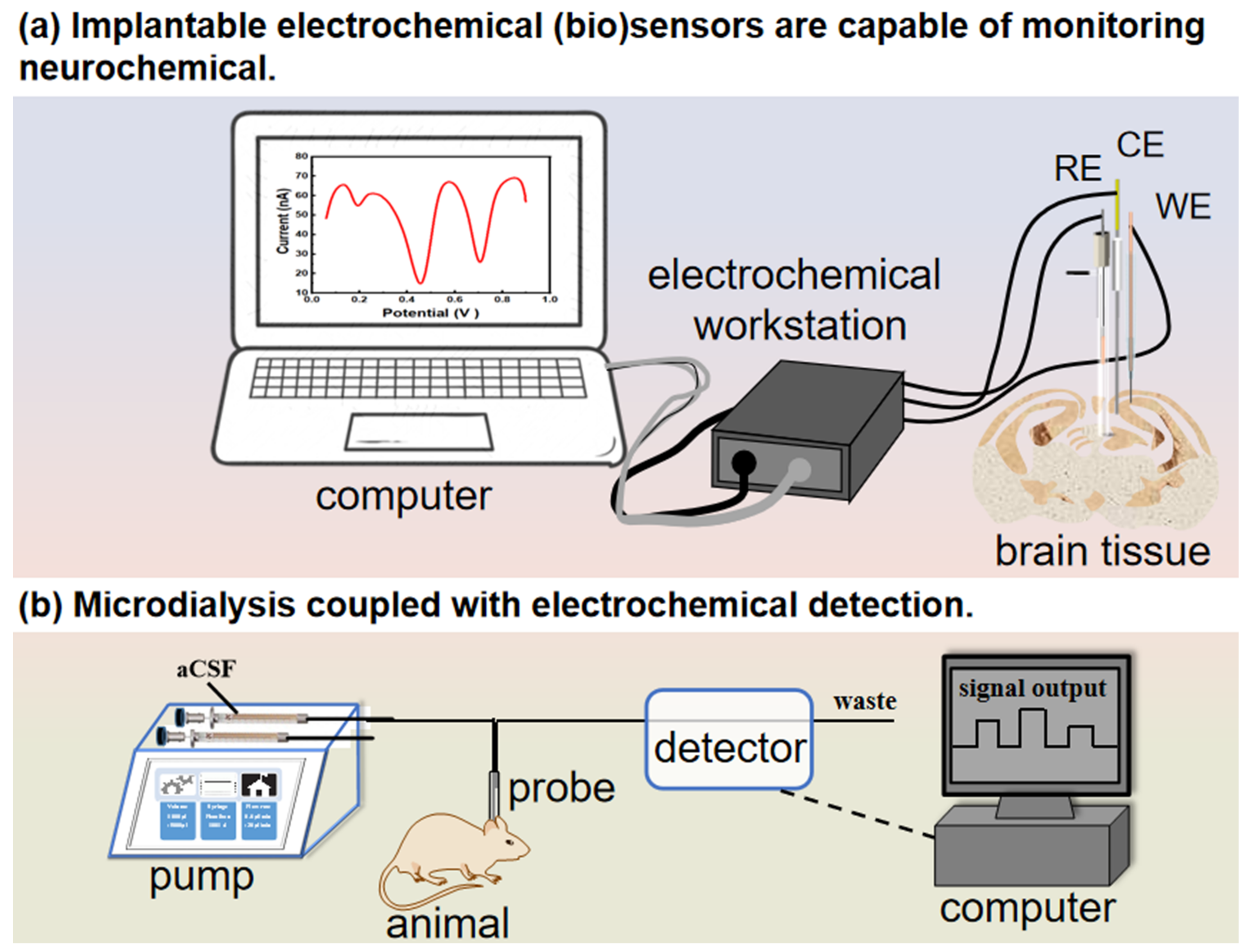
Figure 6.
Online capillary liquid chromatography with electrochemical detection. (a) In vitro online microdialysis-HPLC-EC experimental system. (b) Online in vivo DA measurements. Electrical stimulations were carried out prior to the administration of nomifensine. (The black dots represent the data from the test, and the red lines represent the overall trend). Reprinted from Ref. [178] with permission. Copyright 2015 American Chemical Society.

Figure 8.
(a) Illustration of a measurement using the LLIM in the rat brain. (b) Typical amperometric responses obtained using the LLIM in the cortex of anesthetized rats. Reprinted from Ref. [218] with permission. Copyright 2021 American Chemical Society.
Figure 8.
(a) Illustration of a measurement using the LLIM in the rat brain. (b) Typical amperometric responses obtained using the LLIM in the cortex of anesthetized rats. Reprinted from Ref. [218] with permission. Copyright 2021 American Chemical Society.
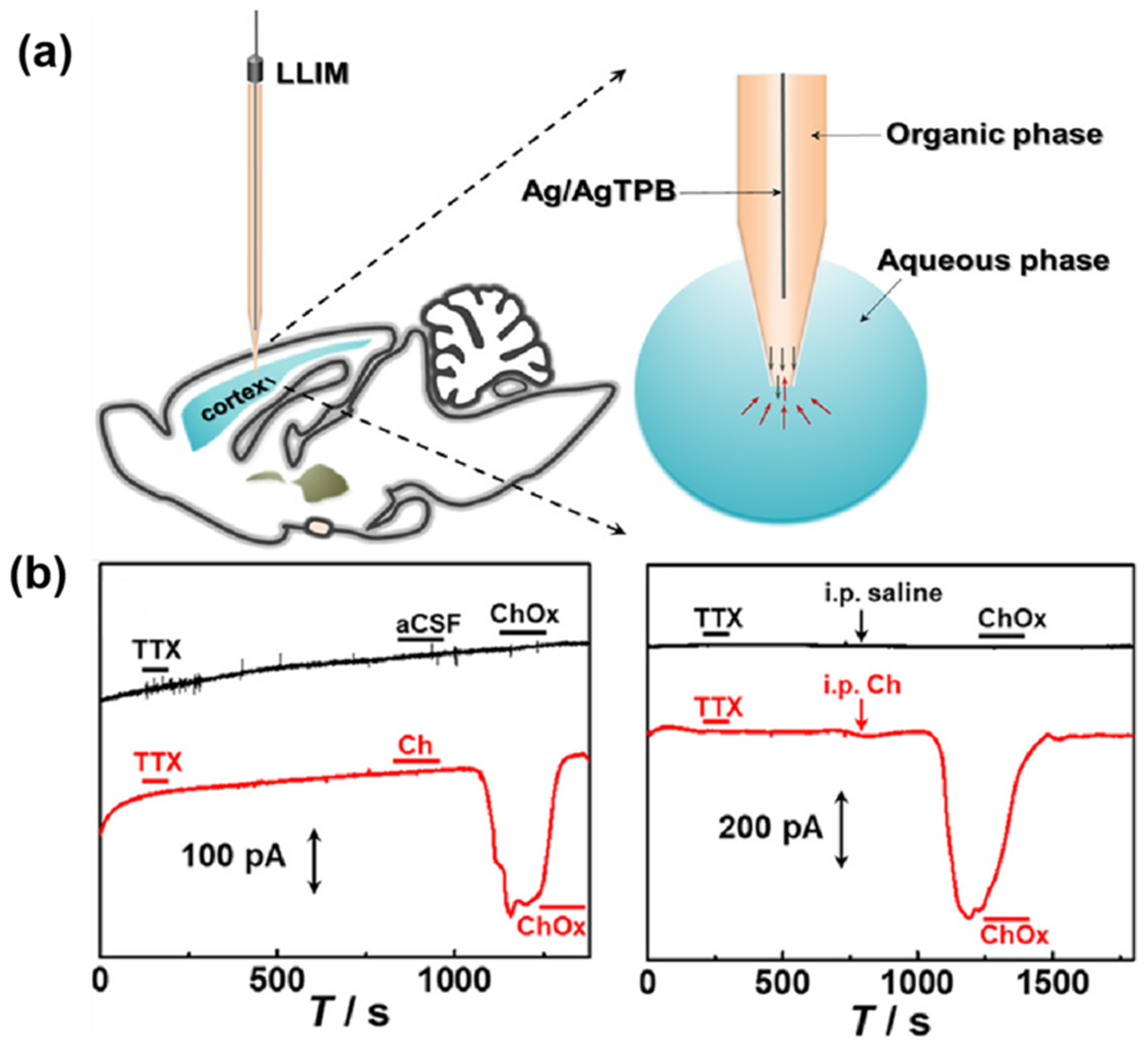
Figure 9.
Schematic illustration of the fabrication of DNA-cho-M–CS–CFE and the DA measurement in the micromorphology of the electrode surface. (The red dotted box is the binding sites of chitosan to phospholipids and membrane proteins, respectively.) Reprinted from Ref. [228] with permission. Copyright 2023 Elsevier B.V.

Table 1.
Implantable electrochemical biosensors to monitor non-electroactive neurochemicals in the brain tissue in vivo.
Table 1.
Implantable electrochemical biosensors to monitor non-electroactive neurochemicals in the brain tissue in vivo.
| Neurochemicals | Sensor Structure | Linear Range | LOD (μM) | Detection Area | Ref. |
|---|---|---|---|---|---|
| ATP | Au/Apt/3-MPA | 0.001–100 µM | 0.5 | Cortex | [73] |
| Cl− | CFME/OxGO/Ti3C2Tx/Ag | 1–700 mM | 10 | Hippocampus | [74] |
| Cl− | CFME/GO/TNWs/Ag/MB | 1–300 mM | 10 | Hippocampus | [75] |
| H+ | CFME/EOGO/PMe | 0.5–600 μM | 0.036 | Hippocampus | [76] |
| H+ | CFNE/CNTs/PoPD | 4.5–8.2 pH | - | Hippocampus | [77] |
| H+ | Hemin-Fc/CNF | 5.5–8.0 pH | - | Striatum, Cortex | [78] |
| H+ | Cat + Fc/SWNT/CFME | 5.91–7.81 pH | - | Striatum | [79] |
| K+ | NO/K+ dual microsensor | 0.01–100 mM | - | Cortex | [80] |
| H+, K+, Ca2+, and Na+ | Mesoporous SiO2/carbon/Co(II) phthalocyanine | 0.1–70.79 μM | - | Hippocampus | [81] |
| 50 μM–140 mM | |||||
| 1 μM–160 mM | |||||
| 130 μM–200 mM | |||||
| O2•− | CFME/SWCNT/MB + ND | 2–200 μM | 0.52 | Striatum, Cortex, Hippocampus | [82] |
| Glucose | Gox/PB/PANI/MWNT/CFE | 50−4000 μM | 40 | Cortex | [83] |
| Lactate | Pt-ceria biosensors | 100 pM−15.5 mM | 0.1 | Hippocampus | [84] |
| K+ | NO/K+ dual microsensor | 10 μM−100 mM | - | Cortex | [80] |
| Glutamate | Pt-Ir/PPD/GlutOx/AsOx/BSA | 5–150 µM | 0.044 | Subthalamic nucleus | [11] |
| Glutamate | GluOx/pDAB/polyimide | Up to 150 µM | 0.22 | Cortex | [85] |
ATP: adenosine triphosphate; Apt: aptamer; 3-MPA: 3-Mercaptopropionic acid; CFME: carbon fiber microelectrode; OxGO: electro-oxidized graphene oxide; Ti3C2Tx: two-dimensional MXene titanium carbide; GO: graphene oxide.; TNWs: titanate nanowires; MB: methylene blue; EOGO: electrochemically oxidized graphene oxide; PMe: poly-(melamine); CFNE: carbon fiber nanotip electrode; CNTs: carbon nanotubes; PoPD: poly-o-phenylenediamine; Hemin-Fc: hemin-aminoferrocene; CNF: carbon nanotube fiber; Cat: catalase; Fc: ferrocene; SWNT: single-walled carbon nanotube; ND: diphenylphosphonate-2-naphthol ester; Gox: glucoseoxidase; PB: Prussian blue; PANI: polyaniline; MWNTs: multi-walled carbon nanotubes; CFE: carbon fiber electrode; PPD: poly-o-phenylenediamine; GlutOx: glutamate oxidase; AsOx: ascorbate oxidase; BSA: bovine serum albumin; pDAB: poly(1,3-diaminobenzene).
Table 2.
Implantable microsensors for the detection of electrochemically active neurochemicals in the brain tissue in vivo.
Table 2.
Implantable microsensors for the detection of electrochemically active neurochemicals in the brain tissue in vivo.
| Neurochemicals | Sensor Structure | Linear Range | LOD | Detection Area | Ref. |
|---|---|---|---|---|---|
| DA | PTA-PANI-coated/CFE | 5–30 µM | - | Striatum | [16] |
| DA | PB/PEDOT/CFdisk | 0.5–10 mM | 0.18 µM | Striatum | [135] |
| DA | PEDOT/GO/CFE | 6.25–212.5 µM | - | Dorsal Striatum | [136] |
| DA | Nafion-Au/GCNE | 0.02–5.6 µM | 0.01 µM | Striatum | [137] |
| DA | AuNPs-rGO/Pt | 0.05–3 µM | 0.01675 µM | Striatum | [129] |
| 5-HT | CFMEA/DS-SWCNT | 0.10–3.40 µM | 5.1 µM | Striatum | [10] |
| 5-HT | CFEA/GR-FeTSPc | 0.05–60 µM | 0.02 µM | Hippocampus | [138] |
| 5-HT | PEDOT/CNT-coated | 0.01–1 µM | - | Hippocampus | [139] |
| AA | SWCNT/CFE | 10–1000 µM | 1000 µM | Cortex | [140] |
| AA | PEDOT/EOGO/CFE | 20–1000 µM | 500 µM | Striatum, Cortex, Hippocampus | [141] |
| Cu2+ | CFME/SWNT + AQ + NS4-C1 + ABTS | 0.5–9.5 µM | 500 nM | Striatum, Cortex, Hippocampus | [142] |
| Cu2+ | CFME/Au/E2Zn2SOD/Ni-NTA | 0.01–35 µM | 3 nM | Striatum | [143] |
| H2O2 | PDA/PB/CNT/CFE | 0−2775 μM | 0.12 μM | Cortex | [144] |
| H2O2 | Cat/ Nafion-PPD/Pt | 25−1000 μM | 1.0 μM | Striatum | [145] |
| H2O2 | Cat + Fc/SWNT/CFME | 1.0–230 mM | - | Striatum | [79] |
| H2S | CFE/mAu/MPS-1 + MHS-1 | 0.2–40 μM | 47 ± 4 μM | Cortex, Striatum, Hippocampus | [9] |
| H2Sn | CFE/Au/FP2 + FcBT | 0.25–20 μM | 50 μM | Cortex, Striatum, Hippocampus | [146] |
| NO | NO/K+ dual microsensor | 0–3.13 μM | - | Cortex | [80] |
| NO | CFE/Ni-P/17-FTMS | 1–3 μM | 12.1 ± 3.4 nM | Cortex | [147] |
PTA: polytannic acid; PANI: polyaniline; CFE: carbon fiber electrode; PB: Prussian blue; PEDOT: poly(3,4-ethylenedioxythiophene); CFdisk: carbon fiber disk; GCNE: glass capillary nanoelectrode; AuNPs: gold nanoparticles; rGO: reduced graphene oxide; CFMEAs: carbon fiber microelectrode arrays; DS: diazonium salt; SWCNTs: single-walled carbon nanotubes; CFEAs: carbon fiber electrode arrays; GR-FeTSPc: graphene-iron-tetrasulfophthalocyanine; AQ: 9,10-anthraquinone; NS4-C1: N,N-bis(2-[2-(ethylthio)ethyl])-2-naphthamide; ABTS: 2,2′-azino-bis(3-ethylbenzthiazoline-6-sulfonic acid); E2Zn2SOD: Cu-free derivative of bovine erythrocyte copper–zinc superoxide dismutase; PDA: polydopamine; PPD: poly-o-phenylenediamine; Cat: catalase; Fc: ferrocene; mAu: mesoporous gold film; MPS-1: 3,4-bis((2-fluoro-5-nitrobenzoyl)oxy)-benzoic acid; MHS-1: N-(4-(2,5-dinitrophenoxy) phenyl)-5-(1, 2-dithiolan-3-yl)pentanamide (MHS-1); FP2: 4-(5-(1,2-dithiolan-3-yl)pentanamido)-1,2-phenylene bis(2-fluoro-5-nitrobenzoate); FcBT: α-lipoic acid ferrocenylamide; Ni-P: nickel(II)Tetrakis 3-methoxy-4-hydroxyphenyl-porphyrin; 17-FTMS: (heptadecafluoro-1,1,2,2-Tetrahydrodecyl) trimethoxysilane.
Disclaimer/Publisher’s Note: The statements, opinions and data contained in all publications are solely those of the individual author(s) and contributor(s) and not of MDPI and/or the editor(s). MDPI and/or the editor(s) disclaim responsibility for any injury to people or property resulting from any ideas, methods, instructions or products referred to in the content. |
© 2024 by the authors. Licensee MDPI, Basel, Switzerland. This article is an open access article distributed under the terms and conditions of the Creative Commons Attribution (CC BY) license (https://creativecommons.org/licenses/by/4.0/).
Share and Cite
MDPI and ACS Style
Xu, X.; Zuo, Y.; Chen, S.; Hatami, A.; Gu, H. Advancements in Brain Research: The In Vivo/In Vitro Electrochemical Detection of Neurochemicals. Biosensors 2024, 14, 125. https://doi.org/10.3390/bios14030125
AMA Style
Xu X, Zuo Y, Chen S, Hatami A, Gu H. Advancements in Brain Research: The In Vivo/In Vitro Electrochemical Detection of Neurochemicals. Biosensors. 2024; 14(3):125. https://doi.org/10.3390/bios14030125
Chicago/Turabian StyleXu, Xiaoxuan, Yimei Zuo, Shu Chen, Amir Hatami, and Hui Gu. 2024. "Advancements in Brain Research: The In Vivo/In Vitro Electrochemical Detection of Neurochemicals" Biosensors 14, no. 3: 125. https://doi.org/10.3390/bios14030125
Note that from the first issue of 2016, this journal uses article numbers instead of page numbers. See further details here.