
Sensing of p53 and EGFR Biomarkers Using High Efficiency SERS Substrates

Abstract
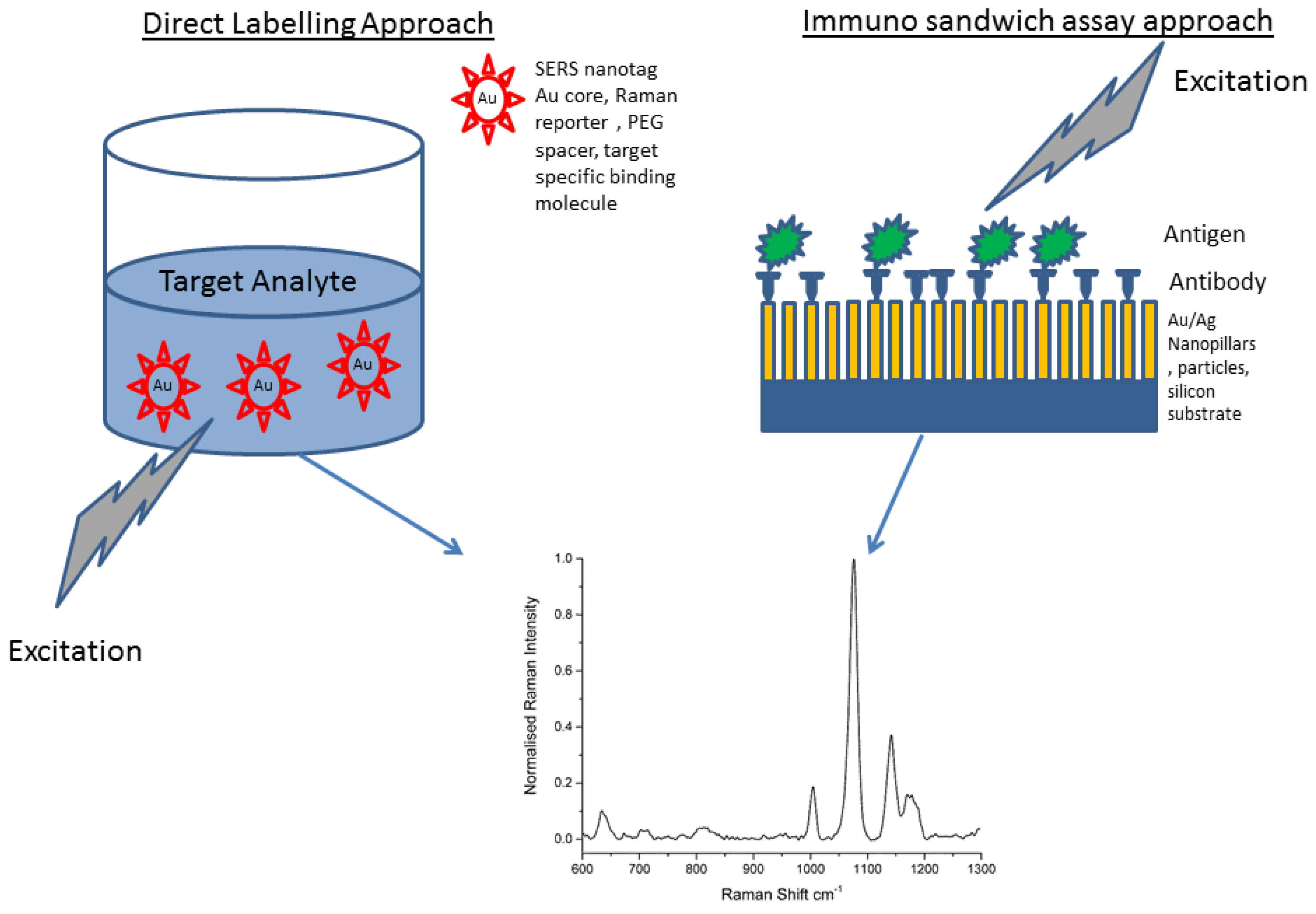
:1. Introduction

2. Experimental Section
2.1. Functionalisation of Substrates


| Protein | Raman Active Linker |
|---|---|
| p53 | ATP |
| EGFR | 6MP |
| EGFR | ATP |
2.2. Raman Microscopy
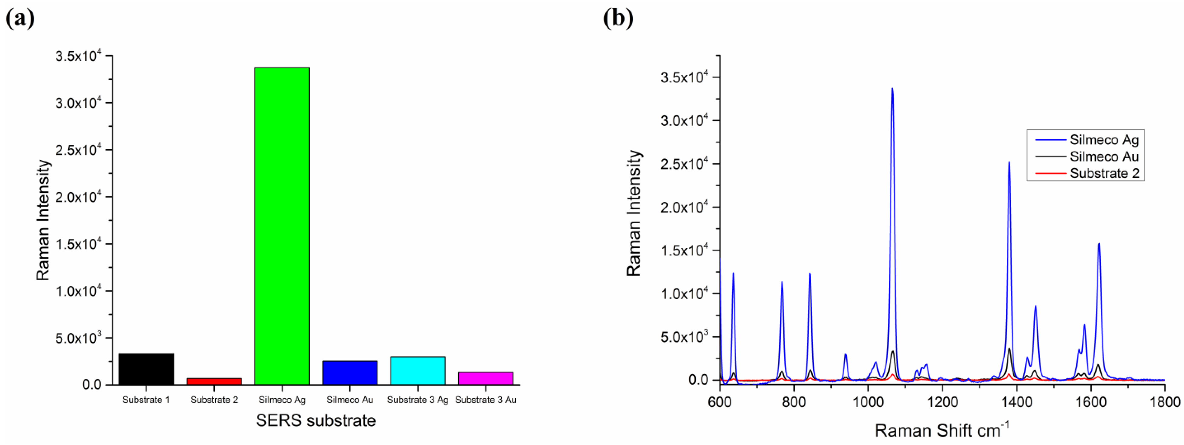
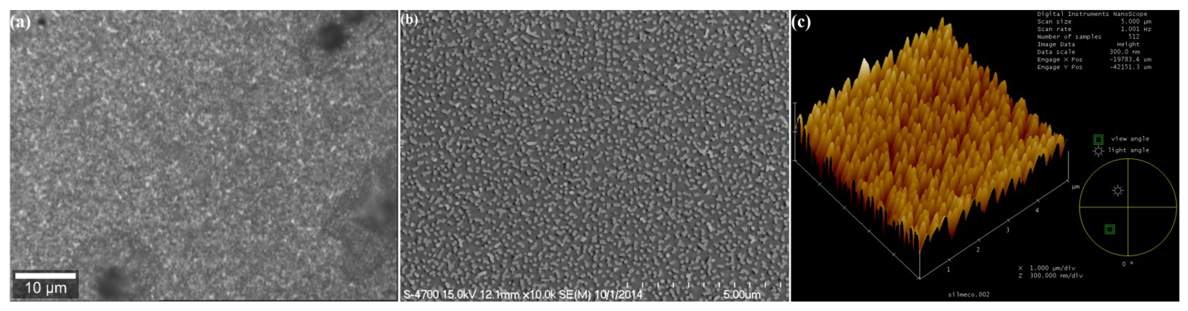
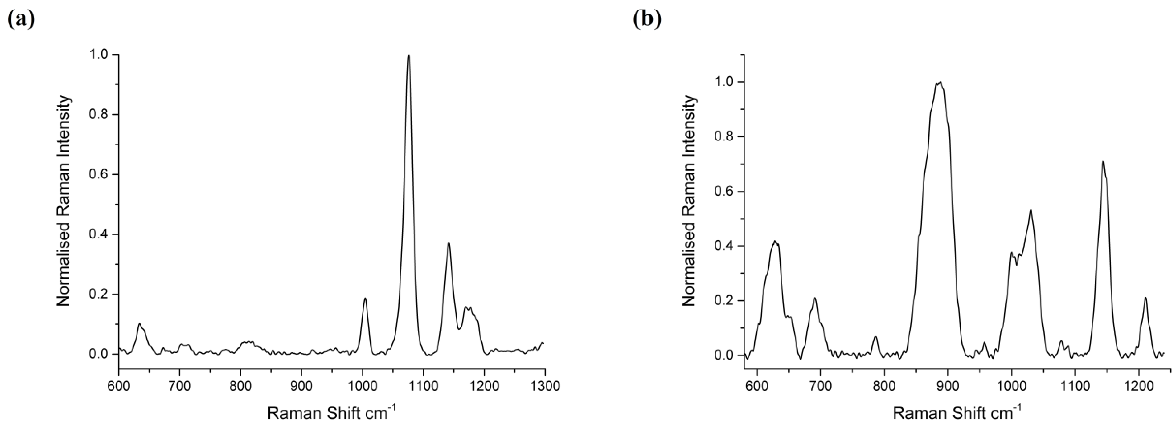
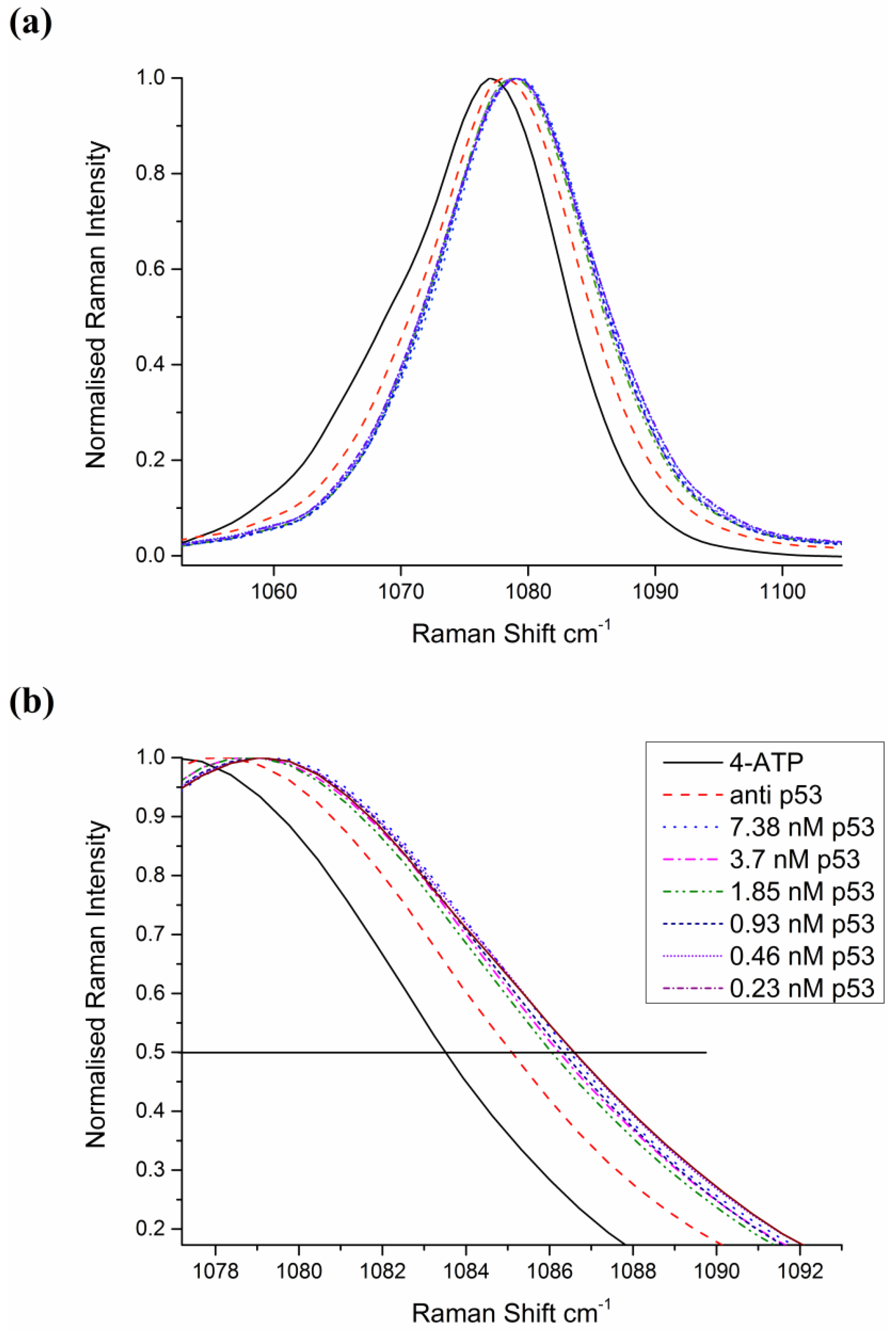
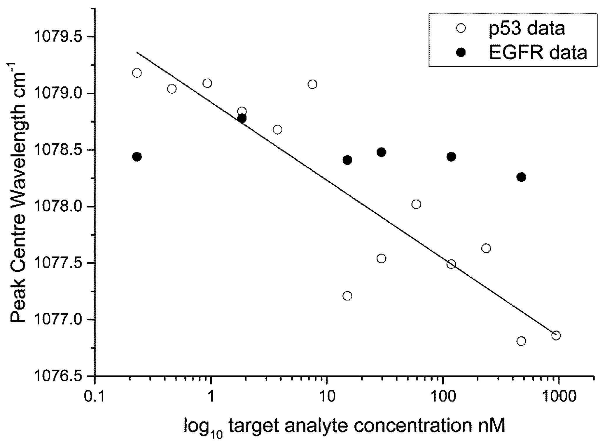
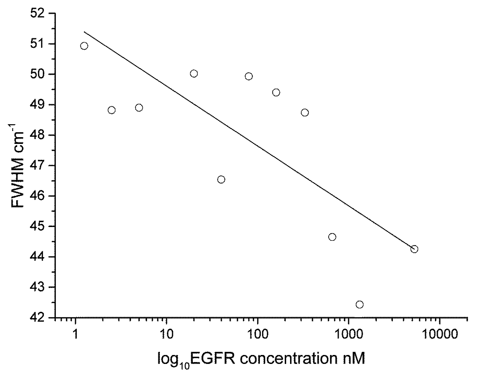
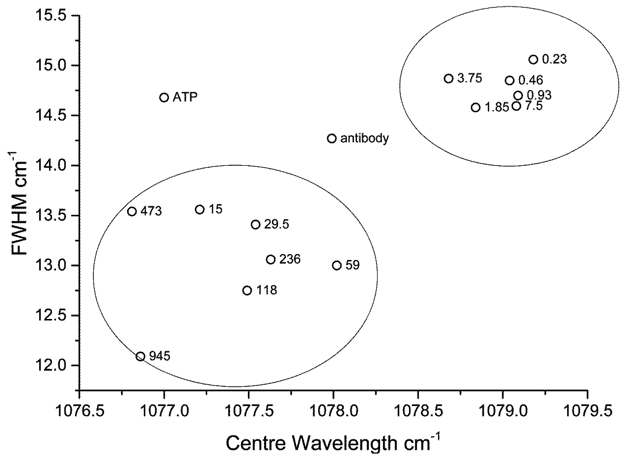
3. Results and Discussion


4. Conclusions
Supplementary Files
Supplementary File 1Acknowledgments
Author Contributions
Conflicts of Interest
References
- Domenici, F.; Bizzarri, A.R.; Cannistraro, S. Sers-based nanobiosensing for ultrasensitive detection of the p53 tumor suppressor. Int. J. Nanomed. 2011, 6, 2033–2042. [Google Scholar]
- Chan, J.; Fore, S.; Wachsmann-Hogiu, S.; Huser, T. Raman spectroscopy and microscopy of individual cells and cellular components. Laser Photonics Rev. 2008, 2, 325–349. [Google Scholar] [CrossRef]
- Bantz, K.C.; Meyer, A.F.; Wittenberg, N.J.; Im, H.; Kurtuluş, Ö.; Lee, S.H.; Lindquist,, N.C.; Oh, S.-H.; Haynes, C.L. Recent progress in sers biosensing. Phys. Chem. Chem. Phys. 2011, 13, 11551–11567. [Google Scholar] [CrossRef] [PubMed]
- Dietzek, B.; Cialla, D.; Scmitt, M.; Popp, J. Introduction to the fundamentals of Raman spectroscopy. In Confocal Raman Microscopy; Dieing, T., Hollricher, O., Toporski, J., Eds.; Springer-Verlag: Berlin/Heidelberg, Germany, 2011; pp. 21–40. [Google Scholar]
- Vendrell, M.; Maiti, K.K.; Dhaliwal, K.; Chang, Y.T. Surface-enhanced Raman scattering in cancer detection and imaging. Trends Biotechnol. 2013, 31, 249–257. [Google Scholar] [CrossRef] [PubMed]
- Fales, A.M.; Yuan, H.; Vo-Dinh, T. Cell-penetrating peptide enhanced intracellular raman imaging and photodynamic therapy. Mol. Pharm. 2013, 10, 2291–2298. [Google Scholar] [CrossRef] [PubMed]
- Maiti, K.K.; Dinish, U.S.; Samanta, A.; Vendrell, M.; Soh, K.S.; Park, S.J.; Olivo, M.; Chang, Y.T. Multiplex targeted in vivo cancer detection using sensitive near-infrared sers nanotags. Nano Today 2012, 7, 85–93. [Google Scholar] [CrossRef]
- Tu, Q.; Chang, C. Diagnostic applications of Raman spectroscopy. Nanomed. Nanotechnol. Biol. Med. 2012, 8, 545–558. [Google Scholar] [CrossRef] [PubMed]
- Lucas, L.; Chen, X.K.; Smith, A.; Korbelik, M.; Zeng, H.; Lee, P.W.K.; Hewitt, K.C. Imaging EGFR distribution using surface-enhanced Raman spectroscopy. In Proceedings of Plasmonics in Biology and Medicine VI (Proc. SPIE 7192), San Jose, CA, USA, 24 January 2009; Volume 7192.
- Li, X.; Yang, T.; Lin, J. Spectral analysis of human saliva for detection of lung cancer using surface-enhanced Raman spectroscopy. J. Biomed. Opt. 2012, 17, 0370031–0370035. [Google Scholar] [CrossRef] [PubMed]
- Inscore, F.; Shende, C.; Sengupta, A.; Huang, H.; Farquharson, S. Detection of drugs of abuse in saliva by surface-enhanced Raman spectroscopy (SERS). Appl. Spectrosc. 2011, 65, 1004–1008. [Google Scholar] [CrossRef] [PubMed]
- Farquharson, S.; Gift, A.D.; Shende, C.; Maksymiuk, P.; Inscore, F.E.; Murran, J. Detection of 5-fluorouracil in saliva using surface-enhanced Raman spectroscopy. Vib. Spectrosc. 2005, 38, 79–84. [Google Scholar] [CrossRef]
- Samanta, A.; Maiti, K.K.; Soh, K.S.; Liao, X.; Vendrell, M.; Dinish, U.S.; Yun, S.W.; Bhuvaneswari, R.; Kim, H.; Rautela, S.; et al. Ultrasensitive near-infrared Raman reporters for SERS-based in vivo cancer detection. Angew. Chem. Int. Ed. 2011, 50, 6089–6092. [Google Scholar] [CrossRef] [PubMed]
- Antonio, K.A.; Schultz, Z.D. Advances in biomedical raman microscopy. Anal. Chem. 2014, 86, 30–46. [Google Scholar] [CrossRef] [PubMed]
- Harper, M.M.; McKeating, K.S.; Faulds, K. Recent developments and future directions in SERS for bioanalysis. Phys. Chem. Chem. Phys. 2013, 15, 5312–5328. [Google Scholar] [CrossRef] [PubMed]
- Maiti, K.K.; Dinish, U.S.; Fu, C.Y.; Lee, J.J.; Soh, K.S.; Yun, S.W.; Bhuvaneswari, R.; Olivo, M.; Chang, Y.T. Development of biocompatible SERS nanotag with increased stability by chemisorption of reporter molecule for in vivo cancer detection. Biosens. Bioelectron. 2010, 26, 398–403. [Google Scholar] [CrossRef] [PubMed]
- Kah, J.C.Y.; Kho, K.W.; Lee, C.G.L.; Richard, C.J.; Sheppard, R.; Shen, Z.X.; Soo, K.C.; Olivo, M.C. Early diagnosis of oral cancer based on the surface plasmon resonance of gold nanoparticles. Int. J. Nanomed. 2007, 2, 785–798. [Google Scholar]
- Huefner, A.; Kuan, W.-L.; Barker, R.A.; Mahajan, S. Intracellular SERS nanoprobes for distinction of different neuronal cell types. Nano Lett. 2013, 13, 2463–2470. [Google Scholar] [CrossRef] [PubMed]
- Dinish, U.S.; Balasundaram, G.; Chang, Y.T.; Olivo, M. Actively targeted in vivo multiplex detection of intrinsic cancer biomarkers using biocompatible SERS nanotags. Sci. Rep. 2014, 4. [Google Scholar] [CrossRef] [PubMed]
- Wang, Y.; Schlucker, S. Rational design and synthesis of SERS labels. Analyst 2013, 138, 2224–2238. [Google Scholar] [CrossRef] [PubMed] [Green Version]
- Perumal, J.B.G.; Mahyuddin, A.P.; Choolani, M.; Olivo, M. SERS-based quantitative detection of ovarian cancer prognostic factor haptoglobin. Int. J. Nanomed. 2015, 10, 1831–1840. [Google Scholar] [CrossRef] [PubMed]
- Girish, C.M.; Iyer, S.; Thankappan, K.; Rani, V.V.D.; Gowd, G.S.; Menon, D.; Nair, S.; Koyakutty, M. Rapid detection of oral cancer using Ag-TiO2 nanostructured surface-enhanced raman spectroscopic substrates. J. Mater. Chem. B 2014, 2, 989–998. [Google Scholar] [CrossRef]
- Dinish, U.S.; Yaw, F.C.; Agarwal, A.; Olivo, M. Development of highly reproducible nanogap sers substrates: Comparative performance analysis and its application for glucose sensing. Biosens. Bioelectron. 2011, 26, 1987–1992. [Google Scholar] [CrossRef] [PubMed]
- Domenici, F.; Bizzarri, A.R.; Cannistraro, S. Surface-enhanced Raman scattering detection of wild-type and mutant p53 proteins at very low concentration in human serum. Anal. Biochem. 2012, 421, 9–15. [Google Scholar] [CrossRef] [PubMed]
- Han, X.X.; Ozaki, Y.; Zhao, B. Label-free detection in biological applications of surface-enhanced Raman scattering. Trac Trends Anal. Chem. 2012, 38, 67–78. [Google Scholar] [CrossRef]
- Kho, K.W.; Dinish, U.S.; Kumar, A.; Olivo, M. Frequency shifts in SERS for biosensing. ACS Nano 2012, 6, 4892–4902. [Google Scholar] [CrossRef] [PubMed]
- Perumal, J.; Kong, K.V.; Dinish, U.S.; Bakker, R.M.; Olivo, M. Design and fabrication of random silver films as substrate for SERS based nano-stress sensing of proteins. RSC Adv. 2014, 4, 12995–13000. [Google Scholar] [CrossRef]
- Vousden, K.H.; Ryan, K.M. P53 and metabolism. Nat. Rev. Cancer 2009, 9, 691–700. [Google Scholar] [CrossRef] [PubMed]
- Vousden, K.H.; Prives, C. Blinded by the light: The growing complexity of p53. Cell 2009, 137, 413–431. [Google Scholar] [CrossRef] [PubMed]
- Yewale, C.; Baradia, D.; Vhora, I.; Patil, S.; Misra, A. Epidermal growth factor receptor targeting in cancer: A review of trends and strategies. Biomaterials 2013, 34, 8690–8707. [Google Scholar] [CrossRef] [PubMed]
- Herbst, R.S. Review of epidermal growth factor receptor biology. Int. J. Radiat. Oncol. Biol. Phys. 2004, 59, S21–S26. [Google Scholar] [CrossRef] [PubMed]
- Silmeco. Available online: www.silmeco.com (accessed on 10 August 2015).
- Yang, J.; Palla, M.; Bosco, F.G.; Rindzevicius, T.; Alstrøm, T.S.; Schmidt, M.S.; Boisen, A.; Ju, J.; Lin, Q. Surface-enhanced Raman spectroscopy based quantitative bioassay on aptamer-functionalized nanopillars using large-area raman mapping. ACS Nano 2013, 7, 5350–5359. [Google Scholar] [CrossRef] [PubMed]
- Wu, K.; Rindzevicius, T.; Schmidt, M.S.; Mogensen, K.B.; Hakonen, A.; Boisen, A. Wafer-scale leaning silver nanopillars for molecular detection at ultra-low concentrations. J. Phys. Chem. C 2015, 119, 2053–2062. [Google Scholar] [CrossRef] [Green Version]
- Hong, S.; Li, X. Optimal size of gold nanoparticles for surface-enhanced Raman spectroscopy under different conditions. J. Nanomater. 2013, 2013. [Google Scholar] [CrossRef]
- Szeghalmi, A.V.; Leopold, L.; Pînzaru, S.; Chis, V.; Silaghi-Dumitrescu, I.; Schmitt, M.; Popp, J.; Kiefer, W. Adsorption of 6-mercaptopurine and 6-mercaptopurine riboside on silver colloid: A pH dependent surface enhanced Raman spectroscopy and density functional theory study. Part I. 6-mercaptopurine. J. Mol. Struct. 2005, 735–736, 103–113. [Google Scholar] [CrossRef]
- Han, G.; Liu, R.; Han, M.Y.; Jiang, C.; Wang, J.; Du, S.; Liu, B.; Zhang, Z. Label-free surface-enhanced Raman scattering imaging to monitor the metabolism of antitumor drug 6-mercaptopurine in living cells. Anal. Chem. 2014, 86, 11503–11507. [Google Scholar] [CrossRef] [PubMed]
- Zabel, J.; Nair, R.R.; Ott, A.; Georgiou, T.; Geim, A.K.; Novoselov, K.S.; Casiraghi, C. Raman spectroscopy of graphene and bilayer under biaxial strain: Bubbles and balloons. Nano Lett. 2012, 12, 617–621. [Google Scholar] [CrossRef] [PubMed]
- Dieing, T.; Ibach, W. Software requirements and data analysis in confocal raman microscopy. In Confocal Raman Microscopy; Dieing, T., Hollricher, O., Toporski, J., Eds.; Springer: Berlin/Heidelberg, Germany, 2011; Volume 158, pp. 61–89. [Google Scholar]
- Kitagawa, T.; Yabuki, K.; Young, R.J. An investigation into the relationship between processing, structure and properties for high-modulus pbo fibres. Part 1. Raman band shifts and broadening in tension and compression. Polymer 2001, 42, 2101–2112. [Google Scholar] [CrossRef]
© 2015 by the authors; licensee MDPI, Basel, Switzerland. This article is an open access article distributed under the terms and conditions of the Creative Commons Attribution license (http://creativecommons.org/licenses/by/4.0/).
Share and Cite
Owens, P.; Phillipson, N.; Perumal, J.; O’Connor, G.M.; Olivo, M. Sensing of p53 and EGFR Biomarkers Using High Efficiency SERS Substrates. Biosensors 2015, 5, 664-677. https://doi.org/10.3390/bios5040664
Owens P, Phillipson N, Perumal J, O’Connor GM, Olivo M. Sensing of p53 and EGFR Biomarkers Using High Efficiency SERS Substrates. Biosensors. 2015; 5(4):664-677. https://doi.org/10.3390/bios5040664
Chicago/Turabian StyleOwens, Peter, Nigel Phillipson, Jayakumar Perumal, Gerard M. O’Connor, and Malini Olivo. 2015. "Sensing of p53 and EGFR Biomarkers Using High Efficiency SERS Substrates" Biosensors 5, no. 4: 664-677. https://doi.org/10.3390/bios5040664