
Beneficial Effects of Betaine: A Comprehensive Review
by
 , and
, and
Madan Kumar Arumugam
1,2 ,
,
Matthew C. Paal
1,2,
Terrence M. Donohue, Jr.
1,2,3,
Murali Ganesan
1,2,
Natalia A. Osna
1,2 and
Kusum K. Kharbanda
1,2,3,* 1
Research Service, Veterans Affairs Nebraska-Western Iowa Health Care System, Omaha, NE 68105, USA
2
Department of Internal Medicine, University of Nebraska Medical Center, Omaha, NE 68198, USA
3
Department of Biochemistry & Molecular Biology, University of Nebraska Medical Center, Omaha, NE 68198, USA
*
Author to whom correspondence should be addressed.
Biology 2021, 10(6), 456; https://doi.org/10.3390/biology10060456
Submission received: 31 March 2021
/
Revised: 6 May 2021
/
Accepted: 19 May 2021
/
Published: 22 May 2021
(This article belongs to the Special Issue Non-alcoholic Liver Injury)
Abstract
:Simple Summary
A large number of studies report that medicinal herbs and many food ingredients protect against the development of liver disease because they possess antioxidant, anti-inflammatory, or anti-necrotic activities. This review focuses on the biological and beneficial effects of dietary betaine (trimethylglycine), a naturally occurring and crucial methyl donor, that restores methionine homeostasis in cells. We describe recent studies on betaine’s mechanism(s) of action as a therapeutic agent for improving indices of alcohol-induced and metabolic- associated liver disease. Due to its low cost, high tolerability, and efficacy, we suggest betaine as a promising therapeutic for clinical use to treat these aforementioned diseases as well as other liver-/non-liver-related diseases and conditions.
Abstract
Medicinal herbs and many food ingredients possess favorable biological properties that contribute to their therapeutic activities. One such natural product is betaine, a stable, nontoxic natural substance that is present in animals, plants, and microorganisms. Betaine is also endogenously synthesized through the metabolism of choline or exogenously consumed through dietary intake. Betaine mainly functions as (i) an osmolyte and (ii) a methyl-group donor. This review describes the major physiological effects of betaine in whole-body health and its ability to protect against both liver- as well as non-liver-related diseases and conditions. Betaine’s role in preventing/attenuating both alcohol-induced and metabolic-associated liver diseases has been well studied and is extensively reviewed here. Several studies show that betaine protects against the development of alcohol-induced hepatic steatosis, apoptosis, and accumulation of damaged proteins. Additionally, it can significantly prevent/attenuate progressive liver injury by preserving gut integrity and adipose function. The protective effects are primarily associated with the regulation of methionine metabolism through removing homocysteine and maintaining cellular SAM:SAH ratios. Similarly, betaine prevents metabolic-associated fatty liver disease and its progression. In addition, betaine has a neuroprotective role, preserves myocardial function, and prevents pancreatic steatosis. Betaine also attenuates oxidant stress, endoplasmic reticulum stress, inflammation, and cancer development. To conclude, betaine exerts significant therapeutic and biological effects that are potentially beneficial for alleviating a diverse number of human diseases and conditions.
1. Introduction
Many medicinal herbs and food ingredients possess therapeutic properties and a few of these have been developed as functional foods [1]. Numerous scientific reports have shown that many of these natural products possess favorable biological properties that contribute to their therapeutic activities [2]. One such natural product is betaine, also known as trimethylglycine, a stable, nontoxic natural substance that is present in animals, plants, and microorganisms. While betaine was first identified in the 19th century in beets (Beta vulgaris) [3,4], it is also found at high concentrations in other food sources including wheat bran, wheat germ, and spinach, as well as in microorganisms and aquatic invertebrates [3]. Betaine is endogenously synthesized through the metabolism of choline [3,4], or exogenously consumed through dietary intake [5]. Due to its essential biochemical functions, many microorganisms utilize betaine and have evolved different metabolic pathways for its biosynthesis and catabolism [6].
2. Dietary Betaine Uptake
Betaine is a short-chain, neutral, amino acid derivative. Daily betaine intake in the human diet ranges from an average of 1 g/day to a high of 2.5 g/day in individuals who consume a diet high in whole wheat and/or shellfish [7]. Dietary betaine is thought to be absorbed across the enterocytes primarily through the sodium-dependent amino acid transport system A, however sodium-independent transport also occurs [5]. The membrane-bound betaine/γ-aminobutyric acid transporter 1 (BGT-1) is also involved in the uptake of betaine following hypertonic stress [4,5,8]. Betaine consumed either from food sources or as dietary supplements presents similar bioavailability and is catabolized to dimethylglycine and finally to sarcosine in mitochondria of liver and kidney cells [4,9].
3. Important Roles of Betaine
Betaine mainly functions as (i) an osmolyte and (ii) a methyl-group donor. Because it possesses N+ and COO− in its molecular structure, as shown in (Figure 1), betaine helps to maintain the intracellular osmotic pressure similar to other electrolytes. By exhibiting little or no binding to protein surfaces, betaine allows cells to control the surface tension of water, stabilizing both protein structure and function [4,5,8,10]. It thereby protects cells, proteins, and enzymes subjected to osmotic stress. This is particularly relevant in the kidney due to its high concentration of electrolytes and urea [11]. Betaine is the most effective osmolyte studied for the hydration of albumin [12], forming almost a complete monolayer of water around the protein and it can also maintain hemoglobin solvation [13]. In the Kupffer cells, the resident macrophages of the liver, betaine serves as an osmolyte and prevents the hyperosmolarity-induced (i) suppression of tumor necrosis factor α release and (ii) induction of prostaglandin formation and cyclooxygenase 2 expression, thereby modulating its immune function [14]. The mRNA encoding for the betaine transporter are significantly upregulated by hyperosmolarity [14]. Betaine also has an important regulatory role of organic osmolytes on human RBC membrane ATPases and it protects against hypoosmotic stress [15]. Further, by protecting skeletal muscle myosin ATPase, betaine prevents myosin structural changes due to urea [16]. Additionally, betaine affects the movement of water across the intestinal epithelium and has a role in the osmoregulation of the small intestine of broiler chicks [17,18]. In particular, betaine prevents coccidiosis (parasitic infection), an expensive disease with an estimated cost to the world’s poultry industry of USD 3.2 billion per year [19,20]. Coccidial infection disrupts osmotic balance in cells of the alimentary tract causing ionic imbalances, defective absorption, and dehydration in infected poultry These defects were reversed, and food utilization was restored after dietary betaine intake, indicating its importance in regulating colonic fluid balance and transport across the intestinal epithelium [21].
The other major function of betaine is that it donates its methyl group to the toxic metabolite, homocysteine, converting it to methionine. This reaction in catalyzed by betaine-homocysteine methyltransferase (BHMT), an enzyme that was first thought to be present primarily in the liver and kidneys [4]. Recent studies from our laboratory revealed that BHMT is also expressed in other important organs such as white adipose tissue and the intestine [22,23]. These findings have prompted additional research to evaluate betaine’s role in disease prevention [4] as well as human wellness [24].
4. Disease Prevention by Betaine Administration
Hepatic steatosis, defined as accumulation of excess fat in the liver [25], occurs when the mechanisms that normally utilize or remove lipids become impaired. Steatosis is the earliest manifestation of alcohol misuse or high caloric intake, but it can also be caused by insulin resistance, malnutrition, anorexia, sleep apnea, or exposure to toxins/drugs such as carbon tetrachloride, diphtheria toxins, aspirin, or tetracyclines [26]. Prolonged hepatic fat storage eventually results in metabolic dysfunction, inflammation, and advanced forms of liver disease [27,28,29,30]. The progression of liver disease from simple steatosis to hepatitis to cirrhosis and hepatocellular carcinoma is correlated with many factors, including excessive alcohol consumption, bacterial/viral infections, high body mass index, fat distribution, diabetes, race, ethnicity, genetics, and gender [31,32,33].
Alcohol-related and metabolism-associated fatty liver disease (ALD and MAFLD, respectively) are the most common causes of chronic liver disease worldwide [25,34,35]. The World Health Organization estimates that 2.3 billion people actively consumed alcohol in 2018 [36]. Excessive alcohol consumption is the third-leading preventable cause of death in the United States [37,38]. It is the most common cause of end-stage liver disease with 50% of cirrhosis-related mortality attributed either directly or indirectly to excessive alcohol use [39]. In particular, alcoholic hepatitis, a severe syndrome of ALD that is characterized by rapid onset of jaundice, malaise, tender hepatomegaly, and subtle features of systemic inflammatory response, represents a significant public health burden with almost 0.1% of all admissions related to this condition [40].
MAFLD, previously known as non-alcoholic fatty liver disease (NAFLD), is an umbrella term for liver disease unrelated to alcohol consumption and is most commonly associated with metabolic syndrome. MAFLD is characterized primarily by storage of excess macrovesicular fat due to an imbalance between the homeostatic mechanisms that regulate synthesis versus utilization of fat in liver cells [41] and is histologically indistinguishable from alcohol-induced hepatic steatosis. Studies show diabetes is a major risk factor for MAFLD [42]. The progression of MAFLD to metabolic-associated steatohepatitis (MASH), characterized by inflammatory changes in the liver, is accompanied by an increase in all-cause mortality as well as liver-related mortality. MASH is rapidly emerging as a leading cause of liver transplantation worldwide [43]. Diverse experimental animal models were examined to establish the characteristics and pathogenesis of ALD and MAFLD initiation and progression (Table 1), thus providing significant clues to the critical molecular targets to treat these fatty liver diseases [4,27,28,44,45]. Studies have focused on betaine as a treatment due to its classification as a lipotrope, i.e., an agent that reduces or prevents the accumulation of fat in the liver [46,47]. In this review, we summarize the role of betaine in restoring normal hepatic function in diseases of diverse etiologies, with special references to ALD and MAFLD.
4.1. ALD
Alcohol globally accounts for 3.3 million deaths each year [39,48,49,50,51,52]. An estimated 2.4 billion individuals worldwide consume alcoholic beverages [39] in social settings without experiencing harmful effects [53,54]. However, harmful use of alcohol is responsible for 5.1% of the global burden of disease and long-term excessive consumption is inextricably linked to liver disease [55], resulting in a costly socioeconomic and medical burden [54]. The liver is the major target organ with the greatest degree of tissue injury from excessive drinking because it is the primary site of ethanol metabolism [33,56]. Alcohol metabolism occurs by three distinct pathways. In the first major pathway, alcohol is oxidized to acetaldehyde via alcohol dehydrogenase (ADH), a NAD+-requiring enzyme expressed at high levels in hepatocytes. The second major pathway involved in alcohol oxidization is the microsomal ethanol oxidizing system (MEOS), largely catalyzed by cytochrome P450-2E1 (CYP2E1). In the third pathway, alcohol oxidation is catalyzed by catalase in peroxisomes [57]. Acetaldehyde, generated by these three pathways, then enters the mitochondrion and is oxidized to acetate by aldehyde dehydrogenase (ALDH)-mediated catalysis [58,59].
4.1.1. Stages of ALD
ALD is a disease spectrum, consisting of three major stages: steatosis or fatty liver, steatohepatitis, and fibrosis and/or cirrhosis [31]. Steatosis is characterized by intrahepatic deposition of mostly triglycerides and cholesterol esters in the form of lipid droplets [60,61,62]. While initially in perivenular hepatocytes that surround the central vein of the liver lobule, steatosis progresses to mid-lobular hepatocytes, and then extends to the periportal hepatocytes that surround the hepatic portal vein [63,64,65]. Steatosis is a reversible state that can resolve upon cessation of alcohol consumption [66]. If left untreated, steatosis can advance to steatohepatitis, characterized by inflammation and neutrophil infiltration [67]. An additional pathological feature observed in the steatohepatitis stage is “ballooning” hepatocytes, which appear swollen and reveal cellular damage [31,68,69]. The activation of Kupffer cells and their subsequent loss, the degeneration of sinusoidal endothelial cell fenestrations, and the infiltration of circulating macrophages and neutrophils, define the inflammatory changes [70,71] and subsequent hepatocyte damage observed during the development of steatohepatitis. This progressive injury, in-turn, activates hepatic stellate cells (HSCs) which are key players in the development of fibrosis [72,73]. Activated HSCs proliferate and become the principal source for the increased and irregular deposition of extracellular matrix components which replace the normal matrix with dense basement-membrane-like collagen, characteristic of fibrosis [74,75,76,77]. Furthermore, HSCs accelerate inflammatory cytokine production, drawing even more inflammatory cells and amplifying hepatocyte damage [77,78] and fibrotic changes that alter hepatic lobular organization, characteristic of hepatic cirrhosis [71].
Several mechanisms have been proposed for the development and progression of ALD including acetaldehyde toxicity, oxidative stress, increased intestinal permeability-induced endotoxemia, Kupffer cell activation, production of cytokines and chemokines, a compromised immune system, nutritional deficiencies, and altered methionine metabolism [33,52]. Previous reports from many laboratories, including ours, have demonstrated that ethanol consumption impairs several of the steps in methionine metabolism [23,60,79,80,81,82,83]. Methionine is an essential amino acid that is not only needed to initiate protein synthesis but is equally important for generating the universal methyl-group donor, S-adenosylmethionine (SAM). Chronic alcohol consumption decreases SAM levels in the liver [23,84,85] and elevates both homocysteine [83,86] and S-adenosylhomocysteine (SAH) levels [23,87,88] to ultimately decrease the hepatic SAM:SAH ratio [23]. These alterations primarily occur because of the ethanol-induced inhibition of methionine synthase (MS) which is involved in removing SAH by remethylating homocysteine to generate SAM [23,60,81,82]. The consequence of the reduced SAM:SAH ratio is impaired function of several crucial hepatic methylation reactions catalyzed by specific methyltransferases [89]. Their impaired function ultimately results in the generation of hallmark features of ALD, including steatosis, apoptosis, accumulation of damaged protein, and proteasome inhibition [23,33,60,81,82,90,91,92,93].
4.1.2. Betaine Protects against the Development of Alcohol-Induced Hepatic Steatosis
Alcohol-induced fat accumulation in the liver is caused by increased uptake of adipose derived free fatty acids, accelerated de novo lipogenesis, decelerated mitochondrial fatty acid oxidation, and reduced very low-density lipoprotein (VLDL) export [94,95,96]. These alterations result from the alcohol-induced change in activities of several enzymes, transcription factors, and signaling events.
Phosphatidylethanolamine N-methyltransferase (PEMT) is an important liver enzyme that catalyzes the three successive methylations of phosphatidylethanolamine (PE) to form phosphatidylcholine (PC) [97]. The PC species generated by this pathway is an essential constituent of VLDL and hence impairment in PEMT-catalyzed PC generation reduces VLDL synthesis and secretion to retain lipids in the hepatocytes, causing their accumulation [97,98,99,100]. The transcription factor sterol regulatory element-binding protein (SREBP) regulates lipid synthesis in liver and other tissues [101]. Peroxisome proliferator-activated receptor-α (PPARα) belongs to the nuclear hormone receptor superfamily and, in the liver, functions as a lipid sensor to regulate the genes that encode the enzymes for oxidation, transport, and export of free fatty acids [102].
Alcohol consumption enhances the fatty acid synthesis proteins and the levels of SREBP and fatty acid synthase (FAS) in the liver [103,104]. In contrast, alcohol downregulates the lipid metabolism regulatory proteins, PPARα, AMP-dependent protein kinase (AMPK), and adiponectin receptor-mediated signaling, which play significant roles in lipid homeostasis [95,104]. Studies have also shown that PPARα activity/expression is downregulated in livers of ethanol-fed mice [105,106]. In addition, alcohol treatment inhibits AMPK activity, which occurs via increased intrahepatic-ceramide-levels-induced protein phosphatase 2A (PP2A) activation in mice and cultured hepatoma cells [107,108]. Zhang et al., however, reported that alcohol-induced reduction in PP2A methylation promotes the phosphorylation of forkhead box O1 (FOXO1), ultimately leading to triglyceride accumulation in the liver [109]. Long-term alcohol exposure also diminishes mitochondrial oxidative phosphorylation, which promotes hepatocyte damage by decreasing respiratory efficiency and promoting oxidant stress [110,111]. Further, alcohol administration has also shown to impair PEMT activity, resulting in reduction in VLDL secretion [90,112]. Studies conducted in several rodent models have also characterized how alcohol-induced changes in the adipose–liver axis promote hepatic steatosis. One of the most well-documented phenomena is the reduced secretion of the adipokine and adiponectin, and the impaired expression of hepatic adiponectin receptors, contributing to the development of alcohol-induced liver steatosis [113].
Interestingly, all the above-mentioned events that promote hepatic fat accumulation are (i) indirectly or directly related to alterations in methionine metabolism and (ii) mitigated by betaine treatment as shown in multiple investigations [23,60,61,81,82,83,90,91,114,115,116], as schematically represented in Figure 2. Betaine treatment exerts protection against ethanol-induced injury by restoring the intrahepatic SAM:SAH ratio [23] and maintaining normal methylation activity [60,81,82] by providing a methyl group to homocysteine. This reaction is catalyzed by an alternate enzyme, BHMT, that, like MS, remethylates homocysteine [86] to remove SAH [23,88] and generate methionine necessary for SAM synthesis [23,117,118,119,120], and thereby maintain the hepatic SAM:SAH ratio [23]. Consequently, the activity of PEMT is preserved, leading to normal levels of VLDL secretion [90]. In addition, betaine prevents/attenuates alcohol-induced hepatic steatosis by restoring FOXO1 transcriptional activity via methylating and activating PP2A [109] and suppressing the synthesis of the rate-limiting enzyme in triglyceride synthesis, diacylglycerol acyltransferase 2 [121,122]. Song et al. reported that betaine restores the serum adiponectin levels in ethanol-fed rats by increasing its production in adipose tissue [123]. Betaine administration activates AMPK, which enhances genes encoding proteins involved in fatty acid transport and fatty acid oxidation, while decreasing fatty acid synthesis [124,125], thereby preventing triglyceride and cholesterol accumulation in the liver [115]. Betaine treatment also blocks alcohol-induced nitric oxide synthase 2 (NOS2) and nitric oxide generation, which preserves mitochondrial function [61]. Thus, betaine administration protects against the development of alcohol-induced liver injury by restoring methylation potential, increasing mitochondrial oxidation, and decreasing both the uptake of adipose derived free fatty acid and de novo lipogenesis.
4.1.3. Betaine Prevents Other Indices of Early Alcohol-Induced Liver Damage
Our laboratory has also demonstrated that alcohol-induced alterations in the rat hepatocellular SAM:SAH ratio and the resulting impairment in the activities of isoprenyl carboxyl methyltransferase (ICMT), L-isoaspartyl methyltransferase (PIMT), and protein arginine N- methyltransferases (PRMT), which, respectively, regulate apoptosis [91], cause accumulation of damaged proteins [92] and inhibit proteasome activities [126]. Betaine, by restoring the methylation potential and normalizing the activities of the three methyltransferases, mitigates or eliminates these defects [23,81,82,90,91,92].
4.1.4. Betaine Prevents Oxidative Stress and Inflammation in ALD
Oxidative metabolism primarily occurs in the mitochondrion, where reactive oxygen species (ROS) are generated as byproducts of biological energy-generating reactions. The body has antioxidant enzymes and antioxidants that comprise the detoxification system to remove or neutralize ROS and free radicals under normal physiological conditions [127,128]. Increased ROS generation that surpasses the capacity of the detoxification systems alters the stability of nucleic acids, proteins, and the lipid membranes of cells, compromising cellular function and promoting inflammation [129]. Alcohol consumption induces NADPH oxidase in Kupffer cells, which, by generating greater quantities of ROS causes activation of the transcription factor, nuclear factor-κB (NF-kB), that enhances tumor necrosis factor alpha (TNF-α) production to promote liver damage [130]. The MEOS pathway is also induced by alcohol consumption [131] that generates higher quantities of ROS causing oxidant stress and progressive hepatocyte injury [132]. Betaine is anti-inflammatory by its ability to upregulate antioxidant defense system [133]. Ethanol-induced increases in several factors involved in the development of inflammation, such as cluster of differentiation 14 (CD14), TNFα, cyclooxygenase-2 (COX2), growth arrest and DNA-damage-inducible 45β (GADD45β), LPS-induced TN factor (LITAF), janus kinase 3 (JAK3), toll-like receptor 2 (TLR2), toll-like receptor 4 (TLR4), interleukin 1β (IL1β), programmed cell death 4 (PDCD4), and NOS2, are all suppressed by betaine supplementation [115,133,134,135,136]. In addition, betaine supplementation also prevents alcohol-induced depletion of hepatic cysteine and glutathione (GSH). Altogether, betaine supplementation improves oxyradical scavenging activity in liver tissues altered by chronic alcohol consumption [133]. Treatment with betaine reduces the alcohol-induced elevations in serum ALT and AST [115]. Importantly, betaine administration also prevents the blood-alcohol-level cycle and significantly reduces the blood alcohol level by promoting the phenylethanolamine N-methyltransferase-mediated conversion of norepinephrine to epinephrine and increasing the metabolic rate [135].
Alcohol induces epigenetic modifications such as histone modifications (acetylation/phosphorylation/methylation/ubiquitylation/sumoylation), methylation status of DNA (hypomethylation/hypermethylation), and changes in miRNAs [137]. Epigenetic regulation of genes relevant to ALD disease pathogenesis is closely related to the underlying ethanol-induced reduction in the hepatic SAM:SAH ratio and reduced gene body methylation in all autosomes and in specific gene body sites in NOS, each of which were prevented by betaine administration [134]. Betaine prevented the formation of Mallory–Denk bodies through epigenetically attenuating the decrease of methionine adenosyltransferase 1A (MAT1A), S-adenosylhomocysteine hydrolase (SAHH), BHMT, and adenosylmethionine decarboxylase 1 (AMD1) expression and inhibiting the increase of methylenetetrahydrofolate reductase expression [138].
4.1.5. Betaine Protects against the Detrimental Effects of HCV and Ethanol on Innate Immunity
Interferon type 1 (IFN type 1) response is crucial for protection of the host from many viruses including hepatitis C virus (HCV). HCV is a hepatotropic virus [139]. The progression of hepatitis is regulated by susceptibility of hepatocytes to viral infections that depends on activation of innate immunity, namely transduction of the IFN signal to activate anti-viral genes. IFN type I binds to the receptors on the cell surface of hepatocytes to induce signal transducer and activator of transcription (STAT1) and STAT2 phosphorylation followed by IRF9 (an additional factor) and their attachment to the interferon-stimulated response element (ISRE) area of DNA to activate interferon-stimulated genes (ISGs). There are certain inhibitors that block IFN signaling. One of them is protein inhibitor of activated STAT 1 (PIAS1), which attaches to STAT1 when STAT1 is not methylated by PRMT1.
In our studies, we found that IFN response in hepatocytes is suppressed by HCV, and this effect is potentiated by ethanol metabolism [140,141,142,143]. As schematically shown (Figure 3), acetaldehyde interferes with the attachment of STAT1 to DNA due to complex formation between phosphorylated/non-methylated STAT1 and an inhibitor of IFN signaling, PIAS1. This is attributed to impaired arginine and lysine methylation of STAT1 by PRMT1, which allows STAT1 to bind PIAS1, thereby preventing activation of anti-viral ISGs. This effect was reversed by betaine [143]. Furthermore, treatment with AMI (an arginine-methylation inhibitor), BIX (a lysine-methylation inhibitor) and tubercidin (a pan-methylation inhibitor), all mimicked the effects of acetaldehyde by suppressing the attachment of STAT1 to DNA. This led to reduced activation of ISGs with anti-viral properties, such as OAS1, OASL, viperin, and protein kinase R, which were restored by betaine co-treatment [143]. In addition to impaired PRMT1-mediated STAT1 methylation suppressing IFN signaling in hepatocytes, we also observed that ethanol metabolite induced an increase in levels of a demethylase, jumonji domain-containing 6 protein (JMJD6), generating demethylated STAT1. Betaine attenuated ethanol metabolite-induced upregulation of JMJD6, thereby increasing protective effects of anti-viral ISGs in HCV-infected hepatocytes [141]. Thus, betaine reverses alcohol-induced suppression in STAT1 methylation by PRMT1 and decreases de-methylation of STAT1 by JMJD6, thereby restoring IFN signaling and inducing anti-viral effects via stimulation of ISGs in HCV-infected alcohol-exposed hepatocytes.
4.1.6. Betaine Protects against Fulminant Liver Failure and Toxin-Induced Liver Damage
Fulminant hepatic failure is characterized as severe liver injury with impairment of synthetic capability of liver cells and encephalopathy (decline in brain function) in patients with previous normal liver or, at least, well-compensated liver disease [144]. Rasineni et al. showed that betaine could prevent fulminant liver failure induced by LPS-galactosamine in mice by attenuating the activation of caspase-3 and apoptosis [145]. The toxic effects of carbon tetrachloride (CCl4) on hepatocytes are manifested histologically as hepatic steatosis, centrilobular necrosis, and ultimately cirrhosis. Betaine supplementation to CCl4 -injected rats significantly reduced hepatic lipidosis [146] and reduced the toxic effects of CCl4 on cell organelles [147]. Betaine supplementation also alleviated CCl4-induced fibrosis by inhibiting lipid peroxidation, hepatic inflammation, and expression of transforming growth factor-β1 [148].
4.2. MAFLD
MAFLD is defined by macrovesicular steatosis in hepatocytes, in the absence of a secondary cause such as alcohol or drugs. It is a leading cause of chronic liver disease worldwide [149]. MAFLD progresses from simple liver steatosis to steatohepatitis, and in more severe cases, to liver fibrosis, cirrhosis, and hepatocellular carcinoma [150,151]. The cause for hepatic fat accumulation in MAFLD includes increased fat accumulation in the liver from high caloric intake with persistent adipocyte derived FFA delivery and uptake by the liver, increased de novo hepatic lipogenesis, and decreased VLDL export from hepatocytes [27,152,153]. The molecular mechanism is very similar to what was discussed before in the context of ALD pathogenesis, including upregulation of key transcription factors including SREBP-1, which enhances expression of lipogenic enzymes including FAS, acetyl-CoA carboxylase, and stearoyl-CoA desaturase [154].
Lu et al. reported that a diet deficient in one-carbon methyl groups such as betaine, choline, folate, and methionine results in the development and progression of fatty liver disease by affecting specific changes in genes involved in one-carbon metabolism [155,156]. Betaine supplementation to male mice fed a high-fat diet prevented betaine deficiency, insulin resistance, and fatty liver, and normalized serum ALT levels [27]. It has also been shown that betaine supplementation to rats fed a high-fat diet upregulates the mRNAs encoding BHMT, GNMT, and MGAT, all key enzymes of one-carbon metabolism involved in regulating fat metabolism [157]. Further, betaine supplementation decreased hepatic lipid accumulation by slowing lipogenesis and enhancing lipophagy in ApoE−/− mouse models through enhanced expression of PPARα and elevated fatty acid oxidation by upregulating expression of mitochondrial and extra-mitochondrial fatty acid oxidation enzymes [158]. In addition, betaine increased AMPK, fibroblast growth factor 10, and adipose triglyceride lipase levels while suppressing lipid-metabolism-related genes in ApoE−/− mice fed a high-fat diet [159]. Mice fed a high-sucrose diet also exhibit significant fat accumulation and increased lipogenic activity in the liver similar to what was seen in high-fat diet administration, which were attenuated with betaine treatment via upregulation of AMPK [160]. Betaine supplementation to mice with MAFLD induced by methionine- and choline-deficient diet alleviated steatosis, inflammation, apoptosis, and oxidative stress, normalized mitochondrial size and respiratory chain function, stimulated β-oxidation of fatty acids, increased the number of autophagosomes, and restored both glutathione content and antioxidant enzyme activities in livers [28,161,162,163].
It is interesting to note that betaine deficiency as seen in animal models of MAFLD has been correlated with increased disease severity, a similar trend to that seen in patients [164]. Importantly, betaine treatment decreased the grade of histological steatosis, inflammation, and fibrosis in MAFLD patients [165,166]. Migilo et al. reported that oral administration of betaine for 8 weeks to MAFLD patients reduced hepatomegaly and liver-injury-marker enzymes [167]. It is evident that the efficacy of betaine needs to be tested in better-designed clinical trials for the treatment for MAFLD as well as for ALD. Betaine is indeed an attractive model compound for alleviating fatty liver diseases [168] due to its low cost, high tolerability, high solubility, and a variety of other beneficial effects as schematically shown in Figure 2.
4.3. Alterations in Gut–Liver and Adipose–Liver Axes in Promoting Hepatic Damage
Crosstalk between the gut and liver plays a prominent role in the pathogenesis of ALD and MAFLD [169,170]. Alcohol consumption induces intestinal dysbiosis (microbial imbalance in the gut) and increases intestinal permeability which lead to translocation of microbes and their products into the portal circulation. These products are recognized by immune receptors on resident liver macrophages (Kupffer cells) and hepatic stellate cells (HSCs) to initiate an inflammatory cascade that triggers a fibrotic response [171,172,173]. Alcohol-induced dysbiosis also promotes steatosis development [174].
White adipose tissue (WAT) plays an important role in regulating whole-body lipid and energy homeostasis [175]. It not only acts as a reservoir for energy storage, but also as a complex, essential, and highly active metabolic and endocrine organ. WAT communicates with the liver and other tissues to control lipid distribution [176] and its dysfunction is a key feature in the pathophysiology of ALD, MAFLD, and obesity-related chronic metabolic and cardiovascular diseases [177,178,179]. Most notably, during ALD and MAFLD pathogenesis, it is the accelerated WAT lipolysis and the uptake of the mobilized free fatty acids by the liver and their esterification into triglycerides which contributes to hepatic steatosis development [180,181,182]. The latter process is regulated by altered secretion of adipokines observed in animal models or in patients with metabolic disease associated with high caloric intake or alcohol misuse [183,184,185,186,187,188,189,190]. Ethanol consumption increases WAT TNF-α expression, which, by inhibiting the release of the anti-inflammatory adipokine, adiponectin [191], impairs hepatic lipid metabolism [113]. Similarly, high-caloric-intake-induced WAT lipolysis is accompanied by necrosis and inflammation in this organ, along with aberrant secretion of adipokines which contribute to hepatic damage [192,193]. Further studies have reported that chronic alcohol consumption results in impaired methionine metabolism in adipose tissue, characterized by increased accumulation of homocysteine [123] and SAH levels and a consequent decrease in the SAM:SAH ratio [194]. This loss in the methylation potential has been shown to enhance hormone-sensitive lipase (HSL) activation to promote lipolysis in WAT [123,195].
4.3.1. Betaine Maintains Intestinal Epithelial Barrier Integrity
The intestinal epithelial barrier is mainly provided by the highly specialized intercellular multiprotein junctional complex, tight junctions, located at the apical end of epithelial cells which allow minimal leakage of luminal contents into the portal circulation. If the barrier is breached, it can cause significant inflammation and, if sustained, liver damage [196]. Betaine improved the intestinal mucosal barrier by upregulating expression of zonula occludens-1 (ZO1) and occluding-tight junction proteins as well as maintained the normal gut microbiota composition in an acute liver failure model by inhibiting the TLR4/MyD88 signaling pathway [197]. Similar results were also reported on attenuation of LPS-induced decreases of the tight junction structural proteins, occluding, and claudin-1, by betaine administration to restore barrier function of porcine intestinal epithelial cells [198]. Others previously reported similar protective effects of betaine in stabilizing intestinal epithelium in coccidia-infected broiler chicks [199]. Additional beneficial effects of betaine include activation of digestive enzymes and restoring intestinal morphology and microbial diversity in high salt stressed rats [200]. Studies from our laboratory showed that betaine prevents accumulation of intracellular SAH, which is associated with intestinal barrier disruption [22]. The protective functions of betaine in the intestine are represented schematically in Figure 4.
4.3.2. Betaine Maintains Adipose Function
Previous studies showed that betaine corrected abnormal adipokine (adiponectin, resistin, and leptin) levels, enhanced insulin sensitivity by improving extracellular signal-regulated protein kinase (ERK1/2) and protein kinase B, reduced endoplasmic stress, enhanced fatty acid oxidation, and restored mitochondrial function and N6-methyladenosine mRNA methylation in WAT of mice fed a high-fat diet [201,202]. Further studies showed that betaine enhanced the conversion of existing WAT to brown adipose tissue through stimulated mitochondrial biogenesis in mice fed a high-fat diet [203]. Similar protective effects of betaine have been shown in restoring alcohol-induced adipose dysfunction. Betaine restored the impaired methylation status in WAT to alleviate PP2A inhibition and prevent the persistent HSL activation and lipolysis [194] and improved circulating adiponectin levels in alcohol-fed mice [123]. Betaine treatment was also reported to reduce the hypoxia-induced expression of inflammatory adipokines, IL6, TNFα, and leptin in human adipocytes [204].
4.4. Protective Effects of Betaine on Other Tissues
Alcohol-induced pancreatic steatosis in rat models was effectively prevented with betaine supplementation by suppressing SREBP-1c and FAS expression [125]. Betaine also inhibited intramyocellular lipid accumulation and improved insulin resistance in mice fed a high-fat diet [203]. In addition, betaine prevented the development of isoprenaline-induced myocardial dysfunction via its antioxidant effects and by preserving mitochondrial function [205]. Further, betaine protected against cadmium nephrotoxicity by inhibiting lipid peroxidation, increasing total antioxidant status, and reducing caspase signaling cascade in renal tissues [206]. Similarly, the antioxidant properties of betaine not only prevented oxidative stress in the kidneys and liver in an experimental allergic asthma model but also improved airway inflammation of lung tissue [207].
Betaine treatment led to a substantial rise in the motor unit activity and recovery of residual power in weakened muscle tissues of patients with acute anterior poliomyelitis, which resulted in improved sense of well-being, less fatigue, and greater strength and endurance during treatment [4,7,208,209]. Singhal et al. reported that betaine via the BHMT-catalyzed pathway exerted epigenetic control and activated neuroprotective transcriptional programs in the brain of mice with multiple sclerosis by restoring the methylation potential (SAM:SAH ratio) and preventing axonal damage [210].
4.5. Anti-Cancer Effect of Betaine
Previous studies reported that alcohol consumption is associated with an increased risk of breast cancer development [211,212]. Hong et al. documented the anti-cancer role of betaine in alcohol-associated breast cancer cell growth and colony formation by decreasing the induction of transcription factor II-B-related factor 1 and Pol III gene transcription [213]. Further, choline supplementation (which increases circulating choline and betaine concentrations) given in conjunction with an HFD and a chemical carcinogen 7,12-dimethylbenz[a]anthracene resulted in a 55% decline in hepatocellular carcinoma tumor numbers and a 67% decrease in tumor surface area compared to non-choline-treated mice [214].
5. Other Beneficial Effects of Betaine
5.1. Effects of Betaine on General Well-Being
Chen et al. reported that circulating betaine was closely associated with better body composition and fat distribution with lower fat mass in the trunk regions of Chinese adults [215]. A recent study reported that vitamin B12 deficiency is associated with altered lipid profile and is predictive of metabolic risk [216]. Betaine administration could protect against low-vitamin-B12-induced defects given that low or no vitamin B12 elevates homocysteine levels, reduces SAM:SAH ratio and, by modulating SREBF1 and low-density lipoprotein receptor (LDLR) genes, induces cholesterol biosynthesis in human adipocytes [217].
Betaine has been shown to induce resilience to anhedonia (the inability to feel pleasure) in mice subjected to chronic social defeat stress indicating that betaine could be used as a prophylactic nutrient to prevent stress-related psychiatric disorders [218]. Decreased circulating plasma levels of betaine were also reported in patients with schizophrenia and bipolar disorder [219]. Perhaps these disorders could be treated by restoring the depleted levels given the neuroprotective role of betaine [210]. Hassanpour et al. showed decreased malondialdehyde and improved levels of both superoxide dismutase and glutathione peroxidase, all of which are characteristic of an antioxidant effect, in brains of adult mice fed a betaine-supplemented diet. They also reported betaine has an anti-nociceptive and a sedative role through interactions with opioidergic and γ-aminobutyric acid (GABA) receptors [220]. In cockerels (young roosters), betaine affected the central cholesterol metabolism by decreasing the hypothalamic content of total cholesterol and cholesterol esters and downregulating the expression of cholesterol biosynthetic genes related to brain function [221].
Betaine improves athletic performance as shown by the fact that its addition to a carbohydrate–electrolyte fluid-replacement beverage resulted in improved mean sprint time to exhaustion and enhanced anaerobic/aerobic metabolism [4]. A study also reports that betaine increases tolerance to hypertonic and thermal stressors at the cellular level by stimulating heat shock protein expression, reducing oxidative damage and exercise-induced gut permeability, and protecting against bacterial translocation and endotoxemia [5]. At the systemic level, chronic betaine intake lowers core temperature by reducing inflammation markers and changing blood chemistry as shown in several animal models exposed to heat stress [5,222,223]. Leng et al. stated that betaine supplementation did not affect growth performance of broilers (chickens raised for meat production), but it effectively reduced abdominal fat deposition by decreasing fatty acid synthesis and increasing β-oxidation [224]. He et al. showed significant changes in triglyceride, free fatty acid, and LDL-and HDL-cholesterol after betaine treatment of heat stressed broilers, thus improving carcass composition via modulating lipid metabolism [225]. Also, betaine supplementation in water reduced rectal temperature in broiler chickens exposed to cyclical heat stress [226] and decreased core and skin temperature of sheep exposed to the same treatment [227].
5.2. Effects of Maternal Betaine Supplementation on Offspring
Higher plasma betaine concentrations of pregnant women at 26–28 weeks of gestation is associated with smaller infant birth size and lower abdominal fat mass of their offspring [228]. Generally, maternal betaine supplementation normalizes fetal growth and adiposity of progeny of obese mice by reducing glucose and fatty acid transporters and the growth-promoting insulin-like growth factor 2 in the placenta [229]. Maternal betaine supplementation during gestation improved twin lamb survival and shortened time interval from birth to first suck, potentially due to increased creatine production [230]. Further, betaine supplementation to pregnant rats exposed to glucocorticoids normalized adipose lipolysis and circulating free fatty acids, and prevented ectopic lipid deposition in liver and skeletal muscle by modifying DNA methylation on the promoter sites of lipolytic genes [231]. Betaine administration decreased hepatic cholesterol deposition through epigenetic regulation of genes involved in cholesterol metabolism in juvenile chickens [232]. Betaine exerted a transgenerational effect on estrogen-responsive genes in rat offspring, which was associated with corresponding alterations in DNA methylation and the promoter of affected genes [233].
6. Safety Studies with Betaine
Betaine is approved for human consumption based on the effectiveness with therapeutic equivalence by the FDA under sections 505 of the Federal Food, Drug, and Cosmetic Act [236]. Oral 3 or 6 g single doses of betaine administrated in orange juice after a 12 h overnight fast to healthy human volunteers resulted in an acute and dose-dependent increase in serum betaine levels and a reduction in plasma homocysteine concentrations within 2 h [237]. Human intervention studies showed no adverse effects with 4 g/day supplemental administration of betaine in healthy subjects, however overweight subjects with metabolic syndrome showed a significant increase in total and LDL-cholesterol concentrations. These effects were not observed with 3 g/day of betaine administration [238]. Other toxicity studies reported that the LD50 in mice for betaine is 10.8 g/kg [239] when injected subcutaneously and 0.83 g/kg with intravenous injection [240]. Burnett et al. reported a similar oral betaine LD50 value of 11.1 g/kg in rats [241]. An administration of betaine to piglets at dose levels up to 20 g/kg feed for 6 weeks showed no adverse effects [238].
7. Conclusions and Future Perspectives
This review described the major physiological effects of betaine as a preventive agent for the treatment for various diseases (Table 1), including ALD, MAFLD, and cancer due to its properties as an osmoprotectant and a methyl-group donor. Betaine also attenuates oxidative stress, endoplasmic reticulum stress, inflammation, and cancer development. The protective effects are primarily associated with the regulation of methionine metabolism, by removing homocysteine and maintaining cellular SAM:SAH ratio. As a result, it is worthwhile to further investigate betaine because it exerts significant therapeutic and biological effects that are potentially beneficial for alleviating a diverse number of human diseases.
Author Contributions
Conceptualization, MK.A. and K.K.K.; resources, MK.A. and K.K.K.; writing—review and editing, MK.A., M.C.P., M.G., and K.K.K.; visualization, MK.A. and K.K.K.; supervision, N.A.O., T.M.D.J., and K.K.K.; project administration, K.K.K.; funding acquisition, K.K.K. All authors have read and agreed to the published version of the manuscript.
Funding
This work was supported by the National Institutes of Health grant R01 AA026723 (KKK) and Merit Review grant BX004053 (KKK) from the U.S. Department of Veterans Affairs, Biomedical Laboratory Research and Development Service.
Institutional Review Board Statement
Not applicable.
Informed Consent Statement
Not applicable.
Data Availability Statement
Not applicable.
Conflicts of Interest
The authors declare no conflict of interest. The funders had no role in the design of the study and writing of the manuscript, or in the decision to publish.
Abbreviations
ADH, alcohol dehydrogenase; AHCY, adenosyl homocysteinase; ALD, alcohol-associated fatty liver disease; ALDH, aldehyde dehydrogenase; AMD1, adenosylmethionine decarboxylase 1; AMPK, AMP-dependent protein kinase; BGT-1, betaine/γ-aminobutyric acid transporter 1; BHMT, betaine-homocysteine methyltransferase; CD14, cluster of differentiation 14; COX-2, cyclooxygenase-2; CYP2E1, cytochrome P450 2E1; ERK. extracellular-regulated kinase; FAS, fatty acid synthase; GABA, gamma aminobutyric acid; GADD45β, growth arrest and DNA damage-inducible beta; GSH, glutathione; HCV, hepatitis C virus; HSL, hormone-sensitive lipase; ICMT, isoprenyl carboxyl methyltransferase; IFN, interferon; IL1β, interleukin 1β; NOS, inducible nitric oxide synthase; ISGs, interferon-stimulated genes; ISRE, interferon-stimulated response element; JAK3, janus kinase 3; JMJD6, jumonji domain-containing 6 protein; LDLR, low-density lipoprotein receptor; LITAF, LPS-induced TN factor; MAFLD, metabolism-associated fatty liver disease; MASH, metabolic-associated steatohepatitis; MAT1A, methionine adenosyltransferase 1A; MEOS, microsomal ethanol oxidizing system; MS, methionine synthase; MTHFR, methylene tetrahydrofolate reductase; NAFLD, non-alcoholic fatty liver disease; NF-kB, nuclear factor kappa B; OAS1, 2′-5′ oligoadenylate synthetase-1; PC, phosphatidylcholine; PDCD4, programmed cell death 4; PEMT, phosphatidylethanolamine N-methyltransferase; PIAS1, protein inhibitor of activated STAT 1; PIMT- L, isoaspartyl methyltransferase; ROS, reactive oxygen species; PP2A, protein phosphatase 2A; PPARα, peroxisome proliferator-activated receptor-α; PRMT1, protein methyltransferase 1; SAH, S-adenosylhomocysteine; SAM, S-adenosylmethionine; SREBP, sterol regulatory element binding protein; STAT, signal transducer and activator of transcription; TNF-α, tumor necrosis factor alpha; TLR2, toll-like receptor 2; TLR4, toll-like receptor 4; VLDL, very low-density lipoprotein; WAT, white adipose tissue.
References
- Zhou, Y.; Li, Y.; Zhou, T.; Zheng, J.; Li, S.; Li, H.-B. Dietary Natural Products for Prevention and Treatment of Liver Cancer. Nutrients 2016, 8, 156. [Google Scholar] [CrossRef] [Green Version]
- Domitrović, R.; Potočnjak, I. A comprehensive overview of hepatoprotective natural compounds: Mechanism of action and clinical perspectives. Arch. Toxicol. 2016, 90, 39–79. [Google Scholar] [CrossRef]
- Zhao, G.; He, F.; Wu, C.; Li, P.; Li, N.; Deng, J.; Zhu, G.; Ren, W.; Peng, Y. Betaine in Inflammation: Mechanistic Aspects and Applications. Front. Immunol. 2018, 9, 1070. [Google Scholar] [CrossRef] [PubMed] [Green Version]
- Craig, S.A.S. Betaine in human nutrition. Am. J. Clin. Nutr. 2004, 80, 539–549. [Google Scholar] [CrossRef] [Green Version]
- Willingham, B.D.; Ragland, T.J.; Ormsbee, M.J. Betaine Supplementation May Improve Heat Tolerance: Potential Mechanisms in Humans. Nutrients 2020, 12, 2939. [Google Scholar] [CrossRef] [PubMed]
- Zou, H.; Chen, N.; Shi, M.; Xian, M.; Song, Y.; Liu, J. The metabolism and biotechnological application of betaine in microorganism. Appl. Microbiol. Biotechnol. 2016, 100, 3865–3876. [Google Scholar] [CrossRef]
- Hoffman, J.R.; Ratamess, N.A.; Kang, J.; Rashti, S.L.; Faigenbaum, A.D. Effect of betaine supplementation on power performance and fatigue. J. Int. Soc. Sports Nutr. 2009, 6, 7–10. [Google Scholar] [CrossRef] [PubMed] [Green Version]
- Wettstein, M.; Weik, C.; Holneicher, C.; Häussinger, D. Betaine as an osmolyte in rat liver: Metabolism and cell-to-cell interactions. Hepatology 1998, 27, 787–793. [Google Scholar] [CrossRef] [PubMed]
- Cholewa, J.M.; Guimarães-Ferreira, L.; Zanchi, N.E. Effects of betaine on performance and body composition: A review of recent findings and potential mechanisms. Amino Acids 2014, 46, 1785–1793. [Google Scholar] [CrossRef] [PubMed]
- Konstantinova, S.V.; Tell, G.S.; Vollset, S.E.; Nygård, O.; Bleie, Ø.; Ueland, P.M. Divergent associations of plasma choline and betaine with components of metabolic syndrome in middle age and elderly men and women. J. Nutr. 2008, 138, 914–920. [Google Scholar] [CrossRef] [Green Version]
- Horio, M.; Ito, A.; Matsuoka, Y.; Moriyama, T.; Orita, Y.; Takenaka, M.; Imai, E. Apoptosis induced by hypertonicity in Madin Darley canine kidney cells: Protective effect of betaine. Nephrol. Dial. Transplant. 2001, 16, 483–490. [Google Scholar] [CrossRef] [Green Version]
- Courtenay, E.S.; Capp, M.W.; Anderson, A.C.F.; Record, M.T., Jr. Vapor Pressure Osmometry Studies of Osmolyte−Protein Interactions: Implications for the Action of Osmoprotectants in Vivo and for the Interpretation of “Osmotic Stress” Experiments in Vitro. Biochemistry 2000, 39, 4455–4471. [Google Scholar] [CrossRef]
- Hundahl, C.; Fago, A.; Malte, H.; Weber, R.E. Allosteric Effect of Water in Fish and Human Hemoglobins. J. Biol. Chem. 2003, 278, 42769–42773. [Google Scholar] [CrossRef] [Green Version]
- Zhang, F.; Warskulat, U.; Wettstein, M.; Häussinger, D. Identification of betaine as an osmolyte in rat liver macrophages (Kupffer cells). Gastroenterology 1996, 110, 1543–1552. [Google Scholar] [CrossRef]
- Moeckel, G.W.; Shadman, R.; Fogel, J.M.; Sadrzadeh, S.M.H. Organic osmolytes betaine, sorbitol and inositol are potent inhibitors of erythrocyte membrane ATPases. Life Sci. 2002, 71, 2413–2424. [Google Scholar] [CrossRef]
- Ortiz-Costa, S.; Sorenson, M.; Sola-Penna, M. Counteracting effects of urea and methylamines in function and structure of skeletal muscle myosin. Arch. Biochem. Biophys. 2002, 408, 272–278. [Google Scholar] [CrossRef]
- Kettunen, H.; Peuranen, S.; Tiihonen, K. Betaine aids in the osmoregulation of duodenal epithelium of broiler chicks, and affects the movement of water across the small intestinal epithelium in vitro. Comp. Biochem. Physiol. Part A Mol. Integr. Physiol. 2001, 129, 595–603. [Google Scholar] [CrossRef]
- Kempson, S.A.; Vovor-Dassu, K.; Day, C. Betaine Transport in Kidney and Liver: Use of Betaine in Liver Injury. Cell. Physiol. Biochem. 2013, 32, 32–40. [Google Scholar] [CrossRef] [Green Version]
- Dalloul, R.A.; Lillehoj, H.S. Poultry coccidiosis: Recent advancements in control measures and vaccine development. Expert Rev. Vaccines 2006, 5, 143–163. [Google Scholar] [CrossRef] [PubMed]
- Amerah, A.M.; Ravindran, V. Effect of coccidia challenge and natural betaine supplementation on performance, nutrient utilization, and intestinal lesion scores of broiler chickens fed suboptimal level of dietary methionine. Poult. Sci. 2015, 94, 673–680. [Google Scholar] [CrossRef] [PubMed]
- Davies, S.E.; Chalmers, R.A.; Randall, E.W.; Iles, R.A. Betaine metabolism in human neonates and developing rats. Clin. Chim. Acta 1988, 178, 241–249. [Google Scholar] [CrossRef]
- Thomes, P.G.; Osna, N.A.; Bligh, S.M.; Tuma, D.J.; Kharbanda, K.K. Role of defective methylation reactions in ethanol-induced dysregulation of intestinal barrier integrity. Biochem. Pharmacol. 2015, 96, 30–38. [Google Scholar] [CrossRef]
- Kharbanda, K.K.; Mailliard, M.E.; Baldwin, C.R.; Beckenhauer, H.C.; Sorrell, M.F.; Tuma, D.J. Betaine attenuates alcoholic steatosis by restoring phosphatidylcholine generation via the phosphatidylethanolamine methyltransferase pathway. J. Hepatol. 2007, 46, 314–321. [Google Scholar] [CrossRef] [PubMed]
- Ismaeel, A. Effects of Betaine Supplementation on Muscle Strength and Power: A Systematic Review. J. Strength Cond. Res. 2017, 31, 2338–2346. [Google Scholar] [CrossRef] [PubMed]
- Singh, S.; Osna, N.A.; Kharbanda, K.K. Treatment options for alcoholic and non-alcoholic fatty liver disease: A review. World J. Gastroenterol. 2017, 23, 6549–6570. [Google Scholar] [CrossRef] [PubMed]
- Bakir, M.B.; Salama, M.A.; Refaat, R.; Ali, M.A.; Khalifa, E.A.; Kamel, M.A. Evaluating the therapeutic potential of one-carbon donors in nonalcoholic fatty liver disease. Eur. J. Pharmacol. 2019, 847, 72–82. [Google Scholar] [CrossRef]
- Kathirvel, E.; Morgan, K.; Nandgiri, G.; Sandoval, B.C.; Caudill, M.A.; Bottiglieri, T.; French, S.W.; Morgan, T.R. Betaine improves nonalcoholic fatty liver and associated hepatic insulin resistance: A potential mechanism for hepatoprotection by betaine. Am. J. Physiol. Liver Physiol. 2010, 299, G1068–G1077. [Google Scholar] [CrossRef] [Green Version]
- Veskovic, M.; Mladenovic, D.; Milenkovic, M.; Tosic, J.; Borozan, S.; Gopcevic, K.; Labudovic-Borovic, M.; Dragutinovic, V.; Vucevic, D.; Jorgacevic, B.; et al. Betaine modulates oxidative stress, inflammation, apoptosis, autophagy, and Akt/mTOR signaling in methionine-choline deficiency-induced fatty liver disease. Eur. J. Pharmacol. 2019, 848, 39–48. [Google Scholar] [CrossRef]
- Di Ciaula, A.; Baj, J.; Garruti, G.; Celano, G.; De Angelis, M.; Wang, H.H.; Di Palo, D.M.; Bonfrate, L.; Wang, D.Q.-H.; Portincasa, P. Liver Steatosis, Gut-Liver Axis, Microbiome and Environmental Factors. A Never-Ending Bidirectional Cross-Talk. J. Clin. Med. 2020, 9, 2648. [Google Scholar] [CrossRef]
- Nassir, F.; Rector, R.S.; Hammoud, G.M.; Ibdah, J.A. Pathogenesis and Prevention of Hepatic Steatosis. Gastroenterol. Hepatol. 2015, 11, 167–175. [Google Scholar]
- Lefkowitch, J.H. Morphology of Alcoholic Liver Disease. Clin. Liver Dis. 2005, 9, 37–53. [Google Scholar] [CrossRef] [PubMed]
- Lonardo, A.; Nascimbeni, F.; Maurantonio, M.; Marrazzo, A.; Rinaldi, L.; Adinolfi, L.E. Nonalcoholic fatty liver disease: Evolving paradigms. World J. Gastroenterol. 2017, 23, 6571–6592. [Google Scholar] [CrossRef] [PubMed]
- Osna, N.A.; Donohue, T.M., Jr.; Kharbanda, K.K. Alcoholic Liver Disease: Pathogenesis and Current Management. Alcohol. Res. 2017, 38, 147–161. [Google Scholar] [PubMed]
- Younossi, Z.M.; Koenig, A.B.; Abdelatif, D.; Fazel, Y.; Henry, L.; Wymer, M. Global epidemiology of nonalcoholic fatty liver disease-Meta-analytic assessment of prevalence, incidence, and outcomes. Hepatology 2016, 64, 73–84. [Google Scholar] [CrossRef] [PubMed] [Green Version]
- Mitra, S.; De, A.; Chowdhury, A. Epidemiology of non-alcoholic and alcoholic fatty liver diseases. Transl. Gastroenterol. Hepatol. 2020, 5, 16. [Google Scholar] [CrossRef] [PubMed]
- World Health Organization (WHO). Global status report on alcohol and health 2018. WHO Alcohol. Drugs Addict. Behav. 2018, 8, e010454. [Google Scholar]
- Shah, N.J.; Royer, A.; John, S. Alcoholic Hepatitis; Statpearls: Treasure Island, FL, USA, 2020. [Google Scholar]
- Rehm, J.; Shield, K.D. Global Burden of Alcohol Use Disorders and Alcohol Liver Disease. Biomedicines 2019, 7, 99. [Google Scholar] [CrossRef] [Green Version]
- Asrani, S.K.; Devarbhavi, H.; Eaton, J.; Kamath, P.S. Burden of liver diseases in the world. J. Hepatol. 2019, 70, 151–171. [Google Scholar] [CrossRef] [PubMed]
- Im, G.Y.; Cameron, A.M.; Lucey, M.R. Liver transplantation for alcoholic hepatitis. J. Hepatol. 2019, 70, 328–334. [Google Scholar] [CrossRef] [Green Version]
- Diehl, A.M.; Day, C. Cause, Pathogenesis, and Treatment of Nonalcoholic Steatohepatitis. N. Engl. J. Med. 2017, 377, 2063–2072. [Google Scholar] [CrossRef]
- Leite, N.C.; Salles, G.F.; Araujo, A.L.E.; Villela-Nogueira, C.A.; Cardoso, C.R.L. Prevalence and associated factors of non-alcoholic fatty liver disease in patients with type-2 diabetes mellitus. Liver Int. 2009, 29, 113–119. [Google Scholar] [CrossRef]
- Rinella, M.E. Nonalcoholic fatty liver disease: A systematic review. JAMA 2015, 313, 2263–2273. [Google Scholar] [CrossRef] [PubMed]
- Sanyal, A.J.; Campbell–Sargent, C.; Mirshahi, F.; Rizzo, W.B.; Contos, M.J.; Sterling, R.K.; Luketic, V.A.; Shiffman, M.L.; Clore, J.N. Nonalcoholic steatohepatitis: Association of insulin resistance and mitochondrial abnormalities. Gastroenterology 2001, 120, 1183–1192. [Google Scholar] [CrossRef]
- Abdelmalek, M.F.; Sanderson, S.O.; Angulo, P.; Soldevila-Pico, C.; Liu, C.; Peter, J.; Keach, J.; Cave, M.; Chen, T.; McClain, C.J.; et al. Betaine for nonalcoholic fatty liver disease: Results of a randomized placebo-controlled trial. Hepatology 2009, 50, 1818–1826. [Google Scholar] [CrossRef]
- Best, C.H.; Huntsman, M.E. The effects of the components of lecithine upon deposition of fat in the liver. J. Physiol. 1932, 75, 405–412. [Google Scholar] [CrossRef] [PubMed] [Green Version]
- Jiang, M.; Wu, N.; Chen, X.; Wang, W.; Chu, Y.; Liu, H.; Li, W.; Chen, D.; Li, X.; Xu, B. Pathogenesis of and major animal models used for nonalcoholic fatty liver disease. J. Int. Med. Res. 2019, 47, 1453–1466. [Google Scholar] [CrossRef] [PubMed] [Green Version]
- Hosseini, N.; Shor, J.; Szabo, G. Alcoholic Hepatitis: A Review. Alcohol Alcohol. 2019, 54, 408–416. [Google Scholar] [CrossRef]
- World Health Organization (WHO). Global Status Report on Alcohol and Health. P. Xiv. 2014 ed. Available online: http://www.who.int/substance_abuse/publications/global_alcohol_report/msb_gsr_2014_1.pdf?ua=1.2017 (accessed on 18 January 2017).
- Sudhinaraset, M.; Wigglesworth, C.; Takeuchi, D.T. Social and cultural contexts of alcohol use: Influences in a social-ecological framework. Alcohol Res. 2016, 38, 35–45. [Google Scholar] [PubMed]
- Rahman, M.A.; Patters, B.J.; Kodidela, S.; Kumar, S. Extracellular Vesicles: Intercellular Mediators in Alcohol-Induced Pathologies. J. Neuroimmune Pharmacol. 2019, 15, 409–421. [Google Scholar] [CrossRef]
- Seitz, H.K.; Bataller, R.; Cortez-Pinto, H.; Gao, B.; Gual, A.; Lackner, C.; Mathurin, P.; Mueller, S.; Szabo, G.; Tsukamoto, H. Alcoholic liver disease. Nat. Rev. Dis. Prim. 2018, 4, 16. [Google Scholar] [CrossRef]
- Sayette, M.A. The effects of alcohol on emotion in social drinkers. Behav. Res. Ther. 2017, 88, 76–89. [Google Scholar] [CrossRef] [Green Version]
- Schulze, R.J.; Ding, W.-X. Lipid droplet dynamics in alcoholic fatty liver disease. Liver Res. 2019, 3, 185–190. [Google Scholar] [CrossRef] [PubMed]
- Rehm, J. The Risks Associated With Alcohol Use and Alcoholism. Alcohol Res. Health 2011, 34, 135–143. [Google Scholar] [PubMed]
- NHS. Alcohol-related liver disease. NHS 2018, 74, 280. [Google Scholar]
- Oshino, N.; Oshino, R.; Chance, B. The characteristics of the ‘peroxidatic’ reaction of catalase in ethanol oxidation. Biochem. J. 1973, 131, 555–563. [Google Scholar] [CrossRef] [Green Version]
- Cederbaum, A.I. Alcohol Metabolism. Clin. Liver Dis. 2012, 16, 667–685. [Google Scholar] [CrossRef] [Green Version]
- Zakhari, S. Overview: How Is Alcohol Metabolized by the Body? Alcohol Res. Health 2006, 29, 245–254. [Google Scholar]
- Kharbanda, K.K. Role of transmethylation reactions in alcoholic liver disease. World J. Gastroenterol. 2007, 13, 4947–4954. [Google Scholar] [CrossRef] [Green Version]
- Kharbanda, K.K.; Todero, S.L.; King, A.L.; Osna, N.A.; McVicker, B.L.; Tuma, D.J.; Wisecarver, J.L.; Bailey, S.M. Betaine Treatment Attenuates Chronic Ethanol-Induced Hepatic Steatosis and Alterations to the Mitochondrial Respiratory Chain Proteome. Int. J. Hepatol. 2011, 2012, 1–10. [Google Scholar] [CrossRef]
- Listenberger, L.; Townsend, E.; Rickertsen, C.; Hains, A.; Brown, E.; Inwards, E.G.; Stoeckman, A.K.; Matis, M.P.; Sampathkumar, R.S.; Osna, N.A.; et al. Decreasing Phosphatidylcholine on the Surface of the Lipid Droplet Correlates with Altered Protein Binding and Steatosis. Cells 2018, 7, 230. [Google Scholar] [CrossRef] [Green Version]
- Fernando, H.; Wiktorowicz, J.E.; Soman, K.V.; Kaphalia, B.S.; Khan, M.F.; Ansari, G.A.S. Liver proteomics in progressive alcoholic steatosis. Toxicol. Appl. Pharmacol. 2013, 266, 470–480. [Google Scholar] [CrossRef] [Green Version]
- Donohue, T.M., Jr. Alcohol-induced steatosis in liver cells. World J. Gastroenterol. 2007, 13, 4974–4978. [Google Scholar] [PubMed]
- Lieber, C.S.; Rubin, E. Alcoholic Fatty Liver. N. Engl. J. Med. 1969, 280, 705–708. [Google Scholar] [CrossRef] [PubMed]
- Bergheim, I.; McClain, C.J.; Arteel, G.E. Treatment of Alcoholic Liver Disease. Dig. Dis. 2005, 23, 275–284. [Google Scholar] [CrossRef] [PubMed] [Green Version]
- McClain, C.J.; Barve, S.; Deaciuc, I.; Kugelmas, M.; Hill, D. Cytokines in Alcoholic Liver Disease. Semin. Liver Dis. 1999, 19, 205–219. [Google Scholar] [CrossRef] [PubMed]
- Celli, R.; Zhang, X. Pathology of Alcoholic Liver Disease. J. Clin. Transl. Hepatol. 2014, 2, 103–109. [Google Scholar] [CrossRef]
- Patel, R.; Mueller, M. Alcoholic Liver Disease; Statpearls: Treasure Island, FL, USA, 2020. [Google Scholar]
- Van Der Heide, D.; Weiskirchen, R.; Bansal, R. Therapeutic Targeting of Hepatic Macrophages for the Treatment of Liver Diseases. Front. Immunol. 2019, 10, 2852. [Google Scholar] [CrossRef] [PubMed] [Green Version]
- Sharma, B.; John, S. Hepatic Cirrhosis; Statpearls: Treasure Island, FL, USA, 2020. [Google Scholar]
- Li, D.; Friedman, S. Liver fibrogenesis and the role of hepatic stellate cells: New insights and prospects for therapy. J. Gastroenterol. Hepatol. 1999, 14, 618–633. [Google Scholar] [CrossRef]
- Ankoma-Sey, V.; Friedman, S.L. Hepatic stellate cells. Liver Growth Repair. 1998, 14, 512–537. [Google Scholar] [CrossRef]
- Teli, M.; Day, C.; James, O.; Burt, A.; Bennett, M. Determinants of progression to cirrhosis or fibrosis in pure alcoholic fatty liver. Lancet 1995, 346, 987–990. [Google Scholar] [CrossRef]
- Nieto, N.; Greenwel, P.; Friedman, S.L.; Zhang, F.; Dannenberg, A.J.; Cederbaum, A.I. Ethanol and Arachidonic Acid Increase α2(I) Collagen Expression in Rat Hepatic Stellate Cells Overexpressing Cytochrome P450 2E1. J. Biol. Chem. 2000, 275, 20136–20145. [Google Scholar] [CrossRef] [Green Version]
- Nieto, N.; Friedman, S.L.; Greenwel, P.; Cederbaum, A.I. CYP2E1-mediated oxidative stress induces collagen type I expression in rat hepatic stellate cells. Hepatology 1999, 30, 987–996. [Google Scholar] [CrossRef] [PubMed]
- Vera, M.; Nieto, N. Hepatic stellate cells and alcoholic liver disease. Rev. Esp. Enferm. Dig. 2006, 98, 674–684. [Google Scholar] [CrossRef] [PubMed] [Green Version]
- Chan, Y.-T.; Wang, N.; Tan, H.Y.; Li, S.; Feng, Y. Targeting Hepatic Stellate Cells for the Treatment of Liver Fibrosis by Natural Products: Is It the Dawning of a New Era? Front. Pharmacol. 2020, 11, 548. [Google Scholar] [CrossRef]
- Purohit, V.; Abdelmalek, M.F.; Barve, S.; Benevenga, N.J.; Halsted, C.H.; Kaplowitz, N.; Kharbanda, K.K.; Liu, Q.-Y.; Lu, S.C.; McClain, C.J.; et al. Role of S-adenosylmethionine, folate, and betaine in the treatment of alcoholic liver disease: Summary of a symposium. Am. J. Clin. Nutr. 2007, 86, 14–24. [Google Scholar] [CrossRef] [Green Version]
- Halsted, C.H.; Villanueva, J.A.; Devlin, A.M. Folate deficiency, methionine metabolism, and alcoholic liver disease. Alcohol 2002, 27, 169–172. [Google Scholar] [CrossRef]
- Kharbanda, K.K. Alcoholic Liver Disease and Methionine Metabolism. Semin. Liver Dis. 2009, 29, 155–165. [Google Scholar] [CrossRef] [PubMed]
- Kharbanda, K.K. Methionine metabolic pathway in alcoholic liver injury. Curr. Opin. Clin. Nutr. Metab. Care 2013, 16, 89–95. [Google Scholar] [CrossRef] [PubMed] [Green Version]
- Ji, C.; Kaplowitz, N. Betaine decreases hyperhomocysteinemia, endoplasmic reticulum stress, and liver injury in alcohol-fed mice. Gastroenterology 2003, 124, 1488–1499. [Google Scholar] [CrossRef]
- Song, Z.; Zhou, Z.; Chen, T.; Hill, D.; Kang, J.; Barve, S.; McClain, C. S-adenosylmethionine (SAMe) protects against acute alcohol induced hepatotoxicity in mice. J. Nutr. Biochem. 2003, 14, 591–597. [Google Scholar] [CrossRef]
- Li, Z.; Wang, F.; Liang, B.; Su, Y.; Sun, S.; Xia, S.; Shao, J.; Zhang, Z.; Hong, M.; Zhang, F.; et al. Methionine metabolism in chronic liver diseases: An update on molecular mechanism and therapeutic implication. Signal Transduct. Target. Ther. 2020, 5, 1–9. [Google Scholar] [CrossRef]
- Barak, A.J.; Beckenhauer, H.C.; Kharbanda, K.K.; Tuma, D.J. Chronic ethanol consumption increases homocysteine accumulation in hepatocytes. Alcohol 2001, 25, 77–81. [Google Scholar] [CrossRef]
- Trimble, K.C.; Molloy, A.; Scot, J.M.; Weir, D.G. The effect of ethanol on one-carbon metabolism: Increased methionine catabolism and lipotrope methyl-group wastage. Hepatology 1993, 18, 984–989. [Google Scholar] [CrossRef]
- Barak, A.J.; Beckenhauer, H.C.; Mailliard, M.E.; Kharbanda, K.K.; Tuma, D.J. Betaine Lowers Elevated S-Adenosylhomocysteine Levels in Hepatocytes from Ethanol-Fed Rats. J. Nutr. 2003, 133, 2845–2848. [Google Scholar] [CrossRef] [PubMed]
- Clarke, S.; Banfield, K. S-adenosylmethionine-dependent methyltransferases. In Homocysteine in Health and Disease; Carmel, R., Jacobsen, D.W., Eds.; Cambridge University Press: Cambridge, UK, 2001; pp. 63–78. [Google Scholar]
- Kharbanda, K.K.; Todero, S.L.; Ward, B.W.; Cannella, J.J., 3rd; Tuma, D.J. Betaine administration corrects ethanol-induced defective VLDL secretion. Mol. Cell. Biochem. 2009, 327, 75–78. [Google Scholar] [CrossRef] [PubMed]
- Kharbanda, K.K.; Rogers, D.D., 2nd; Mailliard, M.E.; Siford, G.L.; Barak, A.J.; Beckenhauer, H.C.; Sorrell, M.F.; Tuma, D.J. Role of elevated s-adenosylhomocysteine in rat hepatocyte apoptosis: Protection by betaine. Biochem. Pharmacol. 2005, 70, 1883–1890. [Google Scholar] [CrossRef] [PubMed] [Green Version]
- Kharbanda, K.K.; Mailliard, M.E.; Baldwin, C.R.; Sorrell, M.F.; Tuma, D.J. Accumulation of proteins bearing atypical isoaspartyl residues in livers of alcohol-fed rats is prevented by betaine administration: Effects on protein-l-isoaspartyl methyltransferase activity. J. Hepatol. 2007, 46, 1119–1125. [Google Scholar] [CrossRef]
- Kharbanda, K.K.; Todero, S.L.; Moats, J.C.; Harris, R.M.; Osna, N.A.; Thomes, P.G.; Tuma, D.J. Alcohol Consumption Decreases Rat Hepatic Creatine Biosynthesis Via Altered Guanidinoacetate Methyltransferase Activity. Alcohol. Clin. Exp. Res. 2013, 38, 641–648. [Google Scholar] [CrossRef]
- You, M.; Arteel, G.E. Effect of ethanol on lipid metabolism. J. Hepatol. 2019, 70, 237–248. [Google Scholar] [CrossRef] [Green Version]
- Rasineni, K.; Casey, C.A. Molecular mechanism of alcoholic fatty liver. Indian J. Pharmacol. 2012, 44, 299–303. [Google Scholar] [CrossRef]
- Jump, D.B. Fatty acid regulation of hepatic lipid metabolism. Curr. Opin. Clin. Nutr. Metab. Care 2011, 14, 115–120. [Google Scholar] [CrossRef] [Green Version]
- Watkins, S.M.; Zhu, X.; Zeisel, S.H. Phosphatidylethanolamine-N-methyltransferase Activity and Dietary Choline Regulate Liver-Plasma Lipid Flux and Essential Fatty Acid Metabolism in Mice. J. Nutr. 2003, 133, 3386–3391. [Google Scholar] [CrossRef] [PubMed]
- Liu, M.; Chung, S.; Shelness, G.S.; Parks, J.S. Hepatic ABCA1 and VLDL triglyceride production. Biochim. Biophys. Acta (BBA) Mol. Cell Biol. Lipids 2012, 1821, 770–777. [Google Scholar] [CrossRef] [PubMed] [Green Version]
- Nishimaki-Mogami, T.; Yao, Z.; Fujimori, K. Inhibition of phosphatidylcholine synthesis via the phosphatidylethanolamine methylation pathway impairs incorporation of bulk lipids into VLDL in cultured rat hepatocytes. J. Lipid Res. 2002, 43, 1035–1045. [Google Scholar] [CrossRef] [PubMed] [Green Version]
- Noga, A.A.; Zhao, Y.; Vance, D.E. An Unexpected Requirement for PhosphatidylethanolamineN-Methyltransferase in the Secretion of Very Low Density Lipoproteins. J. Biol. Chem. 2002, 277, 42358–42365. [Google Scholar] [CrossRef] [Green Version]
- Moslehi, A.; Hamidi-Zad, Z. Role of SREBPs in Liver Diseases: A Mini-review. J. Clin. Transl. Hepatol. 2018, 6, 1–7. [Google Scholar] [CrossRef] [PubMed] [Green Version]
- Yu, S.; Rao, S.; Reddy, J.K. Peroxisome Proliferator-Activated Receptors, Fatty Acid Oxidation, Steatohepatitis and Hepatocarcinogenesis. Curr. Mol. Med. 2003, 3, 561–572. [Google Scholar] [CrossRef]
- You, M.; Crabb, D.W. Recent Advances in Alcoholic Liver Disease II. Minireview: Molecular mechanisms of alcoholic fatty liver. Am. J. Physiol. Liver Physiol. 2004, 287, G1–G6. [Google Scholar] [CrossRef] [Green Version]
- You, M.; Matsumoto, M.; Pacold, C.M.; Cho, W.K.; Crabb, D.W. The role of AMP-activated protein kinase in the action of ethanol in the liver. Gastroenterology 2004, 127, 1798–1808. [Google Scholar] [CrossRef]
- Meng, F.-G.; Zhang, X.-N.; Liu, S.-X.; Wang, Y.-R.; Zeng, T. Roles of peroxisome proliferator-activated receptor α in the pathogenesis of ethanol-induced liver disease. Chem. Interact. 2020, 327, 109176. [Google Scholar] [CrossRef]
- Nanji, A.A.; Dannenberg, A.J.; Jokelainen, K.; Bass, N.M. Alcoholic liver injury in the rat is associated with reduced expression of peroxisome proliferator-alpha (pparalpha)-regulated genes and is ameliorated by pparalpha activation. J. Pharmacol. Exp. Ther. 2004, 310, 417–424. [Google Scholar] [CrossRef] [Green Version]
- Liangpunsakul, S.; Wou, S.-E.; Zeng, Y.; Ross, R.A.; Jayaram, H.N.; Crabb, D.W. Effect of ethanol on hydrogen peroxide-induced AMPK phosphorylation. Am. J. Physiol. Liver Physiol. 2008, 295, G1173–G1181. [Google Scholar] [CrossRef] [Green Version]
- Liangpunsakul, S.; Sozio, M.S.; Shin, E.; Zhao, Z.; Xu, Y.; Ross, R.A.; Zeng, Y.; Crabb, D.W. Inhibitory effect of ethanol on AMPK phosphorylation is mediated in part through elevated ceramide levels. Am. J. Physiol. Liver Physiol. 2010, 298, G1004–G1012. [Google Scholar] [CrossRef] [PubMed] [Green Version]
- Zhang, Y.; Yuan, T.; Su, Z.; Wang, X.; Wang, Y.; Ni, Y.; Zuo, Y.; Gu, H. Reduced methylation of PP2Ac promotes ethanol–induced lipid accumulation through FOXO1 phosphorylation in vitro and in vivo. Toxicol. Lett. 2020, 331, 65–74. [Google Scholar] [CrossRef] [PubMed]
- Cahill, A.; Cunningham, C.C.; Adachi, M.; Ishii, H.; Bailey, S.M.; Fromenty, B.; Davies, A. Effects of alcohol and oxidative stress on liver pathology: The role of the mitochondrion. Alcohol. Clin. Exp. Res. 2002, 26, 907–915. [Google Scholar] [CrossRef] [PubMed]
- Hoek, J.B.; Cahill, A.; Pastorino, J.G. Alcohol and mitochondria: A dysfunctional relationship. Gastroenterology 2002, 122, 2049–2063. [Google Scholar] [CrossRef] [PubMed] [Green Version]
- Venkatesan, S.; Ward, R.; Peters, T. Effect of chronic ethanol feeding on the hepatic secretion of very-low-density lipoproteins. Biochim. Biophys. Acta (BBA) Lipids Lipid Metab. 1988, 960, 61–66. [Google Scholar] [CrossRef]
- Xu, A.; Wang, Y.; Keshaw, H.; Xu, L.Y.; Lam, K.S.; Cooper, G.J. The fat-derived hormone adiponectin alleviates alcoholic and nonalcoholic fatty liver diseases in mice. J. Clin. Investig. 2003, 112, 91–100. [Google Scholar] [CrossRef] [Green Version]
- Kharbanda, K.K.; Ronis, M.J.J.; Shearn, C.T.; Petersen, D.R.; Zakhari, S.; Warner, D.R.; Feldstein, A.E.; McClain, C.J.; Kirpich, I.A. Role of nutrition in alcoholic liver disease: Summary of the symposium at the esbra 2017 congress. Biomolecules 2018, 8, 16. [Google Scholar] [CrossRef] [Green Version]
- Yang, W.; Huang, L.; Gao, J.; Wen, S.; Tai, Y.; Chen, M.; Huang, Z.; Liu, R.; Tang, C.; Li, J. Betaine attenuates chronic alcohol-induced fatty liver by broadly regulating hepatic lipid metabolism. Mol. Med. Rep. 2017, 16, 5225–5234. [Google Scholar] [CrossRef] [Green Version]
- Bingül, I.; Başaran-Küçükgergin, C.; Aydın, A.; Çoban, J.; Doğan-Ekici, I.; Doğru-Abbasoğlu, S.; Uysal, M. Betaine treatment decreased oxidative stress, inflammation, and stellate cell activation in rats with alcoholic liver fibrosis. Environ. Toxicol. Pharmacol. 2016, 45, 170–178. [Google Scholar] [CrossRef]
- Mato, J.M.; Martínez-Chantar, M.L.; Lu, S.C. S-adenosylmethionine metabolism and liver disease. Ann. Hepatol. 2013, 12, 183–189. [Google Scholar] [CrossRef]
- Ji, C.; Shinohara, M.; Vance, D.; Than, T.A.; Ookhtens, M.; Chan, C.; Kaplowitz, N. Effect of Transgenic Extrahepatic Expression of Betaine-Homocysteine Methyltransferase on Alcohol or Homocysteine-Induced Fatty Liver. Alcohol. Clin. Exp. Res. 2008, 32, 1049–1058. [Google Scholar] [CrossRef] [PubMed]
- Barak, A.J.; Beckenhauer, H.C.; Tuma, D.J. Betaine effects on hepatic methionine metabolism elicited by short-term ethanol feeding. Alcohol 1996, 13, 483–486. [Google Scholar] [CrossRef]
- Barak, A.J.; Beckenhauer, H.C.; Badakhsh, S.; Tuma, D.J. The effect of betaine in reversing alcoholic steatosis. Alcohol. Clin. Exp. Res. 1997, 21, 1100–1102. [Google Scholar] [CrossRef] [PubMed]
- Wang, Z.; Yao, T.; Song, Z. Involvement and mechanism of DGAT2 upregulation in the pathogenesis of alcoholic fatty liver disease. J. Lipid Res. 2010, 51, 3158–3165. [Google Scholar] [CrossRef] [PubMed] [Green Version]
- Wang, Z.; Yao, T.; Song, Z. Extracellular signal-regulated kinases 1/2 suppression aggravates transforming growth factor-beta1 hepatotoxicity: A potential mechanism for liver injury in methionine-choline deficient-diet-fed mice. Exp. Biol. Med. 2010, 235, 1347–1355. [Google Scholar] [CrossRef] [PubMed] [Green Version]
- Song, Z.; Zhou, Z.; Deaciuc, I.; Chen, T.; McClain, C.J. Inhibition of adiponectin production by homocysteine: A potential mechanism for alcoholic liver disease. Hepatology 2007, 47, 867–879. [Google Scholar] [CrossRef] [PubMed]
- Li, S.; Wang, H.; Wang, X.; Wang, Y.; Feng, J. Betaine affects muscle lipid metabolism via regulating the fatty acid uptake and oxidation in finishing pig. J. Anim. Sci. Biotechnol. 2017, 8, 72. [Google Scholar] [CrossRef] [Green Version]
- Yang, W.; Gao, J.; Tai, Y.; Chen, M.; Huang, L.; Wen, S.; Huang, Z.; Liu, R.; Li, J.; Tang, C. Betaine Attenuates Alcohol-Induced Pancreatic Steatosis. Pancreas 2016, 45, 836–845. [Google Scholar] [CrossRef]
- Osna, N.A.; White, R.L.; Donohue, T.M., Jr.; Beard, M.R.; Tuma, D.J.; Kharbanda, K.K. Impaired methylation as a novel mechanism for proteasome suppression in liver cells. Biochem. Biophys. Res. Commun. 2010, 391, 1291–1296. [Google Scholar] [CrossRef] [Green Version]
- Sies, H. Oxidative stress: Oxidants and antioxidants. Exp. Physiol. 1997, 82, 291–295. [Google Scholar] [CrossRef] [PubMed]
- Davies, K.J. Oxidative stress: The paradox of aerobic life. Biochem. Soc. Symp. 1995, 61, 1–31. [Google Scholar] [CrossRef]
- Freitas, I.; Boncompagni, E.; Tarantola, E.; Gruppi, C.; Bertone, V.; Ferrigno, A.; Milanesi, G.; Vaccarone, R.; Tira, M.E.; Vairetti, M. In SituEvaluation of Oxidative Stress in Rat Fatty Liver Induced by a Methionine- and Choline-Deficient Diet. Oxidative Med. Cell. Longev. 2016, 2016, 1–14. [Google Scholar] [CrossRef] [PubMed] [Green Version]
- Ceni, E.; Mello, T.; Galli, A. Pathogenesis of alcoholic liver disease: Role of oxidative metabolism. World J. Gastroenterol. 2014, 20, 17756–17772. [Google Scholar] [CrossRef] [PubMed]
- Lieber, C.S. The Discovery of the Microsomal Ethanol Oxidizing System and Its Physiologic and Pathologic Role. Drug Metab. Rev. 2004, 36, 511–529. [Google Scholar] [CrossRef]
- Cichoz-Lach, H.; Michalak, A. Oxidative stress as a crucial factor in liver diseases. World J. Gastroenterol. 2014, 20, 8082–8091. [Google Scholar] [CrossRef] [PubMed]
- Jung, Y.S.; Kim, S.J.; Kwon, D.Y.; Ahn, C.W.; Kim, Y.S.; Choi, D.W. Alleviation of alcoholic liver injury by betaine involves an enhancement of antioxidant defense via regulation of sulfur amino acid metabolism. Food Chem. Toxicol. 2013, 62, 292–298. [Google Scholar] [CrossRef]
- Medici, V.; Schroeder, D.I.; Woods, R.; LaSalle, J.M.; Geng, Y.; Shibata, N.M.; Peerson, J.; Hodzic, E.; Dayal, S.; Tsukamoto, H.; et al. Methylation and gene expression responses to ethanol feeding and betaine supplementation in the cystathionine beta synthase-deficient mouse. Alcohol. Clin. Exp. Res. 2014, 38, 1540–1549. [Google Scholar] [CrossRef] [Green Version]
- Li, J.; Li, X.; Caudill, M.; Malysheva, O.; Bardag-Gorce, F.; Oliva, J.; French, B.; Gorce, E.; Morgan, K.; Kathirvel, E.; et al. Betaine feeding prevents the blood alcohol cycle in rats fed alcohol continuously for 1month using the rat intragastric tube feeding model. Exp. Mol. Pathol. 2011, 91, 540–547. [Google Scholar] [CrossRef] [PubMed] [Green Version]
- Shi, Q.-Z.; Wang, L.-W.; Zhang, W.; Gong, Z.-J. Betaine inhibits Toll-like receptor 4 expression in rats with ethanol-induced liver injury. World J. Gastroenterol. 2010, 16, 897–903. [Google Scholar]
- Jangra, A.; Sriram, C.S.; Pandey, S.; Choubey, P.; Rajput, P.; Saroha, B.; Bezbaruah, B.K.; Lahkar, M. Epigenetic Modifications, Alcoholic Brain and Potential Drug Targets. Ann. Neurosci. 2016, 23, 246–260. [Google Scholar] [CrossRef] [PubMed] [Green Version]
- Oliva, J.; Bardag-Gorce, F.; Li, J.; French, B.A.; Nguyen, S.K.; Lu, S.C.; French, S.W. Betaine prevents Mallory-Denk body formation in drug-primed mice by epigenetic mechanisms. Exp. Mol. Pathol. 2009, 86, 77–86. [Google Scholar] [CrossRef] [PubMed] [Green Version]
- Belouzard, S.; Danneels, A.; Fénéant, L.; Séron, K.; Rouillé, Y.; Dubuisson, J. Entry and Release of Hepatitis C Virus in Polarized Human Hepatocytes. J. Virol. 2017, 91, e00478-17. [Google Scholar] [CrossRef] [Green Version]
- Ganesan, M.; Poluektova, L.Y.; Tuma, D.J.; Kharbanda, K.K.; Osna, N.A. Acetaldehyde Disrupts Interferon Alpha Signaling in Hepatitis C Virus-Infected Liver Cells by Up-Regulating USP18. Alcohol. Clin. Exp. Res. 2016, 40, 2329–2338. [Google Scholar] [CrossRef] [PubMed]
- Ganesan, M.; Tikhanovich, I.; Vangimalla, S.S.; Dagur, R.S.; Wang, W.; Poluektova, L.I.; Sun, Y.; Mercer, D.F.; Tuma, D.; Weinman, S.A.; et al. Demethylase JMJD6 as a New Regulator of Interferon Signaling: Effects of HCV and Ethanol Metabolism. Cell. Mol. Gastroenterol. Hepatol. 2018, 5, 101–112. [Google Scholar] [CrossRef] [Green Version]
- Ganesan, M.; Hindman, J.; Tillman, B.; Jaramillo, L.; Poluektova, L.I.; French, B.A.; Kharbanda, K.K.; French, S.W.; Osna, N.A. FAT10 suppression stabilizes oxidized proteins in liver cells: Effects of HCV and ethanol. Exp. Mol. Pathol. 2015, 99, 506–516. [Google Scholar] [CrossRef] [PubMed]
- Ganesan, M.; Zhang, J.; Bronich, T.; Poluektova, L.I.; Donohue, T.M., Jr.; Tuma, D.J.; Kharbanda, K.K.; Osna, N.A. Acetaldehyde accelerates HCV-induced impairment of innate immunity by suppressing methylation reactions in liver cells. Am. J. Physiol. Liver Physiol. 2015, 309, G566–G577. [Google Scholar] [CrossRef]
- Gotthardt, D.; Riediger, C.; Weiss, K.H.; Encke, J.; Schemmer, P.; Schmidt, J.; Sauer, P. Fulminant hepatic failure: Etiology and indications for liver transplantation. Nephrol. Dial. Transplant. 2007, 22 (Suppl. S8), viii5–viii8. [Google Scholar] [CrossRef] [Green Version]
- Rasineni, K.; Lee, S.M.L.; McVicker, B.L.; Osna, N.A.; Casey, C.A.; Kharbanda, K.K. Susceptibility of Asialoglycoprotein Receptor-Deficient Mice to Lps/Galactosamine Liver Injury and Protection by Betaine Administration. Biology 2020, 10, 19. [Google Scholar] [CrossRef]
- Junnila, M.; Barak, A.J.; Beckenhauer, H.C.; Rahko, T. Betaine reduces hepatic lipidosis induced by carbon tetrachloride in Sprague-Dawley rats. Vet. Hum. Toxicol. 1998, 40, 263–266. [Google Scholar]
- Junnila, M.; Rahko, T.; Sukura, A.; Lindberg, L.-A. Reduction of Carbon Tetrachloride-Induced Hepatotoxic Effects by Oral Administration of Betaine in Male Han-Wistar Rats: A Morphometric Histological Study. Vet. Pathol. 2000, 37, 231–238. [Google Scholar] [CrossRef] [PubMed]
- Tsai, M.-T.; Chen, C.-Y.; Pan, Y.-H.; Wang, S.-H.; Mersmann, H.J.; Ding, S.-T. Alleviation of Carbon-Tetrachloride-Induced Liver Injury and Fibrosis by Betaine Supplementation in Chickens. Evid. Based Complement. Altern. Med. 2015, 2015, 1–12. [Google Scholar] [CrossRef] [Green Version]
- Maurice, J.; Manousou, P. Non-alcoholic fatty liver disease. Clin. Med. 2018, 18, 245–250. [Google Scholar] [CrossRef] [PubMed]
- Da Silva, R.P.; Eudy, B.J.; Deminice, R. One-Carbon Metabolism in Fatty Liver Disease and Fibrosis: One-Carbon to Rule Them All. J. Nutr. 2020, 150, 994–1003. [Google Scholar] [CrossRef] [PubMed]
- Lau, J.K.C.; Zhang, X.; Yu, J. Animal models of non-alcoholic fatty liver disease: Current perspectives and recent advances. J. Pathol. 2017, 241, 36–44. [Google Scholar] [CrossRef] [PubMed]
- Schwarz, J.-M.; Linfoot, P.; Dare, D.; Aghajanian, K. Hepatic de novo lipogenesis in normoinsulinemic and hyperinsulinemic subjects consuming high-fat, low-carbohydrate and low-fat, high-carbohydrate isoenergetic diets. Am. J. Clin. Nutr. 2003, 77, 43–50. [Google Scholar] [CrossRef]
- Kwanten, W.J.; Martinet, W.; Michielsen, P.P.; Francque, S.M. Role of autophagy in the pathophysiology of nonalcoholic fatty liver disease: A controversial issue. World J. Gastroenterol. 2014, 20, 7325–7338. [Google Scholar] [CrossRef] [PubMed]
- Paglialunga, S.; Dehn, C.A. Clinical assessment of hepatic de novo lipogenesis in non-alcoholic fatty liver disease. Lipids Health Dis. 2016, 15, 159. [Google Scholar] [CrossRef] [Green Version]
- Lu, S.C.; Alvarez, L.; Huang, Z.-Z.; Chen, L.; An, W.; Corrales, F.J.; Avila, M.A.; Kanel, G.; Mato, J.M. Methionine adenosyltransferase 1A knockout mice are predisposed to liver injury and exhibit increased expression of genes involved in proliferation. Proc. Natl. Acad. Sci. USA 2001, 98, 5560–5565. [Google Scholar] [CrossRef] [Green Version]
- Radziejewska, A.; Muzsik, A.; Milagro, F.I.; Martínez, J.A.; Chmurzynska, A. One-Carbon Metabolism and Nonalcoholic Fatty Liver Disease: The Crosstalk between Nutrients, Microbiota, and Genetics. Lifestyle Genom. 2019, 13, 53–63. [Google Scholar] [CrossRef]
- Deminice, R.; Da Silva, R.P.; Lamarre, S.G.; Kelly, K.B.; Jacobs, R.L.; Brosnan, M.E.; Brosnan, J.T. Betaine supplementation prevents fatty liver induced by a high-fat diet: Effects on one-carbon metabolism. Amino Acids 2015, 47, 839–846. [Google Scholar] [CrossRef] [PubMed]
- Wang, L.; Chen, L.; Tan, Y.; Wei, J.; Chang, Y.; Jin, T.; Zhu, H. Betaine supplement alleviates hepatic triglyceride accumulation of apolipoprotein E deficient mice via reducing methylation of peroxisomal proliferator-activated receptor alpha promoter. Lipids Health Dis. 2013, 12, 34. [Google Scholar] [CrossRef] [Green Version]
- Chen, W.; Zhang, X.; Xu, M.; Jiang, L.; Zhou, M.; Liu, W.; Chen, Z.; Wang, Y.; Zou, Q.; Wang, L. Betaine prevented high-fat diet-induced nafld by regulating the fgf10/ampk signaling pathway in ApoE−/− mice. Eur. J. Nutr. 2020, 60, 1655–1668. [Google Scholar] [CrossRef] [PubMed]
- Song, Z.; Deaciuc, I.; Zhou, Z.; Song, M.; Chen, T.; Hill, D.; McClain, C.J. Involvement of AMP-activated protein kinase in beneficial effects of betaine on high-sucrose diet-induced hepatic steatosis. Am. J. Physiol. Liver Physiol. 2007, 293, G894–G902. [Google Scholar] [CrossRef]
- Vesković, M.; Labudović-Borović, M.; MladenoviĆ, D.; Jadžić, J.; Jorgačević, B.; Vukićević, D.; Vučević, D.; Radosavljević, T. Effect of Betaine Supplementation on Liver Tissue and Ultrastructural Changes in Methionine–Choline-Deficient Diet-Induced NAFLD. Microsc. Microanal. 2020, 26, 1–10. [Google Scholar] [CrossRef]
- Abu Ahmad, N.; Raizman, M.; Weizmann, N.; Wasek, B.; Arning, E.; Bottiglieri, T.; Tirosh, O.; Troen, A.M. Betaine attenuates pathology by stimulating lipid oxidation in liver and regulating phospholipid metabolism in brain of methionine-choline–deficient rats. FASEB J. 2019, 33, 9334–9349. [Google Scholar] [CrossRef] [PubMed] [Green Version]
- Kwon, D.Y.; Jung, Y.S.; Kim, S.J.; Park, H.K.; Park, J.H.; Kim, Y.C. Impaired Sulfur-Amino Acid Metabolism and Oxidative Stress in Nonalcoholic Fatty Liver Are Alleviated by Betaine Supplementation in Rats. J. Nutr. 2008, 139, 63–68. [Google Scholar] [CrossRef] [Green Version]
- Sookoian, S.; Puri, P.; Castaño, G.O.; Scian, R.; Mirshahi, F.; Sanyal, A.J.; Pirola, C.J. Nonalcoholic steatohepatitis is associated with a state of betaine-insufficiency. Liver Int. 2017, 37, 611–619. [Google Scholar] [CrossRef]
- Mukherjee, S.; Tamara, B.; Kharbanda, K.; Barak, A.J.; Sorrell, M.F.; Tuma, D.J. Impact of betaine on hepatic fibrosis and homocysteine in nonalcoholic steatohepatitis—A prospective, cohort study. Open Transl. Med. J. 2011, 3, 1–4. [Google Scholar] [CrossRef] [Green Version]
- Abdelmalek, M.F.; Angulo, P.; Jorgensen, R.A.; Sylvestre, P.B.; Lindor, K.D. Betaine, a promising new agent for patients with nonalcoholic steatohepatitis: Results of a pilot study. Am. J. Gastroenterol. 2001, 96, 2711–2717. [Google Scholar] [CrossRef]
- Miglio, F.; Rovati, L.; Santoro, A.; Setnikar, I. Efficacy and Safety of Oral Betaine Glucuronate in Non-alcoholic Steatohepatitis. A double-blind, randomized, parallel-group, placebo-controlled prospective clinical study. Arzneimittelforschung 2000, 50, 722–727. [Google Scholar] [CrossRef] [Green Version]
- Mukherjee, S. Role of betaine in liver disease-worth revisiting or has the die been cast? World J. Gastroenterol. 2020, 26, 5745–5748. [Google Scholar] [CrossRef] [PubMed]
- Zhu, L.; Baker, S.S.; Gill, C.; Liu, W.; Alkhouri, R.; Baker, R.D.; Gill, S.R. Characterization of gut microbiomes in nonalcoholic steatohepatitis (NASH) patients: A connection between endogenous alcohol and NASH. Hepatology 2013, 57, 601–609. [Google Scholar] [CrossRef]
- Henao-Mejia, J.; Elinav, E.; Jin, C.; Hao, L.; Mehal, W.Z.; Strowig, T.; Thaiss, C.A.; Kau, A.L.; Eisenbarth, S.; Jurczak, M.J.; et al. Inflammasome-mediated dysbiosis regulates progression of NAFLD and obesity. Nat. Cell Biol. 2012, 482, 179–185. [Google Scholar] [CrossRef] [PubMed] [Green Version]
- Csak, T.; Ganz, M.; Pespisa, J.; Kodys, K.; Dolganiuc, A.; Szabo, G. Fatty acid and endotoxin activate inflammasomes in mouse hepatocytes that release danger signals to stimulate immune cells. Hepatology 2011, 54, 133–144. [Google Scholar] [CrossRef] [PubMed] [Green Version]
- Seki, E.; Schnabl, B. Role of innate immunity and the microbiota in liver fibrosis: Crosstalk between the liver and gut. J. Physiol. 2012, 590, 447–458. [Google Scholar] [CrossRef]
- Campo, L.; Eiseler, S.; Apfel, T.; Pyrsopoulos, N. Fatty Liver Disease and Gut Microbiota: A Comprehensive Update. J. Clin. Transl. Hepatol. 2018, 7, 1–5. [Google Scholar] [CrossRef] [Green Version]
- Bajaj, J.S. Alcohol, liver disease and the gut microbiota. Nat. Rev. Gastroenterol. Hepatol. 2019, 16, 235–246. [Google Scholar] [CrossRef] [PubMed]
- Ahima, R.S.; Flier, J.S. Adipose Tissue as an Endocrine Organ. Trends Endocrinol. Metab. 2000, 11, 327–332. [Google Scholar] [CrossRef]
- Wang, Z.-G.; Dou, X.-B.; Zhou, Z.-X.; Song, Z.-Y. Adipose tissue-liver axis in alcoholic liver disease. World J. Gastrointest. Pathophysiol. 2016, 7, 17–26. [Google Scholar] [CrossRef]
- Li, Y.; Ding, W.-X. Adipose tissue autophagy and homeostasis in alcohol-induced liver injury. Liver Res. 2017, 1, 54–62. [Google Scholar] [CrossRef] [PubMed]
- Longo, M.; Zatterale, F.; Naderi, J.; Parrillo, L.; Formisano, P.; Raciti, G.A.; Beguinot, F.; Miele, C. Adipose tissue dysfunction as determinant of obesity-associated metabolic complications. Int. J. Mol. Sci. 2019, 20, 2358. [Google Scholar] [CrossRef] [PubMed] [Green Version]
- Pestana, D.; Teixeira, D.; Meireles, M.; Marques, C.; Norberto, S.; Sá, C.; Fernandes, V.C.; Correia-Sá, L.; Faria, A.; Guardão, L.; et al. Adipose tissue dysfunction as a central mechanism leading to dysmetabolic obesity triggered by chronic exposure to p,p’-DDE. Sci. Rep. 2017, 7, 2738. [Google Scholar] [CrossRef] [PubMed]
- Wei, X.; Shi, X.; Zhong, W.; Zhao, Y.; Tang, Y.; Sun, W.; Yin, X.; Bogdanov, B.; Kim, S.; McClain, C.; et al. Chronic Alcohol Exposure Disturbs Lipid Homeostasis at the Adipose Tissue-Liver Axis in Mice: Analysis of Triacylglycerols Using High-Resolution Mass Spectrometry in Combination with In Vivo Metabolite Deuterium Labeling. PLoS ONE 2013, 8, e55382. [Google Scholar] [CrossRef]
- Kawano, Y.; Cohen, D.E. Mechanisms of hepatic triglyceride accumulation in non-alcoholic fatty liver disease. J. Gastroenterol. 2013, 48, 434–441. [Google Scholar] [CrossRef] [Green Version]
- Fabbrini, E.; Mohammed, B.S.; Magkos, F.; Korenblat, K.M.; Patterson, B.W.; Klein, S. Alterations in Adipose Tissue and Hepatic Lipid Kinetics in Obese Men and Women With Nonalcoholic Fatty Liver Disease. Gastroenterology 2008, 134, 424–431. [Google Scholar] [CrossRef] [Green Version]
- Kang, L.; Chen, X.; Sebastian, B.M.; Pratt, B.T.; Bederman, I.R.; Alexander, J.C.; Previs, S.F.; Nagy, L.E. Chronic ethanol and triglyceride turnover in white adipose tissue in rats: Inhibition of the anti-lipolytic action of insulin after chronic ethanol contributes to increased triglyceride degradation. J. Biol. Chem. 2007, 282, 28465–28473. [Google Scholar] [CrossRef] [Green Version]
- Zhao, C.; Liu, Y.; Xiao, J.; Liu, L.; Chen, S.; Mohammadi, M.; McClain, C.J.; Li, X.; Feng, W. FGF21 mediates alcohol-induced adipose tissue lipolysis by activation of systemic release of catecholamine in mice. J. Lipid Res. 2015, 56, 1481–1491. [Google Scholar] [CrossRef] [Green Version]
- Zhong, W.; Zhao, Y.; Tang, Y.; Wei, X.; Shi, X.; Sun, W.; Sun, X.; Yin, X.; Sun, X.; Kim, S.; et al. Chronic Alcohol Exposure Stimulates Adipose Tissue Lipolysis in Mice: Role of Reverse Triglyceride Transport in the Pathogenesis of Alcoholic Steatosis. Am. J. Pathol. 2012, 180, 998–1007. [Google Scholar] [CrossRef] [Green Version]
- Parker, R. The role of adipose tissue in fatty liver diseases. Liver Res. 2018, 2, 35–42. [Google Scholar] [CrossRef]
- Parker, R.; Kim, S.-J.; Gao, B. Alcohol, adipose tissue and liver disease: Mechanistic links and clinical considerations. Nat. Rev. Gastroenterol. Hepatol. 2018, 15, 50–59. [Google Scholar] [CrossRef] [PubMed]
- Steiner, J.L.; Lang, C.H. Alcohol, Adipose Tissue and Lipid Dysregulation. Biomolecules 2017, 7, 16. [Google Scholar] [CrossRef]
- Tan, X.; Sun, X.; Li, Q.; Zhao, Y.; Zhong, W.; Sun, X.; Jia, W.; McClain, C.J.; Zhou, Z. Leptin Deficiency Contributes to the Pathogenesis of Alcoholic Fatty Liver Disease in Mice. Am. J. Pathol. 2012, 181, 1279–1286. [Google Scholar] [CrossRef] [Green Version]
- Stern, J.H.; Rutkowski, J.M.; Scherer, P.E. Adiponectin, Leptin, and Fatty Acids in the Maintenance of Metabolic Homeostasis through Adipose Tissue Crosstalk. Cell Metab. 2016, 23, 770–784. [Google Scholar] [CrossRef] [Green Version]
- He, Z.; Li, M.; Zheng, D.; Chen, Q.; Liu, W.; Feng, L. Adipose tissue hypoxia and low-grade inflammation: A possible mechanism for ethanol-related glucose intolerance? Br. J. Nutr. 2015, 113, 1355–1364. [Google Scholar] [CrossRef] [PubMed] [Green Version]
- Kaelin, B.R.; McKenzie, C.M.; Hempel, K.W.; Lang, A.L.; Arteel, G.E.; Beier, J.I. Adipose tissue-liver crosstalk during pathologic changes caused by vinyl chloride metabolites in mice. Toxicol. Appl. Pharmacol. 2020, 399, 115068. [Google Scholar] [CrossRef] [PubMed]
- Duwaerts, C.C.; Amin, A.M.; Siao, K.; Her, C.; Fitch, M.; Beysen, C.; Turner, S.M.; Goodsell, A.; Baron, J.L.; Grenert, J.P.; et al. Specific Macronutrients Exert Unique Influences on the Adipose-Liver Axis to Promote Hepatic Steatosis in Mice. Cell. Mol. Gastroenterol. Hepatol. 2017, 4, 223–236. [Google Scholar] [CrossRef] [PubMed] [Green Version]
- Dou, X.; Xia, Y.; Chen, J.; Qian, Y.; Li, S.; Zhang, X.; Song, Z. Rectification of impaired adipose tissue methylation status and lipolytic response contributes to hepatoprotective effect of betaine in a mouse model of alcoholic liver disease. Br. J. Pharmacol. 2014, 171, 4073–4086. [Google Scholar] [CrossRef] [Green Version]
- Wang, Z.; Dou, X.; Yao, T.; Song, Z. Homocysteine inhibits adipogenesis in 3T3-L1 preadipocytes. Exp. Biol. Med. 2011, 236, 1379–1388. [Google Scholar] [CrossRef] [PubMed] [Green Version]
- Arrieta, M.C.; Bistritz, L.; Meddings, J.B. Alterations in intestinal permeability. Gut 2006, 55, 1512–1520. [Google Scholar] [CrossRef] [PubMed] [Green Version]
- Chen, Q.; Wang, Y.; Jiao, F.; Shi, C.; Pei, M.; Wang, L.; Gong, Z. Betaine inhibits Toll-like receptor 4 responses and restores intestinal microbiota in acute liver failure mice. Sci. Rep. 2020, 10, 21850. [Google Scholar] [CrossRef] [PubMed]
- Wu, J.; He, C.; Bu, J.; Luo, Y.; Yang, S.; Ye, C.; Yu, S.; He, B.; Yin, Y.; Yang, X. Betaine attenuates LPS-induced downregulation of Occludin and Claudin-1 and restores intestinal barrier function. BMC Vet. Res. 2020, 16, 75. [Google Scholar] [CrossRef] [PubMed] [Green Version]
- Kettunen, H.; Tiihonen, K.; Peuranen, S.; Saarinen, M.; Remus, J. Dietary betaine accumulates in the liver and intestinal tissue and stabilizes the intestinal epithelial structure in healthy and coccidia-infected broiler chicks. Comp. Biochem. Physiol. Part A Mol. Integr. Physiol. 2001, 130, 759–769. [Google Scholar] [CrossRef]
- Wang, H.; Li, S.; Fang, S.; Yang, X.; Feng, J. Betaine Improves Intestinal Functions by Enhancing Digestive Enzymes, Ameliorating Intestinal Morphology, and Enriching Intestinal Microbiota in High-salt stressed Rats. Nutrients 2018, 10, 907. [Google Scholar] [CrossRef] [PubMed] [Green Version]
- Wang, Z.; Yao, T.; Pini, M.; Zhou, Z.; Fantuzzi, G.; Song, Z. Betaine improved adipose tissue function in mice fed a high-fat diet: A mechanism for hepatoprotective effect of betaine in nonalcoholic fatty liver disease. Am. J. Physiol. Liver Physiol. 2010, 298, G634–G642. [Google Scholar] [CrossRef] [Green Version]
- Zhou, X.; Chen, J.; Chen, J.; Wu, W.; Wang, X.; Wang, Y. The beneficial effects of betaine on dysfunctional adipose tissue and N6-methyladenosine mRNA methylation requires the AMP-activated protein kinase α1 subunit. J. Nutr. Biochem. 2015, 26, 1678–1684. [Google Scholar] [CrossRef]
- Du, J.; Shen, L.; Tan, Z.; Zhang, P.; Zhao, X.; Xu, Y.; Gan, M.; Yang, Q.; Ma, J.; Jiang, A.; et al. Betaine Supplementation Enhances Lipid Metabolism and Improves Insulin Resistance in Mice Fed a High-Fat Diet. Nutrients 2018, 10, 131. [Google Scholar] [CrossRef] [Green Version]
- Olli, K.; Lahtinen, S.; Rautonen, N.; Tiihonen, K. Betaine reduces the expression of inflammatory adipokines caused by hypoxia in human adipocytes. Br. J. Nutr. 2013, 109, 43–49. [Google Scholar] [CrossRef] [Green Version]
- Ganesan, B.; Anandan, R. Protective effect of betaine on changes in the levels of lysosomal enzyme activities in heart tissue in isoprenaline-induced myocardial infarction in Wistar rats. Cell Stress Chaperones 2009, 14, 661–667. [Google Scholar] [CrossRef] [Green Version]
- Hagar, H.; Al Malki, W. Betaine supplementation protects against renal injury induced by cadmium intoxication in rats: Role of oxidative stress and caspase-3. Environ. Toxicol. Pharmacol. 2014, 37, 803–811. [Google Scholar] [CrossRef]
- Pourmehdi, A.; Sakhaei, Z.; Alirezaei, M.; Dezfoulian, O. Betaine effects against asthma-induced oxidative stress in the liver and kidney of mice. Mol. Biol. Rep. 2020, 47, 1–7. [Google Scholar] [CrossRef]
- Borsook, M.E.; Billig, H.K.; Golseth, J.G. Betaine and glycocyamine in the treatment of disability resulting from acute anterior poliomyelitis. Ann. West. Med. Surg. 1952, 6, 423–427. [Google Scholar] [PubMed]
- Lee, E.C.; Maresh, C.M.; Kraemer, W.J.; Yamamoto, L.M.; Hatfield, D.L.; Bailey, B.L.; Armstrong, L.E.; Volek, J.S.; McDermott, B.P.; Craig, S.A. Ergogenic effects of betaine supplementation on strength and power performance. J. Int. Soc. Sports Nutr. 2010, 7, 27. [Google Scholar] [CrossRef] [PubMed] [Green Version]
- Singhal, N.K.; Sternbach, S.; Fleming, S.; Alkhayer, K.; Shelestak, J.; Popescu, D.; Weaver, A.; Clements, R.; Wasek, B.; Bottiglieri, T.; et al. Betaine restores epigenetic control and supports neuronal mitochondria in the cuprizone mouse model of multiple sclerosis. Epigenetics 2020, 15, 871–886. [Google Scholar] [CrossRef] [PubMed]
- Liu, Y.; Nguyen, N.; Colditz, G.A. Links between Alcohol Consumption and Breast Cancer: A Look at the Evidence. Women’s Health 2015, 11, 65–77. [Google Scholar] [CrossRef] [Green Version]
- McDonald, J.A.; Goyal, A.; Terry, M.B. Alcohol Intake and Breast Cancer Risk: Weighing the Overall Evidence. Curr. Breast Cancer Rep. 2013, 5, 208–221. [Google Scholar] [CrossRef] [Green Version]
- Hong, Z.; Lin, M.; Zhang, Y.; He, Z.; Zheng, L. Role of betaine in inhibiting the induction of RNA Pol III gene transcription and cell growth caused by alcohol. Chem. Interact. 2020, 325, 109129. [Google Scholar] [CrossRef]
- Brown, A.L.; Conrad, K.; Allende, D.S.; Gromovsky, A.D.; Zhang, R.; Neumann, C.K.; Owens, A.P.; Tranter, M.; Helsley, R.N. Dietary Choline Supplementation Attenuates High-Fat-Diet–Induced Hepatocellular Carcinoma in Mice. J. Nutr. 2019, 150, 775–783. [Google Scholar] [CrossRef]
- Chen, Y.-M.; Liu, Y.; Wang, X.; Guan, K.; Zhu, H.-L. Higher serum concentrations of betaine rather than choline is associated with better profiles of DXA-derived body fat and fat distribution in Chinese adults. Int. J. Obes. 2014, 39, 465–471. [Google Scholar] [CrossRef]
- Al-Musharaf, S.; Aljuraiban, G.S.; Hussain, S.D.; Alnaami, A.M.; Saravanan, P.; Al-Daghri, N. Low Serum Vitamin B12 Levels Are Associated with Adverse Lipid Profiles in Apparently Healthy Young Saudi Women. Nutrients 2020, 12, 2395. [Google Scholar] [CrossRef]
- Adaikalakoteswari, A.; Finer, S.; Voyias, P.D.; McCarthy, C.M.; Vatish, M.; Moore, J.; Smart-Halajko, M.; Bawazeer, N.; Al-Daghri, N.M.; McTernan, P.G.; et al. Vitamin B12 insufficiency induces cholesterol biosynthesis by limiting s-adenosylmethionine and modulating the methylation of SREBF1 and LDLR genes. Clin. Epigenet. 2015, 7, 1–14. [Google Scholar] [CrossRef] [Green Version]
- Qu, Y.; Zhang, K.; Pu, Y.; Chang, L.; Wang, S.; Tan, Y.; Wang, X.; Zhang, J.; Ohnishi, T.; Yoshikawa, T.; et al. Betaine supplementation is associated with the resilience in mice after chronic social defeat stress: A role of brain–gut–microbiota axis. J. Affect. Disord. 2020, 272, 66–76. [Google Scholar] [CrossRef]
- Murata, Y.; Ikegame, T.; Koike, S.; Saito, T.; Ikeda, M.; Sasaki, T.; Iwata, N.; Kasai, K.; Bundo, M.; Iwamoto, K. Global DNA hypomethylation and its correlation to the betaine level in peripheral blood of patients with schizophrenia. Prog. Neuro Psychopharmacol. Biol. Psychiatry 2020, 99, 109855. [Google Scholar] [CrossRef] [PubMed]
- Hassanpour, S.; Rezaei, H.; Razavi, S.M. Anti-nociceptive and antioxidant activity of betaine on formalin- and writhing tests induced pain in mice. Behav. Brain Res. 2020, 390, 112699. [Google Scholar] [CrossRef]
- Idriss, A.A.; Hu, Y.; Sun, Q.; Hou, Z.; Yang, S.; Omer, N.A.; Abobaker, H.; Zhao, R. Fetal betaine exposure modulates hypothalamic expression of cholesterol metabolic genes in offspring cockerels with modification of promoter DNA methylation. Poult. Sci. 2020, 99, 2533–2542. [Google Scholar] [CrossRef]
- Ala, F.S.; Hassanabadi, A.; Golian, A. Effects of dietary supplemental methionine source and betaine replacement on the growth performance and activity of mitochondrial respiratory chain enzymes in normal and heat-stressed broiler chickens. J. Anim. Physiol. Anim. Nutr. 2018, 103, 87–99. [Google Scholar] [CrossRef] [Green Version]
- Zhang, L.; Ying, S.; An, W.; Lian, H.; Zhou, G.; Han, Z. Effects of dietary betaine supplementation subjected to heat stress on milk performances and physiology indices in dairy cow. Genet. Mol. Res. 2014, 13, 7577–7586. [Google Scholar] [CrossRef]
- Leng, Z.; Fu, Q.; Yang, X.; Ding, L.; Wen, C.; Zhou, Y. Increased fatty acid beta-oxidation as a possible mechanism for fat-reducing effect of betaine in broilers. Anim. Sci. J. 2016, 87, 1005–1010. [Google Scholar] [CrossRef] [PubMed]
- He, S.; Zhao, S.; Dai, S.; Liu, D.; Bokhari, S.G. Effects of dietary betaine on growth performance, fat deposition and serum lipids in broilers subjected to chronic heat stress. Anim. Sci. J. 2015, 86, 897–903. [Google Scholar] [CrossRef]
- Zulkifli, I.; Mysahra, S.A.; Jin, L.Z. Dietary Supplementation of Betaine (Betafin®) and Response to High Temperature Stress in Male Broiler Chickens. Asian Australas. J. Anim. Sci. 2004, 17, 244–249. [Google Scholar] [CrossRef]
- Digiacomo, K.; Simpson, S.; Leury, B.J.; Dunshea, F.R. Dietary Betaine Impacts the Physiological Responses to Moderate Heat Conditions in a Dose Dependent Manner in Sheep. Animals 2016, 6, 51. [Google Scholar] [CrossRef] [Green Version]
- Van Lee, L.; Tint, M.T.; Aris, I.M.; Quah, P.L.; Fortier, M.V.; Lee, Y.S.; Yap, F.K.; Saw, S.M.; Godfrey, K.M.; Gluckman, P.D.; et al. Prospective associations of maternal betaine status with offspring weight and body composition at birth: The Growing Up in Singapore Towards healthy Outcomes (GUSTO) cohort study. Am. J. Clin. Nutr. 2016, 104, 1327–1333. [Google Scholar] [CrossRef] [Green Version]
- Joselit, Y.; Nanobashvili, K.; Jack-Roberts, C.; Greenwald, E.; Malysheva, O.V.; Caudill, M.A.; Saxena, A.; Jiang, X. Maternal betaine supplementation affects fetal growth and lipid metabolism of high-fat fed mice in a temporal-specific manner. Nutr. Diabetes 2018, 8, 41. [Google Scholar] [CrossRef] [PubMed]
- Brougham, B.-J.; Weaver, A.C.; Swinbourne, A.M.; Baida, B.E.L.; Kelly, J.M.; Walker, S.K.; Kleemann, D.O.; Van Wettere, W.H. Maternal Supplementation with Dietary Betaine during Gestation to Improve Twin Lamb Survival. Animals 2020, 10, 1749. [Google Scholar] [CrossRef] [PubMed]
- Zhao, N.; Yang, S.; Sun, B.; Feng, Y.; Zhao, R. Maternal betaine protects rat offspring from glucocorticoid-induced activation of lipolytic genes in adipose tissue through modification of DNA methylation. Eur. J. Nutr. 2019, 59, 1707–1716. [Google Scholar] [CrossRef] [PubMed]
- Hu, Y.; Feng, Y.; Ding, Z.; Lv, L.; Sui, Y.; Sun, Q.; Abobaker, H.; Cai, D.; Zhao, R. Maternal betaine supplementation decreases hepatic cholesterol deposition in chicken offspring with epigenetic modulation of SREBP2 and CYP7A1 genes. Poult. Sci. 2020, 99, 3111–3120. [Google Scholar] [CrossRef]
- Yang, Y.; Jiang, W.; Yang, S.; Qi, F.; Zhao, R. Transgenerational Inheritance of Betaine-Induced Epigenetic Alterations in Estrogen-Responsive IGF-2/IGFBP2 Genes in Rat Hippocampus. Mol. Nutr. Food Res. 2020, 64, e1900823. [Google Scholar] [CrossRef]
- Kharbanda, K.K.; Rogers, D.D., 2nd; Mailliard, M.E.; Siford, G.L.; Barak, A.J.; Beckenhauer, H.C.; Sorrell, M.F.; Tuma, D.J. A comparison of the effects of betaine and s-adenosylmethionine on ethanol-induced changes in methionine metabolism and steatosis in rat hepatocytes. J. Nutr. 2005, 135, 519–524. [Google Scholar] [CrossRef] [Green Version]
- Barak, A.J.; Beckenhauer, H.C.; Junnila, M.; Tuma, D.J. Dietary Betaine Promotes Generation of Hepatic S-Adenosylmethionine and Protects the Liver from Ethanol-Induced Fatty Infiltration. Alcohol. Clin. Exp. Res. 1993, 17, 552–555. [Google Scholar] [CrossRef]
- DHHS/FDA, Dhhs/fda. Electronic Orange Book-Approved Drug Products with Therapeutic Equivalence Evaluations. Cancer Epidemiol. Biomark. Prev. 2006, 15. Available online: https://www.accessdata.fda.gov/scripts/cder/ob/index.cfm (accessed on 15 November 2006).
- Schwab, U.; Törrönen, A.; Meririnne, E.; Saarinen, M.; Alfthan, G.; Aro, A.; Uusitupa, M. Orally Administered Betaine Has an Acute and Dose-Dependent Effect on Serum Betaine and Plasma Homocysteine Concentrations in Healthy Humans. J. Nutr. 2006, 136, 34–38. [Google Scholar] [CrossRef] [PubMed] [Green Version]
- EFSA Panel on Dietetic Products; Nutrition and Allergies (NDA); Turck, D.; Bresson, J.L.; Burlingame, B.; Dean, T.; Fairweather-Tait, S.; Heinonen, M.; Hirsch-Ernst, K.I.; Mangelsdorf, I.; et al. Safety of betaine as a novel food pursuant to regulation (ec) no 258/97. EFSA J. 2017, 15, e05057. [Google Scholar]
- Rotzsch, W.; Lorenz, I.; Strack, E. On the toxicity of carnitine and some related substances. Acta Boil. Med. Ger. 1959, 3, 28–36. [Google Scholar]
- Dechezlepretre, S.; Portet, R.; Cheymol, J. Comparative toxicity of trimethylamine (TMA), of its oxide trimethylaminoxide (TMAO), and of their combination. Med. Pharmacol. Exp. Int. J. Exp. Med. 1967, 16, 529–535. [Google Scholar] [PubMed]
- Burnett, C.L.; Bergfeld, W.F.; Belsito, D.V.; Hill, R.A.; Klaassen, C.D.; Liebler, D.C.; Marks, J.G., Jr.; Shank, R.C.; Slaga, T.J.; Snyder, P.W.; et al. Safety assessment of alkyl betaines as used in cosmetics. Int. J. Toxicol. 2018, 37, 28S–46S. [Google Scholar] [CrossRef]
Figure 1.
Structure of betaine.

Figure 2.
Schematic representation for the functions of betaine in liver.

Figure 3.
Schematic representation for effects of betaine on HCV and ethanol-mediated innate immunity.
Figure 3.
Schematic representation for effects of betaine on HCV and ethanol-mediated innate immunity.

Figure 4.
Schematic representation for the functions of betaine in the intestine.


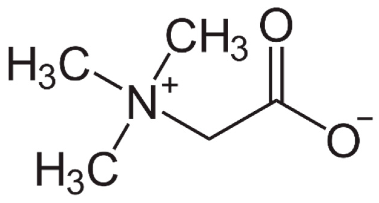
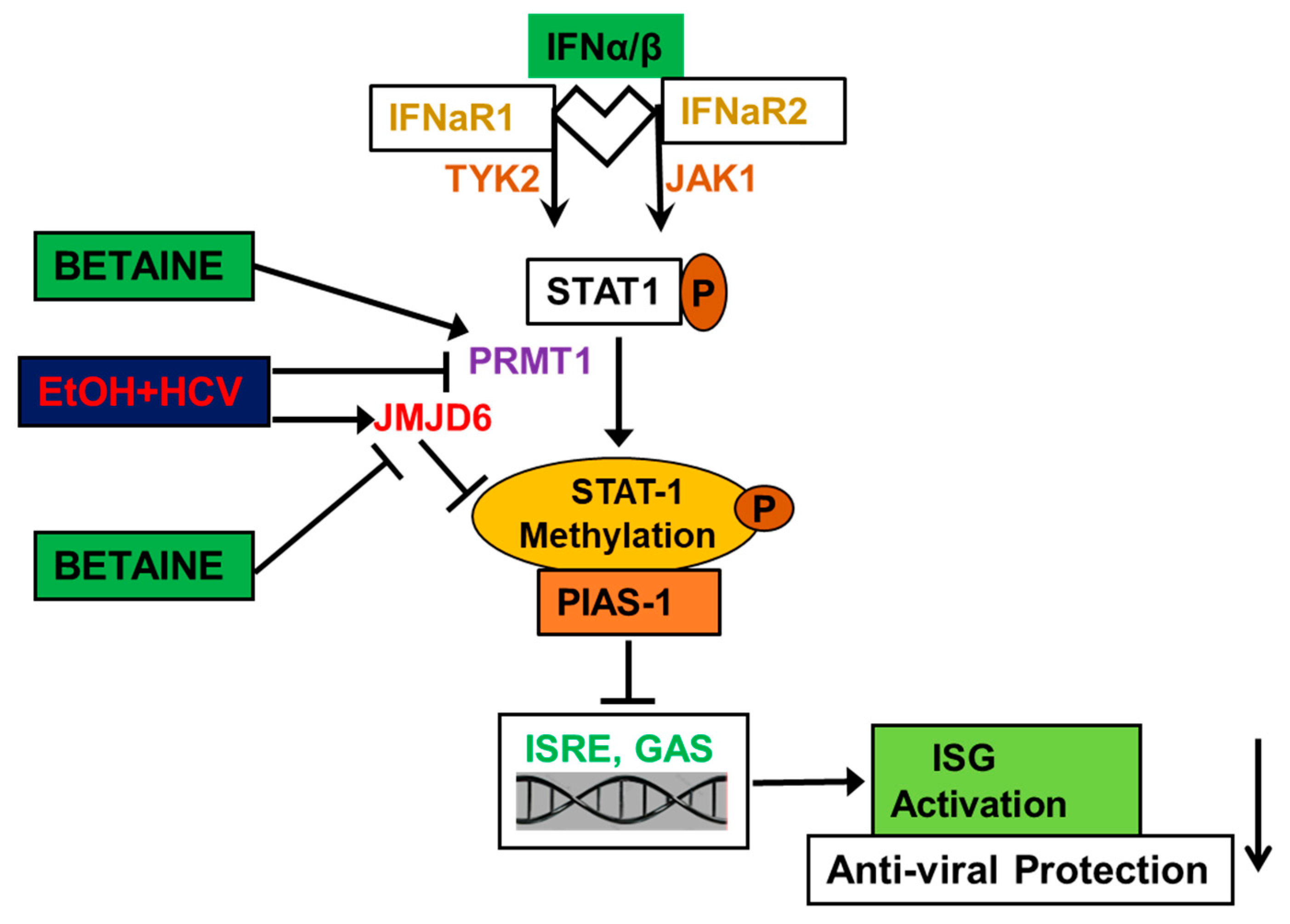
Table 1.
Protective effects of betaine in experimental animal models, cell culture systems, and clinical studies.
Table 1.
Protective effects of betaine in experimental animal models, cell culture systems, and clinical studies.
| Therapeutic Effects of Betaine Administration | Experimental Model | Authors |
|---|---|---|
| Prevents hepatic fat accumulation in ALD | Male Wistar rats; C57BL/6 mice; Balb/c mice | [23,27,83,115,121,157,158,160] |
| Preserves/restores hepatic SAM: SAH ratios by regenerating SAM and lowering SAH and homocysteine levels in ALD | Male Wistar rats; hepatocytes; male C57BL/6 mice | [23,60,61,81,82,83,84,86,88,91,92,117,119,121,234,235] |
| Restores activities of various liver methyltransferases (PEMT, ICMT, PIMT, PRMT) to increase phosphatidylcholine levels, preventing apoptosis and accumulation of damaged proteins, and restoring proteasome activity | Male Wistar rats; hepatocytes | [23,90,91,92] |
| Suppresses the synthesis of DGAT2, a rate-limiting enzyme in triglyceride synthesis, by alleviating ERK1/2 inhibition in ALD | Male C57BL/6 mice | [121] |
| Upregulates antioxidant defense system and improves oxyradical scavenging activity in ALD | Male Wistar rats | [133] |
| Prevents/attenuates ER stress in ALD | Male C57BL/6 mice | [83] |
| Exerts hepatoprotection by preserving mitochondrial function in ALD | Male Wistar rats | [61] |
| Restores the serum adiponectin levels in ALD | Mice | [123] |
| Prevents elevations of CD14, TNFα, COX2, GADD45β, LITAF, JAK3, TLR2, TLR4, IL1β, and PDCD4 and NOS2 mRNA levels in alcoholic liver injury | Male Wistar rats | [115,133] |
| Prevents serum ALT and AST activity elevations in models of ALD and MAFLD | Male Wistar rats | [27,115,121] |
| Reduces liver oxidant stress, inflammation, and apoptosis in MAFLD | Male C57BL/6 mice | [28] |
| Remethylates homocysteine, protecting from oxidant stress and restoring phosphatidylcholine generation in MAFLD | C57BL/6 mice | [161] |
| Stimulates β-oxidation in livers of MCD diet-induced MAFLD | Male Sprague-Dawley rats | [162] |
| Alleviates steatosis and increases autophagosomes numbers in mouse livers with MAFLD | Male C57BL/6 mice; rats | [120,161] |
| Enhances the conversion of existing WAT to brown adipose tissue through stimulating mitochondrial biogenesis in MAFLD | Mice | [203] |
| Alleviates ROS-induced mitochondrial respiratory chain dysfunction in MAFLD | Male Sprague-Dawley rats | [163]. |
| Attenuates different grades of steatosis, inflammation, and fibrosis in MAFLD patients | Human trials | [45,165,166,167] |
| Prevents adipose tissue dysfunction in ALD | Male C57BL/6 mice | [194] |
| Reduces the inflammatory adipokines, IL6, TNFα, and leptin in human adipocytes | Human visceral adipocytes | [204] |
| Inhibits lipid peroxidation, hepatic inflammation, and expression of transforming growth factor-β1 in liver fibrosis | Male chicks | [148] |
| Suppresses alcoholic liver fibrosis | Rats | [116] |
| Prevents the formation of Mallory–Denk bodies through epigenetic means by attenuating the decrease of MAT1A, SAHH, BHMT, and AMD1 expression | C3H male mice | [138] |
| Reverses the inhibitory effects of acetaldehyde on IFN signaling and decreases de-methylation of STAT1 by JMJD6 | HCV-infected Huh7.5 CYP2E1 (+) cells and human hepatocytes | [141,143] |
| Enhances expression of PPARα and elevates fatty acid catabolism | Male C57BL/6 and ApoE−/− mice | [158]. |
| Inhibits lipogenic activity in liver by activation of AMPK | ApoE−/− mice; Male C57BL/6 mice | [159,160] |
| Regulates colonic fluid balance | Rats | [21,200] |
| Improves intestinal barrier function and maintains the gut microbiota | Porcine epithelial cells; Caco-2 cells; rat small intestinal cell line IEC-18 | [22,197,198] |
| Activates GI digestive enzymes and ameliorates intestinal morphology and microbiota dysbiosis | Male Sprague Dawley rats | [200] |
| Attenuates alcoholic-induced pancreatic steatosis | Male Wistar rats | [125] |
| Associated with resilience to anhedonia and prevention of stress-related psychiatric disorders | Male C57BL/6 mice | [218] |
| Treats asthma-induced oxidative stress, thus improving airway function of lung tissue | BALB/C mice | [207] |
| Protects against cadmium nephrotoxicity | Male Wistar rats | [206] |
| Protects against isoprenaline-induced myocardial dysfunction | Male Wistar rats | [205] |
| Anti-nociceptive and sedative role via interactions with opioidergic and GABA receptors | Male albino mice | [220] |
| Normalizes fetal growth and reduces adiposity of progeny from obese mice | C57BL/6J mice | [229] |
| Anti-cancer effect in alcohol-associated breast cancer cell growth and development | Breast adenocarcinoma cell line (MCF-7) | [213] |
| Reduces rectal temperature in broiler chickens | Chickens | [226,227] |
| Improves post-natal lamb survival | Lambs | [230] |
Publisher’s Note: MDPI stays neutral with regard to jurisdictional claims in published maps and institutional affiliations. |
© 2021 by the authors. Licensee MDPI, Basel, Switzerland. This article is an open access article distributed under the terms and conditions of the Creative Commons Attribution (CC BY) license (https://creativecommons.org/licenses/by/4.0/).
Share and Cite
MDPI and ACS Style
Arumugam, M.K.; Paal, M.C.; Donohue, T.M., Jr.; Ganesan, M.; Osna, N.A.; Kharbanda, K.K. Beneficial Effects of Betaine: A Comprehensive Review. Biology 2021, 10, 456. https://doi.org/10.3390/biology10060456
AMA Style
Arumugam MK, Paal MC, Donohue TM Jr., Ganesan M, Osna NA, Kharbanda KK. Beneficial Effects of Betaine: A Comprehensive Review. Biology. 2021; 10(6):456. https://doi.org/10.3390/biology10060456
Chicago/Turabian StyleArumugam, Madan Kumar, Matthew C. Paal, Terrence M. Donohue, Jr., Murali Ganesan, Natalia A. Osna, and Kusum K. Kharbanda. 2021. "Beneficial Effects of Betaine: A Comprehensive Review" Biology 10, no. 6: 456. https://doi.org/10.3390/biology10060456
Note that from the first issue of 2016, this journal uses article numbers instead of page numbers. See further details here.