Article

Mesenchymal Stem Cell-Derived Exosomes Loaded with Selenium or Nano Selenium as a Novel Therapeutic Paradigm for Streptozotocin-Induced Type 1 Diabetes in Rats

by
Dlovan Y. Khalil
1,2,*,
Ridah H. Hussein
2 and
Wafaa M. El-Kholy
3
1
Virology Department, Central Health Laboratory, Ministry of Health, Sulaymaniyah 46012, Iraq
2
Department of Biology, College of Science, Slaimani University, Sulaymaniyah 46001, Iraq
3
Zoology Department, Faculty of Science, Mansoura University, Mansoura P.O. Box 11432, Egypt
*
Author to whom correspondence should be addressed.
Biology 2024, 13(4), 253; https://doi.org/10.3390/biology13040253
Submission received: 18 March 2024 / Revised: 8 April 2024 / Accepted: 9 April 2024 / Published: 11 April 2024
(This article belongs to the Special Issue Animal Models of Pancreatic Diseases)

Abstract

:

Simple Summary

Type 1 diabetes mellitus is a metabolic disorder characterized by hyperglycemia due to insulin insufficiency as a consequence of the pancreatic β-cells’ auto-immune attack. A potential novel remedy, counting on using mesenchymal stem cell (MSC)-derived exosomes loaded with an antioxidant known for its insulin-mimetic effect like selenium could be useful to address this condition and its related complications. The results indicated a hypoglycemic and antiapoptotic superiority for nano selenium-loaded exosomes rather than elemental selenium.

Abstract

Type 1 diabetes mellitus (T1DM) is a metabolic disorder characterized by hyperglycemia due to insulin insufficiency as a consequence of the pancreatic β-cells’ auto-immune attack. Nowadays, the application of mesenchymal stem cell-derived exosomes (MSCs-Exs) as the main cell-free therapy for diabetes treatment is becoming more and more extensive. In non-autologous therapy, researchers are moving towards a new strategy based on loading MSC-Exs with certain drugs, aimed at maintaining and maximizing the function of exosomes at the function site and enhancing their efficiency and safety. This study aims to explore and compare the therapeutic potentialities of mesenchymal stem cell-derived exosomes (MSCs-Exs) loaded with either selenium (Se) or nano selenium (NSe), a natural antioxidant micronutrient, in the management of T1DM in rats. In our 4-week experiment, six rat groups were included, namely, control, Ex+Se, Ex+NSe, STZ-diabetic (D), D+ Ex+Se, and D+Ex+NSe groups. Both diabetic-treated groups showed marked pancreatic regenerative antioxidant, immunomodulatory, anti-inflammatory, and anti-apoptotic capacities, with the D+Ex+NSe injection showing superiority in managing diabetes hazards, as evidenced by various biochemical and histological assessments.

1. Introduction

Type 1 diabetes mellitus (T1DM) is a metabolic disorder characterized by hyperglycemia due to insulin insufficiency as a consequence of the pancreatic β-cells’ auto-immune attack [1,2]. The most common therapeutic strategies were and still are exogenous insulin injection and oral hypoglycemics, which only manage hyperglycemia temporarily but cannot cure diabetic complications [3]. Therefore, a potential novel remedy, counting on using mesenchymal stem cells (MSCs), has emerged for DM treatment, because of their immunomodulatory capability. However, MSC-based therapy highlighted various safety concerns, including their undesired differentiation and the possibility of malignant transformation [4,5]. Of note, the therapeutic effects of MSCs are exerted mainly via paracrine signaling, by secreting various extracellular vesicles (EVs), including microvesicles, apoptotic bodies, and exosomes [3,6].
Exosomes are nano-sized EVs (30–200 nm) that exert their biological effects through the release of many soluble factors, lipids, and RNA, either in a paracrine or endocrine manner [7]. Recently, MSC-derived exosomes (MSC-Exs) have represented a promising cell-free therapy in regenerative medicine [5], where they have been proven to be equally effective [8,9] or even superior [5,6] in the alleviation of T1DM and its complications, compared with their parental MSCs. This excellence may be attributed to their easier quantification, bioactivity maintenance during transportation and storage, intercellular communication roles, and multiple bioactivities, besides the suggestion of a safer therapeutic protocol, because of the lack of direct tumorigenicity and posing of fewer membrane-bound proteins [5,10]. However, exosomes can be developed to perform crucial therapeutic functions by being employed as natural carriers to deliver certain drugs with diverse therapeutic effects, for them to reach full clinical potential [11,12].
Selenium (Se) is an essential micronutrient, that could be administrated as a dietary supplement and plays important roles in normal thyroid function, DNA synthesis, redox regulation, and fertility in humans [13]. In addition, Se was suggested for T1DM management, because of its confirmed strong antidiabetic and insulin-mimetic properties [14,15]. It was reported to protect β-cells from inflammation and oxidative damage in the body, by being involved in the synthesis of 35 different selenoproteins, including glutathione peroxidase (GPx) [13], which can regulate proper insulin-signaling cascade and inhibit β-cells apoptosis [16]. In this line, the treatment of diabetic animals with Se was reported to increase the viability of pancreatic β-cells and insulin secretion, by inhibiting pancreatic apoptosis and necrosis and upregulating the expression of the insulin gene [17,18]. However, Se has a narrow therapeutic window and the toxicity margins are very delicate, whereas the nanoparticles of Se (SeNPs) possess remarkably reduced toxicity [19]. Nanosized Se (NSe) was suggested as a superior effective molecular compound, with higher antidiabetic, antioxidant, and anti-apoptotic activities compared to ordinary Se [20,21], which could be attributed to its improved bio-activity, bioavailability, cellular uptake, delivery, and very limited toxicity [22].
Hence, this study aims to explore and compare the anti-apoptotic and regenerative capabilities of MSCs-EXs loaded with either Se or NSe particles to alleviate hyperglycemia and restore normal insulin secretion from the pancreas of T1DM rats.

2. Materials and Methods

2.1. Chemicals

Streptozotocin (STZ), selenium (Se), and nano selenium (NSe) were purchased from Sigma Aldrich Co. (St. Louis, MO, USA), while exosomes (500 μg/mL) were purchased from NAWAH Co. (El Mokattam, Egypt).

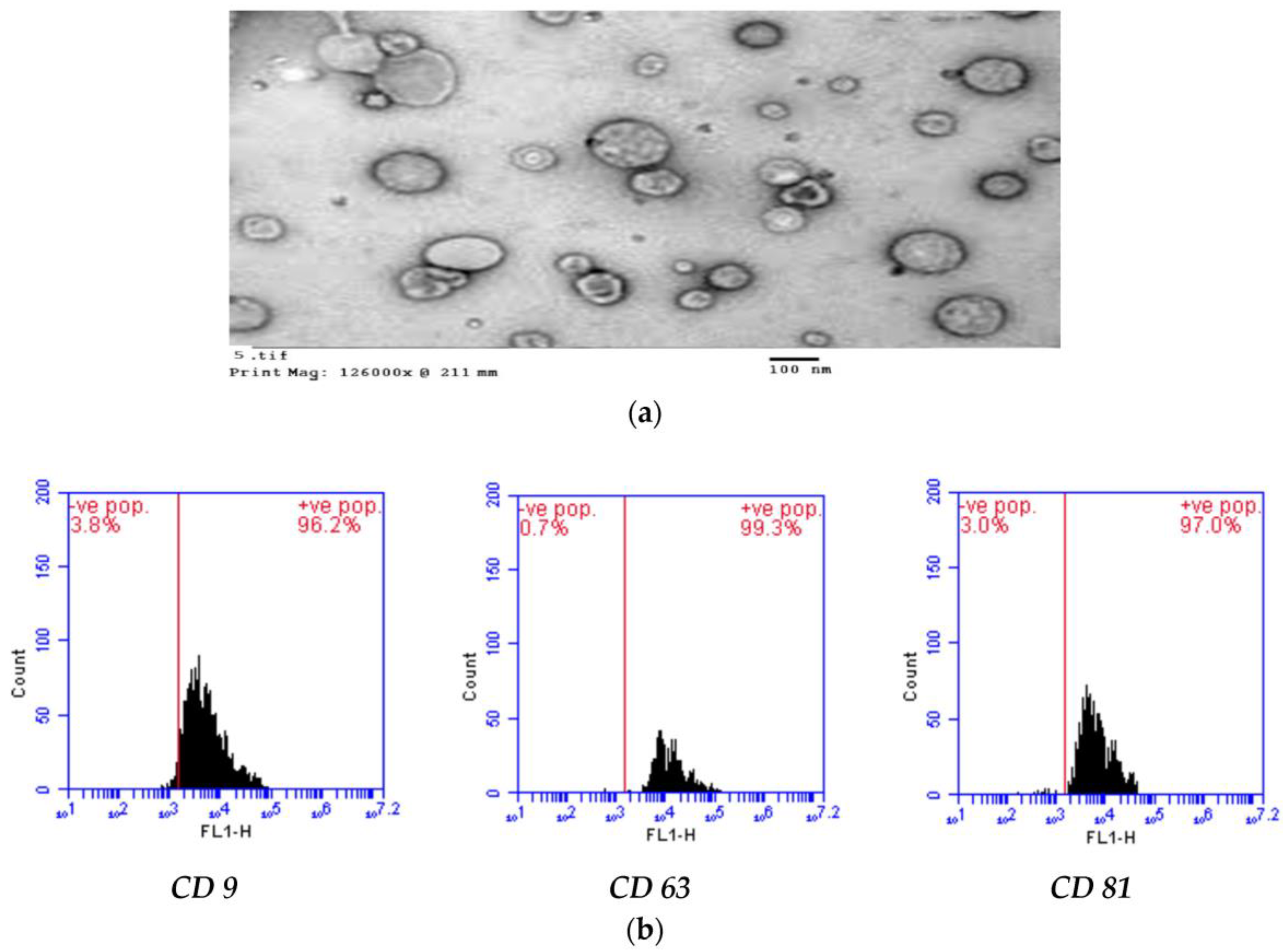
2.2. AD-MSCs-Exs Preparation and Characterization

The exosomes derived from mesenchymal stem cell stock (500 μg/mL) were sonicated in phosphate-buffered saline (Pbs, pH 7.4) for 10 min, to ensure their complete solubilization. The Ex morphological identity was confirmed using a transmission electron microscope (TEM, Zeiss, LEO 906E, Jena, Germany). A FACS Caliber flow cytometer (Becton Dickinson, Franklin Lakes, NJ, USA) was applied for the characterization of specific surface markers (CD9, CD63, and CD81) (Sigma Aldrich, USA), by using specific rats’ antibodies purchased from Sigma Aldrish, USA [4,6].

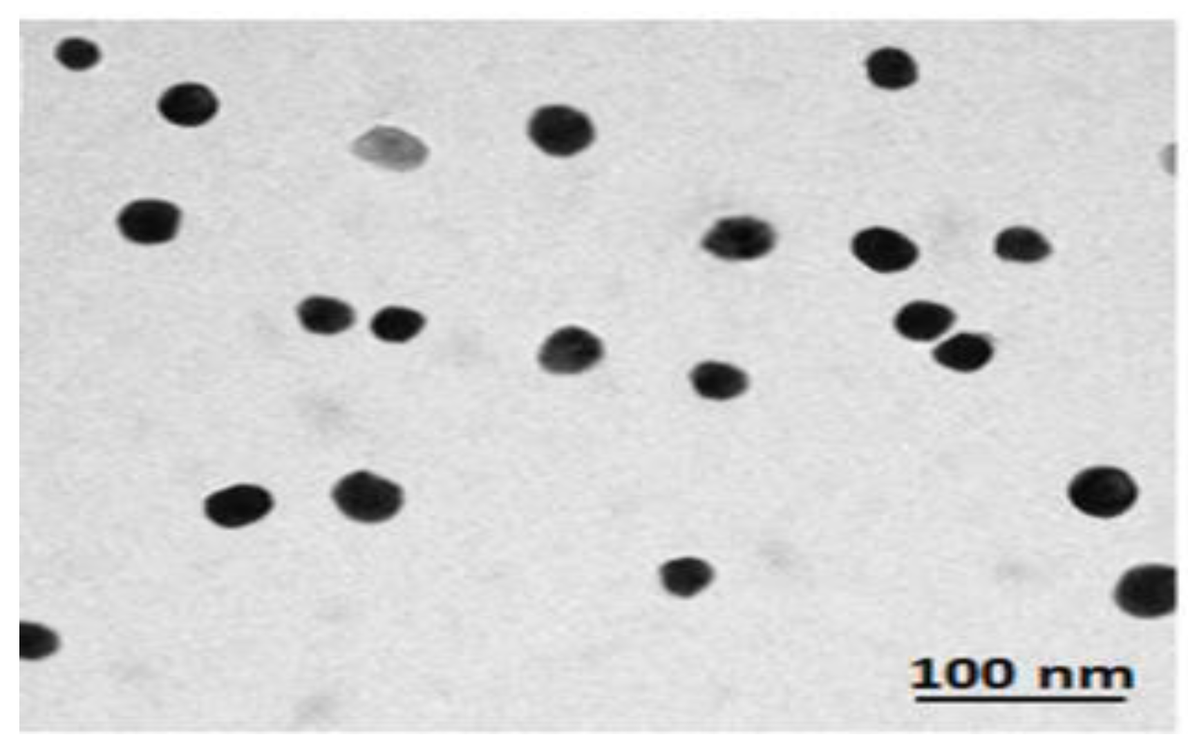
2.3. Nano-Selenium (NSe) Preparation and Characterization

NSe particles (5–50 nm) were purchased from Sigma Aldrich (USA), separated by centrifugation, and dispersed in an aqueous medium by means of sonication. Meanwhile, TEM (Zeiss, LEO 906E, Germany) was used to examine the NSe particles’ shape and size [21].

2.4. Loading of EXs with Either Se or NSe

Se or NSe particle incorporation into exosomes was performed using a sonication-aided exosome drug-loading technique according to Haney et al. [23], with modifications. Exosomes were dispersed in 0.1 mg/mL PBS. Either 20 mL of Se solution in DMSO (200 ppm) or 20 mL of NSe in PBS were added to 20 mL of exosome solution forming a Drug-EXs mixture, which was then sonicated using a probe sonicator (750 v, 20% power, 6 cycles by 4 s pulse/2 s pause (total time 24 s), and cooled down for 2 min on ice, before being further sonicated. These steps were repeated 3 times. The drug-loaded exosomes were enclosed in a cellulose dialysis sac (12–14 KDa) for purification. The dialysis bag was sealed properly both from top and bottom and inserted into 7.5 mL of PBS 7.4 at 50 r.p.m. using a shaking incubator at room temperature. After 1 h, the media were collected and replaced with new 7.5 mL of PBS for another 30 min. On the other hand, the same steps were performed to create unloaded exosomes but without adding active ingredients and using PBS only. The obtained sample stocks were divided into insulin syringes (each of 0.5 mL), covered with foil, and stored at −20 °C until use.

2.5. Induction of Diabetes

Rats were fasted overnight and then received an intraperitoneal (IP) single dose of streptozotocin (STZ, 60 mg/kg bw) solution dissolved in cold sodium citrate buffer (0.1 M, pH 4.6). After 3 days, the tail vein fasting blood glucose (FBG) level was assessed using an ACCU–CHEK Go glucometer (Roche Co, Mannheim, Germany). Only rats with FBG over 200 mg/dL were considered diabetics [24].

2.6. Experimental Design

The 4-week experimental design was approved by the Animal Ethics Committee of the Faculty of Science, Mansoura University, Mansoura, Egypt [MU-ACUC (SC.PhD.23.03.6)]. Sixty-four male albino Wistar rats (180–210 g) were housed in plastic cages with water and chow free access. Animals were split into 6 groups each of 8 rats, as follows:
A.
Control groups
1. 
Normal group: Rats received an IP single dose of citrate buffer (2 mL/kg, pH 4.6).
2. 
Ex group: Rats received an IV single dose of 0.5 mL unloaded EXs.
3. 
Ex+Se group: Rats received an IV single dose of 0.5 mL EXs loaded with Se.
4. 
Ex+NSe group: Rats received an IV single dose of 0.5 mL EXs loaded with NSe.
B.
Diabetic groups
5. 
Diabetic (D) untreated group: Diabetic rats received an IP single dose of STZ (60 mg/kg).
6. 
Ex-treated group: Diabetic rats received an IV single dose of 0.5 mL unloaded EXs.
7. 
Diabetic Ex+Se-treated group: Diabetic rats received an IV single dose of 0.5 mL EXs loaded with Se.
8. 
Diabetic Ex+NSe-treated group: Diabetic rats received an IV single dose of 0.5 mL EXs loaded with NSe.

2.7. Sample Collection

Diethyl ether was used to anesthetize overnight fasted rats before dissecting them. A cardiac puncture was performed to collect blood, where a few droplets, for the assessment of glycosylated hemoglobin (HbA1c), were placed in heparinized tubes. Blood samples were centrifuged (1000× g for 15 min) to obtain plasma, and sera were carefully separated and stored at −20 °C. On the other hand, pancreas specimens were harvested and divided into three appropriate parts; one part for forming 10% (w/v) homogenate (kept at −20 °C), another fresh part for flow cytometric analysis, and the third part for histopathological examination (stored in 10% formalin solution).

2.8. Histochemical Examination

Pancreas samples were cut (1 cm thickness), fixed in 10% neutral buffer formalin, and embedded in paraffin wax to form sample blocks. A microtome was used to cut the wax blocks into 3–5 µm thickness sections, which were later mounted on labeled glass slides. A Leica Auto Stainer was then used to stain the slides with Hematoxylin and Eosin. Afterward, a light microscope was used to examine the pancreatic samples [25].

2.9. Biochemical Assays

Bio Vision Company, Milpitas, CA, USA, ELISA kits were used to detect serum insulin and C-peptide levels. The estimation of serum glucose, glycosylated hemoglobin (HbA1c), and amylase levels was performed using SPINREACT diagnostics kits in Spain. The pancreatic content of some antioxidants markers, including catalase (CAT), superoxide dismutase (SOD), glutathione (GSH), and glutathione peroxidase (GPx), in addition to some oxidative stress markers, including nitric oxide (NO), malondialdehyde (MDA), and hydrogen peroxide (H2O2), were all assessed using diagnostic kits obtained from Bio Diagnostic Company, Egypt.

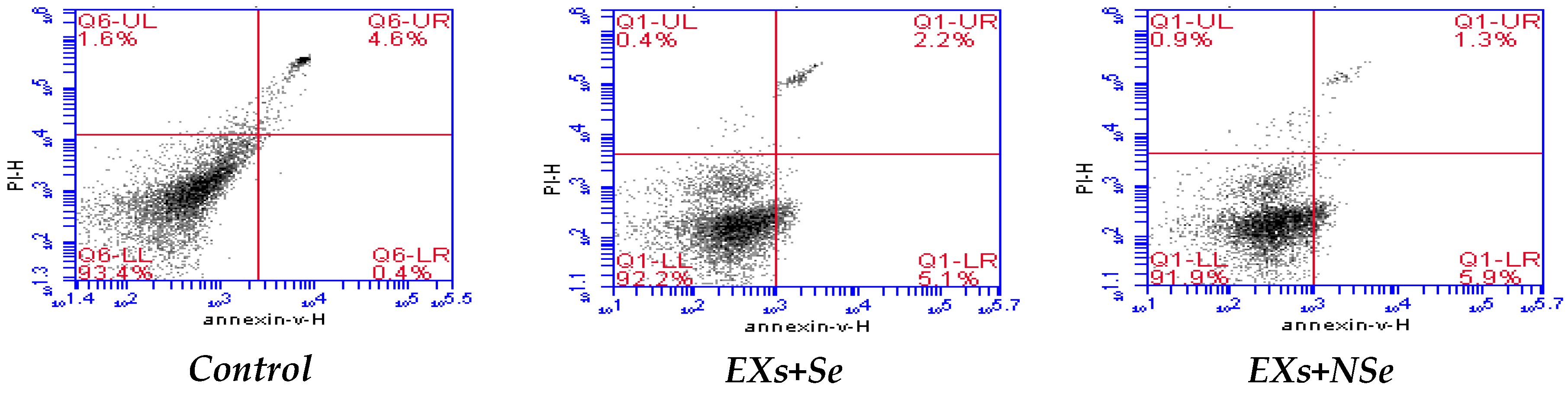
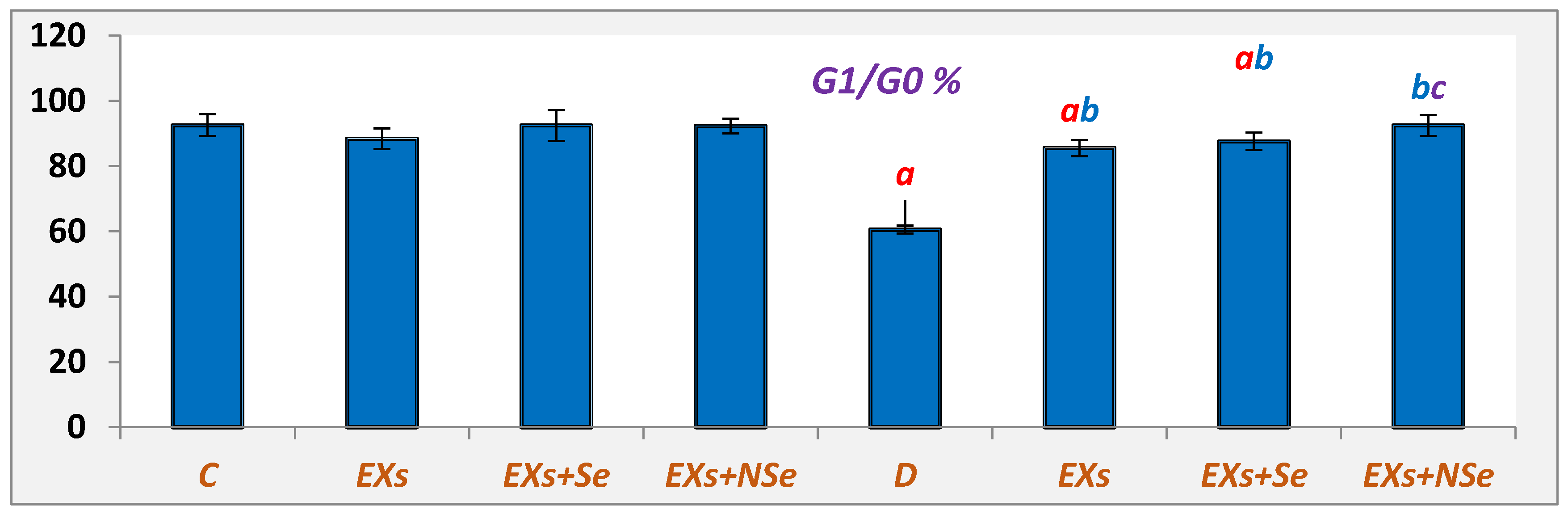
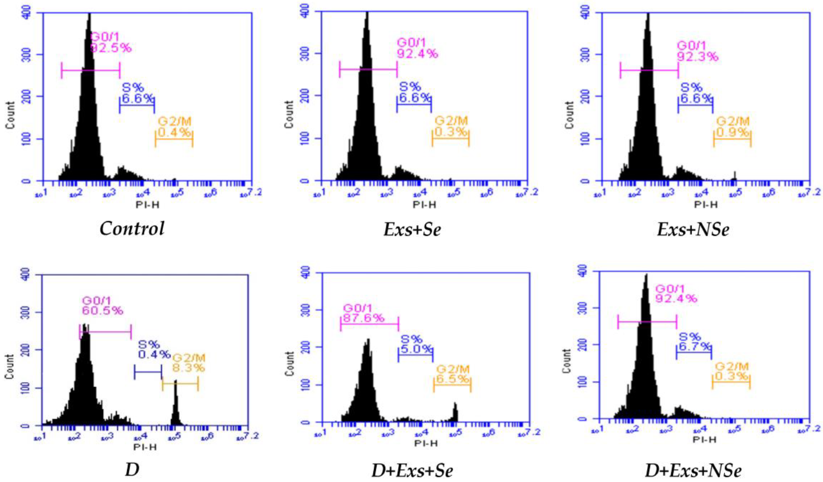
2.10. Flow Cytometric Analysis

Antibody kits obtained from Sigma Aldrich Company (USA) were used to determine various pancreatic inflammatory indicators % (IL-6, TGF-β, and TNF-α), apoptotic (annexin V, P53, caspase-3) and anti-apoptotic (Bcl-2) incidences, and various cell cycle phases, in addition to AD-MSCs-EXs surface markers (CD9, CD63, and CD81), according to the method of Tribukait et al. [26]. Briefly, a fresh tissue specimen was dissolved in 250 mL of distilled water and centrifuged at 1800× g rpm for 10 min. the supernatant was aspirated, and cells were fixed in ice-cold 96–100% ethanol. The cells were stained by linking the fluorochrome directly to the primary antibody. Then, the cells were washed with 2 mL of PBS/BSA and centrifuged, where the supernatant was discarded and the cells were resuspended in 0.2 mL of PBS/BSA. Finally, data were acquired by means of flow cytometry using a FACS Caliber flow cytometer (Becton Dickinson, USA) [27].

2.11. Statistical Analysis
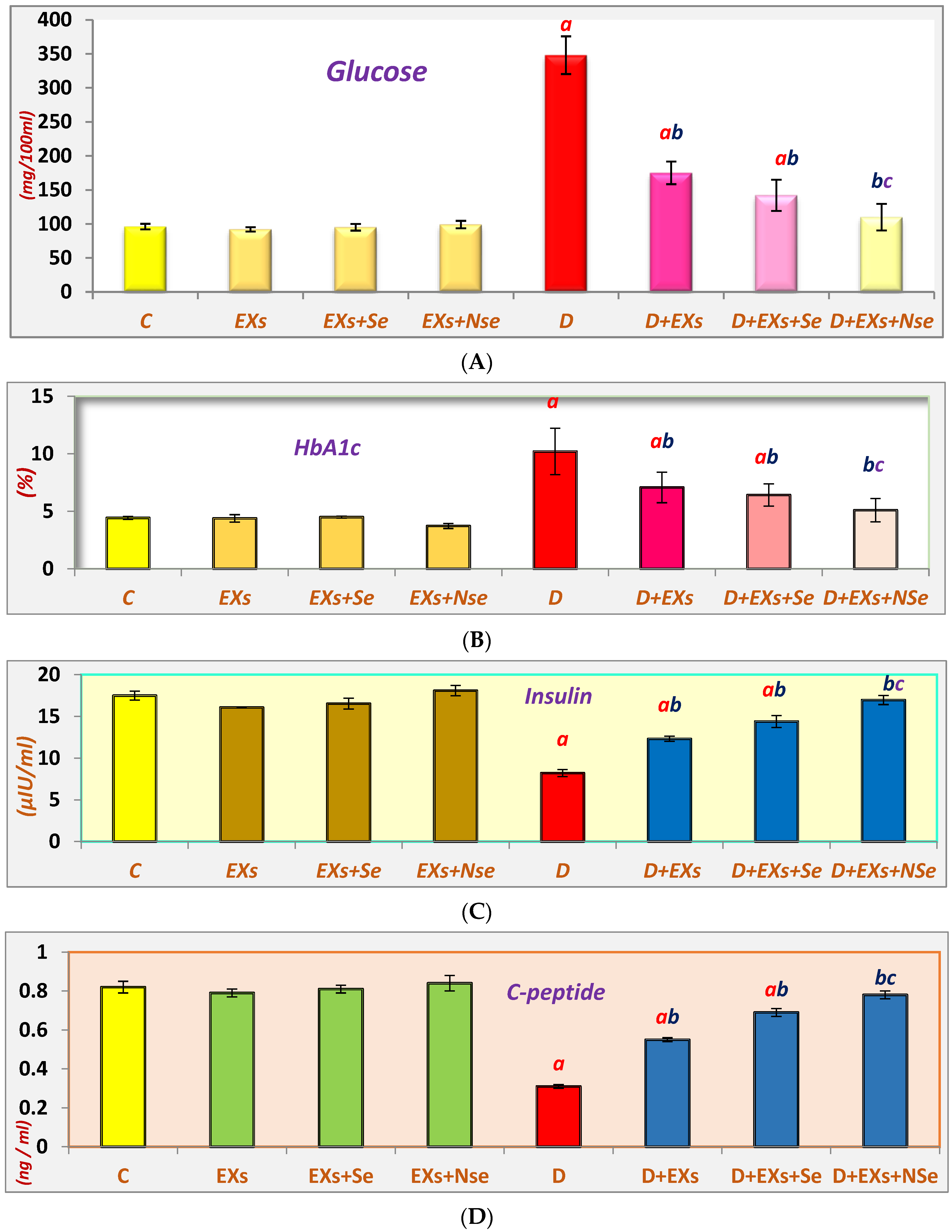
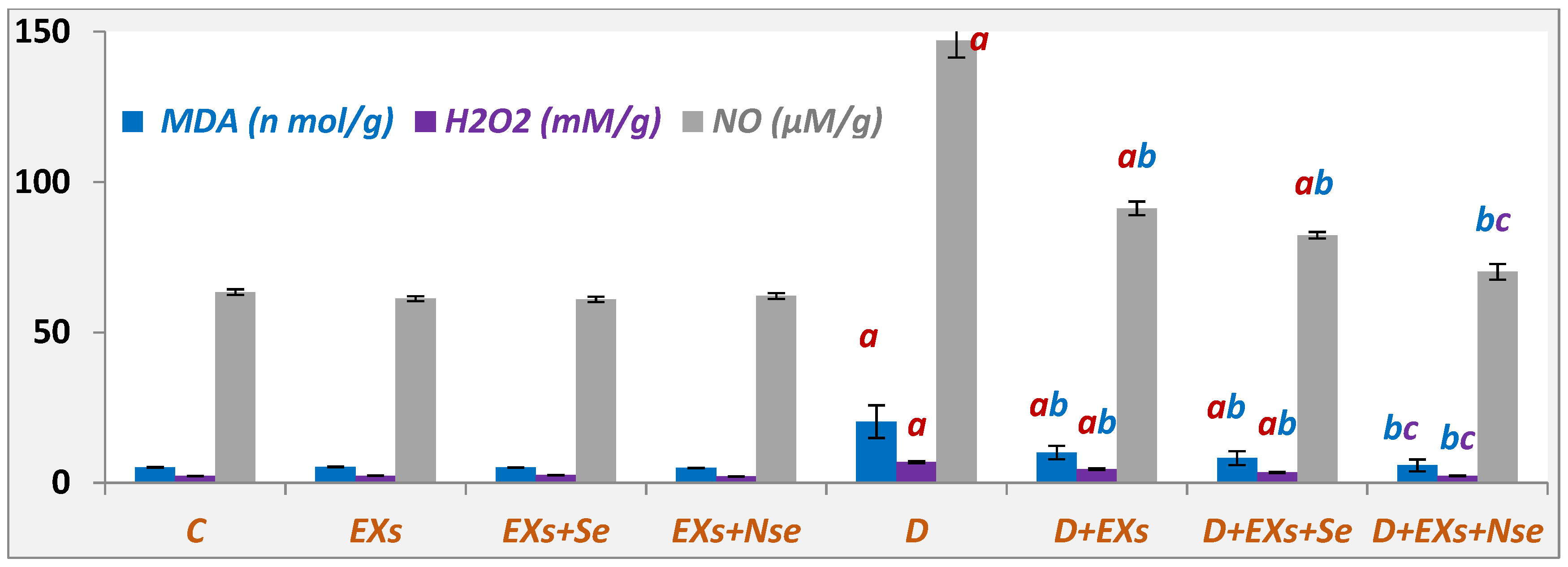
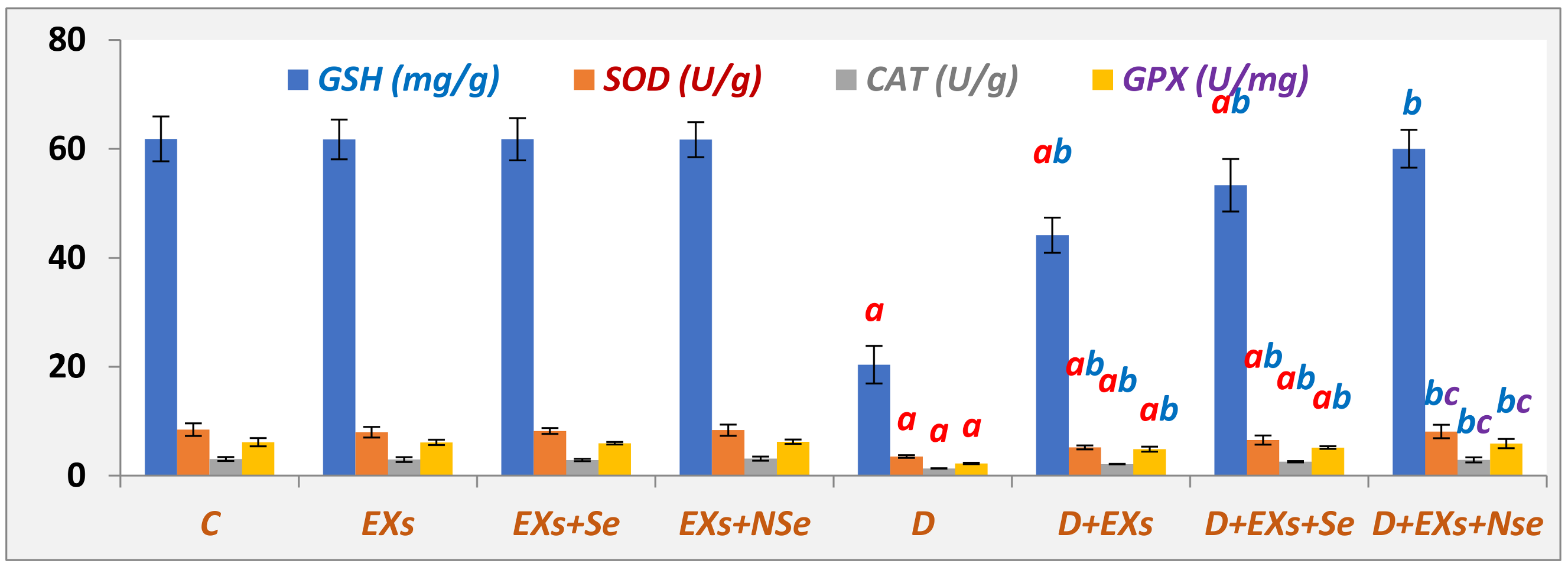
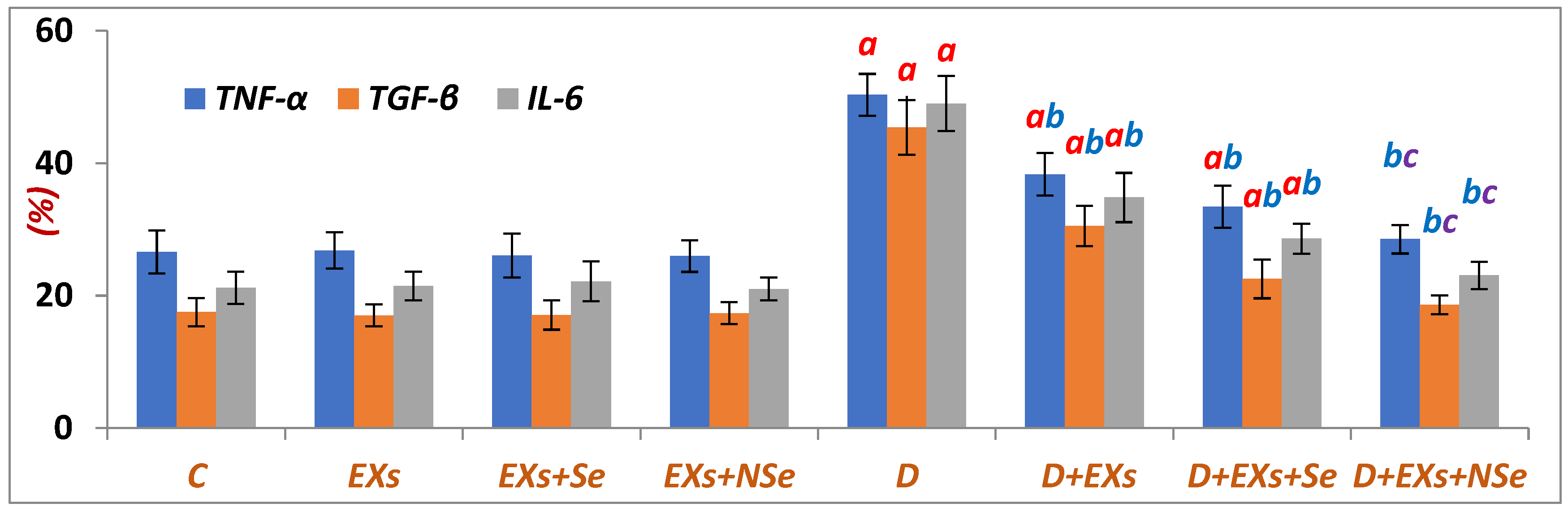
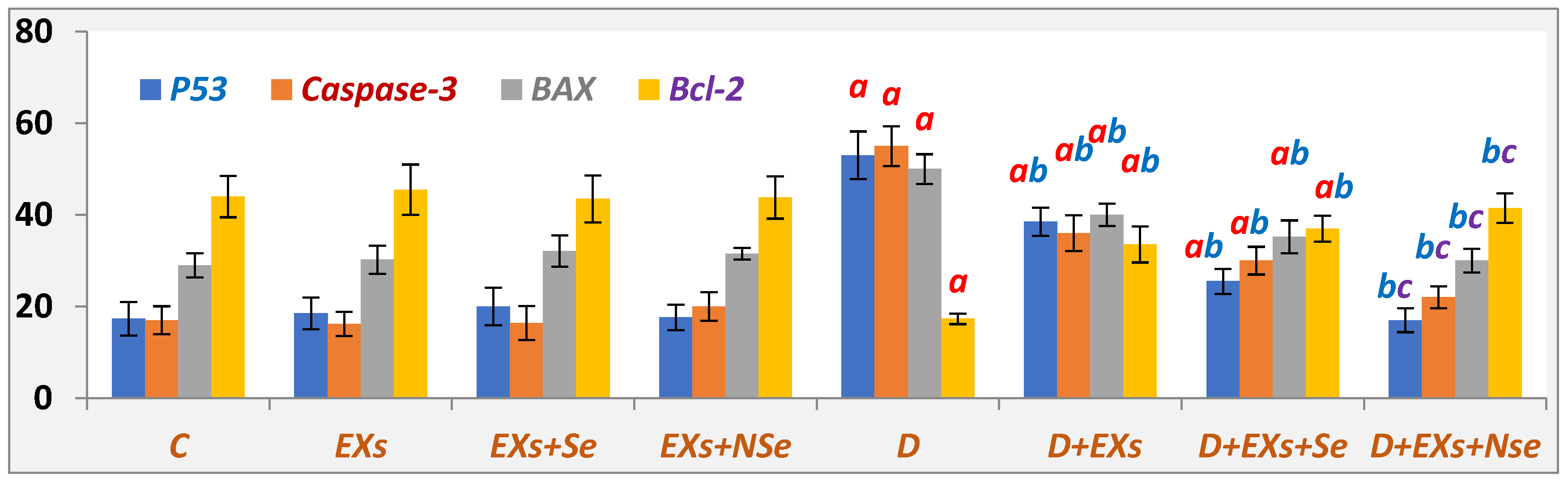
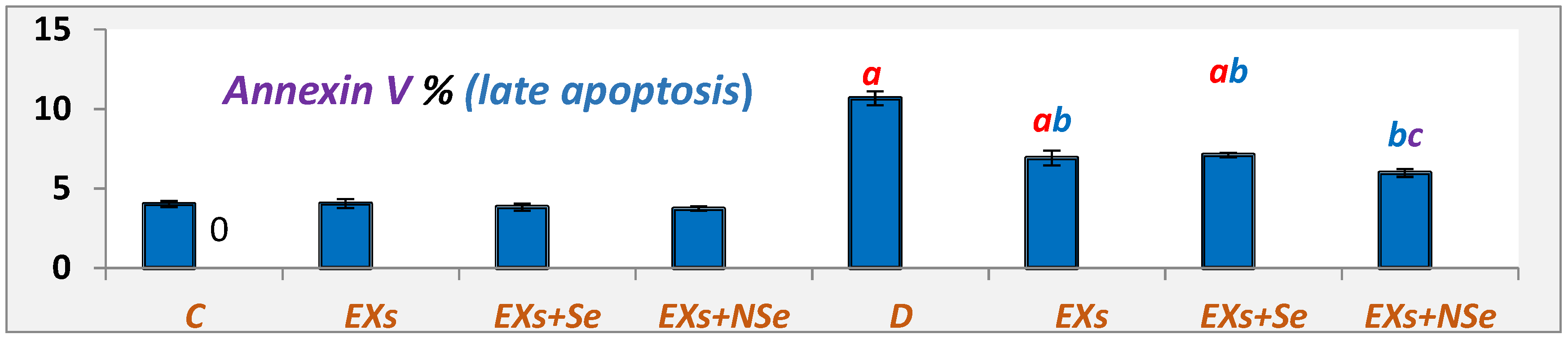
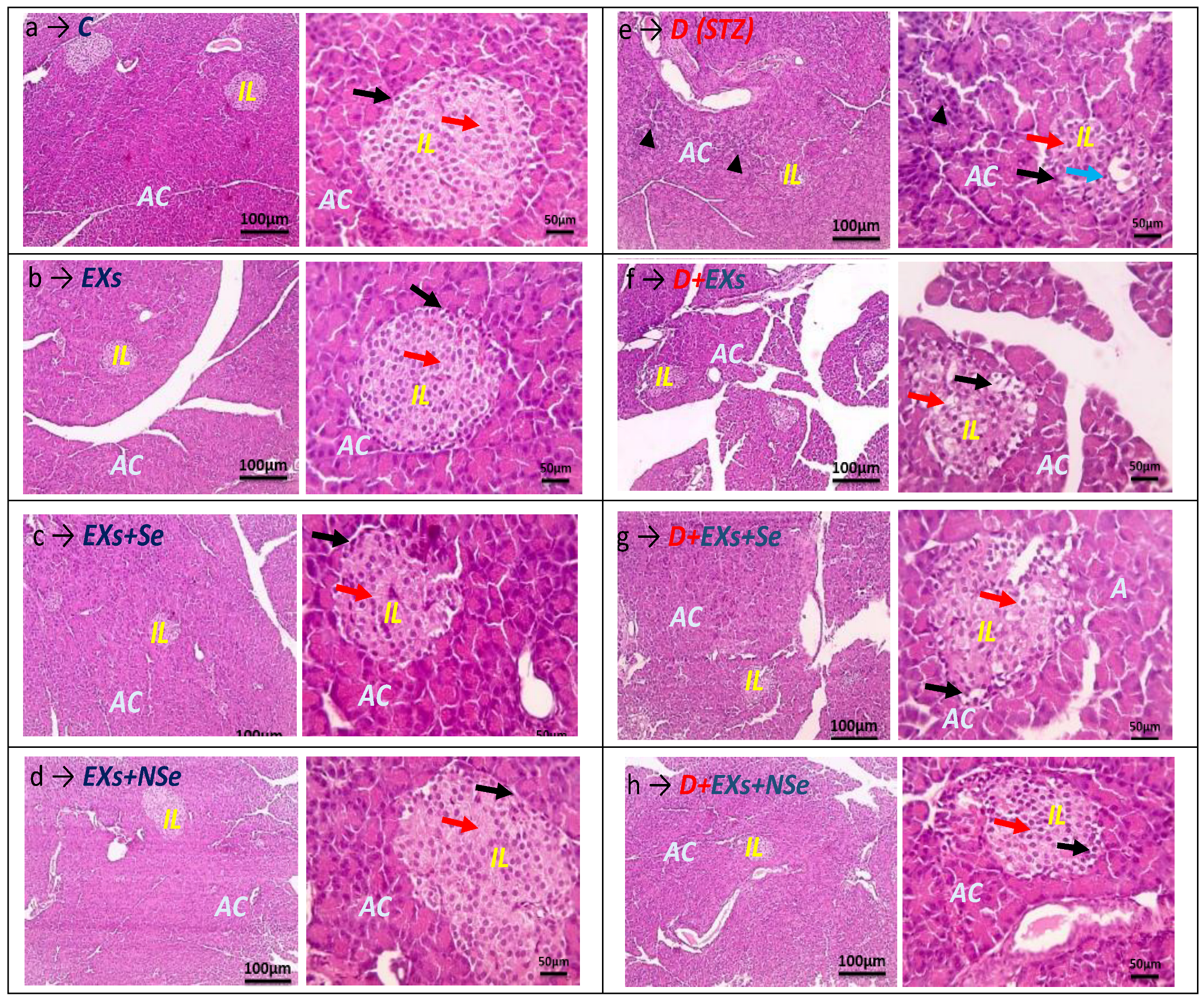
The obtained data were evaluated using a Statistical Package for the Social Sciences (SPSS/21 software version) for Windows, by applying ANOVA followed by post hoc Tukey multiple range tests. All results were expressed as the mean ± standard error (SEM), where n = 8. The level of significance was set at p ≤ 0.05 for all statistical tests.

3. Results

TEM for the observation of exosome morphology showed circular and intact vesicles with a size range of 100–150 nm (Figure 1a). The flow cytometric analysis showed markedly high CD9 (96.2%), CD63 (99.3%), and CD81 (97.0%) expression, confirming the AD-MSCs-EXs phenotype identity (Figure 1b).
The TEM examination of NSe morphology (Figure 2) indicated a clear, spherical shape, with a size range of 5–50 nm.
A marked elevation in serum levels of both glucose and HbA1c was detected in diabetic rats, as shown in Figure 3A–D, while insulin and C-peptide levels were significantly decreased, compared to control rats. However, a noticeable enhancement was recorded in all diabetic-treated groups, relative to untreated diabetics, with an obvious NSe-loaded Ex hypoglycemic superiority relative to Exs loaded with ordinary Se.
The diabetic rats’ results represented in Figure 4 highlight marked elevations in pancreatic levels of oxidative stress markers (MDA, H2O2, and NO), relative to the control rats. On the other hand, all treated rat groups reflected a significant oxidative marker decrease, with a marked Ex+NSe treatment excellence.
In comparison with the control rats, the Figure 5 data demonstrate a marked antioxidant content decline in the pancreas of diabetic rats. However, the injection of diabetic rats with either Exs or Exs+Se or Exs+NSe showed critical antioxidant content upregulation compared to the untreated diabetic group. Nevertheless, the Ex+NSe therapy revealed an excellent antioxidant potential compared to the other treatment.
Compared to control rats, the Figure 6 data clearly show a marked progress in the pancreatic inflammatory status of diabetic rats, which was significantly suppressed following the injection with unloaded Exs or Se- and NSe-loaded Exs. Notably, the NSe-loaded Exs revealed the highest anti-inflammatory efficacy.
Relative to control, substantial progress in the apoptotic status of diabetic rats’ pancreas was suggested, as seen in Figure 7, by the upregulation of marked apoptotic markers (P53, Bax, and caspase-3%), coupled with the noted anti-apoptotic Bcl-2% diminishing. In contrast, all therapy protocols revealed a tremendous pancreatic anti-apoptotic effect compared to the untreated diabetic rats, with a superior efficacy for the Ex+NSe protocol.
Figure 8 and Figure 9 showed various pancreatic annexin V % indications in different rat groups. Untreated diabetic rats exhibited a marked decline in viable cell % coupled with significant elevations in apoptotic and necrotic cell %, compared to control. Following different protocols of treatment, diabetic rats showed a notable increase in viable cell %, with a marked decrease in both apoptotic and necrotic cell %. However, the NSe-loaded Ex protocol reflected the greatest improvement in pancreatic cell status.
The Figure 10 and Figure 11 data summarize different pancreatic cell cycle phases in different rat groups. Untreated diabetic rats showed a significant downregulation in the G0/G1 cell %, which reflects the pancreatic viable cell %, compared to control rats. On the contrary, relative to untreated diabetic rats, the Ex, Ex+Se, or Ex+NSe injection to diabetic rats exhibited a marked upregulation in the pancreatic viable cell %. However, the Ex+NSe injection recorded a superior enhancement compared to Ex+Se-treated diabetic rats.
Figure 12a–d demonstrate a representative image for pancreatic tissues of different normal rat groups (C, Ex, Ex+Se, and Ex+NSe, respectively), which showed a standard architecture without any notable pathological changes, where prominent nuclei and clear borders of Langerhans’ islets, with intact construction separated by a normal connective tissue septum, were illustrated, indicating the safe usage of EXs loaded with either Se or Nse. In contrast to the previous groups, the pancreatic tissues of diabetic-untreated rats (D) represented in Figure 12e clarify the destructive effect of the STZ injection, confirmed by the marked islets’ mass size shrinkage, pyknotic nuclei, and congested blood vessels, besides the limited islet vacuolation. However, Figure 12f–h show that diabetic animals treated with unloaded Exs or Exs loaded with either Se (D+Exs+Se) or Nse (D+Exs+Nse) indicated a notable variable degree of amelioration in the islets’ architecture, relative to the untreated diabetic group (D), represented by the obvious restoration of the normal islets’ mass size and the rare occurrence of either congested blood vessels or apoptotic nuclei. Notably, diabetic rats injected with NSe-loaded Exs displayed superior pancreatic architecture amelioration, compared to other treatment protocols.

4. Discussion

Since the principle mechanism of DM induction relies on the huge induction of oxidative stress in the pancreas, administering hypoglycemic agents with known antioxidant potency could effectively restore normal glycemic control and counteract the resulting diabetic hazards in treated subjects. Hence, we suggested a somewhat complicated mechanism of treatment involving the loading of mesenchymal stem cell-derived exosomes (MSC-Exs) with either selenium (Se) or nano selenium (NSe). Our novel protocol greatly upregulated the pancreatic antioxidant contents of diabetic rats, with a direct subsequent suppression of the oxidative stress status. Such events were strongly suggested to decrease the pancreatic inflammatory progress, leading to a pancreatic apoptosis marked arrest, coupled with a significant elevation in the number of viable pancreatic cells, resulting in the restoration of normal insulin secretion and blood glucose management. Similar our results, Sun et al. [3] and He et al. [5] found that MSCs-Exs effectively decreased FBG, increased insulin release, and alleviated hyperglycemia in STZ-treated diabetic rats [28,29] as well as STZ-treated diabetic mice [30], likely via restoring both normal β-islet structure and function, inhibiting β-cell apoptosis and promoting their proliferation, promoting glycolysis/glycogen synthesis, and inhibiting gluconeogenesis.
Interestingly, co-culturing pancreatic β-islets with MSCs-Exs greatly promotes the transplanted islets’ vascularization, angiogenesis, and survival after engraftment, and reduces their apoptosis and auto-immunity, evidenced by the downregulation of some apoptotic genes (Caspase-3, P53, and BAX), while upregulating the anti-apoptotic Bcl-2 gene expression [31,32,33]. Some earlier studies reported that MSCs-Exs can slow or even suppress the progression of T1DM patients by inhibiting the autoimmune targeting of pancreatic β-islets, by promoting autoreactive T-cell apoptosis and regulatory T-cell proliferation; this leads to enhancing the islets’ regeneration, angiogenesis, and survival, resulting in a rise in insulin secretion and the management of hyperglycemia and its complications [7,33,34]. In addition, there is even a possibility that the reparative properties of MSCs-Exs could cause a functional recovery of the already-existing islets, making its injection safer and potentially equally effective or even superior to the MSCs themselves in T1DM therapy and its complications [3,33,35,36].
On the other hand, selenium (Se)’s antidiabetic action appears to result from its behavior as an insulin-mimetic and as an antioxidant nutrient, since both insulin signaling and secretion are associated with the cellular redox state [14,37]. The administration of sodium selenite to diabetic rats for 4 weeks (5 ppm in drinking water), according to Barakat et al. [17,38], or for 2 weeks (0.173 mg/kg, IP injection), according to Iukuza et al. [39], was found to favor a marked FBG and HbA1c decline and the restoration of normal β-cell architecture, likely by suppressing the synthesis of hepatic glucose, promoting glucose uptake by peripheral tissues and glycogen synthesis. Interestingly, the findings of Harmon et al. [40], Lei and Vatamaniuk [41], and Steinbrenner et al. [42] were suggestive of the possible role of Se in the regulation of the redox state of pancreatic β-cells, by increasing the synthesis of other antioxidant selenoproteins including GPx, and participating in the protection of β-islets’ cell membranes from oxidative damage, with a marked restoration of the normal β-cell mass and insulin release. In addition, Labunskyy et al. [43] reported that Se supplementation increased other major antioxidant contents (GSH, SOD, and CAT). However, other mechanisms were suggested to be partly responsible for Se hypoglycemic action, including the renal glucose excretion acceleration and the suppression of intestinal glucose transport [14]. Regarding nano selenium (NSe), Al-Quraishy et al. [44] reported that the treatment of STZ-diabetic rats with NSe (0.1 mg/kg orally for 28 days) greatly alleviated hyperglycemia, possibly by eliciting insulin-mimetic activity, evidenced by the significant decline in FBG levels coupled with a marked elevation in insulin secretion and liver glycogen content, through enhancing the activity of glucose-6-phosphate dehydrogenase while reversing the abnormal activity of some gluconeogenic and glycolytic liver enzymes.

5. Conclusions

The injection of MSCs-EXs either unloaded or loaded with Se or NSe resulted in a marked blood glucose management, in rats with T1DM. However, the injection of NSe-loaded MSCs-Exs exhibited a marked hypoglycemic superiority, with notable pancreatic immunomodulatory, anti-inflammatory, anti-apoptotic, antioxidant, and regenerative potentialities. Such behavior is explained, at least in part, by NSe’s recorded antioxidant efficacy in scavenging the pancreatic free radicals, in addition to this metal’s well-known insulin-mimetic effect. However, some limitations should be addressed in future work including (1) the addition of a positive treatment control with a standard anti-diabetic drug such as insulin or metformin, (2) knowing the actual quantity of NSe that was successfully entrapped/loaded into the exosomes, (3) a robust physicochemical characterization, and, finally, (4) ascertaining the therapeutic limits of safety, therapeutic efficacy, and toxicity of the drug, since only ‘one volume, 0.5 mL’ was tested.

Author Contributions

D.Y.K., R.H.H. and W.M.E.-K. conceived and designed the study. D.Y.K. was responsible for lab work, data tabulating and acquisition, literature search, and preparing the first draft of the manuscript. All authors have read and agreed to the published version of the manuscript.

Funding

This research received no external funding.

Institutional Review Board Statement

This design was approved by the Animal Ethics Committee of the Faculty of Science, Mansoura University, Mansoura, Egypt [MU-ACUC (SC.PhD.23.03.6)].

Informed Consent Statement

Not applicable.

Data Availability Statement

Data are contained within the article.

Conflicts of Interest

The authors declare that they have no known competing financial interests or personal relationships that could have appeared to influence the work reported in this paper.

Abbreviations

AD-MSCs: Adipose-derived mesenchymal stem cells; CAT: Catalase; CD: Cluster of differentiation; D: Diabetic; DM: Diabetes mellitus; DMEM: Dulbecco’s modified Eagle’s medium; Exs: Exosomes; FBG: Fasting blood glucose; FBS: Fetal bovine serum; GPx: Glutathione peroxidase; GSH: Glutathione; HbA1c: glycosylated hemoglobin; H2O2: Hydrogen peroxide; MDA: Malondialdehyde; MSCs: Mesenchymal stem cells; NSe: Nanoselenuim; PBS: Phosphate-buffered saline; ROS: Reactive oxygen species; Se: Selenuim; SEM: Standard error of the mean; SOD: Superoxide dismutase; SPSS: Statistical Package for the Social Sciences; STZ: Streptozotocin; T1DM: Type 1 diabetes mellitus; T-reg: Regulatory T-cells.

References

  1. Yang, M.; Chen, J.; Chen, L. The roles of mesenchymal stem cell-derived exosomes in diabetes mellitus and its related complications. Front. Endocrinol. 2022, 13, 1027686. [Google Scholar] [CrossRef] [PubMed]
  2. Zhu, M.; Wu, J.; Gao, J. Exosomes for diabetes syndrome: Ongoing applications and perspective. Biomater. Sci. 2022, 10, 2154. [Google Scholar] [CrossRef] [PubMed]
  3. Sun, Y.; Tao, Q.; Wu, X.; Zhang, L.; Liu, Q.; Wang, L. The Utility of Exosomes in Diagnosis and Therapy of Diabetes Mellitus and Associated Complications. Front. Endocrinol. 2021, 12, 756581. [Google Scholar] [CrossRef] [PubMed]
  4. Harrell, C.R.; Jovicic, N.; Djonov, V.; Volarevic, V. Therapeutic Use of Mesenchymal Stem Cell-Derived Exosomes: From Basic Science to Clinics. Pharmaceutics 2020, 12, 474. [Google Scholar] [CrossRef]
  5. He, Q.; Wang, L.; Zhao, R.; Yan, F.; Sha, S.; Cui, C.; Song, J.; Hu, H.; Guo, X.; Yang, M.; et al. Retraction Note: Mesenchymal stem cell-derived exosomes exert ameliorative effects in type 2 diabetes by improving hepatic glucose and lipid metabolism via enhancing autophagy. Stem Cell Res. Ther. 2022, 13, 505. [Google Scholar] [CrossRef] [PubMed]
  6. Keshtkar, S.; Kaviani, M.; Sarvestani, F.S.; Ghahremani, M.H.; Aghdaei, M.H.; Al-Abdullah, I.H.; Azarpira, N. Exosomes derived from human mesenchymal stem cells preserve mouse islet survival and insulin secretion function. EXCLI J. 2020, 19, 1064–1080. [Google Scholar] [CrossRef] [PubMed]
  7. Harrell, C.R.; Jovicic, N.; Djonov, V.; Arsenijevic, N.; Volarevic, V. Mesenchymal stem cell-derived exosomes and other extracellular vesicles as new remedies in the therapy of inflammatory diseases. Cells 2019, 8, 1605. [Google Scholar] [CrossRef]
  8. Mathew, B.; Ravindran, S.; Liu, X.; Torres, L.; Chennakesavalu, M.; Huang, C.-C.; Feng, L.; Zelka, R.; Lopez, J.; Sharma, M.; et al. Mesenchymal stem cell-derived extracellular vesicles and retinal ischemia-reperfusion. Biomaterials 2019, 197, 146–160. [Google Scholar] [CrossRef]
  9. Nakano, M.; Kubota, K.; Kobayashi, E.; Chikenji, T.S.; Saito, Y.; Konari, N.; Fujimiya, M. Bone marrow-derived mesenchymal stem cells improve cognitive impairment in an Alzheimer’s disease model by increasing the expression of microRNA-146a in the hippocampus. Sci. Rep. 2020, 10, 10772. [Google Scholar] [CrossRef]
  10. Keshtkar, S.; Azarpira, N.; Ghahremani, M.H. Mesenchymal stem cell-derived extracellular vesicles: Novel frontiers in regenerative medicine. Stem Cell Res. Ther. 2018, 9, 63–70. [Google Scholar] [CrossRef]
  11. Lv, Q.; Deng, J.; Chen, Y.; Wang, Y.; Liu, B.; Liu, J. Engineered human adipose stem-cell-derived exosomes loaded with miR-21-5p to promote diabetic cutaneous wound healing. Mol. Pharm. 2020, 17, 1723–1733. [Google Scholar] [CrossRef] [PubMed]
  12. Nguyen, D.D.; Lai, J. Synthesis, bioactive properties, and biomedical applications of intrinsically therapeutic nanoparticles for disease treatment. Chem. Eng. J. 2022, 435, 134970. [Google Scholar] [CrossRef]
  13. Moon, S.; Oh, C.M. Selenium and Risk of Diabetes. Biomarkers in Diabetes. In Biomarkers in Disease: Methods, Discoveries, and Applications; Springer: Cham, Switzerland, 2023. [Google Scholar] [CrossRef]
  14. Fontenelle, L.C.; Feitosa, M.M.; Morais, J.B.S.; Severo, J.S.; de Freitas, T.E.C.; Beserra, J.B.; Henriques, G.S.; Marreiro, D.N. The role of selenium in insulin resistance. Braz. J. Pharm. Sci. 2018, 54, e00139. [Google Scholar] [CrossRef]
  15. Steinbrenner, H.; Duntas, L.H.; Rayman, M.P. The role of selenium in type-2 diabetes mellitus and its metabolic comorbidities. Redox Biol. 2022, 50, 102236. [Google Scholar] [CrossRef] [PubMed]
  16. Brigelius-Flohé, R.; Flohé, L. Selenium, and redox signaling. Arch. Biochem. Biophys. 2017, 617, 48–59. [Google Scholar] [CrossRef]
  17. Barakat, G.M.; Moustafa, M.E.; Khalifeh, I.; Hodroj, M.H.; Bikhazi, A.; Rizk, S. Effects of exendin-4 and selenium on the expression of GLP-1R, IRS-1, and preproinsulin in the pancreas of diabetic rats. J. Physiol. Biochem. 2016, 73, 387–394. [Google Scholar] [CrossRef]
  18. Meng, X.-L.; Zhang, H.-L.; Feng, L.-L.; Chen, M.-L.; Liu, Y.-Y.; Yu, X.; Huan, F.-N.; Lu, J.; Wang, D.; Liu, H.-S.; et al. Selenoprotein SelK increases the secretion of insulin from MIN6 β cells. RSC. Adv. 2017, 7, 35038–35047. [Google Scholar] [CrossRef]
  19. Khurana, A.; Tekula, S.; Saifi, M.A.; Venkatesh, P.; Godugu, C. Therapeutic applications of selenium nanoparticles. Biomed. Pharmacother. 2019, 111, 802–812. [Google Scholar] [CrossRef]
  20. Alkhudhayri, A.; Al-Shaebi, E.M.; Qasem, A.A.; Murshed, M.; Mares, M.H.; Al-Quraishy, S.; Dkhil, M.A. Antioxidant and anti-apoptotic effects of selenium nanoparticles against murine eimeriosis. An. Acad. Bras. Ciênc. 2020, 92, e20191107. [Google Scholar] [CrossRef]
  21. Fatima, S.; Alfrayh, R.; Alrashed, M.; Alsobaie, S.; Ahmad, R.; Mahmood, A. Selenium Nanoparticles by Moderating Oxidative Stress Promote Differentiation of Mesenchymal Stem Cells to Osteoblasts. Int. J. Nanomed. 2021, 16, 331–343. [Google Scholar] [CrossRef]
  22. Shahidi, S.; Asl, S.S.; Gholamigeravand, B.; Afshar, S.; Hashemi-Firouzi, N.; Samzadeh-Kermani, A.; Majidi, M.; Amiri, K. Effect of co-treatment with mesenchymal stem cells and polyvinyl alcohol-coated selenium nanoparticles on rats with streptozotocin-induced Alzheimer’s disease. Res. Sq. 2022. [Google Scholar] [CrossRef]
  23. Haney, M.J.; Klyachko, N.L.; Zhao, Y.; Gupta, R.; Plotnikova, E.G.; He, Z.; Patel, T.; Piroyan, A.; Sokolsky, M.; Kabanov, A.V.; et al. Exosomes as drug delivery vehicles for Parkinson’s disease therapy. J. Control. Release 2015, 207, 18–30. [Google Scholar] [CrossRef] [PubMed]
  24. Kodidela, S.; BegumShaik, F.; Chinta, V.; Mohammad, S.; Pasala, C.; Mittameedi, S.; Maddu, N.; Wudayagiri, R.; Nallanchakravarthula, V. Possible ameliorative role of green tea on chronic alcohol mediated renal toxicity of STZ-induced diabetic rats. Clin. Nutr. Exp. 2020, 34, 1–25. [Google Scholar] [CrossRef]
  25. Suvarna, S.K.; Layton, C.; Bancroft, J.D. Bancroft’s Theory and Practice of Histological Techniques, 7th ed.; Elsevier: Amsterdam, The Netherlands, 2013. [Google Scholar]
  26. Tribukait, B.; Moberger, G.; Zetterberg, A. Methodological aspects of rapid-flow cytoflurometry for DNA analysis of human urinary bladder cells. Pulse Cytophotom. 2013, 1, 50–60. [Google Scholar]
  27. Wang, L.; Shi, S.; Bai, R.; Wang, Y.; Guo, Z.; Li, D. Biological properties of bone marrow stem cells and adipose-derived stem cells derived from T2DM rats: A comparative study. Cell Biosci. 2020, 10, 102. [Google Scholar] [CrossRef]
  28. Sun, Y.; Shi, H.; Yin, S.; Ji, C.; Zhang, X.; Zhang, B.; Wu, P.; Shi, Y.; Mao, F.; Yan, Y.; et al. Human mesenchymal stem cell-derived exosomes alleviate type 2 diabetes mellitus by reversing peripheral insulin resistance and relieving beta-cell destruction. ACS Nano 2018, 12, 7613–7628. [Google Scholar] [CrossRef]
  29. Yap, S.K.; Tan, K.L.; Abd Rahaman, N.Y.; Saulol Hamid, N.F.; Ooi, D.J.; Tor, Y.S.; Looi, Q.H.D.; Tan, L.K.S.; How, C.W.; Foo, J.B. Human umbilical cord mesenchymal stem cell-derived small extracellular vesicles ameliorated insulin resistance in type 2 diabetes mellitus rats. Pharmaceutics 2022, 14, 649. [Google Scholar] [CrossRef]
  30. Sharma, R.; Kumari, M.; Mishra, S.; Chaudhary, D.K.; Kumar, A.; Avni, B.; Tiwari, S. Exosomes Secreted by Umbilical Cord Blood-Derived Mesenchymal Stem Cell Attenuate Diabetes in Mice. J. Diabetes Res. 2021, 2021, 9534574. [Google Scholar] [CrossRef]
  31. Xue, C.; Shen, Y.; Li, X.; Li, B.; Zhao, S.; Gu, J.; Chen, Y.; Ma, B.; Wei, J.; Han, Q.; et al. Exosomes Derived from Hypoxia-Treated Human Adipose Mesenchymal Stem Cells Enhance Angiogenesis through the PKA Signaling Pathway. Stem Cells Dev. 2018, 27, 456–465. [Google Scholar] [CrossRef]
  32. Mahdipour, E.; Salmasi, Z.; Sabeti, N. The potential of stem cell-derived exosomes to regenerate β islets through Pdx-1 dependent mechanism in a rat model of type 1 diabetes. J. Cell Physiol. 2019, 234, 20310–20321. [Google Scholar] [CrossRef]
  33. Sabry, D.; Marzouk, S.; Zakaria, R.; Ibrahim, H.A.; Samir, M. The effect of exosomes derived from mesenchymal stem cells in the treatment of induced type 1 diabetes mellitus in rats. Biotechnol. Lett. 2020, 42, 1597–1610. [Google Scholar] [CrossRef] [PubMed]
  34. Nojehdehi, S.; Soudi, S.; Hesampour, A.; Rasouli, S.; Soleimani, M.; Hashemi, S.M. Immunomodulatory effects of mesenchymal stem cell-derived exosomes on experimental type-1 autoimmune diabetes. J. Cell Biochem. 2018, 119, 9433–9443. [Google Scholar] [CrossRef] [PubMed]
  35. Li, M.; Ke, Q.F.; Tao, S.C.; Guo, S.C.; Rui, B.Y.; Guo, Y.P. Fabrication of hydroxyapatite/chitosan composite hydrogels loaded with exosomes derived from miR-126-3p overexpressed synovial mesenchymal stem cells for diabetic chronic wound healing. J. Mater. Chem. B 2016, 4, 6830–6841. [Google Scholar] [CrossRef]
  36. Shi, Y.; Wang, Y.; Li, Q.; Liu, K.; Hou, J. Immunoregulatory mechanisms of mesenchymal stem and stromal cells in inflammatory diseases. Nat. Rev. Nephrol. 2018, 14, 493–507. [Google Scholar] [CrossRef]
  37. Karalis, D.T. The Beneficiary Role of Selenium in Type II Diabetes: A Longitudinal Study. Cureus 2019, 11, e6443. [Google Scholar] [CrossRef]
  38. Barakat, G.M.; Moustafa, M.E.; Bikhazi, A.B. Effects of selenium and exendin-4 on glucagon-like peptide-1 receptor, IRS-1, and Raf-1 in the liver of diabetic rats. Biochem. Genet. 2012, 50, 922–935. [Google Scholar] [CrossRef]
  39. Iizuka, Y.; Ueda, Y.; Yagi, Y.; Sakurai, E. Significant improvement of insulin resistance of GK rats by treatment with sodium selenate. Biol. Trace Elem. Res. 2010, 138, 265–271. [Google Scholar] [CrossRef]
  40. Harmon, J.S.; Bogdani, M.; Parazzoli, S.D.; Mak, S.S.M.; Oseid, E.A.; Berghmans, M.; LeBoeuf, R.C.; Robertson, R.P. Beta-cell-specific overexpression of glutathione peroxidase preserves intranuclear MafA and reverses diabetes in db/db mice. Endocrinology 2009, 150, 4855–4862. [Google Scholar] [CrossRef] [PubMed]
  41. Lei, X.G.; Vatamaniuk, M.Z. Two tales of antioxidant enzymes on β cells and diabetes. Antioxid. Redox Signal. 2011, 14, 489–503. [Google Scholar] [CrossRef] [PubMed]
  42. Steinbrenner, H.; Hotze, A.-L.; Speckmann, B.; Pinto, A.; Sies, H.; Schott, M.; Ehlers, M.; Scherbaum, W.A.; Schinner, S. Localization and regulation of pancreatic selenoprotein P. J. Mol. Endocrinol. 2013, 50, 31–42. [Google Scholar] [CrossRef]
  43. Labunsky, V.M.; Lee, B.C.; Handy, D.E.; Loscalzo, J.; Hatfield, D.L.; Gladyshev, V.N. Both maximal expression of selenoproteins and selenoprotein deficiency can promote the development of type 2 diabetes-like phenotype in mice. Antioxid. Redox Signal. 2011, 14, 2327–2336. [Google Scholar] [CrossRef] [PubMed]
  44. Al-Quraishy, S.; Dkhil, M.A.; Moneim, A.E.A. Anti-hyperglycemic activity of selenium nanoparticles in streptozotocin-induced diabetic rats. Int. J. Nanomed. 2015, 10, 6741–6756. [Google Scholar] [CrossRef]
Figure 1. (a) Exs morphology. (b) MSCs-Exs surface marker flow cytometric analysis.
Biology 13 00253 g001
Figure 2. NSe morphology.
Biology 13 00253 g002
Figure 3. Values expressed as mean ± SEM (n = 8). (A) Serum glucose levels. (B) Serum HbA1c levels. (C) Serum insulin levels. (D) Serum C-peptide levels. Letters a, b, and c indicate significant differences (p ≤ 0.05) compared to control, untreated diabetic, and Ex+Se-treated diabetic groups, respectively.
Biology 13 00253 g003
Figure 4. Pancreatic MDA, NO, and H2O2 contents. Values expressed as mean ± SEM (n = 8). Letters a, b, and c indicate significant differences (p ≤ 0.05) compared to control, untreated diabetic, and Ex+Se-treated diabetic groups, respectively.
Biology 13 00253 g004
Figure 5. Pancreatic GSH, SOD, CAT, and GPx contents. Values expressed as mean ± SEM (n = 8). Letters a, b, and c indicate significant differences (p ≤ 0.05) compared to control, untreated diabetic, and Exs+Se-treated diabetic groups, respectively.
Biology 13 00253 g005
Figure 6. Pancreatic TNF-α, TGF-β, and IL-6 contents. Values expressed as mean ± SEM (n = 8). Letters a, b, and c indicate significant differences (p ≤ 0.05) compared to control, untreated diabetic, and Ex+Se-treated diabetic groups, respectively.
Biology 13 00253 g006
Figure 7. Pancreatic P53, BAX, Caspase-3, and Bcl-2%. Values expressed as mean ± SEM (n = 8). Letters a, b, and c indicate significant differences (p ≤ 0.05) compared to control, untreated diabetic, and Ex+Se-treated diabetic groups, respectively.
Biology 13 00253 g007
Figure 8. Pancreatic Annexin V (late apoptosis) %. Values expressed as mean ± SEM (n = 8). Letters a, b, and c indicate significant differences (p ≤ 0.05) compared to control, untreated diabetic, and EX+Se-treated diabetic groups, respectively.
Biology 13 00253 g008
Figure 9. Pancreatic Annexin V % indications. LL = −ve for both stains = viable cells %. LR = +ve for annexin V = early apoptosis %. UR = +ve for both stains = late apoptosis %. UL = +ve for propidium iodide (PI) = necrosis %.
Biology 13 00253 g009aBiology 13 00253 g009b
Figure 10. Pancreatic G1/G0%. Values expressed as mean ± SEM (n = 8). Letters a, b, and c indicate significant differences (p ≤ 0.05) compared to control, untreated diabetic, and Ex+Se-treated diabetic groups, respectively.
Biology 13 00253 g010
Figure 11. Pancreatic cell cycle (viable cells = G0/G1%).
Biology 13 00253 g011
Figure 12. Photomicrograph of HE-stained pancreatic sections in different rat groups. (Low magnification → 100 X: bar 100 and high magnification → 400 X: bar 50). (ad) Control, Ex, Ex+Se and Ex+NSe normal groups, respectively. These groups showed no histopathological alteration in the normal structure of either the exocrine portion, represented by acinar cells (AC), or the endocrine portion, represented by islands of Langerhans (IL), containing α-cells (black arrows) and β-cells (red arrows). (e) D (STZ-treated) group displayed evident IL degeneration, as seen in the severe hydropic degeneration of most α-cells and β-cells, with few apoptotic β-cells (blue arrow), and pyknotic nuclei of epithelial lining acini (arrowheads). (fh) Ex-, Ex+Se-, and Exs+NSe-treated diabetic groups, respectively, demonstrated much less hydropic degeneration. However, pancreatic sections from the D+Ex+NSe group exhibited the most markedly improved histological picture of both AC and IL (α and β-cells).
Biology 13 00253 g012
Disclaimer/Publisher’s Note: The statements, opinions and data contained in all publications are solely those of the individual author(s) and contributor(s) and not of MDPI and/or the editor(s). MDPI and/or the editor(s) disclaim responsibility for any injury to people or property resulting from any ideas, methods, instructions or products referred to in the content.

