Article

First Observations of Buzzards (Buteo) as Definitive Hosts of Sarcocystis Parasites Forming Cysts in the Brain Tissues of Rodents in Lithuania

by
Petras Prakas
,
Marius Jasiulionis
,
Tautvilė Šukytė
,
Evelina Juozaitytė-Ngugu
,
Vitalijus Stirkė
,
Linas Balčiauskas
* and
Dalius Butkauskas
Nature Research Centre, Akademijos Str. 2, 08412 Vilnius, Lithuania
*
Author to whom correspondence should be addressed.
Biology 2024, 13(4), 264; https://doi.org/10.3390/biology13040264
Submission received: 3 April 2024 / Revised: 12 April 2024 / Accepted: 14 April 2024 / Published: 16 April 2024

Abstract

:

Simple Summary

Some species of Sarcocystis parasites form cysts in the brains of small mammals. These parasites have been shown in laboratory experiments to be transmitted by Buteo buzzards. However, there is a lack of studies identifying these parasites in natural definitive hosts. In the current investigation, we examined brain tissues of small mammals and small intestines of two buzzard species collected in Lithuania for Sarcocystis spp. Species of Sarcocystis were confirmed using DNA sequence analysis. Of the eleven small mammal species inspected, only bank voles were infected with cysts of Sarcocystis glareoli. The prevalence of this parasite in the brain of vole hosts reached 9.1%. Based on genetic examination, half of the investigated common buzzards were positive for S. glareoli. Furthermore, two Sarcocystis species, including a putative new species, were detected in the small intestines of rough-legged buzzards. Our results indicate that Buteo buzzards play an important role in transmitting rarely studied Sarcocystis parasites forming cysts in the tissues of small mammals.

Abstract

Representatives of the genus Sarcocystis are worldwide distributed apicomplexan parasites characterised by two-host prey-predator relationships. Sarcocystis spp. produce sarcocysts in the muscles and brains of intermediate hosts and develop sporocysts in the intestines of definitive hosts. Two species, Sarcocystis glareoli and Sarcocystis microti, previously assigned to the genus Frenkelia, form cysts in the brains of rodents and are transmitted through the common buzzard (Buteo buteo). In our study, brain samples of 694 small mammals caught in different regions of Lithuania were examined for Sarcocystis spp. Additionally, 10 B. buteo and two rough-legged buzzards (Buteo lagopus) were tested for sporocysts of the analysed parasites. Sarcocystis species were identified based on 28S rRNA sequence comparison. Of the eleven species of small mammals tested, Sarcocystis parasites were observed only in the bank vole (Clethrionomys glareolus). Cysts of S. glareoli were detected in 34 out of 374 C. glareolus (9.1%, 95% CI = 6.4–12.5%). Molecular investigation showed the presence of only S. glareoli in the intestines of 50% of B. buteo. Furthermore, two species, Sarcocystis sp. Rod3 and Sarcocystis sp. Rod4, were confirmed in B. lagopus. Our results demonstrate the need for further studies on Sarcocystis cycling between rodents and birds.

1. Introduction

The genus Sarcocystis (Apicomplexa: Sarcocystidae) is a widespread group of parasites worldwide, comprising more than 200 species. These parasites infect reptiles, birds and mammals, including humans. Members of the genus Sarcocystis are characterised by a two-host life cycle. Typically, the intermediate host of the parasite is the prey, and the definitive host is the predator. Sarcocysts develop mainly in the muscles and central nervous system (CNS) of the intermediate host, while oocysts sporulate in the small intestine of the definitive host [1,2]. Some species of Sarcocystis are pathogenic, and the pathogenicity of the parasite is expressed in intermediate hosts [3].
Due to their widespread prevalence, small mammals are fundamental components in food webs, playing important roles in ecosystem functioning. They serve as the base of the food chain for many birds of prey [4,5]. Small mammals represent one of the animal groups where the highest number of Sarcocystis species has been identified. To date, over 45 different Sarcocystis species have been reported to use small mammals as intermediate hosts [1], and S. attenuati [6], S. funereus [7], S. kani [8], S. muricoelognathis [9], S. myodes [10], S. ratti [11] and S. scandentiborneensis [12] have been described in recent years. However, as surveys for Sarcocystis spp. in these hosts are fragmentary, the actual number of Sarcocystis species is likely much higher [8,13].
In previous taxonomical reviews, members of the family Sarcocystidae were traditionally categorized into two groups. The genera Sarcocystis and Frenkelia were classified under the subfamily Sarcocystinae, while other genera such as Besnoitia, Hammondia, Neospora, Toxoplasma, and others were attributed to subfamily Toxoplasamatinae. Within the genus Frenkelia, only two species, Frenkelia glareoli and F. microti have been identified that form cysts in the CNS of rodents. The subfamily Sarcocystinae was characterised by obligate development in two hosts, with the sexual sporogony stage occurring in the intestine of the definitive hosts. Traditionally, the Frenkelia and Sarcocystis genera were distinguished based on the localisation and morphology of their asexual stages in the intermediate host. The asexual phase of the life cycle of Frenkelia species occurs in the liver of the intermediate host, with only one generation of merozoites, while cysts are exclusively detected in the CNS [14]. Molecular analyses conducted between 1998 and 2000 suggested the synonymisation of these two genera [15,16,17]. Finally, the species F. glareoli was reclassified as S. glareoli, F. microti was renamed S. microti and a new name S. jaypeedubeyi was proposed for the former S. microti [18,19].
It has been observed that S. microti demonstrates less specificity for intermediate hosts compared to S. glareoli. Cysts of S. microti have been detected in the brains of rodents belonging to multiple genera, including Microtus, Apodemus, Mesocricetus, Rattus, Mus, Mastomys, Cricetus, and Chinchilla [1]. Additionally, it is presumed that S. microti can also form cysts in the Norway lemming (Lemmus lemmus) [20], muskrat (Ondatra zibethica) [21] and North American porcupine (Erethizon dorsatum) [22]. Meanwhile, some authors believe that S. glareoli primarily utilizes the bank vole (Clethrionomys glareolus) as its intermediate host [1]. Nevertheless, reports exist demonstrating the presence of S. glareoli in the European water vole (Arvicola amphibius), common vole (Microtus arvalis), short-tailed vole (M. agrestis) and members of the genus Apodemus [23,24]. Furthermore, based on short fragment of the conservative 18S rRNA gene, an organism similar to S. glareoli has been reported in the broad-eared bat (Nyctinomops laticaudatus) [25].
Sarcocystis microti infections have been documented in small mammals across North America [26], Europe [23,27,28,29], and Japan [30]. Similarly, the presence of S. glareoli has been reported in Europe [23,24,28,29,31,32,33]. Both S. microti and S. glareoli utilize the common buzzard (Buteo buteo) as their definitive host. However, the rough-legged buzzard (Buteo lagopus) is also suspected to serve as a definitive host for S. glareoli [34], while S. microti employs red-tailed hawks (Buteo jamaicensis) [26]. Thus, the available data do not resolve the issue of intermediate and definitive host specificity of S. glareoli and S. microti. Since the identification of S. glareoli and S. microti has primarily relied on morphological studies, further molecular investigations are necessary to identify these Sarcocystis species in different hosts.
Both S. glareoli and S. microti were previously confirmed in Lithuania through microscopic analysis of 560 rodents caught across different regions of the country between 1995 and 2001 [28]. However, Buteo buzzards in Lithuania have not been examined for Sarcocystis parasites. The objectives of our present study are threefold: (i) to establish the prevalence of Sarcocystis spp. in the brains of small mammals collected in Lithuania, (ii) to identify Sarcocystis species in the brain tissues of these animals using molecular methods, and (iii) to investigate the potential role of Buteo buzzards in the transmission of Sarcocystis spp. forming cysts in the brains of small mammals using molecular analysis.

2. Materials and Methods

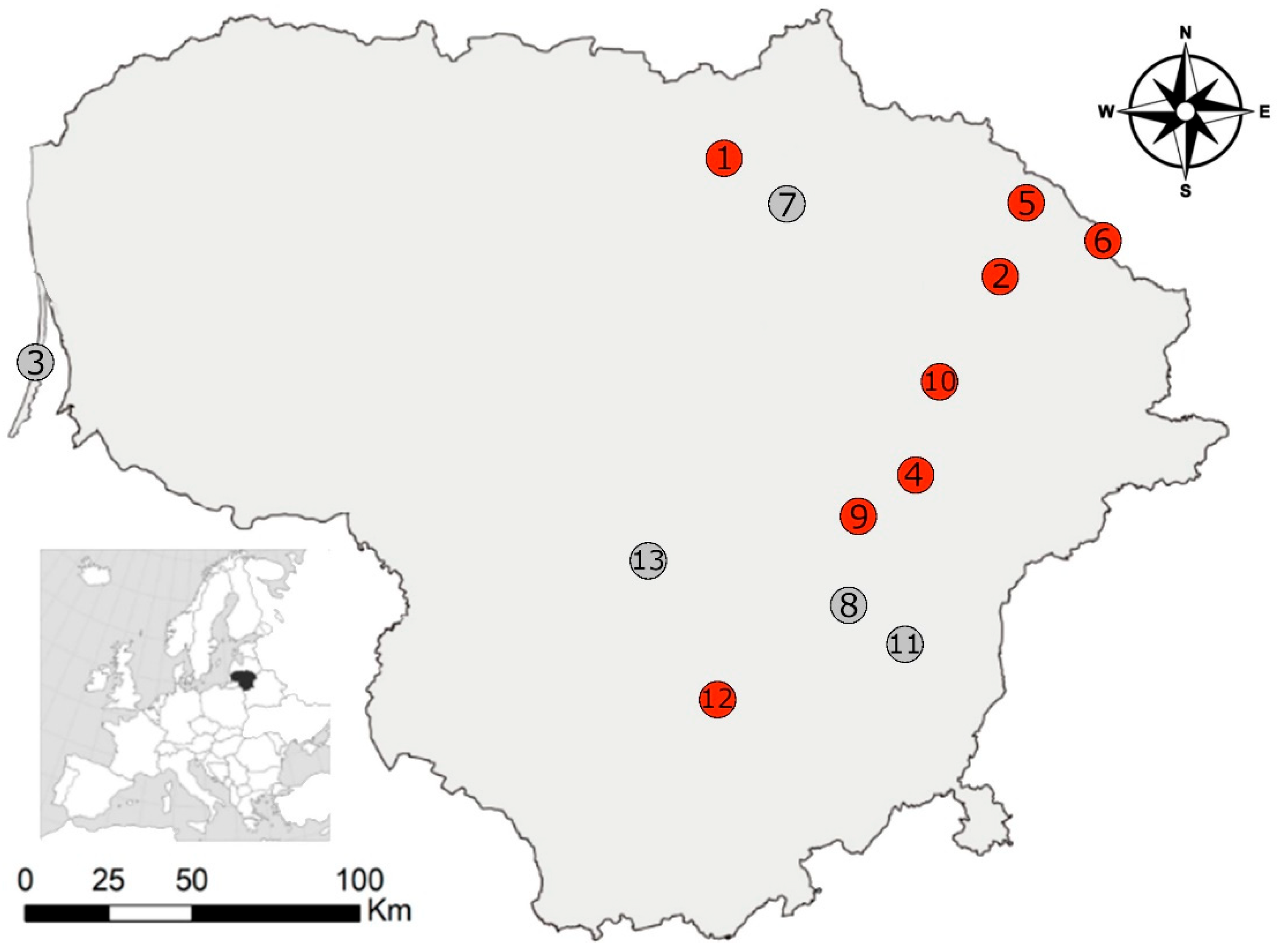
2.1. Trapping of Small Mammals

A standard snap trap line method (25 traps at 5 m intervals) was deployed to capture small mammals at 13 distinct locations within Lithuania during the autumn of the year 2023. In three cases, sites in close proximity were combined into one sample (Figure 1). Subsequently, the study analysed 694 individual small mammals representing 11 species: the striped field mouse (Apodemus agrarius), n = 5, the yellow-necked mouse (A. flavicollis), n = 2, C. glareolus, n = 374, M. agrestis, n = 14, M. arvalis, n = 144 the root vole (Alexandromys oeconomus), n = 11, the harvest mouse (Micromys minutus), n = 3, the house mouse (Mus musculus), n = 33, the water shrew (Neomys fodiens), n = 9, the common shrew (Sorex araneus), n = 45 and the pygmy shrew (S. minutus), n = 54. The first eight species belong to the order Rodentia, while the last three species are members of the order Eulipotyphla.

2.2. Morphological Examination of Sarcocystis spp. from Brain Tissues of Small Mammals

To determine the prevalence of Sarcocystis spp. in the brain of small mammals, fragments of brain tissue (~0.2 g) were stained with 0.2% methylene blue solution for 24 h. Then samples were cleared in 1.5% acetic acid solution for 45–50 min. Afterwards, samples were squeezed between glass compressors and studied under a light microscope (LM) at 40× and 100× magnification. The parasite load was evaluated by counting cysts in ~0.2 g of sample. Eventually, the cysts detected in brain samples were morphologically characterised in squashed fresh preparations under LM. Overall, 10 cysts were excreted from the brain from five individual bank voles (isolates CgLt189.1rp; CgLt189.2rp; CgLt388.1rp; CgLt388.2rp; CgLt.782.1rp; CgLt782.2rp; CgLt.963.1rp; CgLt963.2rp; CgLt1124.1rp; CgLt1124.2rp). Genomic DNA was extracted from the isolated individual cysts without delay.

2.3. Genetic Characterization of Sarcocystis spp. Detected in Small Mammals

The DNA isolation from sarcocysts was conducted using the GeneJET Genomic DNA Purification Kit (Thermo Fisher Scientific Baltics, Vilnius, Lithuania) according to the manufacturer’s recommendations. The partial 28S rRNA was amplified using KL-P1F/KL-P1R primer pairs (Table 1) [35]. Each PCR reaction was carried out in a 25 µL mixture containing 12.5 µL of DreamTaq PCR Master Mix (Thermo Fisher Scientific Baltics, Vilnius, Lithuania), 4 µL of DNA template, 0.5 µM of both forward and reverse primers and nuclease-free water. The PCR was initiated with the initial hot start at 95 °C for 5 min followed by 35 cycles of 94 °C for 45 s, annealing at 52 °C for 60 s and 72 °C for 80 s, and a final extension at 72 °C for 7 min. The visualisation, purification, and sequencing of PCR products followed the previously described protocol [36]. In order to detect essentially similar DNA sequences, and evaluate the interspecific and intraspecific genetic variability of detected Sarcocystis parasites, the 28S rRNA sequences generated in this study were compared with those of various Sarcocystis spp. using the nucleotide BLAST program (http://blast.ncbi.nlm.nih.gov/, accessed on 10 March 2024).

2.4. Collection of Buteo Buzzards

A total of 12 birds (10 B. buteo and two B. lagopus) were collected between 2017 and 2020. All birds, obtained from the Kaunas Tadas Ivanauskas Zoology Museum (the Lithuanian national authority responsible for monitoring dead birds), were found dead as a result of collisions with motor vehicles, power lines, buildings, etc., and were kept frozen at −20 °C until they were dissected.

2.5. Morphological Examination of Sarcocystis spp. from Intestines of Buteo Buzzards

Sarcocystis spp. oocysts/sporocysts were excreted from the entire intestine of each buzzard using a slightly modified method by Verma et al. [38]. Initially, the small intestine was removed from the bird. Then, the faeces from each intestine were squeezed, and the gut was cut lengthwise. The intestinal epithelium was scraped with the help of a scalpel and suspended in 50–100 mL of distilled water. The homogenization was performed in a commercial blender at top speed for 1–2 min with breaks to prevent frothing. The homogenate was centrifuged for 6 min at 1600 rpm, 25 °C in a 50 mL centrifuge tube. The supernatant was discarded, and the sediments were re-suspended in 50 mL water. The above-described process was repeated 5–8 times until most oocysts/sporocysts were released from the host tissue. Thereafter, the sporocyst pellet was suspended in HBSS and filtered through cheesecloth. The homogenate was emulsified in 5.25% sodium hypochlorite (bleach) solution (1:1 ratio) in a cold bath for 30 min. The centrifugation for 6 min at 1600 rpm, 25 °C, and removal of supernatant were repeated until the smell of bleach (chlorine) was gone. Eventually, the oocysts/sporocysts of Sarcocystis spp. were examined under LM at 400× and 1000× magnification. The 400 μL of re-suspended sediments were taken from each sample and used for DNA extraction.

2.6. The Genetic Identification of Sarcocystis spp. from Intestines of Buteo Buzzards

The DNA isolation from the mucosal suspension was performed using the GeneJET Genomic DNA Purification Kit (Thermo Fisher Scientific Baltics, Vilnius, Lithuania). The DNA samples were then stored frozen at −20 °C until further molecular analysis.
Sarcocystis spp. were identified by nested PCR of partial 28S rDNA sequences. In the first step of nested PCR, forward Sgrau281 and reverse Sgrau282 primers were used (Table 1). Whereas in the second step of nested PCR, two primer pairs, GsSglaF1/GsSglaR1 and GsSmicF1/GsSmicR1 were used. The primers of the second round of nested PCR were designed with the help of Primer3Plus program [39]. These primers were designed for amplification of S. glareoli and S. microti. However, due to relatively small variance of Sarcocystis spp. using rodents and birds as their intermediate and definitive hosts, respectively, within rRNA genes [40], it was suspected that GsSglaF1/GsSglaR1 and GsSmicF1/GsSmicR1 might not only be suitable for the amplification of S. glareoli and S. microti. Positive controls (DNA of S. glareoli extracted from single sarcocysts) were used in each set of PCRs. Three negative controls (nuclease free water instead of target DNA) were used: one control for the first amplification step and two controls for the second step of nested PCR. The third negative control was obtained by transferring 1 µL from the negative control of the first amplification step to the negative control of the second amplification step. PCRs were conducted under the conditions described above in Section 2.3 and using annealing temperatures provided in Table 1. For the second PCR assay, 1 μL from the first PCR assay was used.
The visualisation, purification, and sequencing of PCR products were carried out using the previously described protocol [36]. Our 28S rRNA sequences were compared with those of various Sarcocystis spp. with the Nucleotide BLAST program (megablast option) (http://blast.ncbi.nlm.nih.gov/, accessed on 10 March 2024). The 28S rDNA sequences of Sarcocystis spp. isolated from intestinal scrapings of Buteo buzzards were made available in GenBank with PP535696–PP535702 accession numbers.

2.7. Phylogenetic Analysis

Based on the molecular results, a putative new Sarcocystis species was identified in single sample of intestinal mucosa of B. lagopus, prompting further phylogenetic analysis. Phylogenetic relationships were reconstructed using the Maximum Likelihood (ML) method. Multiple alignments of partial 28S rRNA sequences of 20 Sarcocystis taxa was carried out using the MUSCLE algorithm incorporated into MEGA7 version 7.0.26 software [41]. The selection of the evolutionary model was conducted using MEGA7, based on the lowest Bayesian Information Criterion values generated. The robustness of the resulted phylogeny was tested using bootstrap test with 1000 replications.
Furthermore, phylogenetic analysis was performed to evaluate the relationships between the species identified in this study and closely related ones (S. glareoli, S. microti, S. jamaicensis and Sarcocystis sp.). A phylogenetic tree was constructed using the Maximum Likelihood algorithm, as described in the paragraph above. Additionally, phylogenetic relationships among the different sequences were inferred through coalescent simulations using a median-joining model implemented in NETWORK v. 10.2.0.0 [42].

2.8. Statistical Tests

Confidence intervals (95% CI) for the proportions of infection prevalence were estimated using online software [43]. Differences in infection prevalence between study sites were tested using the G-test on an online calculator [44]. The number of cysts in the samples was compared between the different study sites using the Student’s t-test in STATISTICA for Windows, version 6.0 (StatSoft, Inc., Tulsa, OK, USA). The level of significance was set at p < 0.05.

3. Results

3.1. Prevalence and Parasite Load of Sarcocystis spp. in Small Mammals

Cysts of Sarcocystis were observed exclusively in the brains of a single small mammal species, C. glareolus. The prevalence of infection reached 9.1% (34 infected of 374 tested, 95% CI = 6.4–12.5%). The infection rates varied among these animals across different study areas. Sarcocystis spp. were detected in rodents collected from eight out of the 13 localities, accounting for 61.5% of the sampled sites (Table 2). The highest detection rates of cysts of Sarcocystis were found in Kamasta, Zabarauskai, and Aukštikalniai. In other sampling sites the prevalence of Sarcocystis spp. ranged from 2.1% to 12.5%. The prevalence of Sarcocystis spp. infection was significantly different only between the two study sites, Kamasta and Lukštas (G = 7.4, p < 0.05).
The number of cysts in 0.2 g brain samples varied from 1 to 50, with an average of 13.4 ± 1.89 (average number of cysts ± standard error). We compared cyst counts across various locations, focusing on three areas where more than one infected individual was captured. The Kamasta study site exhibited the highest average number of cysts (18.0 ± 4.90), correlating with the highest prevalence of examined parasites. The average number of cysts in Kamasta was higher than in the Bileišiai site (11.3 ± 2.24) and the Utena site (9.6 ± 3.17), though the differences were not statistically significant (t = 1.45, p = 0.16 and t = 1.19, p = 0.26).

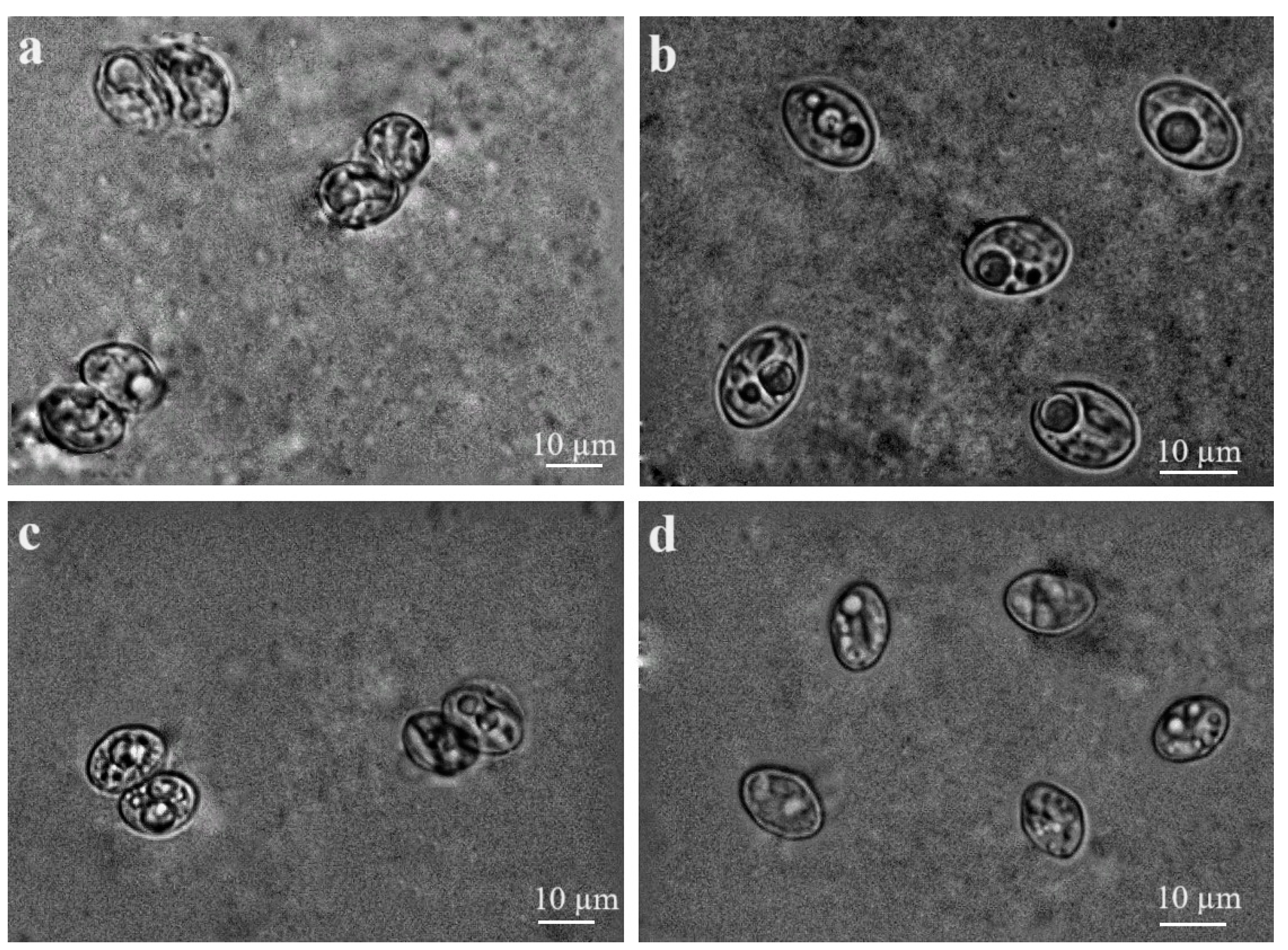
3.2. Morphological and Molecular Characterisation of Sarcocystis spp. in Brain Tissues of Small Mammals

The detailed morphological characterisation of Sarcocystis parasites observed were carried out in five infected C. glareolus. In fresh preparations, the detected cysts greatly varied in shape and size (Figure 2). Sarcocysts were microscopic, appearing as round structures measuring 102.7–279.8 × 102.1–332.8 µm (average size: 189.2 × 207.3 µm; n = 4) (Figure 2a), oval structures measuring 38.9–74.0 × 71.5–135.0 µm (average size: 63.3 × 102.7 µm; n = 4) (Figure 2b), or irregular round structures measuring 48.2–89.0 × 75.7–104.1 µm (average size: 73.6 × 94.0 µm; n = 4) (Figure 2c). Based on molecular analysis, sarcocysts isolated from brain tissues of C. glareolus in Lithuania were identified as S. glareoli.
Our analysis of ten 944 bp long 28S rRNA sequences of S. glareoli isolated from C. glareolus demonstrated 100% identity among them. These sequences have been deposited in GenBank under the accession number PP535695. Despite significant variation in the size and shape of the cysts, the 28S rRNA region tested showed no differences among the S. glareoli isolates.
The 944 bp sequences of S. glareoli obtained in this study exhibited 99.9% identity to S. glareoli sequence AF044251. In our sequences, the C insertion was noticed in 537 nucleotide position to be compared (in TAGGTCCCCCG region, insertion is bolded and underlined). Notably, numerous other related Sarcocystis spp. had the C insertion in a homologous nucleotide position indicating highly probable sequencing error in AF044251 GenBank record.
Additionally, our 28S rRNA sequences of S. glareoli differed from those of S. jamaicensis (KY994650) by two single nucleotide polymorphisms (SNPs), and displayed 98.7% similarity with S. microti (AF044252), and 98.2% similarity with S. wobeseri (LR884239), which forms sarcocysts in muscles of birds [35]. Notably, B. jamaicensis serves as the definitive host and knockout mouse was shown to be laboratory host of S. jamaicensis [40,45].

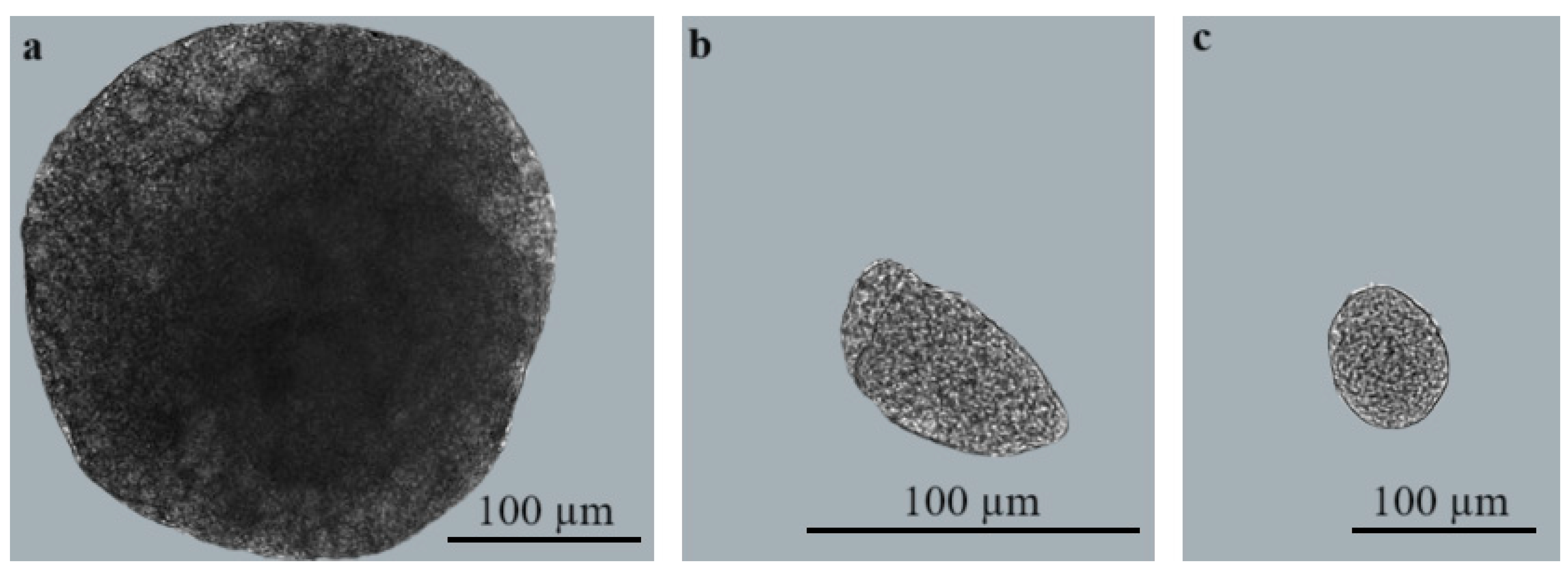
3.3. Morphological Characterisation of Sarcocystis spp. Sporocysts/Oocysts Found in Intestines of Buteo Buzzards

Both examined B. lagopus harboured sporocysts and sporulated oocysts of Sarcocystis spp. (2/2, 100%). Meanwhile, Sarcocystis spp. sporocysts and/or sporulated oocysts were detected in eight of the ten investigated B. buteo (80%). No oocysts were detected in either host species tested.
Upon microscopical analysis, the free sporocysts of Sarcocystis spp. in mucosal scrapings of B. lagopus measured 12.6 × 8.4 μm (range: 10.7–15.2 × 5.5–9.6 μm; n = 55) (Figure 3a), while sporulated oocysts, each containing two sporocysts, measured 12.3 × 18.8 μm (range: 10.6–13.4 × 18.1–19.5; n= 15) in size (Figure 3b). Sporocysts of Sarcocystis spp. in B. buteo measured 13.0 × 8.9 μm (range: 10.1–15.9 × 7.1–11.0; n = 118) (Figure 3c), while sporulated oocysts were 13.0 × 19.0 (range: 11.1–16.3 × 16.1–25.2; n = 59) (Figure 3d).
The examination of sporocysts and sporulated oocysts found in the intestines of B. lagopus and B. buteo indicated that morphological parameters of Sarcocystis spp. overlapped in size.

3.4. Molecular Investigation of Sarcocystis spp. Detected in Intestines of Buteo Hawks

When analysing samples of DNA extracted from scrapings of the intestinal mucosa of two hawk species, amplification was unsuccessful using GsSmicF1 and GsSmicR1 primers in the second round of nested PCR. Out of the 10 examined intestinal mucosa samples from B. buteo, five tested positive (50.0%) by nested PCR using GsSglaF1 and GsSglaR1 as internal primers. The resulting five sequences of 28S rRNA without primer binding sites were 515 bp long. These sequences showed 100% identity with each other and to those of S. glareoli from C. glareolus from Lithuania.
Based on BLAST analysis, the 28S rRNA sequences obtained in this study differed from those of S. jamaicensis (KY994650) by two T/G and A/G SNPs (99.6% similarity), and from those of S. microti (AF044252) by four SNPs and single indel (99.0% similarity), and by ≥2.3% from those of other Sarcocystis spp. available in GenBank. Therefore, molecular investigation confirmed the presence of S. glareoli in the intestinal mucosa of five common buzzards.
Sequencing of the 28S rRNA fragment amplified by the GsSglaF1/GsSglaR1 primer pair, both B. lagopus were positive for Sarcocystis parasites. Comparison of the analysed 515 bp long and 522 bp long sequences revealed the presence of two different Sarcocystis species, Sarcocystis sp. Rod3 and Sarcocystis sp. Rod4.
The BLAST comparison showed that the 515 bp long sequence of Sarcocystis sp. Rod3 differed from those of S. jamaicensis (KY994650), S. glareoli (AF044251) and S. microti (AF044252), by one C/G SNP, by two SNPs and single indel, and by three SNPs and single indel, respectively. Based on investigated 28S rRNA fragment, Sarcocystis sp. Rod4 notably differed from Sarcocystis spp. forming cysts in the brains of rodents (94.1–94.6% similarity). The 522 bp long sequence of Sarcocystis sp. Rod4 displayed 96.4% similarity with that of S. cf. strixi (OQ557459) from A. flavicollis, 96.2% similarity with that of S. funereus (MW349707), and 95.8% similarity with that of S. strixi (MF162316).
It is worth noting that the experimental intermediate host of S. funereus and S. strixi are laboratory mouse, while owls serve as definitive hosts of these Sarcocystis species [7,46,47].
Our 515 bp long 28S rRNA sequences of S. glareoli and Sarcocystis sp. Rod3 exhibited significant similarity to 299–316 bp long sequences (OK576420, OK576425, OK576428-36, OK576438-43, OK576447-49, OK576451, OK576456, OK576459, OK576461-62) of Sarcocystis parasite obtained from intestinal samples of two hosts, B. jamaicensis and red-shouldered hawks (Buteo lineatus) from the USA. This Sarcocystis parasite, as identified by Rogers et al. (2022), was classified as S. jamaicensis [48]. However, in this study, we refer to it as Sarcocystis sp. Based on 28S rRNA sequences, S. glareoli and Sarcocystis sp. Rod3 shared 97.0–99.7% and 97.0–100% similarity with Sarcocystis sp. isolated from B. jamaicensis, and B. lineatus.

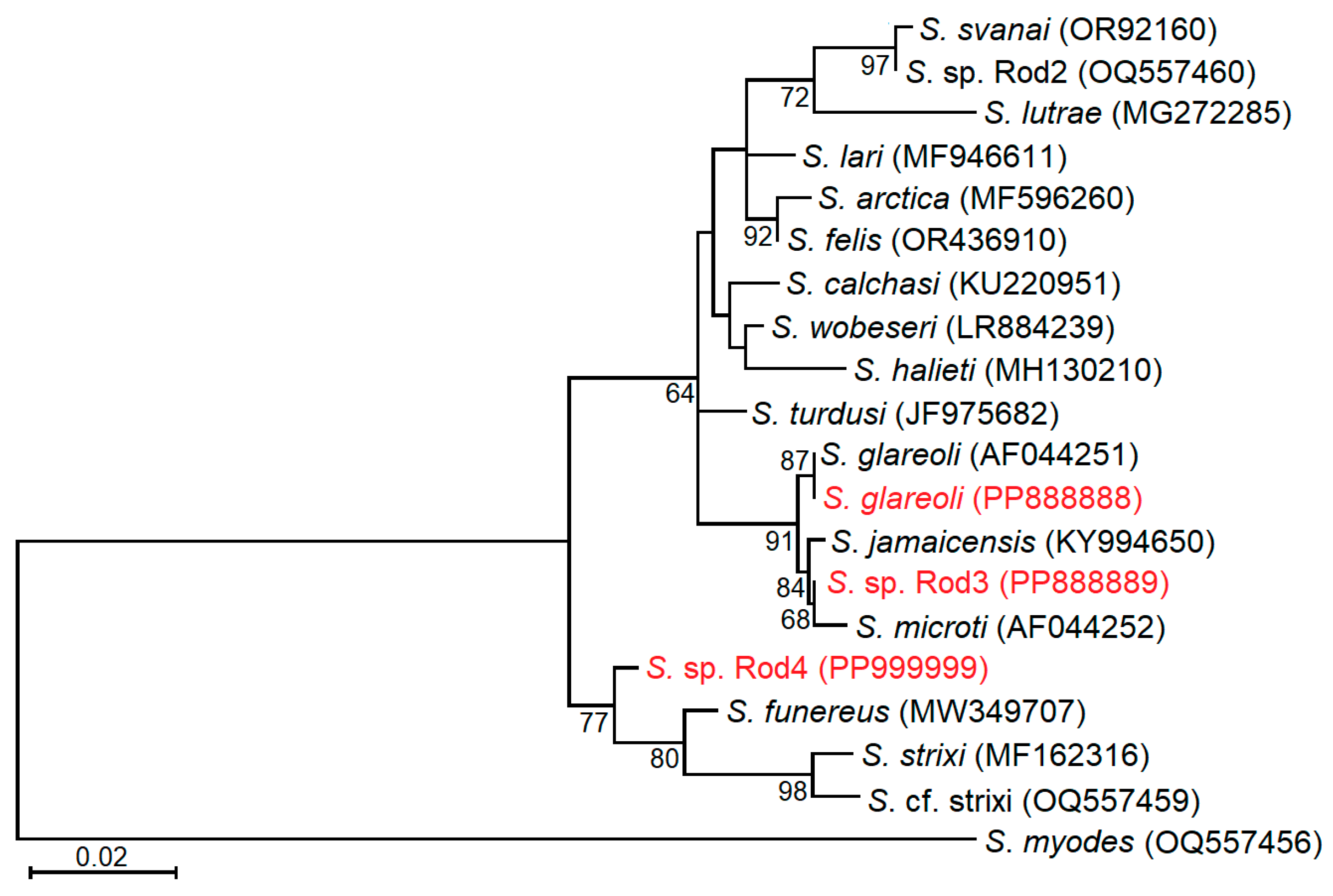
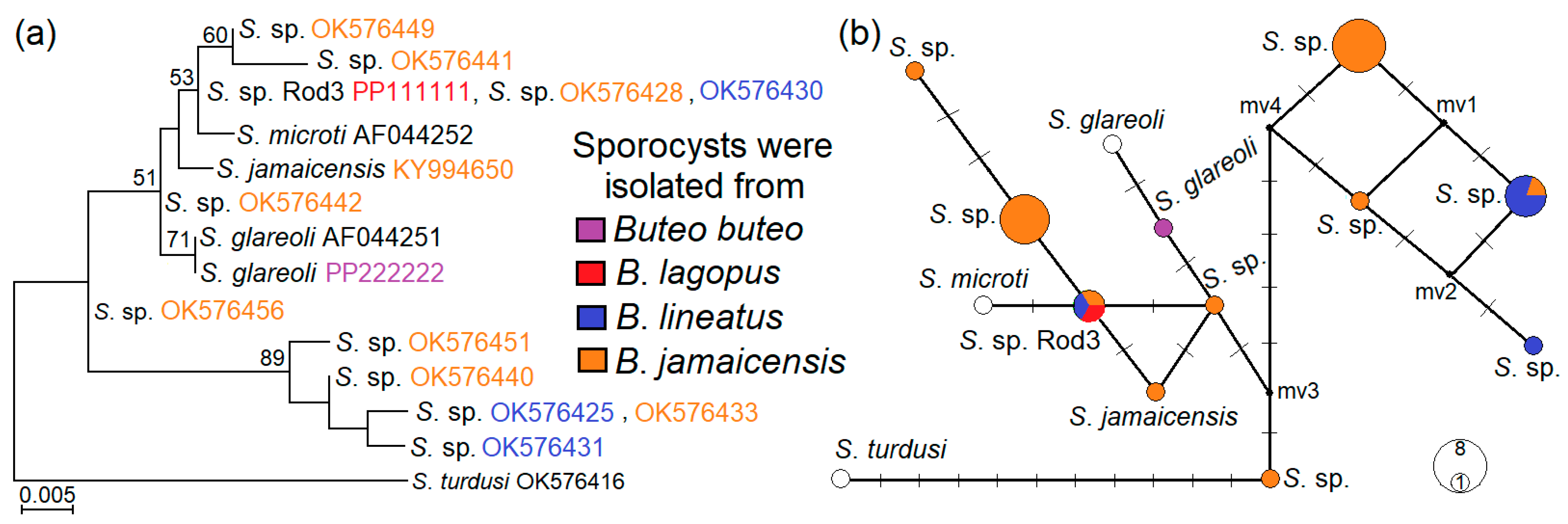
3.5. Phylogenetic Relationships of Examined Sarcocystis Species

Based on the 28S rRNA fragment amplified with GsSglaF1/GsSglaR1 primers, the three Sarcocystis species identified in this study were categorized into two clusters, alongside Sarcocystis species utilizing rodents as intermediate hosts and birds of prey as definitive hosts (Figure 4). Our sequences of S. glareoli grouped together with the sequence of S. glareoli taken from the GenBank AF044251. Sarcocystis sp. Rod3 was found to be most closely related to S. jamaicensis and S. microti. However, the grouping of Sarcocystis sp. Rod3 with S. microti was not well supported (68 bootstrap value). Notably, the branch lengths within the clade comprising S. glareoli, S. jamaicensis, Sarcocystis sp. Rod3 and S. microti was shorter compared to that established in another clade consisting of Sarcocystis spp. using rodents and birds of prey as their intermediate and final hosts. The following clade included S. funereus, S. strixi, S. cf. strixi and Sarcocystis sp. Rod4 from B. lagopus. Sarcocystis funereus formed a sister branch to S. strixi and S. cf. strixi, while Sarcocystis sp. Rod4 formed a sister branch to the aforementioned taxa.
We conducted another phylogenetic analysis by comparing our S. glareoli and Sarcocystis sp. rod3 28S rRNA sequences with those of Sarcocystis sp. from Rogers et al. [48] as well as those of S. glareoli (AF044251), S. microti (AF044252) and S. jamaicensis (KY994650). Out of the 32 analysed sequences, 13 haplotypes were identified, and four haplotypes (sequences: OK576420, OK576425, OK576431, OK576432, OK576433, OK576435, OK576436, OK576440, OK576447, OK576451, OK576459, OK576461, OK576462, OK576464, OK576465) were clustered together with a high support value of 89 (Figure 5a). These four haplotypes were separated from the remaining ones by at least six mutations (Figure 5b) and presumably belonged to a single Sarcocystis species isolated from B. jamaicensis and B. lineatus. However, clustering of other haplotypes belonging to S. glareoli and Sarcocystis sp. rod3, S. jamaicensis, S. microti and remaining haplotypes of Sarcocystis sp. (OK576428, OK576429, OK576430, OK576434, OK576438, OK576439, OK576441, OK576442, OK576443, OK576448, OK576449, OK576456) was not well defined. Thus, the variability of the examined fragment was not sufficient to reveal how many distinct taxa comprised the nine haplotypes. It also should be pointed out that at 307–308 bp 28S rRNA fragment, Sarcocystis sp. rod3 showed 100% identity to OK576428 and OK576430 sequences of Sarcocystis sp. from two Buteo buzzard species from the USA.

4. Discussion

4.1. Sarcocystis Parasites in the Brain Tissues of Rodents

A previous study conducted in Lithuania reported that 72 out of 341 (21.1%) of the analysed C. glareolus had cysts of S. glareoli in their brains [28]. However, in the present examination, the prevalence of S. glareoli was significantly lower (p < 0.001), as only 9.1% (95% CI = 6.4–12.5) of the studied C. glareolus specimens were infected.
According to published data from other European countries, the infection rates of S. glareoli in the C. glareolus ranged from 1.0% to 47.3% [23,24,29,31,32,33]. The prevalence of S. glareoli identified in our study is consistent with data from one study in Germany, which reported that 10.3% (45/445) of studied C. glareolus were positive for S. glareoli [33]. The highest infection rates of S. glareoli in C. glareolus were reported in France (47.3%, 178/376) [23] and the Czech Republic [29], followed by a study in Germany (22.6%, 59/257) [24]. Meanwhile, the lowest prevalence was established in another study conducted in France (1.0%, 1/98) [32] and in a study from the UK (6.3%, 1/16) [31]. Thus, the detection rates of this parasite in C. glareolus vary considerably even within the same countries.
In this study, no cysts of S. microti were detected, despite its previous detection in Lithuania. Formerly, S. microti was identified in two rodent species; however, the prevalence of this parasite was low, with rates of 2.9% (1/35) in A. oeconomus and 4.2% (1/24) in M. agrestis [28]. Two comprehensive investigations (n > 1700) carried out in Europe determined a lower prevalence of S. microti compared to S. glareoli [23,29]. In the study conducted in France, cysts of S. microti were found in M. agrestis, M. arvalis, and C. glareolus, with prevalence ranging from 1.0% to 9.2% in each species [23]. Meanwhile, in the Czech Republic, S. microti was detected in M. arvalis, C. glareolus, and Apodemus sp., with infection rates ranging from 0.6% to 5.0% in all tested species [29]. Notably, S. microti was detected in 20.7% (6/29) of Bedford’s red-backed voles (Clethrionomys rufocanus bedfordiae) in Japan [30].
There are multiple reports that the prevalence of Sarcocystis spp. in small mammals is affected by seasonal changes. A study on Norwegian lemmings (Lemmus lemmus) concluded that S. microti infestation of intermediate hosts depends on the season, with the highest number of parasites found in summer (9%) and the lowest in autumn (0–4%) [27]. Furthermore, research conducted in the Netherlands analysed seasonal dynamics of S. cernae infection in the intermediate host M. arvalis and definitive host the common kestrel (Falco tinnunculus) and concluded that prevalence of S. cernae varies throughout the seasons and was highest in the spring [49]. Similar results were obtained in a study in France, where a variety of small mammals belonging to genera Myodes, Arvicola, Microtus, Apodemus and Sorex were investigated [23]. In our study, rodents were collected in autumn when lower infection rates of Sarcocystis spp. are observed based on aforementioned research. In the previous study conducted in Lithuania [28] rodents were collected not only in autumn, but also in spring and summer thus probably leading to the detection of higher total prevalence of Sarcocystis spp. in the brains of rodents.

4.2. Buteo Buzzards as Definitive Hosts of Sarcocystis spp.

Although detailed studies on S. glareoli and S. microti have been carried out in intermediate hosts [23,28,29], these parasites have been scarcely studied in their natural definitive hosts. Based on morphological examination, “Frenkelia-like” sporocysts were detected in the droppings and intestines of Buteo buzzards in Europe [29,50] and North America [26,51,52]. Without molecular analysis, it is not possible to ascertain whether the sporocysts discovered in the above studies belonged to S. glareoli and/or S. microti, which form cysts in the brains of rodents, or to some other Sarcocystis spp. This is because the morphometrical parameters of sporocysts of many different species of Sarcocystis overlap [1,52].
Due to the lack of data, the prevalence of Sarcocystis spp. sporocysts in different Buteo species, geographical regions, seasons, etc. is still generally unknown. A detailed study conducted in the Czech Republic revealed that the prevalence of Sarcocystis spp. in faeces from nests of B. buteo ranged from 28.3% to 88.6%, depending on the season and locality [29]. Another study performed in North America examined droppings from red-tailed hawks (Buteo borealis) in a rehabilitation centre and determined that half of them contained Sarcocystis spp. sporocysts [52]. In this work, by microscopical examination, 80% of B. buteo and both analysed B. lagopus were infected with oocysts/sporocysts of Sarcocystis spp. Based on 28S rRNA sequence analysis, S. glareoli was confirmed in B. buteo and two Sarcocystis species (Sarcocystis sp. Rod3 and Sarcocystis sp. Rod4) were detected in B. lagopus. Thus, our results indicate that Buteo buzzards in Europe can transmit not only S. glareoli and S. microti, previously assigned to the genus Frenkelia.
The common buzzard nests across nearly the entire European region within the Western Palearctic [53]. This migratory species breeds in Lithuania and is commonly found throughout the country, although it rarely stays for the winter season. It nests in woodlands and small clusters of trees near cultivated fields, displaying remarkable adaptability in choosing its breeding sites [54]. A wide array of prey is consumed, encompassing small mammals, birds, reptiles, amphibians, larger insects, and earthworms [55]. The primary portion (38.3%) of B. buteo diet consists of small mammals, predominantly voles [54,56]. Studies conducted in Poland indicate that these raptors mostly catch common voles [57]. Of the five B. buteo in which S. glareoli were identified, only two of them have a known collection date (February and September). Taking into account that definitive hosts excrete sporocysts of Sarcocystis spp. up to several months [1] and based on migratory data, birds collected in February and September were most likely infected with S. glareoli in Lithuania.
Another species, B. lagopus, only winters in Lithuania. This buzzard is distributed in the northern part of the Western Palearctic. Its range stretches eastward, predominantly across the tundra belt, reaching the Pacific Ocean. From there, it tracks along the coastal areas southward to Sakhalin and extends across the northern reaches of North America on the opposite side of the Pacific [53]. The rough-legged buzzard predominates in the diet, mainly small rodents, accounting typically for 80–95% of the food, where they occur in large numbers [52,54]. In Lithuania, ringed B. lagopus chicks were found in central Europe: Slovakia, Hungary, France, and Italy [54]. In autumn and winter in Lithuania, the ringed B. lagopus come from Norway, Finland, and mostly from Sweden [54]. Both of B. lagopus examined in current work were collected in November of 2018. Based on ecological data, infected B. lagopus were migrants most likely from Scandinavia [54]. It is likely that these buzzards were infected with Sarcocystis sp. Rod3 and Sarcocystis sp. Rod4 before entering Lithuania.

4.3. Genetic Identification of Sarcocystis sp. Rod3 and Sarcocystis sp. Rod4

Based on phylogenetic analysis using 28S rRNA, Sarcocystis sp. Rod3 was found to be closely related to three Sarcocystis species, S. glareoli, S. microti and S. jamaicensis (Figure 4 and Figure 5). These Sarcocystis species produce sarcocysts with thin and smooth cyst walls [1,40]. The natural intermediate host of S. jamaicensis is unknown. However, laboratory experiments have shown that this species circulates between rodents and B. jamaicensis [40,45]. Two IFN-c gene knockout mice orally infected with sporocysts of S. jamaicensis had meningoencephalitis associated with schizonts and merozoites of this parasite, while sarcocysts were detected in the muscles of the animals [45]. Thus, the developmental stages of these three species, S. glareoli, S. microti and S. jamaicensis, are found in the brain. The detection of Sarcocystis sp. Rod3 in the intestine of a single B. lagopus collected in Lithuania suggests that it might be another species in Europe, apart from S. glareoli and S. microti, which can infect rodent brains.
Also, our work demonstrated that 28S rRNA locus is not sufficiently variable for the reliable discrimination of S. glareoli, S. microti, S. jamaicensis and Sarcocystis sp. Rod3. To date, only sequences of 18S rRNA and 28S rRNA of S. glareoli and S. microti are available in GenBank [15,16], while S. jamaicensis has been characterised within 18S rRNA, 28S rRNA, mitochondrial cytochrome oxidase 1 (cox1) and ITS1. The ITS1 genetic marker has been proven to be the best choice for the separation of Sarcocystis species using birds and predatory mammals as their intermediate hosts [58,59]. Although this genetic region has not been commonly applied for the characterization of Sarcocystis spp. from rodents, it has shown more variability compared to cox1 [9,13,59]. In contrast, cox1 is relatively conservative considering Sarcocystis spp. parasitizing small mammals [59]. Therefore, the highly variable ITS1 can be helpful for the distinguishing the aforementioned Sarcocystis species.
Furthermore, the results of the current study and that described by Rogers et al. [48] indicate that different species of the genus Buteo from North America and Europe can transmit the same Sarcocytis species. Intriguingly, based on short at 307–308 bp 28S rRNA sequences, Sarcocystis sp. Rod3 was identified in three different Buteo species. Nevertheless, further studies are needed to clarify the specificity of Sarcocystis species for Buteo buzzards as their definitive hosts.
Sarcocystis sp. Rod 4, identified in a single B. lagopus, phylogenetically clustered with S. funereus, S. strixi, and S. cf. strixi (Figure 4). Two owl species, the Tengmalm’s owl (Aegolius funereus) and the barred owl (Strix varia), are proven definitive hosts of S. funereus [47] and S. strixi [46], respectively. Sarcocysts of these two Sarcocystis species have not yet been discovered in natural intermediate hosts. However, recently the DNA of S. funereus has been detected in blood sample of a single C. glareolus from Lithuania [60]. There is a lack of investigations on the role of the Strigiformes in the transmission of Sarcocystis spp. [37,46,47]. Even so, the close relationships of Sarcocystis sp. Rod 4 to S. funereus and S. strixi imply that Sarcocystis species using Accipitriformes and Strigiformes as their definitive hosts might not have evolved separately. Further examinations are needed to clarify whether the same Sarcocystis species can employ representatives of both orders Accipitriformes and Strigiformes as their definitive hosts.

5. Conclusions

After examining brain samples from 694 small mammals belonging to eight Rodentia and three Eulipotyphla species, cysts of S. glareoli were observed in only 9.1% of the C. glareolus investigated. The literature suggests that this molecularly confirmed Sarcocystis species is more common in Europe compared to another species, S. microti, which also forms cysts in the brains of small mammals. The 28S rRNA sequence analysis confirmed the presence of S. glareoli in half of the B. buteo examined, indicating that these predatory birds are natural definitive hosts of S. glareoli in Lithuania. Our results also highlight the necessity of using other genetic markers, such as ITS1, to distinguish between Sarcocystis spp. parasitizing the brains of small mammals.
Based on molecular examination, two species were identified in B. lagopus: Sarcocystis sp. Rod3, which is closely related to S. jamaicensis, S. glareoli and S. microti, and a putative new species, Sarcocystis sp. Rod4. Ecological data suggest that B. lagopus were likely infected outside Lithuania. Overall, our work underscores the significant role of Buteo buzzards in the transmission of Sarcocystis parasites that produce cysts in small mammals.

Author Contributions

Conceptualization, P.P., M.J. and D.B.; methodology, P.P., L.B. and D.B.; software, P.P. and M.J.; validation, P.P., T.Š. and E.J.-N.; formal analysis, P.P., M.J., T.Š. and E.J.-N.; investigation, M.J., T.Š., E.J.-N., V.S. and L.B.; resources, L.B. and D.B.; data curation, M.J., T.Š. and V.S.; writing—original draft preparation, P.P., M.J., E.J.-N., V.S. and L.B.; writing—review and editing, P.P., M.J., T.Š., E.J.-N., V.S., L.B. and D.B.; visualization, P.P., M.J., T.Š. and E.J.-N.; supervision, D.B.; project administration, P.P.; funding acquisition, P.P. All authors have read and agreed to the published version of the manuscript.

Funding

The research was funded by the Research Council of Lithuania (grant number S-MIP-23-4).

Institutional Review Board Statement

The study was conducted in accordance with Lithuanian legislation (the Republic of Lithuania Law on the Welfare and Protection of Animals No. XI-2271, “Requirements for the Housing, Care and Use of Animals for Scientific and Educational Purposes”, approved by Order No B1-866, 31 October 2012 of the Director of the State Food and Veterinary Service (Paragraph 4 of Article 16) and European legislation (Directive 2010/63/EU) on the protection of animals, and was approved by the Animal Welfare Committee of the Nature Research Centre, protocol No GGT-8, issued 21 March 2021).

Informed Consent Statement

Not applicable.

Data Availability Statement

The 28S rDNA sequences of Sarcocystis spp. generated in the present study were submitted to the GenBank database under PP535695–PP535702.

Acknowledgments

We thank Ida Šaltenienė and Kristijonas Janulaitis for their help with trapping small mammals.

Conflicts of Interest

The authors declare no conflicts of interest. Funders had no role in the design of the study, in the collection, analyses or interpretation of data, in the writing of the manuscript, and in the decision to publish the results.

References

  1. Dubey, J.P.; Calero-Bernal, R.; Rosenthal, B.M.; Speer, C.A.; Fayer, R. Sarcocystosis of Animals and Humans, 2nd ed.; CRC Press: Boca Raton, FL, USA, 2016. [Google Scholar]
  2. Gjerde, B.; Vikøren, T.; Hamnes, I.S. Molecular Identification of Sarcocystis halieti n. sp., Sarcocystis lari and Sarcocystis truncata in the Intestine of a White-Tailed Sea Eagle (Haliaeetus albicilla) in Norway. Int. J. Parasitol. Parasites Wildl. 2018, 7, 1–11. [Google Scholar] [CrossRef]
  3. Miller, M.A.; Barr, B.C.; Nordhausen, R.; James, E.R.; Magargal, S.L.; Murray, M.; Conrad, P.A.; Toy-Choutka, S.; Jessup, D.A.; Grigg, M.E. Ultrastructural and Molecular Confirmation of the Development of Sarcocystis neurona Tissue Cysts in the Central Nervous System of Southern Sea Otters (Enhydra lutris nereis). Int. J. Parasitol. 2009, 39, 1363–1372. [Google Scholar] [CrossRef]
  4. Gryz, J.; Krauze-Gryz, D. Changes in the Tawny Owl Strix aluco Diet Along an Urbanisation Gradient. Biologia 2019, 74, 279–285. [Google Scholar] [CrossRef]
  5. Viteri, M.C.; Stegner, M.A.; Hadly, E.A. Assessing the Reliability of Raptor Pellets in Recording Local Small Mammal Diversity. Quaternary Res. 2022, 106, 1–10. [Google Scholar] [CrossRef]
  6. Hu, J.; Sun, J.; Guo, Y.; Zeng, H.; Zhang, Y.; Tao, J. Infection of the Asian Gray Shrew Crocidura attenuata (Insectivora: Soricidae) with Sarcocystis attenuati n. sp. (Apicomplexa: Sarcocystidae) in China. Parasit. Vectors 2022, 15, 13. [Google Scholar] [CrossRef]
  7. Máca, O.; Kouba, M.; Langrová, I.; Panská, L.; Korpimäki, E.; González-Solís, D. The Tengmalm’s Owl Aegolius funereus (Aves, Strigidae) as the Definitive Host of Sarcocystis funereus sp. nov. (Apicomplexa). Front. Vet. Sci. 2024, 11, 1356549. [Google Scholar] [CrossRef]
  8. Jäkel, T.; Raisch, L.; Richter, S.; Wirth, M.; Birenbaum, D.; Ginting, S.; Khoprasert, Y.; Mackenstedt, U.; Wassermann, M. Morphological and Molecular Phylogenetic Characterization of Sarcocystis kani sp. nov. and Other Novel, Closely Related Sarcocystis spp. Infecting Small Mammals and Colubrid Snakes in Asia. Int. J. Parasitol. Parasites Wildl. 2023, 22, 184–198. [Google Scholar] [CrossRef]
  9. Qin, T.; Ortega-Perez, P.; Wibbelt, G.; Lakim, M.B.; Ginting, S.; Khoprasert, Y.; Wells, K.; Hu, J.; Jäkel, T. A Cyst-Forming Coccidian with Large Geographical Range Infecting Forest and Commensal Rodents: Sarcocystis muricoelognathis sp. nov. Parasit. Vectors 2024, 17, 135. [Google Scholar] [CrossRef]
  10. Rudaitytė-Lukošienė, E.; Jasiulionis, M.; Balčiauskas, L.; Prakas, P.; Stirkė, V.; Butkauskas, D. Morphological and Molecular Description of Sarcocystis myodes n. sp. from the Bank Vole (Clethrionomys glareolus) in Lithuania. Biology 2022, 11, 512. [Google Scholar] [CrossRef]
  11. Prakas, P.; Kirillova, V.; Gavarāne, I.; Grāvele, E.; Butkauskas, D.; Rudaitytė-Lukošienė, E.; Kirjušina, M. Morphological and Molecular Description of Sarcocystis ratti n. sp. from the Black Rat (Rattus rattus) in Latvia. Parasitol. Res. 2019, 118, 2689–2694. [Google Scholar] [CrossRef]
  12. Ortega Pérez, P.; Wibbelt, G.; Brinkmann, A.; Galindo Puentes, J.A.; Tuh, F.Y.Y.; Lakim, M.B.; Nitsche, A.; Wells, K.; Jäkel, T. Description of Sarcocystis scandentiborneensis sp. nov. from Treeshrews (Tupaia minor, T. tana) in Northern Borneo with Annotations on the Utility of COI and 18S rDNA Sequences for Species Delineation. Int. J. Parasitol. Parasites Wildl. 2020, 12, 220–231. [Google Scholar] [CrossRef]
  13. Bentancourt Rossoli, J.V.; Moré, G.; Soto-Cabrera, A.; Moore, D.P.; Morrell, E.L.; Pedrana, J.; Scioli, M.V.; Campero, L.M.; Basso, W.; Hecker, Y.P.; et al. Identification of Sarcocystis spp. in Synanthropic (Muridae) and Wild (Cricetidae) Rodents from Argentina. Parasitol. Res. 2024, 123, 31. [Google Scholar] [CrossRef]
  14. Geisel, O.; Kaiser, E.; Vogel, O.; Krampitz, H.E.; Rommel, M. Pathomorphologic Findings in Short-Tailed Voles (Microtus agrestis) Experimentally-Infected with Frenkelia microti. J. Wildl. Dis. 1979, 15, 267–270. [Google Scholar] [CrossRef]
  15. Votypka, J.; Hypša, V.; Jirku, M.; Flegr, J.; Vavra, J.; Lukes, J. Molecular Phylogenetic Relatedness of Frenkelia spp. (Protozoa, Apicomplexa) to Sarcocystis falcatula Stiles 1893: Is the Genus Sarcocystis Paraphyletic? J. Eukaryot. Microbiol. 1998, 45, 137–141. [Google Scholar] [CrossRef]
  16. Mugridge, N.B.; Morrison, D.A.; Johnson, A.M.; Luton, K.; Dubey, J.P.; Votýpka, J.; Tenter, A.M. Phylogenetic Relationships of the Genus Frenkelia: A Review of its History and New Knowledge Gained from Comparison of Large Subunit Ribosomal Ribonucleic Acid Gene Sequences. Int. J. Parasitol. 1999, 29, 957–972. [Google Scholar] [CrossRef]
  17. Mugridge, N.B.; Morrison, D.A.; Jäkel, T.; Heckeroth, A.R.; Tenter, A.M.; Johnson, A.M. Effects of Sequence Alignment and Structural Domains of Ribosomal DNA on Phylogeny Reconstruction for the Protozoan Family Sarcocystidae. Mol. Biol. Evol. 2000, 17, 1842–1853. [Google Scholar] [CrossRef]
  18. Modrý, D.; Votýpka, J.; Svobodová, M. Note on the Taxonomy of Frenkelia microti (Findlay & Middleton, 1934) (Apicomplexa: Sarcocystidae). Syst. Parasitol. 2004, 58, 185–187. [Google Scholar] [CrossRef]
  19. Odening, K. The Present State of Species-Systematics in Sarcocystis Lankester, 1882 (Protista, Sporozoa, Coccidia). Syst. Parasitol. 1998, 41, 209–233. [Google Scholar] [CrossRef]
  20. Laakkonen, J.; Henttonen, H. Ultrastructure of Frenkelia sp. from a Norwegian Lemming in Finland. J. Wildl. Dis. 2000, 36, 362–366. [Google Scholar] [CrossRef]
  21. Karstad, L. Toxoplasma Microti (The M-Organism) in the Muskrat (Ondatra Zibethica). Can. Vet. J. 1963, 4, 249–251. [Google Scholar]
  22. Kennedy, M.J.; Frelier, P.F. Frenkelia sp. from the Brain of a Porcupine (Erethizon Dorsatum) from Alberta, Canada. J. Wildl. Dis. 1986, 22, 112–114. [Google Scholar] [CrossRef] [PubMed]
  23. Fichet-Calvet, E.; Kia, E.B.; Giraudoux, P.; Quéré, J.P.; Delattre, P.; Ashford, R.W. Frenkelia Parasites in a Small Mammal Community. Dynamics of Infection and Effect on the Host. Parasite 2004, 11, 301–310. [Google Scholar] [CrossRef]
  24. Krücken, J.; Blümke, J.; Maaz, D.; Demeler, J.; Ramünke, S.; Antolová, D.; Schaper, R.; Von Samson-Himmelstjerna, G. Small Rodents as Paratenic or Intermediate Hosts of Carnivore Parasites in Berlin, Germany. PLoS ONE 2017, 12, e0172829. [Google Scholar] [CrossRef]
  25. Cabral, A.D.; Su, C.; Soares, R.M.; Gennari, S.M.; Sperança, M.A.; Da Rosa, A.R.; Pena, H.F.J. Occurrence and Diversity of Sarcocystidae Protozoa in Muscle and Brain Tissues of Bats from São Paulo State, Brazil. Int. J. Parasitol. Parasites Wildl. 2021, 14, 91–96. [Google Scholar] [CrossRef] [PubMed]
  26. Upton, S.J.; McKown, R.D. The Red-Tailed Hawk, Buteo jamaicensis, a Native Definitive Host of Frenkelia microti (Apicomplexa) in North America. J. Wildl. Dis. 1992, 28, 85–90. [Google Scholar] [CrossRef]
  27. Laakkonen, J.; Haukisalmi, V.; Niemimaa, J.; Henttonen, H. Parasite Diversity of Norwegian Lemmings (Lemmus Lemmus). J. Zool. 2001, 253, 549–553. [Google Scholar] [CrossRef]
  28. Grikienienė, J.; Mažeikytė, R.; Balčiauskas, L. The First Data on Brain Parasites of the Genus Frenkelia (Protista: Coccidia) in Some Small Rodent Species in Lithuania. Acta Zoo. Litu. 2003, 13, 21–27. [Google Scholar] [CrossRef]
  29. Svobodová, M.; Vorisek, P.; Votypka, J.; Weidinger, K. Heteroxenous coccidia (Apicomplexa: Sarcocystidae) in the Populations of their Final and Intermediate Hosts: European Buzzard and Small Mammals. Acta Protozool. 2004, 43, 251–260. [Google Scholar]
  30. Fujita, O.; Oku, Y.; Ohbayashi, M. Frenkelia sp. from The Red-Backed Vole, Clethrionomys Rufocanus Bedfordiae, in Hokkaido, Japan. Jpn. J. Vet. Res. 1988, 36, 69–71. [Google Scholar]
  31. Toyé, P.; Tappin, N. Frenkelia glareoli: A Record from Devon. Trans R. Soc. Trop. Med. Hyg. 1972, 66, 529. [Google Scholar]
  32. Deter, J.; Bryja, J.; Chaval, Y.; Galan, M.; Henttonen, H.; Laakkonen, J.; Voutilainen, L.; Vapalahti, O.; Vaheri, A.; Salvador, A.R.; et al. Association between the DQA MHC Class II Gene and Puumala Virus Infection in Myodes Glareolus, the Bank Vole. Infect. Genet. Evol. 2008, 8, 450–458. [Google Scholar] [CrossRef]
  33. Waindok, P.; Özbakış-Beceriklisoy, G.; Janecek-Erfurth, E.; Springer, A.; Pfeffer, M.; Leschnik, M.; Strube, C. Parasites in Brains of Wild Rodents (Arvicolinae and Murinae) in the City of Leipzig, Germany. Int. J. Parasitol. Parasites Wildl. 2019, 10, 211–217. [Google Scholar] [CrossRef]
  34. Tadros, W.; Laarman, J.J. Current Concepts on the Biology, Evolution and Taxonomy of Tissue Cyst-Forming Eimeriid Coccidia. In Advances in Parasitology; Elsevier: Amsterdam, The Netherlands, 1982; Volume 20, pp. 293–468. ISBN 9780120317202. [Google Scholar]
  35. Kutkienė, L.; Prakas, P.; Sruoga, A.; Butkauskas, D. The Mallard Duck (Anas platyrhynchos) as Intermediate Host for Sarcocystis wobeseri sp. nov. from the Barnacle Goose (Branta leucopsis). Parasitol. Res. 2010, 107, 879–888. [Google Scholar] [CrossRef] [PubMed]
  36. Marandykina-Prakienė, A.; Butkauskas, D.; Gudiškis, N.; Juozaitytė-Ngugu, E.; Bagdonaitė, D.L.; Kirjušina, M.; Calero-Bernal, R.; Prakas, P. Sarcocystis Species Richness in Sheep and Goats from Lithuania. Vet. Sci. 2023, 10, 520. [Google Scholar] [CrossRef]
  37. Prakas, P.; Stirkė, V.; Šneideris, D.; Rakauskaitė, P.; Butkauskas, D.; Balčiauskas, L. Protozoan Parasites of Sarcocystis Spp. in Rodents from Commercial Orchards. Animals 2023, 13, 2087. [Google Scholar] [CrossRef] [PubMed]
  38. Verma, S.K.; Lindsay, D.S.; Grigg, M.E.; Dubey, J.P. Isolation, Culture and Cryopreservation of Sarcocystis Species. Curr. Protoc. Microbiol. 2017, 45, 20D.1.1–20D.1.27. [Google Scholar] [CrossRef]
  39. Rozen, S.; Skaletsky, H. Primer3 on the WWW for General Users and for Biologist Programmers. In Bioinformatics Methods and Protocols; Humana Press: Totowa, NJ, USA, 1999; Volume 132, pp. 365–386. [Google Scholar] [CrossRef]
  40. Verma, S.K.; Von Dohlen, A.R.; Mowery, J.D.; Scott, D.; Rosenthal, B.M.; Dubey, J.P.; Lindsay, D.S. Sarcocystis jamaicensis n. sp., from Red-Tailed Hawks (Buteo jamaicensis) Definitive Host and IFN-γ Gene Knockout Mice as Experimental Intermediate Host. J. Parasitol. 2017, 103, 555–564. [Google Scholar] [CrossRef]
  41. Kumar, S.; Stecher, G.; Tamura, K. MEGA7: Molecular evolutionary genetics analysis version 7.0 for bigger datasets. Mol. Biol. Evol. 2016, 33, 1870–1874. [Google Scholar] [CrossRef] [PubMed]
  42. NETWORK v. 10.2.0.0. Available online: https://www.fluxus-engineering.com/sharenet.htm (accessed on 20 March 2024).
  43. Sample Size Calculators. Available online: https://sample-size.net/confidence-interval-proportion/ (accessed on 28 February 2024).
  44. G-Test Calculator. Available online: https://elem.com/~btilly/effective-ab-testing/g-test-calculator.html (accessed on 28 February 2024).
  45. Dubey, J.P.; Cerqueira-Cézar, C.K.; Murata, F.H.A.; Mowery, J.D.; Scott, D.; Dohlen, A.R.V.; Lindsay, D.S. Confirmation of Sarcocystis jamaicensis Sarcocysts in IFN-c Gene Knockout Mice Orally Inoculated with Sporocysts from a Red-Tailed Hawk (Buteo jamaicensis). J. Parasitol. 2019, 105, 143. [Google Scholar] [CrossRef]
  46. Verma, S.K.; Von Dohlen, A.R.; Mowery, J.D.; Scott, D.; Cerqueira-Cézar, C.K.; Rosenthal, B.M.; Dubey, J.P.; Lindsay, D.S. Sarcocystis strixi n. sp. from a Barred Owl (Strix varia) Definitive Host and Interferon Gamma Gene Knockout Mice as Experimental Intermediate Host. J. Parasitol. 2017, 103, 768–777. [Google Scholar] [CrossRef]
  47. Máca, O.; Kouba, M.; Korpimäki, E.; González-Solís, D. Molecular Identification of Sarcocystis sp. (Apicomplexa, Sarcocystidae) in Offspring of Tengmalm’s Owls, Aegolius funereus (Aves, Strigidae). Front. Vet. Sci. 2021, 8, 804096. [Google Scholar] [CrossRef]
  48. Rogers, K.H.; Arranz-Solís, D.; Saeij, J.P.; Lewis, S.; Mete, A. Sarcocystis calchasi and other Sarcocystidae detected in predatory birds in California, USA. Int. J. Parasitol. Parasites Wildl. 2022, 17, 91–99. [Google Scholar] [CrossRef]
  49. Hoogenboom, I.; Dijkstra, C. Sarcocystis Cernae: A Parasite Increasing the Risk of Predation of Its Intermediate Host, Microtus Arvalis. Oecologia 1987, 74, 86–92. [Google Scholar] [CrossRef]
  50. Rommel, M. Vergleichende Darstellung der Entwicklungsbiologie der Gattungen Sarcocystis, Frenkelia, Isospora, Cystoisospora, Hammondia, Toxoplasma und Besnoitia. Z. Parasitenkd. 1978, 57, 269–283. [Google Scholar] [CrossRef]
  51. Lindsay, D.S.; Ambrus, S.I.; Blagburn, B.L. Frenkelia sp.-like infection in the small intestine of a red-tailed hawk. J. Wildl. Dis. 1987, 23, 677–679. [Google Scholar] [CrossRef] [PubMed]
  52. Lindsay, D.S.; Blagburn, B.L. Caryospora uptoni and Frenkelia sp.-like Coccidial lnfections in Red-tailed Hawks (Buteo borealis). J. Wildl. Dis. 1989, 25, 407–409. [Google Scholar] [CrossRef] [PubMed]
  53. Génsbøl, B. Collins Guide to the Birds of Prey of Britain and Europe, North Africa and the Middle East; HarperCollins Publishers: London, UK, 1986. [Google Scholar]
  54. Kontrimavičius, V.L.; Kazlauskas, R.; Logminas, V. Fauna of Lithuania. Birds; Mokslas: Vilnius, Lithuania, 1990. [Google Scholar]
  55. Cramp, S. (Ed.) Handbook of the Birds of Europe, the Middle East and North Africa: The Birds of the Western Palaearctic. Vol. II Hawks to Bustards; Oxford University Press: Oxford, UK, 1980; pp. 1–695. [Google Scholar]
  56. Reif, V.; Tornberg, R.; Jungell, S.; Korpimäki, E. Diet variation of common buzzards in Finland supports the alternative prey hypothesis. Ecography 2001, 24, 267–274. [Google Scholar] [CrossRef]
  57. Panek, M. Does Habitat Diversity Modify the Dietary and Reproductive Response to Prey Fluctuations in a Generalist Raptor Predator, the Eurasian Buzzard Buteo buteo? Birds 2021, 2, 114–126. [Google Scholar] [CrossRef]
  58. Prakas, P.; Butkauskas, D.; Švažas, S.; Juozaitytė-Ngugu, E.; Stanevičius, V. Morphologic and Genetic Identification of Sarcocystis fulicae n. sp. (Apicomplexa: Sarcocystidae) from the Eurasian coot (Fulica atra). J. Wildl. Dis. 2018, 54, 765–771. [Google Scholar] [CrossRef]
  59. Máca, O. Molecular Identification of Sarcocystis lutrae (Apicomplexa: Sarcocystidae) from the Raccoon Dog, Nyctereutes procyonoides, and the Common Raccoon, Procyon lotor, in the Czech Republic. Parasit. Vectors 2020, 13, 231. [Google Scholar] [CrossRef]
  60. Prakas, P.; Gudiškis, N.; Kitrytė, N.; Bagdonaitė, D.L.; Baltrūnaitė, L. Detection of Three Sarcocystis Species (Apicomplexa) in Blood Samples of the Bank Vole and Yellow-Necked Mouse from Lithuania. Life 2024, 14, 365. [Google Scholar] [CrossRef] [PubMed]
Figure 1. Investigation sites in Lithuania where brain tissues of small mammals were examined for the presence of Sarcocystis pathogens: 1: Aukštikalniai; 2: Bileišiai; 3: Juodkrantė; 4: Kamasta; 5: Kukinis; 6: Lukštas (including also Stelmužė locality); 7: Mieliūnai (including Deikiškiai locality); 8: Sudervė (including Brinkiškės and Saldenė localities); 9: Šešuolėliai; 10: Utena; 11: Vilnius; 12: Zabarauskai; and 13: Žiežmariai. Red dots indicate Sarcocystis spp. detected; grey dots indicate not detected.
Biology 13 00264 g001
Figure 2. Sarcocysts in brain tissues of C. glareolus: (a): round form, (b): oval form, (c): irregular round form.
Biology 13 00264 g002
Figure 3. Sporulated oocysts/sporocysts of Sarcocystis spp. found in small intestine mucosal scrapings of buzzards: (a,c): sporulated oocysts; (b,d): sporocysts; (a,b): Sarcocystis spp. from B. lagopus; (c,d): Sarcocystis spp. from B. buteo.
Biology 13 00264 g003
Figure 4. Maximum likelihood phylogenetic tree of Sarcocystis spp. based on 28S rRNA alignment containing 532 nucleotide positions. Sarcocystis myodes was chosen as an outgroup. The HKY + G evolutionary model was used for analysis. GenBank accession numbers of sequences are displayed in parenthesis. Bootstrap values higher than 50 are indicated next to branches. Three Sarcocystis species identified in intestinal mucosa of buzzards from Lithuania are highlighted in red.
Biology 13 00264 g004
Figure 5. The maximum likelihood phylogram (a) and median-joining network (b) of selected Sarcocystis spp. based on 299 bp long fragment of 28S rRNA. Sarcocystis turdusi was used as an outgroup. The Tamura-Nei + G + I evolutionary model was used for ML analysis. GenBank accession numbers are given after species name. The figures next to branches display bootstrap values higher than 50. When multiple sequences were assigned to a single haplotype, only sequences from different hosts are shown in the figure. Hypothetical not determined haplotypes are named mv1–mv4. Dashes indicate mutational steps. Colours respond to Buteo buzzards from which Sarcocystis spp. oocysts/sporocysts were isolated. The S. sp. in our figure correspond to S. jamaicensis in [48].
Biology 13 00264 g005
Table 1. List of oligonucleotides used for the identification of Sarcocystis spp.
Primer NameOrientationPrimer SequenceType of PCRTa LPReference
KL-P1FForward TACCCGCTGAACTTAAGCATconventional52~1000[35]
KL-P1RReverse CCCAAGTTTGACGAACGATTconventional
Sgrau281Forward GAACAGGGAAGAGCTCAAAGTGnested63~900[37]
Sgrau282Reverse GGTTTCCCCTGACTTCATTCTACnested
GsSglaF1Forward GCAAAATGTGTGGTAAGTTTCACATnested61~560Present study
GsSglaR1Reverse CCCTCTAAAAAGATGTTACCCTTCTnested
GsSmicF1Forward TGTGGTAAGTTTCACATAAGGCTAAnested61~550
GsSmicR1Reverse CTTTCTAAAAAGATGTACCTTCTCCTnested
Ta: annealing temperature expressed in °C, LP: length of the product in bp.
Table 2. The quantity of examined specimens of small mammals across 13 study sites, with average detection rates per site and the count of Sarcocystis-infected individuals provided in parentheses. Abbreviations: DR: detection rate, DR CI: 95% confidence interval of detection rate, A. agr: Apodemus agrarius, A. fla: A. flavicollis, C. gla: Clethrionomys glareolus, M. agr: Microtus agrestis, M. arv: M. arvalis, A. oec: A. oeconomus, M. min: Micromys minutus, M. mus: Mus musculus, N. fod: Neomys fodiens, S. ara: Sorex araneus, S. min: S. minutus.
Investigation SitesDR, %DR CISpecies
A. agrA. flaC. glaM. agrM. arvA. oecM. minM. musN. fodS. araS. min
1. Aukštikalniai16.70.4–64.1 6 (1) 7 (0)
2. Bileišiai10.05.7–15.9 1 (0)150 (15)5 (0)16 (0) 1 (0) 1 (0)16 (0)31 (0)
3. Juodkrantė 9 (0)
4. Kamasta22.510.8–38.5 40 (9)1 (0)3 (0) 10 (0)8 (0)
5. Kukinis12.50.3–52.6 8 (1)2 (0)5 (0) 3 (0)1 (0)
6. Lukštas2.10.1–11.3 1 (0)47 (1)1 (0)7 (0)2 (0) 1 (0)2 (0)3 (0)
7. Mieliūnai 5 (0) 2 (0) 53 (0)4 (0) 21 (0) 3 (0)
8. Sudervė 24 (0) 21 (0) 1 (0)5 (0)6 (0)4 (0)1 (0)
9. Šešuolėliai7.70.2–36.0 13 (1)
10. Utena7.62.5–16.8 66 (5)4 (0)30 (0)5 (0)1 (0)2 (0)1 (0)7 (0)10 (0)
11. Vilnius 5 (0)
12. Zabarauskai20.00.5–71.6 5 (1)1 (0)1 (0)
13. Žiežmariai 4 (0) 1 (0)
Total: 5 (0)2 (0)374 (34)14 (0)144 (0)11 (0)3 (0)33 (0)9 (0)45 (0)54 (0)
Disclaimer/Publisher’s Note: The statements, opinions and data contained in all publications are solely those of the individual author(s) and contributor(s) and not of MDPI and/or the editor(s). MDPI and/or the editor(s) disclaim responsibility for any injury to people or property resulting from any ideas, methods, instructions or products referred to in the content.

