Article

Comparison of the Photoautotrophic Growth Regimens of Chlorella sorokiniana in a Photobioreactor for Enhanced Biomass Productivity

by
Elvira E. Ziganshina
,
Svetlana S. Bulynina
and
Ayrat M. Ziganshin
*
Department of Microbiology, Institute of Fundamental Medicine and Biology, Kazan (Volga Region) Federal University, 420008 Kazan, Russia
*
Author to whom correspondence should be addressed.
Biology 2020, 9(7), 169; https://doi.org/10.3390/biology9070169
Submission received: 29 June 2020 / Accepted: 14 July 2020 / Published: 16 July 2020
(This article belongs to the Special Issue The Path to Sustainable Production and Application of Algae)

Abstract

:
Microalgae have a wide industrial potential because of their high metabolic diversity and plasticity. Selection of optimal cultivation methods is important to optimize multi-purpose microalgal biotechnologies. In this research, Chlorella sorokiniana AM-02 that was isolated from a freshwater lake was cultured under various high photosynthetic photon flux density (PPFD) conditions and CO2 gas levels in standard Bold’s basal medium (BBM). Furthermore, a wide range of nitrate levels (180–1440 mg L−1) was tested on the growth of C. sorokiniana. Microalgae growth, pigment concentration, medium pH, exit gas composition, as well as nitrate, phosphate, and sulfate levels were measured during an experimental period. The preferred high PPFD and optimal CO2 levels were found to be 1000–1400 μmol photons m−2 s−1 and 0.5–2.0% (v/v), respectively. The addition of nitrate ions (up to 1440 mg L−1) to the standard growth medium increased final optical density (OD750), cell count, pigment concentration, and total biomass yield but decreased the initial growth rate at high nitrate levels. Our findings can serve as the basis for a robust photoautotrophic cultivation system to maximize the productivity of large-scale microalgal cultures.

1. Introduction

Microalgae-based biotechnologies have received much attention worldwide, and they are presented as complex processes for purification of air and wastewater as well as for the production of substantial amounts of lipids, polysaccharides, proteins, commercially important pigments (such as lutein and astaxanthin), and other useful products. Pharmaceuticals for various purposes, nutraceuticals, fertilizers, biodiesel, bioethanol, and biogas are obtained from different microalgae species [1,2,3,4].
Currently, individual microalgae that are capable of adsorbing or transforming certain pollutants have become excellent candidate species for simultaneously capturing carbon dioxide and removing nutrients, hydrocarbons, pesticides, and cyanide compounds [1,5] as well as for remediation of aquatic ecosystems polluted with heavy metals [6]. The practical application of microalgae metabolic potential can be limited by certain factors, such as photoinhibition and a slow response to different light irradiance, which lead to a low yield of biomass or target products. To increase the cell growth rate, the efficiency of biomass production, and the yield of target products, which means the effectiveness of various microalgae-based technologies, light intensity, photoperiod, temperature, pH, adequacy of macro- and micronutrients, and cultivation regimen (autotrophy, heterotrophy, and mixotrophy) must be thoroughly studied [4,7,8,9].
Multifarious microalgae species, including algae of the genus Chlorella (Chlorophyta; Trebouxiophyceae; Chlorellales; Chlorellaceae), have been studied in various experimental projects to assess their potential for nutrient bioextraction, bioremediation, and bioenergy processes [7,10]. Species in the genus Chlorella are thought to have high potential for industrial use because of their fast and stable growth characteristics [11,12,13]. Chlorella sorokiniana is considered to be one of the promising Chlorella species that is used for wastewater treatments as well as for producing valuable products, including lipids, polysaccharides, proteins, and pigments [10,14,15,16]. Chlorella sorokiniana UTEX 1230 was originally isolated from a stream in Texas in 1951 by Dr. Constantine Sorokin [17], and more detailed information about this species of Chlorella was presented in subsequent research studies [18,19,20]. C. sorokiniana can grow effectively in a wide temperature range (25–40 °C) and under different light intensities [11,18,19,20].
Representatives of freshwater microalgae have the potential for large-scale cultivation. Researchers have developed various methods of cultivation depending on further practical application goals for the object that is being studied. In photoautotrophic regimens, microalgae cultures assimilate inorganic carbon, and light intensity can significantly affect CO2 assimilation and biomass production [3,10,21]. Among cultivation trends for various industrially attractive microalgae, attention to heterotrophic (substrate dependent) or mixotrophic cultivation (auto- and heterotrophic) is given, which explains the choice with a higher growth rate and lower operating costs of green microalgae [7,11,15]. Although microalgae cultivation under heterotrophic and mixotrophic conditions has some advantages, this process is at risk of contamination with heterotrophic microbes because organic compounds are used as carbon and energy sources [22], and this leads to the release of carbon dioxide as one of the major greenhouse gases. The use of gaseous CO2 as a carbon source by photoautotrophic microalgae contributes to sequestration of carbon dioxide, which is one way to reduce the risks that are associated with global warming [23,24]. Furthermore, when synthesis of the target products is the cell’s physiological response to high light stress, it is important to develop and apply effective strategies for photoautotrophic cultivation [10]. Therefore, additional studies are needed on the characteristics of algae metabolism at different light intensities and CO2 concentrations for each microalgae culture. Moreover, the isolation of new strains with high rates of nitrogen, phosphorus, and sulfur removal is important for the development of new wastewater treatment technologies.
Despite recent success in this research area, it is important to develop novel methods and optimize previous methods of growing and harvesting of microalgae so that many attractive species for biotechnology can be grown in a short time and with less corresponding energy use. C. sorokiniana as one of the attractive species for wastewater treatment was selected for the current study. Thus, this study evaluated the optimal high photosynthetic photon flux density (PPFD) conditions and CO2 levels on the growth rate and the ability to utilize important nutrients, such as nitrate, phosphate, and sulfate, by indigenous C. sorokiniana AM-02 that tolerates high irradiance. In addition, based on the optimal growth parameters for C. sorokiniana, the influence of additional nitrogen and sulfur sources on the biomass productivity was investigated. The results presented here offer a new strategy for improving productivity and decreasing resource costs.

2. Materials and Methods

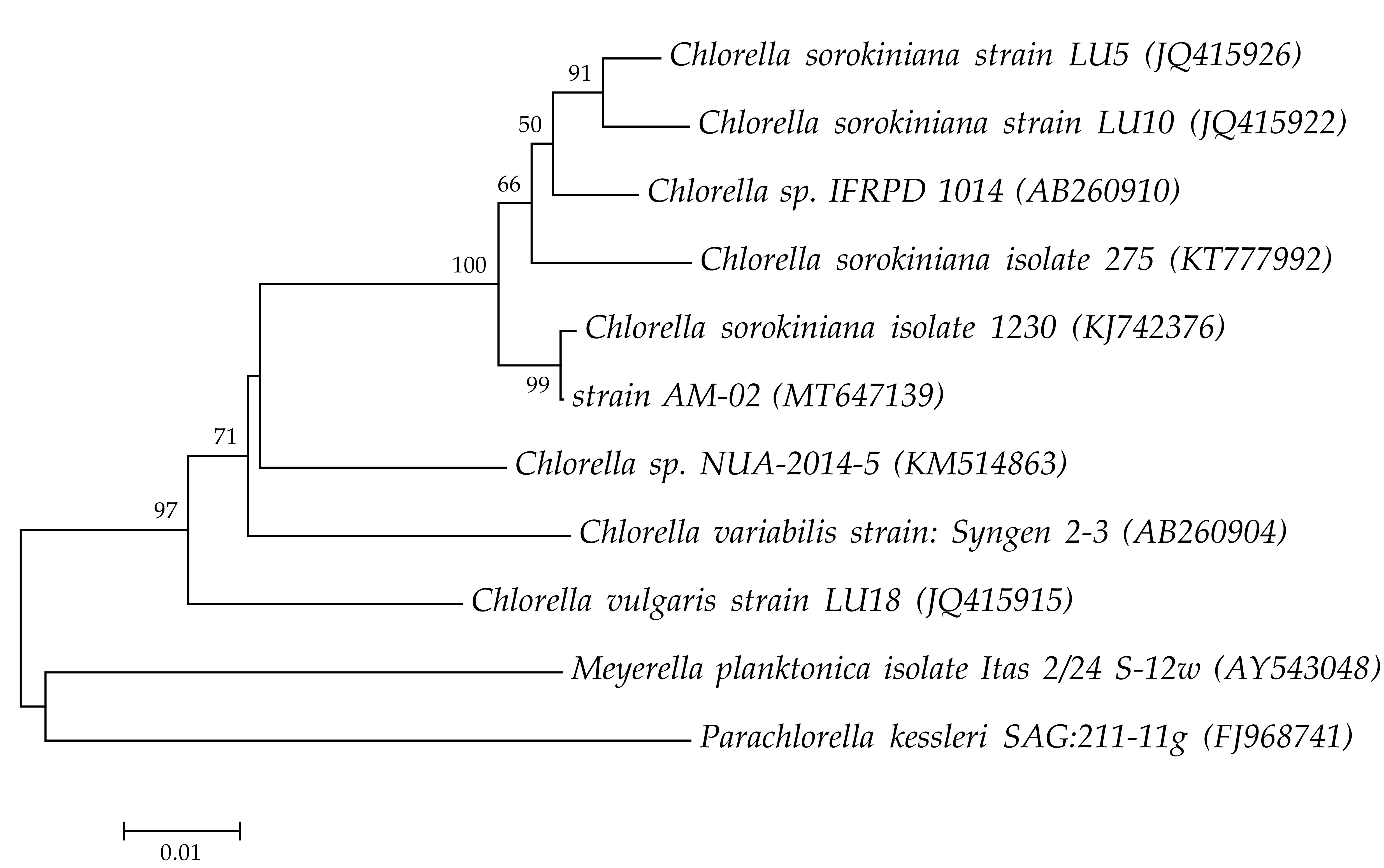
2.1. Isolation and Identification of Microalgal Strain

The microalgae culture of Chlorella sorokiniana AM-02 was isolated from a freshwater lake located in Kazan (Russia) in 2018. The isolated strain was made axenic by continuous sub-culturing using Bold’s basal medium (BBM) [25] on agar plates supplemented with ampicillin and kanamycin (10 and 50 µg mL−1 in the medium, respectively). The axenic culture was identified using molecular analysis of the ribulose bisphosphate carboxylase large subunit (rbcL) gene and translational elongation factor Tu (tufA) gene markers. For this, a FastDNA spin kit (MP Biomedicals, Solon, OH, USA) and a FastPrep-24 homogenizer (MP Biomedicals, Solon, OH, USA) were used to extract DNA from microalgae in accordance with the manufacturer’s protocol. DNA was then quantified with a NanoDrop 2000 spectrophotometer (Thermo Fisher Scientific, Wilmington, DE, USA) and stored at −20 °C until use. Primers for the rbcL and the tufA genes were selected from published studies and were used to identify microalgae (Table 1). Amplicons obtained with these primers were purified using a QIAquick PCR Purification kit (Qiagen, Hilden, Germany) and sequenced on a 3730 DNA Analyzer (Thermo Fisher Scientific, Wilmington, DE, USA) according to the manufacturers’ protocols. Sequencing data was confirmed by BLAST searches in the GenBank’s database and using the BOLD Identification System for the ribulose bisphosphate carboxylase gene. The determined rbcL gene and tufA gene sequences were deposited in the GenBank database under accession numbers MT647139 and MT647140, respectively.

2.2. Cultivation Conditions

Before starting the experiments in a photobioreactor, C. sorokiniana was placed in 250 mL Erlenmeyer flasks containing 30 mL of BBM under sterile conditions to eliminate any contamination. The inoculum was grown with shaking at 120 rpm at +26 °C under continuous illumination (150 μmol photons m−2 s−1). After 5 days, the biomass was concentrated by centrifugation at 5000× g for 5 min and then used as an inoculum for experiments in a photobioreactor.

2.3. Cultivation in a Photobioreactor

In order to study the influence of operational factors on photoautotrophic cultivation of C. sorokiniana for accelerated growth and biomass production, microalga with an initial OD750 of 0.01 (optical density at 750 nm) was grown in a sterilized 3.6 L Labfors 4 Lux photobioreactor (Infors HT, Bottmingen, Switzerland) with a 2.4 L working volume with controlled luminous flux levels. The experiments were divided into three main groups: (1) cultivation under different photosynthetic photon flux density (PPFD) conditions, (2) cultivation at various levels of carbon dioxide, and (3) cultivation in the presence of various levels of nitrate and sulfate ions.
The photobioreactor was illuminated with 1–16 Gro-Lux tubes with high blue and red radiation (per tube: 120 lumen (lm), maximum: 1920 lm) uniformly distributed around the culture vessel. Light intensity was additionally measured using a photosynthetically active radiation meter (PAR meter, Apogee Instruments, Logan, UT, USA). In experiments on the selection of optimal lighting, we tested the growth of culture at different values of illumination. Three treatments were carried out under different PPFD conditions (1000, 1200, and 1400 μmol m−2 s−1). For an optimized strategy for the growth and the accumulation of biomass, we analyzed the following conditions of the carbon dioxide regimen: cultures sparged with atmospheric air only (~0.04% CO2) and cultures sparged simultaneously with atmospheric air and continuously added CO2 (finally 0.5%, 1.0%, and 2.0% CO2 (v/v)). Aeration (0.55 volume of air per volume of liquid per minute) was provided by a compressor via a 0.45 µm filter. Carbon dioxide addition was provided by thermal mass flow meter and controller (Vögtlin Instruments, Aesch, Switzerland). They were mixed and injected into the reactor. The temperature was controlled at +26–26.5 °C. A relatively low temperature was chosen in our experiments since high temperatures lead to higher control costs and higher energy consumption of the process. The reactor was continuously stirred at 120 rpm. In all experiments, light was maintained on a 16:8 light/dark cycle. When observing the foam, a sterile 2% v/v solution of antifoam (Antifoam B, Sigma-Aldrich, St. Louis, MO, USA) was added. The experiments continued in the range of 112–232 h. Light, temperature, medium pH, pressure inside the reactor, carbon dioxide flow, and the percentage of released oxygen and carbon dioxide were measured by the Infors devices and displayed online on a computer screen.
Samples for analysis of algal growth, cell number, pigment content, and ions concentration in the culture medium were collected every day. pH as well percentage of the released molecular oxygen and carbon dioxide were measured throughout the experiments. Two independent experiments were performed to test reproducibility, and the results are presented as the mean.

2.4. Measurement of Algal Growth and Biomass Dry Weight

Optical density was measured using a Lambda 35 spectrophotometer (Perkin Elmer, Singapore) at 750 nm. Cell suspensions were diluted prior to measurements to obtain a final OD750 of less than 0.5 for measurements. Cell number was additionally determined by using a hemocytometer. The determination was conducted in triplicate, and the mean values are presented along with standard deviations. After 112–232 h (depending on the experiment), the centrifuged biomass (at 5000× g for 10 min) was washed twice with sterile deionized water to remove medium salts and centrifuged again. The remaining microalgal pellets were dried at +80 °C for 48 h. The final biomass yield (g L−1) was calculated as the difference between the weight of a centrifuge tube with dried microalgal pellet and the weight of the biomass-free centrifuge tube.

2.5. Measurement of Pigment Concentrations

Chlorophylls a and b, carotenoids, and total pigments (mg L−1) were determined using dimethyl sulfoxide extraction and optical absorption correlation, as previously described by Chai et al. [8] and Wellburn [29]. Optical densities at 480, 649.1, and 665.1 nm were measured with a Lambda 35 spectrophotometer (Perkin Elmer, Singapore). The concentration of pigments was measured in triplicate, and the mean values are presented along with standard deviations.

2.6. Ion Chromatography

Nitrate, phosphate, and sulfate concentrations were measured using a Dionex ICS-900 Ion Chromatography System (Thermo Fisher Scientific, Wilmington, DE, USA) equipped with an IonPac AG22 (4 × 50 mm) guard column, and an IonPac AS22 (4 × 250 mm) analytical column. The mobile phase consisted of 4.5 mM Na2CO3/1.4 mM NaHCO3 at a flow rate of 1.5 mL min−1. Data acquisition and instrument control were performed with the Dionex Chromeleon software (Sunnyvale, CA, USA). Analyses were conducted in triplicate, and the mean values are presented with standard deviations.

2.7. Data Analysis

The effects of PPFD, CO2, and nitrate concentration values on the photoautotrophic growth of C. sorokiniana were evaluated. The influence of these parameters on microalgal biomass was significant when the p value was below 0.05 (based on a two-tailed t test).

3. Results and Discussion

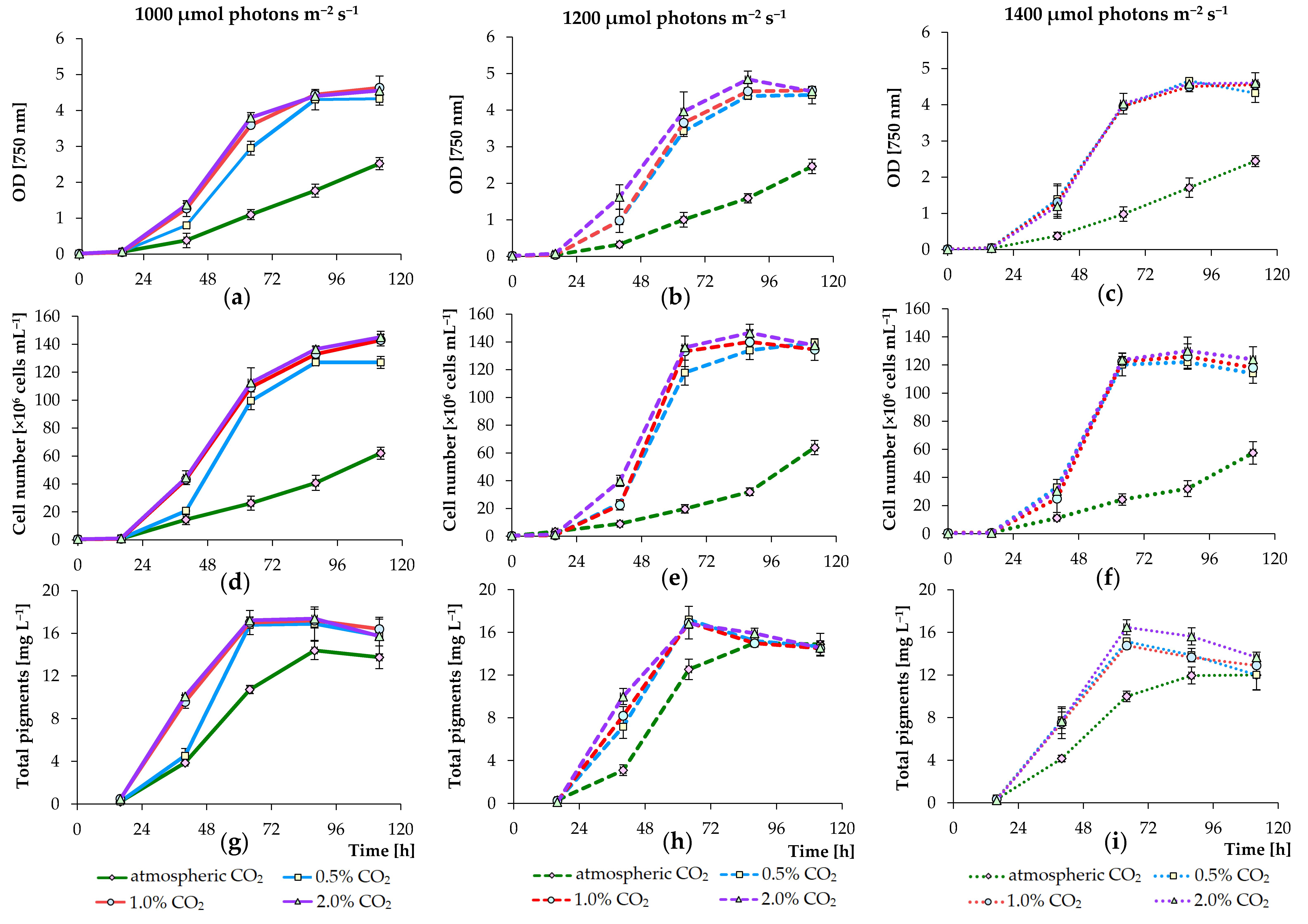
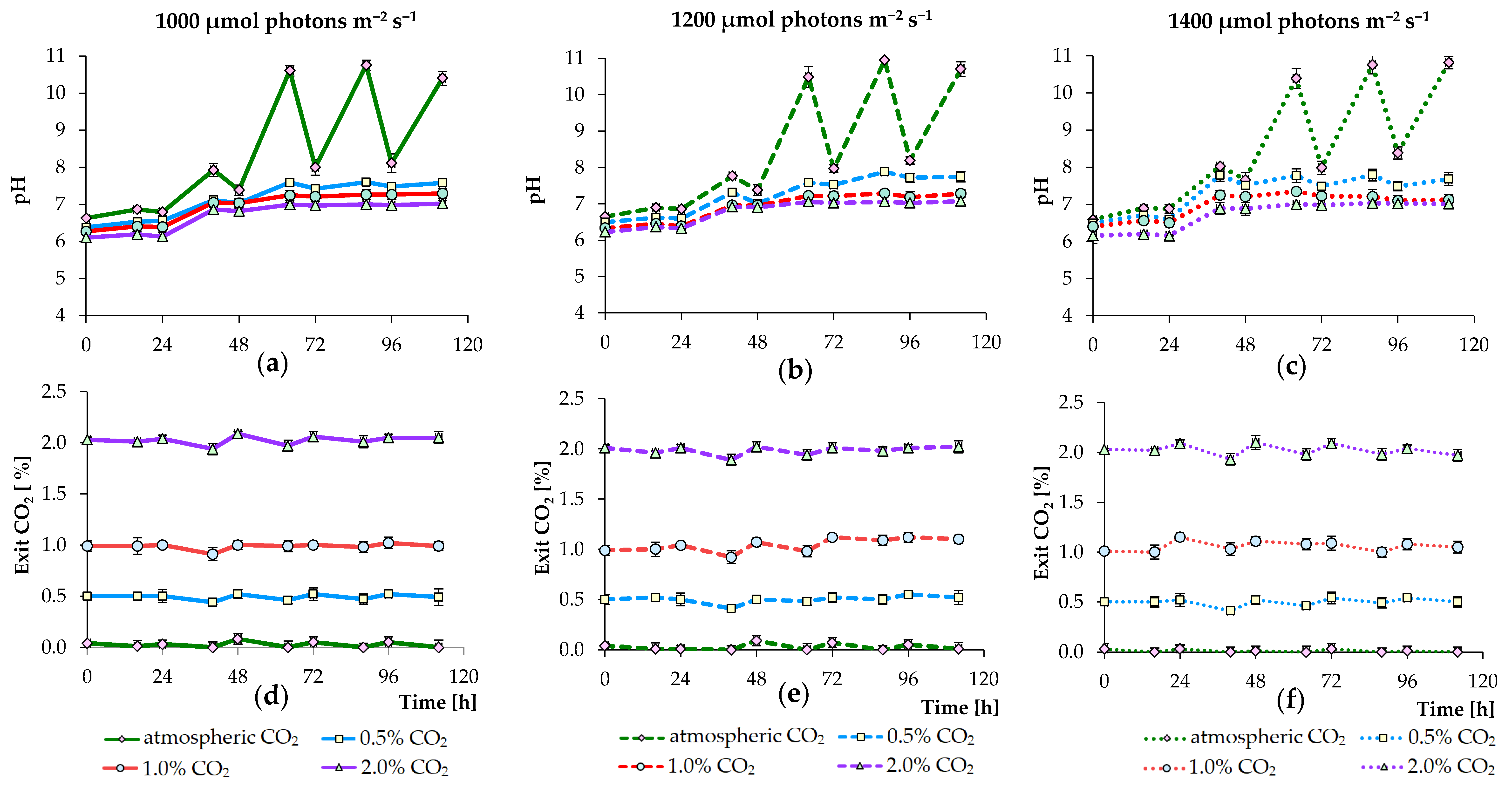
3.1. Effects of PPFD and CO2 Concentration Values on Growth of Algae

In this research study, various PPFD and CO2 concentration values as well as nutrient requirements for maximum growth and biomass accumulation of C. sorokiniana AM-02 were determined. Phylogeny of the strain AM-02 based on rbcL gene is presented in Figure 1. Based on these data, this strain was assigned to C. sorokiniana.
To control the algal growth, OD750 was measured and number of cells was counted (results are summarized in Figure 2). Three high PPFD conditions were chosen (1000, 1200, and 1400 μmol m−2 s−1) for the experiments. We chose high PPFD conditions, because we plan to continue testing the growth of this strain in digested agricultural waste materials (such as pig manure and chicken manure), which are dark in color. In experiments, when cultures were bubbled with air supplemented with final 0.5%, 1.0%, and 2.0% CO2 (for all tested PPFD conditions), a 1.7–2.4-fold increase in growth in standard BBM was observed compared to that detected in treatments with non-CO2 augmented cultures. The mean OD750 values of C. sorokiniana grown at 1000, 1200, and 1400 μmol photons m−2 s−1 and sparged with air after 112 h of cultivation were 2.52, 2.46, and 2.44, respectively, whereas in experiments with the addition of extra CO2, the growth values were above 4.3 (Figure 2a–c).
The cultures in experiments supplied with different concentrations of carbon dioxide (under different PPFD conditions) increased in cell number until stationary growth was achieved at 1.22–1.46 × 10⁸ cells mL−1 (after 88 h), while in treatments supplied with atmospheric carbon dioxide, the cultures increased in cell number to 5.7–6.4 × 10⁷ cells mL−1 only (after 112 h) (Figure 2d–f). Overall, OD750 correlated with the calculated cell numbers (Figure 2). The concentration of total pigments (chlorophyll a, b, and total carotenoids) also increased to 14–17 mg mL−1 with higher accumulation in treatments supplemented with 2% carbon dioxide but decreased thereafter (Figure 2g–i).
Table 2 presents the tested regimens of cultivation and the average yield of dry biomass of microalgae after photoautotrophic cultivation. As can be seen from the cell density, cell number, average biomass yield of dry microalgae, as well as pigment content, the growth had the similar trends under all lighting conditions with an additional CO2 supply; however, the growth of C. sorokiniana was slightly higher in experiments in which PPFD and CO2 concentration values were 1200 μmol m−2 s−1 and 2.0%, respectively. In this regard, we used cultures grown at 1200 μmol photons m−2 s−1 and sparged with 2.0% carbon dioxide for further selection of optimal nutrient concentrations in modified BBM. At 1000 μmol photons m−2 s−1 and 0.5% CO2, the lower growth rate was additionally observed (in treatments with a changing level of CO2). However, at 1400 μmol photons m−2 s−1, the growth was almost identical regardless of the level of CO2 addition (0.5%, 1.0%, or 2%). It is believed that, from an industrial point of view, transportation, storage, and delivery of gaseous carbon dioxide are expensive for large-scale algae production [12]. Therefore, the cultivation of C. sorokiniana AM-02 on an industrial scale can also be achieved with lower levels of CO2 supplementation.
In several previous studies, other PPFD conditions (100–500 μmol photons m−2 s−1) were tested for culturing other C. sorokiniana strains in flasks and bottles, and no significant differences were found between different conditions for photoautotrophic cultures [6,11,13,31]. Due to the shading of the cells one above the other, the design of photobioreactors is also important to ensure adequate light distribution, as reported by Leon-Vaz et al. [32]. An increase in light intensity is not a solution to overcome shading in many photoautotrophic cultures with a high cell density, since many microalgae cannot grow efficiently at extremely high light intensities. However, Cuaresma et al. [33] reported a high growth rate of C. sorokiniana CCAP 211/8K under continuous illumination of 2100 μmol photons m−2 s−1 using red light emitting diodes. In our research, we show that our strain has optimal cultivation conditions in standard BBM at a high surface irradiance between 1000–1400 μmol photons m−2 s−1 with Gro-Lux tubes in a 3.6 L photobioreactor with a working volume of 2.4 L.
Figure 3 demonstrates the changes in pH and the percentage of released CO2 under selected conditions. Thus, the pH increased from an initial ~6.6 to 10.4–10.9 under all lighting conditions in the non-CO2 augmented cultures (autotrophic cultivation leads to hydroxyl formation, which increases the pH), whereas the pH initially dropped to values 6.1–6.4 but ultimately did not increase above 7.8, 7.3, and 7.0 under all lighting conditions with the addition of CO2 up to 0.5%, 1.0%, and 2.0%, respectively (Figure 3a–c). During the light period, pH increased, while cultivation in the dark period led to a slight decrease in pH (in CO2 augmented cultures). In the non-CO2 augmented cultures, pH decreased substantially during the dark period. In the case of CO2 release from the photobioreactor, a decrease and an increase in the CO2 level were observed during the light period and the dark period under all lighting conditions, respectively (Figure 3d–f). The increase in pH found in the culture medium can be explained by the growth of C. sorokiniana and the uptake of carbon from dissolved HCO3, leaving OH ions. Similarly, higher pH levels in the non-CO2 augmented cultures can be attributed to lower dissolved CO2 and less dissociated H+ ions [7,34]. In addition, sodium nitrate nutrition leads to an increase in pH of the medium [35].
Figure 4 shows the nutrient levels in the BBM with nitrate levels of about 180 mg L−1 at the beginning of cultivation. After 64 h and 88 h, nitrogen was completely utilized in standard BBM in the CO2 augmented and the non-CO2 augmented cultures, respectively, and, therefore, decreases in the growth of the microalgae cultures and the pigment concentration were noted. It should also be reported that nitrate, orthophosphate, and sulfate were consumed faster by culture of C. sorokiniana AM-02 when air containing 2.0% CO2 was continuously bubbled into the reactor. Ion chromatography analysis demonstrated that the added amounts of phosphate (around 160 mg L−1) and sulfate (around 35 mg L−1) slightly decreased but were in excess during the entire experimental period, while the nitrate concentration sharply decreased, and NO3 completely was utilized after 64–88 h of cultivation (Figure 4). Thus, the concentration of NO3 was initially insufficient for the active growth and development of the culture in photoautotrophic treatments; therefore, the growth values were low, and the following experiments were performed to identify the effects of increasing NO3 content in the culture medium. These data confirm several reports in the literature on the rapid growth and rate of removal of nutrients but under other, different conditions [11]. In addition, some previous studies have demonstrated that various other C. sorokiniana strains can grow in wastewater with the effective removal of nitrates and phosphates [34,36], providing high potential for their application in developing wastewater treatment technologies.

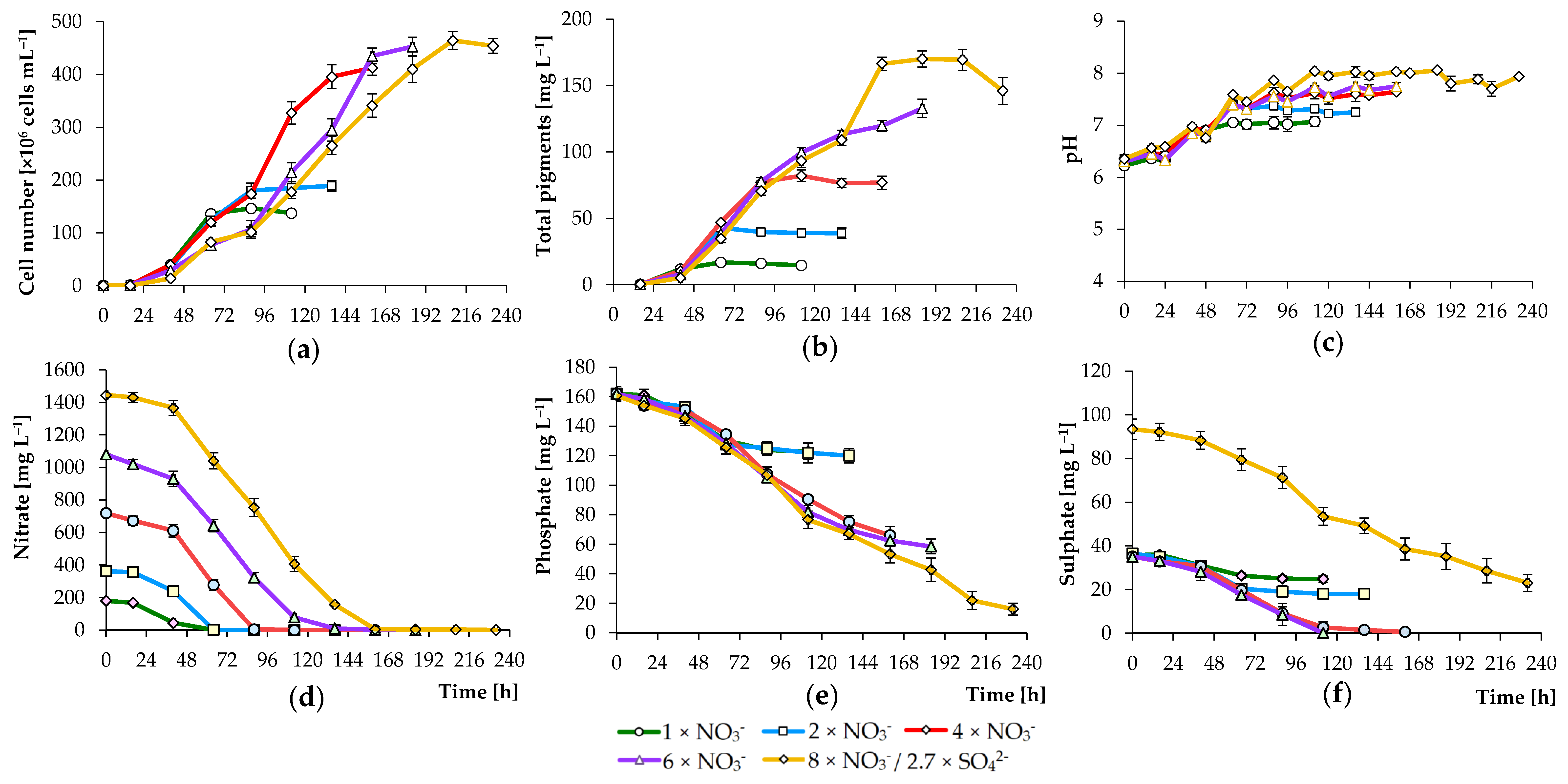
3.2. Effects of Nutrient Concentration on Algal Growth

In standard BBM (a medium that has been used to grow a variety of green algae cultures), NaNO3 is used as a source of nitrogen. The growth of strain AM-02 in treatments containing 180 mg L−1 NO3 (standard concentration) under the selected PPFD and CO2 concentration values (1200 μmol m−2 s−1 and 2% CO2) was low, as measured by low cells number (1.47 × 10⁸ cells mL−1), pigment concentration (16.8 mg L−1), and dry biomass weight (1.10 g L−1) (Figure 5; Table 2). No NO3 was detected in standard BBM sparged with air containing 2.0% CO2 on 66 h and onwards. Figure 5 shows that all key parameters were improved by adding an additional level of NO3. In the 360 mg L−1 NO3-treatments (double NO3 concentration), number of cells, pigment concentration, and dry biomass weight increased significantly (p < 0.001; up to 1.9 × 10⁸ cells mL−1, 42.8 mg L−1, and 1.76 g L−1, respectively). However, even at a double concentration of introduced NO3, nitrogen began to be limited after 66 h, and the cell concentration did not exceed 1.9 × 10⁸ cells mL−1. Under both conditions, PO43– and SO42– levels also decreased but remained abundant for culture growth (Figure 5).
A further increase in the concentration of the nitrogen source to 720 mg L−1 of NO3 led to a significant increase in the number of cells and was reflected in the yield of final dry biomass (p < 0.001; Figure 5a; Table 2). After 160 h, the culture increased in cell concentration until reaching stationary growth at 4.13 × 10⁸ cells mL−1. After 112 h, the concentration of total pigments increased to 82.1 mg mL−1 and remained almost stable during cultivation. As shown in Figure 5b, the pigment concentrations in C. sorokiniana cultivated in the presence of high NO3 levels were generally higher than those observed in cells cultured at lower levels of nitrate ions. A clear positive correlation was observed between chlorophyll levels and the color of the microalgae culture. High concentrations of NO3 promoted the synthesis of pigments. Under these conditions, nitrate and sulfate were completely utilized by 88 h and 160 h, respectively (Figure 5d,f). Phosphate levels also decreased from an initial 160 to 66 mg L−1 by 160 h and remained at around these levels until the end of growth (Figure 5e). In 6×NO3 and 8×NO3-treatments, the final biomass yield and pigment concentration were significantly higher than previously reported (p < 0.001); however, the best growth rate was observed under 4×NO3 conditions. Since SO42– was completely utilized under 4×NO3 and 6×NO3 conditions, we increased its level to 95 mg L−1 in 8×NO3 treatments. Under all conditions, nitrate, phosphate, and sulfate were efficiently utilized by culture of C. sorokiniana (Figure 5). These results clearly demonstrate that C. sorokiniana AM-02 can grow at high nitrate concentrations and can be further tested for wastewater treatment. However, remarkably high NO3 concentrations added to the medium reduced the specific growth, which can be explained by increased osmotic stress and inhibition by the intermediate [37].
The pH increased to 7.1, 7.3, 7.6, 7.8, and 8.0 in 1× NO3, 2× NO3, 4× NO3, 6× NO3, and 8× NO3 -treatments, respectively (Figure 5c). In the case of CO2 emission from the photobioreactor, a decrease and an increase in the CO2 level were observed during the light period and the dark period, respectively, with an increase in the CO2 level by the entire period in experiments with a high nitrogen content (from an initial 2.0% to 2.6%, data not shown). Therefore, based on our results, nitrogen and sulfur are the main nutrients that become insufficient in standard BBM, while the phosphorous level is sufficient to support the growth of microalgae. Our findings also demonstrate that high level of inorganic anions removal is possible (100% for NO3 and SO42– and 91% for PO43– (146 mg L−1)), which indicates the potential use of the strain for wastewater treatment.
Nutrient availability affects the growth of photoautotrophic organisms. One of the important components affecting the growth of microalgae and physiological activity is nitrogen [14,38,39]. For numerous applications of promising photosynthetic microorganisms, an increase in biomass productivity is considered to be an important step. Various potential nitrogen sources and their concentrations, such as urea, nitrate, and ammonium ions, have been evaluated by several other researchers for the cultivation of other C. sorokiniana strains. Lizzul et al. [34] observed that C. sorokiniana UTEX1230 prefers ammonium ions rather than nitrate ions as a source of nitrogen when cultured in wastewater and BBM medium. Ramanna et al. [36] reported that urea provided the highest biomass yield during cultivation of C. sorokiniana strain NIES: 2173 in wastewater. Kim et al. [7] found that growth rate and nitrogen and phosphorus removal rates by C. sorokiniana UTEX 1670 were higher under heterotrophic conditions than under autotrophic conditions. The wastewater nitrogen and phosphate were also effectively utilized by several other C. sorokiniana strains [40]. Another species of the same genus, C. vulgaris, also effectively removed nitrogen and phosphorus from domestic wastewater [41]. In this work, C. sorokiniana AM-02 was isolated from a freshwater lake and therefore can be adapted to different environmental changes and thus potentially provides a competitive advantage when grown in open water.

4. Conclusions

A study was conducted comparing the effect of various PPFD and CO2 concentration values on the growth of the new isolated strain C. sorokiniana strain AM-02. Thus, the photoautotrophic cultures of C. sorokiniana in modified BBM (720 mg L−1 of NO3) showed better growth performance. It was found that the preferred high PPFD and CO2 concentration values are 1000–1400 μmol photons m−2 s−1 and 0.5–2.0%, respectively. In addition, the high growth of C. sorokiniana AM-02 makes this strain particularly suitable for the rapid production of biomass, attractive for bioremediation of aquatic environments contaminated with high levels of nitrate, phosphate, and sulfate. Moreover, our results show that adding an additional nitrogen source can significantly increase final biomass and pigment yield compared to standard BBM. Finally, our findings prove that the new isolated strain C. sorokiniana AM-02 is a fast-growing microalgae strain that can also be used in industry.

Author Contributions

Conceptualization, E.E.Z. and A.M.Z.; methodology, E.E.Z. and A.M.Z.; software, E.E.Z., S.S.B., and A.M.Z.; validation, E.E.Z., S.S.B., and A.M.Z.; formal analysis, E.E.Z., S.S.B., and A.M.Z.; investigation, E.E.Z., S.S.B., and A.M.Z.; resources, A.M.Z.; data curation, E.E.Z. and A.M.Z.; writing—original draft preparation, E.E.Z.; writing—review and editing, S.S.B. and A.M.Z.; visualization, E.E.Z. and S.S.B.; supervision, A.M.Z.; project administration, A.M.Z.; funding acquisition, A.M.Z. All authors have read and agreed to the published version of the manuscript.

Funding

This research was funded by the Russian Foundation for Basic Research, grant number Grant No. 18-34-20002.

Acknowledgments

We would like to thank the technicians within the department of Microbiology at KFU for their support during undertaking of the project.

Conflicts of Interest

The authors declare no conflict of interest.

References

  1. Hammed, A.M.; Prajapati, S.K.; Simsek, S.; Simsek, H. Growth regime and environmental remediation of microalgae. Algae 2016, 31, 189–204. [Google Scholar] [CrossRef] [Green Version]
  2. Chen, B.; Wan, C.; Mehmood, M.A.; Chang, J.S.; Bai, F.; Zhao, X. Manipulating environmental stresses and stress tolerance of microalgae for enhanced production of lipids and value-added products—A review. Bioresour. Technol. 2017, 244, 1198–1206. [Google Scholar] [CrossRef]
  3. Khan, M.I.; Shin, J.H.; Kim, J.D. The promising future of microalgae: Current status, challenges, and optimization of a sustainable and renewable industry for biofuels, feed, and other products. Microb. Cell Fact. 2018, 17, 36. [Google Scholar] [CrossRef]
  4. Saad, M.G.; Dosoky, N.S.; Zoromba, M.S.; Shafik, H.M. Algal biofuels: Current status and key challenges. Energies 2019, 12, 1920. [Google Scholar] [CrossRef] [Green Version]
  5. Wang, L.; Xiao, H.; He, N.; Sun, D.; Duan, S. Biosorption and biodegradation of the environmental hormone nonylphenol by four marine microalgae. Sci. Rep. 2019, 9, 5277. [Google Scholar] [CrossRef] [PubMed]
  6. Kumar, K.S.; Dahms, H.; Won, E.; Lee, J.; Shin, K. Microalgae—A promising tool for heavy metal remediation. Ecotox. Environ. Saf. 2015, 113, 329–352. [Google Scholar] [CrossRef] [PubMed]
  7. Kim, S.; Park, J.E.; Cho, Y.B.; Hwang, S.J. Growth rate, organic carbon and nutrient removal rates of Chlorella sorokiniana in autotrophic, heterotrophic and mixotrophic conditions. Bioresour. Technol. 2013, 144, 8–13. [Google Scholar] [CrossRef] [PubMed]
  8. Chai, S.; Shi, J.; Huang, T.; Guo, Y.; Wei, J.; Guo, M.; Li, L.; Dou, S.; Liu, L.; Liu, G. Characterization of Chlorella sorokiniana growth properties in monosaccharide-supplemented batch culture. PLoS ONE 2018, 17, e0199873. [Google Scholar] [CrossRef] [PubMed] [Green Version]
  9. Cecchin, M.; Benfatto, S.; Griggio, F.; Mori, A.; Cazzaniga, S.; Vitulo, N.; Delledonne, M.; Ballottari, M. Molecular basis of autotrophic vs mixotrophic growth in Chlorella sorokiniana. Sci. Rep. 2018, 8, 6465. [Google Scholar] [CrossRef] [PubMed]
  10. Chen, C.Y.; Liu, C.C. Optimization of lutein production with a two-stage mixotrophic cultivation system with Chlorella sorokiniana MB-1. Bioresour. Technol. 2018, 262, 74–79. [Google Scholar] [CrossRef] [PubMed]
  11. Lizzul, A.M.; Lekuona-Amundarain, A.; Purton, S.; Campos, L.C. Characterization of Chlorella sorokiniana, UTEX 1230. Biology 2018, 7, 25. [Google Scholar] [CrossRef] [PubMed] [Green Version]
  12. Lohman, E.J.; Gardner, R.D.; Pedersen, T.; Peyton, B.M.; Cooksey, K.E.; Gerlach, R. Optimized inorganic carbon regime for enhanced growth and lipid accumulation in Chlorella vulgaris. Biotechnol. Biofuels 2015, 8, 82. [Google Scholar] [CrossRef] [PubMed] [Green Version]
  13. Bohutskyi, P.; Kligerman, D.C.; Byers, N.; Nasr, L.K.; Cua, C.; Chow, S.; Su, C.; Tang, Y.; Betenbaugh, M.J.; Bouwer, E.J. Effects of inoculum size, light intensity, and dose of anaerobic digestion centrate on growth and productivity of Chlorella and Scenedesmus microalgae and their poly-culture in primary and secondary wastewater. Algal Res. 2016, 19, 278–290. [Google Scholar] [CrossRef]
  14. Negi, S.; Barry, A.N.; Friedland, N.; Sudasinghe, N.; Subramanian, S.; Pieris, S.; Holguin, F.O.; Dungan, B.; Schaub, T.; Sayre, R. Impact of nitrogen limitation on biomass, photosynthesis, and lipid accumulation in Chlorella sorokiniana. J. Appl. Phycol. 2016, 28, 803–812. [Google Scholar] [CrossRef]
  15. Kobayashi, N.; Noel, E.A.; Barnes, A.; Watson, A.; Rosenberg, J.N.; Erickson, G.; Oyler, G.A. Characterization of three Chlorella sorokiniana strains in anaerobic digested effluent from cattle manure. Bioresour. Technol. 2013, 150, 377–386. [Google Scholar] [CrossRef] [Green Version]
  16. Lu, S.; Wang, J.; Niu, Y.; Yang, J.; Zhou, J.; Yuan, Y. Metabolic profiling reveals growth related FAME productivity and quality of Chlorella sorokiniana with different inoculum sizes. Biotechnol. Bioeng. 2012, 109, 1651–1662. [Google Scholar] [CrossRef]
  17. Sorokin, C.; Myers, J. A high-temperature strain of Chlorella. Science 1953, 117, 330–331. [Google Scholar] [CrossRef]
  18. Sorokin, C.; Kraus, R.W. Maximum growth rates of Chlorella in steady-state and in synchronized cultures. Proc. Nat. Acad. Sci. USA 1959, 45, 1740–1744. [Google Scholar] [CrossRef] [Green Version]
  19. Sorokin, C.; Krauss, R.W. Effects of temperature and illuminance of Chlorella growth uncoupled from cell division. Plant Physiol. 1962, 37, 37–42. [Google Scholar] [CrossRef] [Green Version]
  20. Sorokin, C.; Krauss, R.W. The dependence of cell division in Chlorella on temperature and light intensity. Am. J. Bot. 1965, 52, 331–339. [Google Scholar] [CrossRef]
  21. Min, M.; Hu, B.; Zhou, W.; Li, Y.; Chen, P.; Ruan, R. Mutual influence of light and CO2 on carbon sequestration via cultivating mixotrophic alga Auxenochlorella protothecoides UMN280 in an organic carbon-rich wastewater. J. Appl. Phycol. 2012, 24, 1099–1105. [Google Scholar] [CrossRef]
  22. Perez-Garcia, O.; Escalante, F.M.E.; de-Bashan, L.E.; Bashan, Y. Heterotrophic cultures of microalgae: Metabolism and potential products. Water Res. 2011, 45, 11–36. [Google Scholar] [CrossRef]
  23. Salih, F.M. Microalgae tolerance to high concentrations of carbon dioxide: A review. J. Environ. Prot. 2011, 2, 648–654. [Google Scholar] [CrossRef] [Green Version]
  24. Pavlik, D.; Zhong, Y.; Daiek, C.; Liao, W.; Morgan, R.; Clary, W.; Liu, Y. Microalgae cultivation for carbon dioxide sequestration and protein production using a high-efficiency photobioreactor system. Algal Res. 2017, 25, 413–420. [Google Scholar] [CrossRef]
  25. Nichols, H.W.; Bold, H.C. Trichosarcina polymorpha Gen. et Sp. Nov. J. Phycol. 1965, 1, 34–38. [Google Scholar] [CrossRef]
  26. Vieira, H.H.; Bagatini, I.L.; Guinart, C.M.; Vieira, A.A.H. tufA gene as molecular marker for freshwater Chlorophyceae. Algae 2016, 31, 155–165. [Google Scholar] [CrossRef] [Green Version]
  27. Hadi, S.I.I.A.; Santana, H.; Brunale, P.P.M.; Gomes, T.G.; Oliveira, M.D.; Matthiensen, A.; Oliveira, M.E.C.; Silva, F.C.P.; Brasil, B.S.A.F. DNA barcoding green microalgae isolated from Neotropical Inland waters. PLoS ONE 2016, 11, e0149284. [Google Scholar] [CrossRef] [PubMed] [Green Version]
  28. Fama, P.; Wysor, B.; Kooistra, W.H.C.F.; Zuccarello, G.C. Molecular phylogeny of the genus Caulerpa (Caulerpales, Chlorophyta) inferred from chloroplast tufA gene. J. Phycol. 2002, 38, 1040–1050. [Google Scholar] [CrossRef] [Green Version]
  29. Wellburn, A.R. The spectral determination of chlorophylls a and b, as well as total carotenoids, using various solvents with spectrophotometers of different resolution. J. Plant Physiol. 1994, 144, 307–313. [Google Scholar] [CrossRef]
  30. Kumar, S.; Stecher, G.; Tamura, K. MEGA7: Molecular evolutionary genetics analysis version 7.0 for bigger datasets. Mol. Biol. Evol. 2016, 33, 1870–1874. [Google Scholar] [CrossRef] [Green Version]
  31. Li, T.; Zheng, Y.; Yu, L.; Chen, S. Mixotrophic cultivation of a Chlorella sorokiniana strain for enhanced biomass and lipid production. Biomass Bioenergy 2014, 66, 204–213. [Google Scholar] [CrossRef]
  32. León-Vaz, A.; León, R.; Díaz-Santos, E.; Vigara, J.; Raposo, S. Using agro-industrial wastes for mixotrophic growth and lipids production by the green microalga Chlorella sorokiniana. New Biotechnol. 2019, 51, 31–38. [Google Scholar] [CrossRef]
  33. Cuaresma, M.; Janssen, M.; Vílchez, C.; Wijffels, R.H. Productivity of Chlorella sorokiniana in a short light-path (SLP) panel photobioreactor under high irradiance. Biotechnol. Bioeng. 2009, 104, 352–359. [Google Scholar] [CrossRef]
  34. Lizzul, A.; Hellier, P.; Purton, S.; Baganz, F.; Ladommatos, N.; Campos, L. Combined remediation and lipid production using Chlorella sorokiniana grown on wastewater and exhaust gases. Bioresour. Technol. 2014, 151, 12–18. [Google Scholar] [CrossRef] [PubMed] [Green Version]
  35. Wang, J.; Zhou, W.; Chen, H.; Zhan, J.; He, C.; Wang, Q. Ammonium nitrogen tolerant Chlorella strain screening and its damaging effects on photosynthesis. Front. Microbiol. 2019, 9, 3250. [Google Scholar] [CrossRef]
  36. Ramanna, L.; Guldhe, A.; Rawat, I.; Bux, F. The optimization of biomass and lipid yields of Chlorella sorokiniana when using wastewater supplemented with different nitrogen sources. Bioresour. Technol. 2014, 168, 127–135. [Google Scholar] [CrossRef]
  37. Gilmour, D.J.; Hipkins, M.F.; Boney, A.D. The effect of osmotic and ionic stress on the primary processes of photosynthesis in Dunaliella tertiolecta. J. Exp. Bot. 1984, 35, 18–27. [Google Scholar] [CrossRef]
  38. Ordog, V.; Stirk, W.A.; Balint, P.; Staden, J.; Lovasz, C. Changes in lipid, protein and pigment concentrations in nitrogen-stressed Chlorella minutissima cultures. J. Appl. Phycol. 2012, 24, 907–914. [Google Scholar] [CrossRef]
  39. Janssen, J.H.; Wijffels, R.H.; Barbosa, M.J. Lipid production in Nannochloropsis gaditana during nitrogen starvation. Biology 2019, 8, 5. [Google Scholar] [CrossRef] [Green Version]
  40. Bohutskyi, P.; Liu, K.; Nasr, L.K.; Byers, N.; Rosenberg, J.N.; Oyler, G.A.; Betenbaugh, M.J.; Bouwer, E.J. Bioprospecting of microalgae for integrated biomass production and phytoremediation of unsterilized wastewater and anaerobic digestion centrate. Appl. Microbiol. Biotechnol. 2015, 99, 6139–6154. [Google Scholar] [CrossRef]
  41. Mayhead, E.; Silkina, A.; Llewellyn, C.A.; Fuentes-Grünewald, C. Comparing nutrient removal from membrane filtered and unfiltered domestic wastewater using Chlorella vulgaris. Biology 2018, 7, 12. [Google Scholar] [CrossRef] [PubMed] [Green Version]
Figure 1. Phylogenetic tree based on rbcL gene sequences. Analysis was conducted in MEGA7 [30] using the neighbor-joining method. The percentages of replicate trees in which the associated taxa clustered together in the bootstrap test (1000 replicates) are shown next to the branches. The evolutionary distances were computed using the Kimura 2-parameter method. Meyerella planktonica and Parachlorella kessleri were used as outgroup.
Biology 09 00169 g001
Figure 2. Growth of C. sorokiniana AM-02 (OD750 (ac) and cell number (df)) and total pigment concentration in cells (gi) cultured under various light and CO2 regimens (1000, 1200, and 1400 μmol photons m−2 s−1 and atmospheric CO2, 0.5, 1.0, and 2.0% CO2) in standard Bold’s basal medium (BBM).
Biology 09 00169 g002
Figure 3. Changes in culture medium pH (ac) and the released carbon dioxide level (df) observed during the growth of C. sorokiniana AM-02 cultured under various light and CO2 regimens (1000, 1200, and 1400 μmol photons m−2 s−1 and atmospheric CO2, 0.5, 1.0, and 2.0% CO2) in standard BBM.
Biology 09 00169 g003
Figure 4. Changes in nitrate (ac), phosphate (df), and sulfate (gi) concentrations in culture medium during the growth of C. sorokiniana AM-02 cultured under various light and CO2 regimens (1000, 1200, and 1400 μmol photons m−2 s−1 and atmospheric CO2, 0.5, 1.0, and 2.0% CO2) in standard BBM.
Biology 09 00169 g004
Figure 5. Growth of C. sorokiniana AM-02 (a), total pigment concentration in cells (b), culture medium pH (c), as well as nitrate (d), phosphate (e), and sulfate (f) removal from the medium in different treatments with modified BBM (PPFD and CO2 concentration values: 1200 μmol photons m−2 s−1 and 2.0% CO2, respectively).
Biology 09 00169 g005
Table 1. List of primers used in this study to identify the strain AM-02.
Molecular MarkerPrimersSequence 5′ → 3′Reference
rbcL (ribulose bisphosphate carboxylase large subunit)rbcL-M379 FGGTTTCAAAGCTYTWCGTGC[26]
rbcLFP RGTAAATACCACGGCTACGRTCTT
Fw_rbcl_192GGTACTTGGACAACWGTWTGGAC[27]
Rv_rbcL_657GAAACGGTCTCKCCARCGCAT
tufA (translational elongation factor Tu)tufA FGGNGCNGCNCAAATGGAYGG[28]
tufA RCCTTCNCGAATMGCRAAWCGC
Table 2. The average dry weight levels of algal biomass at the end of the experiments.
RegimenDry Weight (g L−1)
PPFD (μmol m−2 s−1)Initial NO3 (mg L−1)Initial CO2 (%)
1000180~0.040.43 ± 0.02
10001800.51.07 ± 0.03
10001801.01.08 ± 0.02
10001802.01.02 ± 0.01
1200180~0.040.45 ± 0.02
12001800.51.02 ± 0.02
12001801.01.08 ± 0.04
12001802.01.10 ± 0.03
1400180~0.040.44 ± 0.01
14001800.51.05 ±0.02
14001801.01.08 ±0.01
14001802.01.07 ±0.02
12001802.01.10 ± 0.03
12003602.01.76 ± 0.08
12007202.02.53 ± 0.12
120010802.03.11 ± 0.21
120014402.03.45 ± 0.24
PPFD: photosynthetic photon flux density.

