
Green Nanotechnology Serving the Bioeconomy: Natural Beauty Masks to Save the Environment

Abstract
:1. Introduction
2. Experimental Section
2.1. Materials and Methods
2.1.1. Materials
2.1.2. Methods
Electrospinning
- Voltage: 45–75 kV
- Collecting electrode (CE): cylinder
- Spinning electrode (SE): cylinder
- Distance SE/CE: 10–16 cm
- CE rotation: 2–8 rpm
- Substrate material: Spunbond, 30 gsm, polypropylene 100% with antistatic treatment.
2.2. Measurements
2.2.1. Skin Hydration
2.2.2. Transepidermal Water Loss (TEWL)
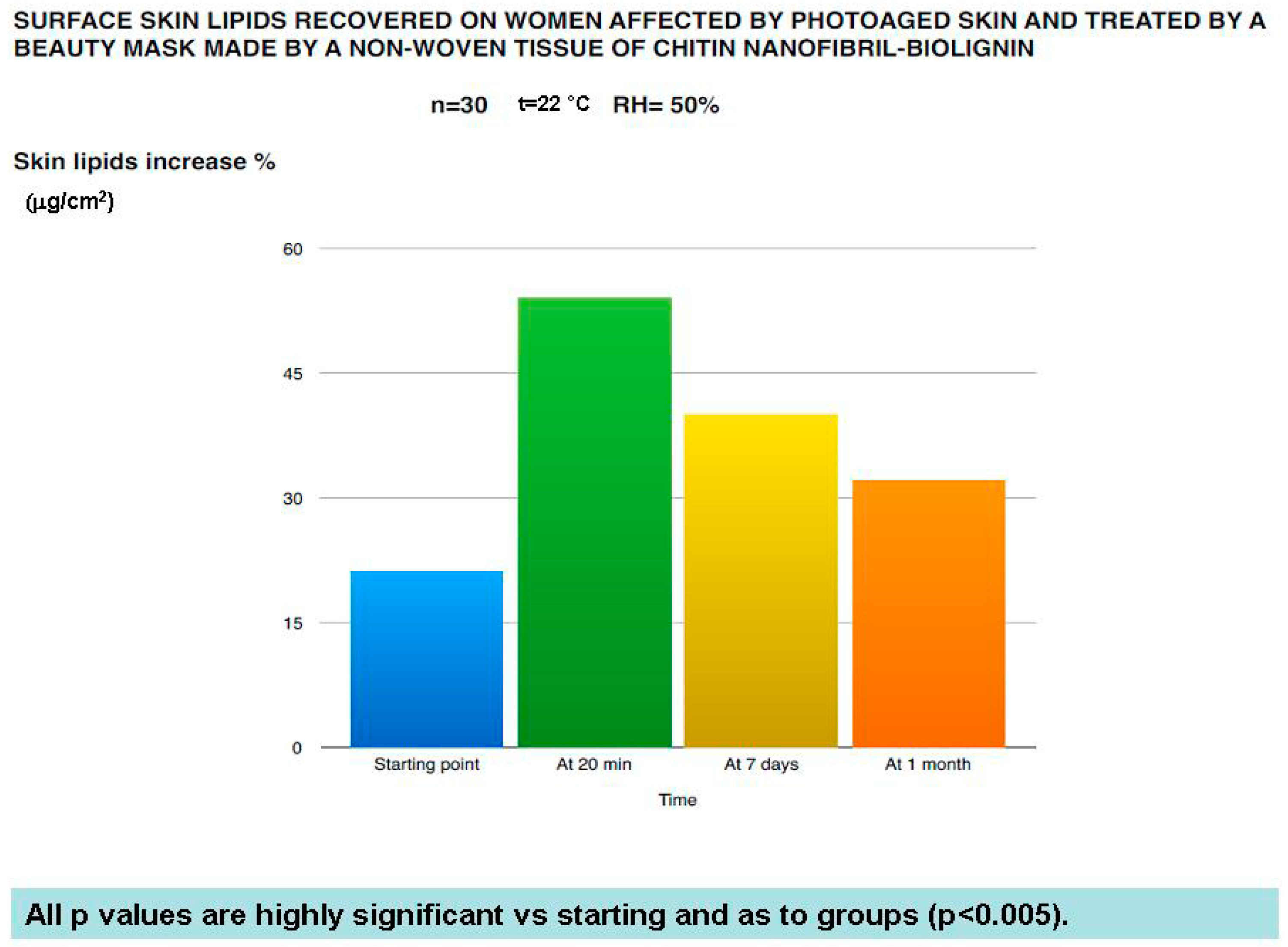
2.2.3. Skin Surface Lipids
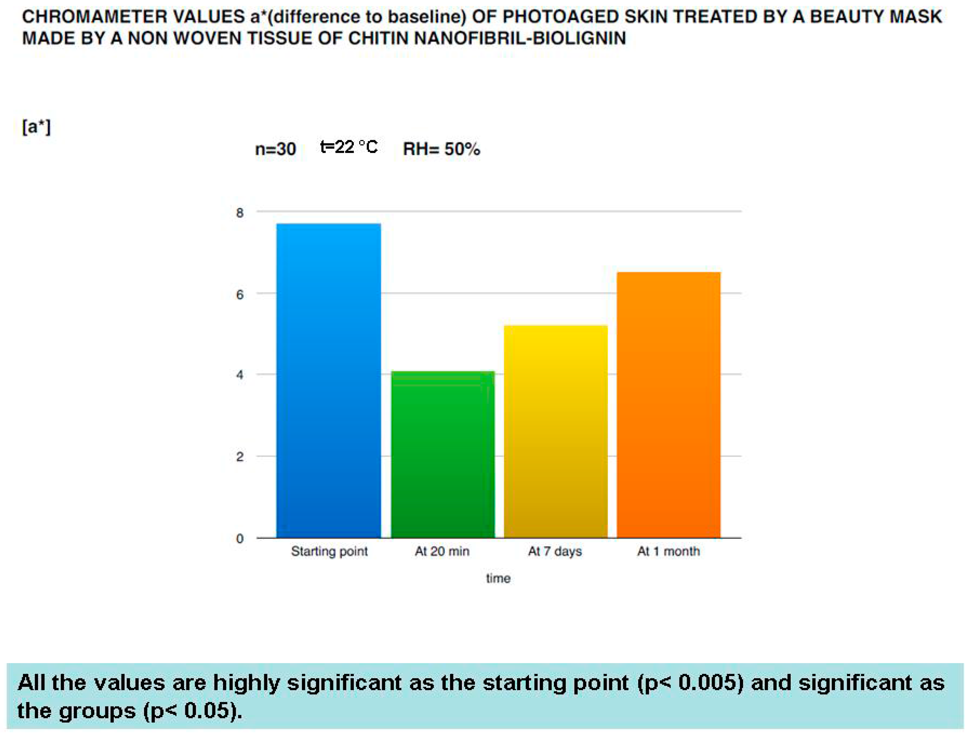
2.2.4. Skin Colour
2.2.5. Characterization of Nanoparticles and Non-Woven Tissue
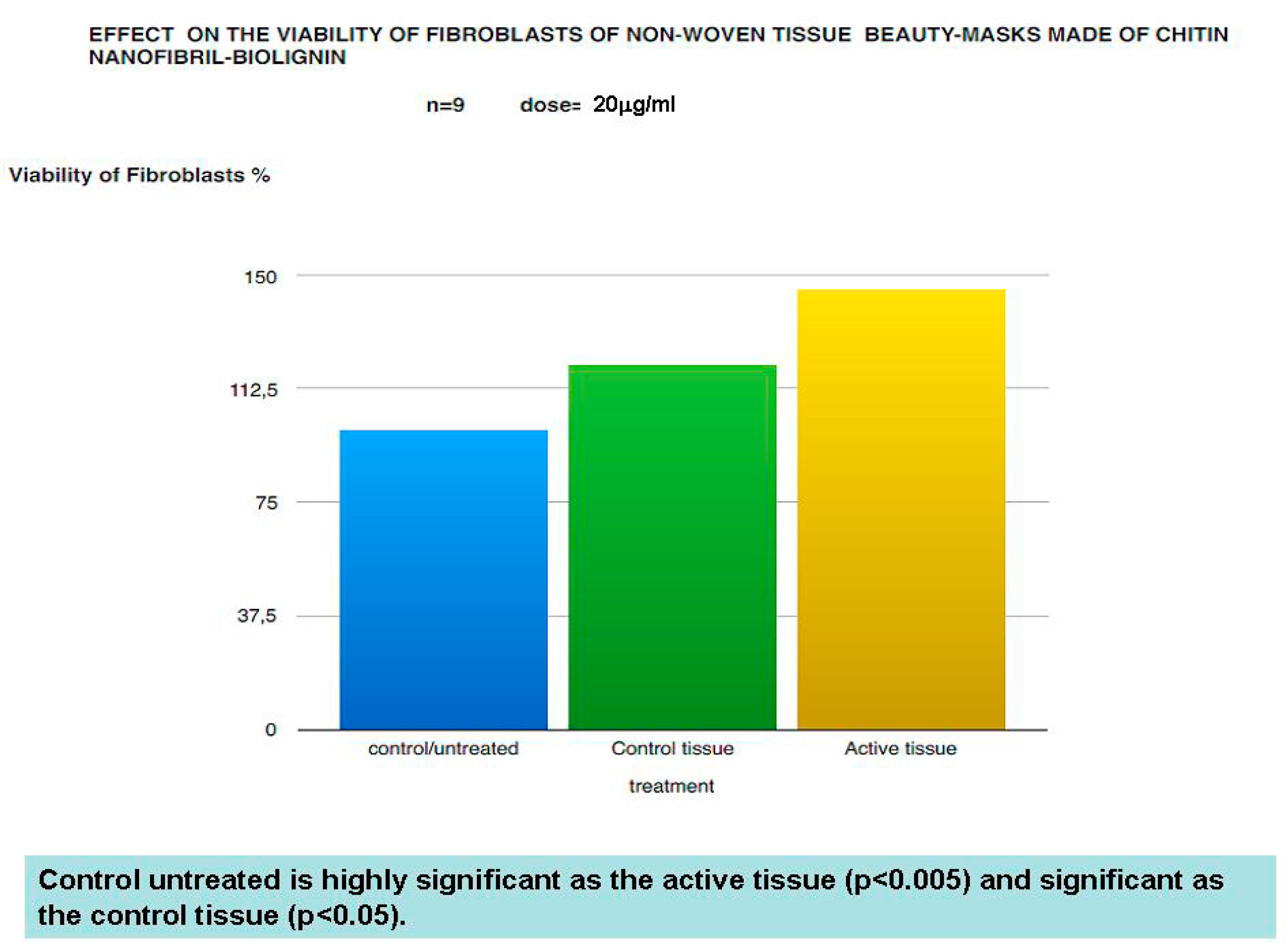
2.2.6. Cytotoxicity Assay and Collagen I Synthesis
2.2.7. Reduction of UV Induces IL-8 and TNF-Alpha Release
2.2.8. Metalloproteinase Release
2.2.9. Collagenase Inhibition
2.2.10. In Vivo Experimental Procedure
2.2.11. Statistical Analysis
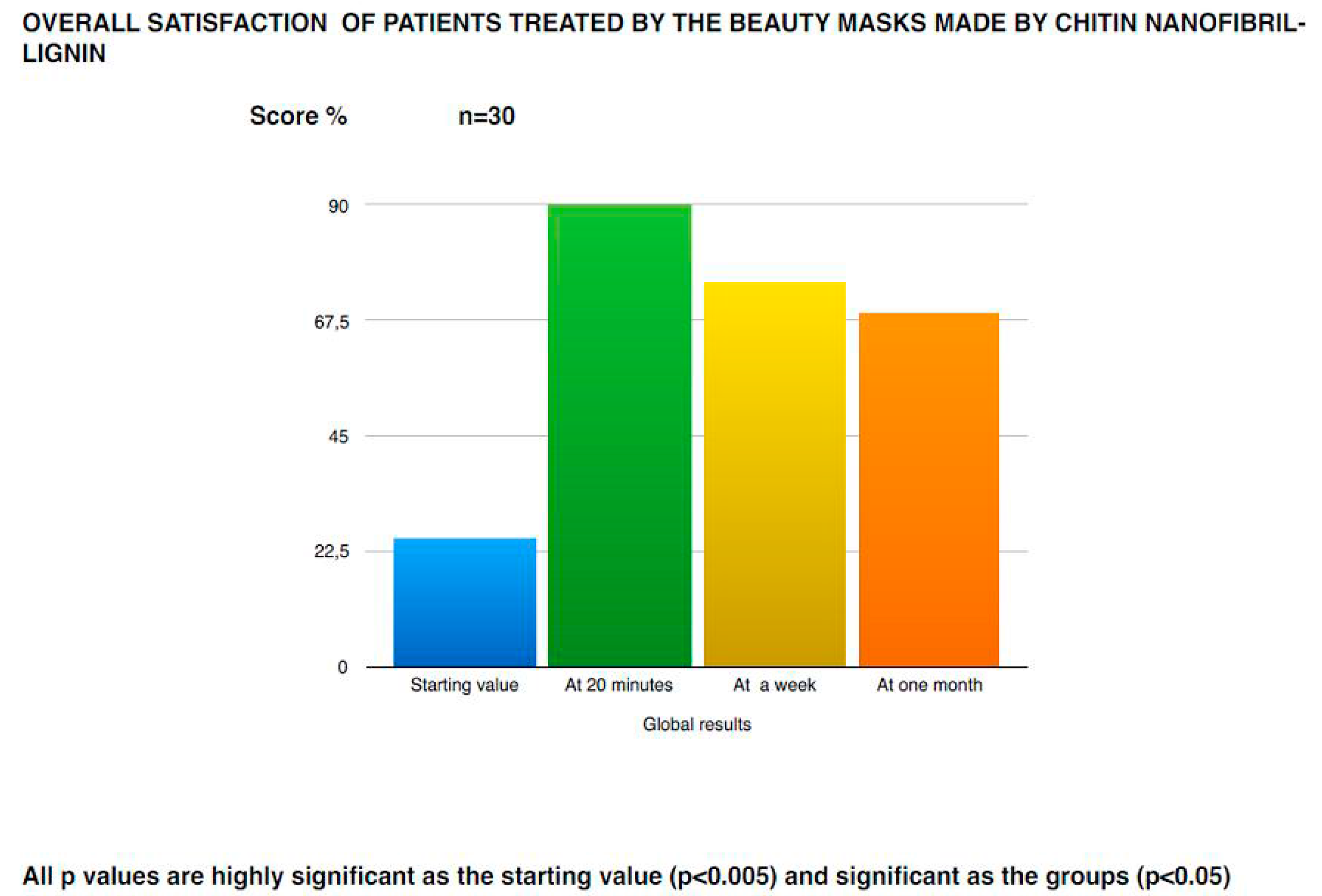
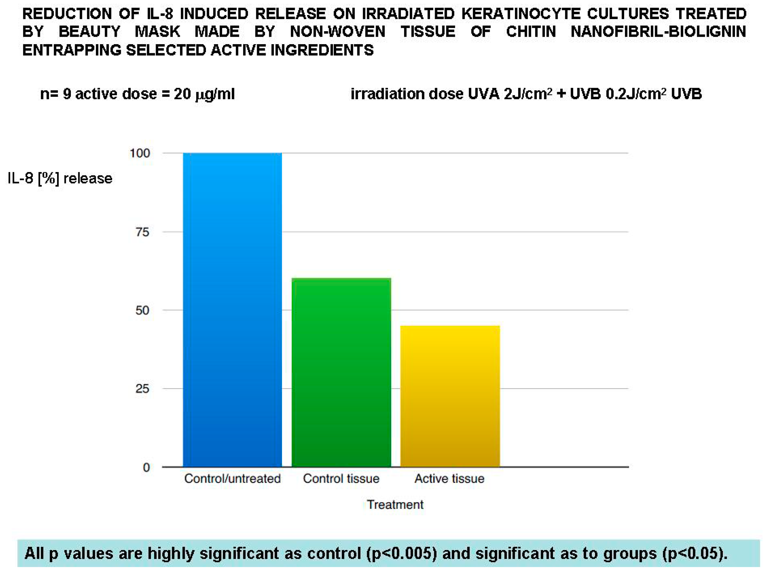
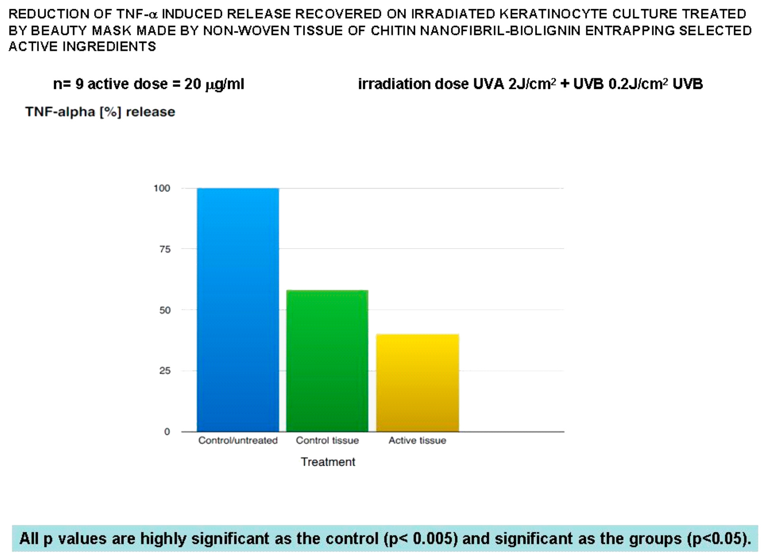
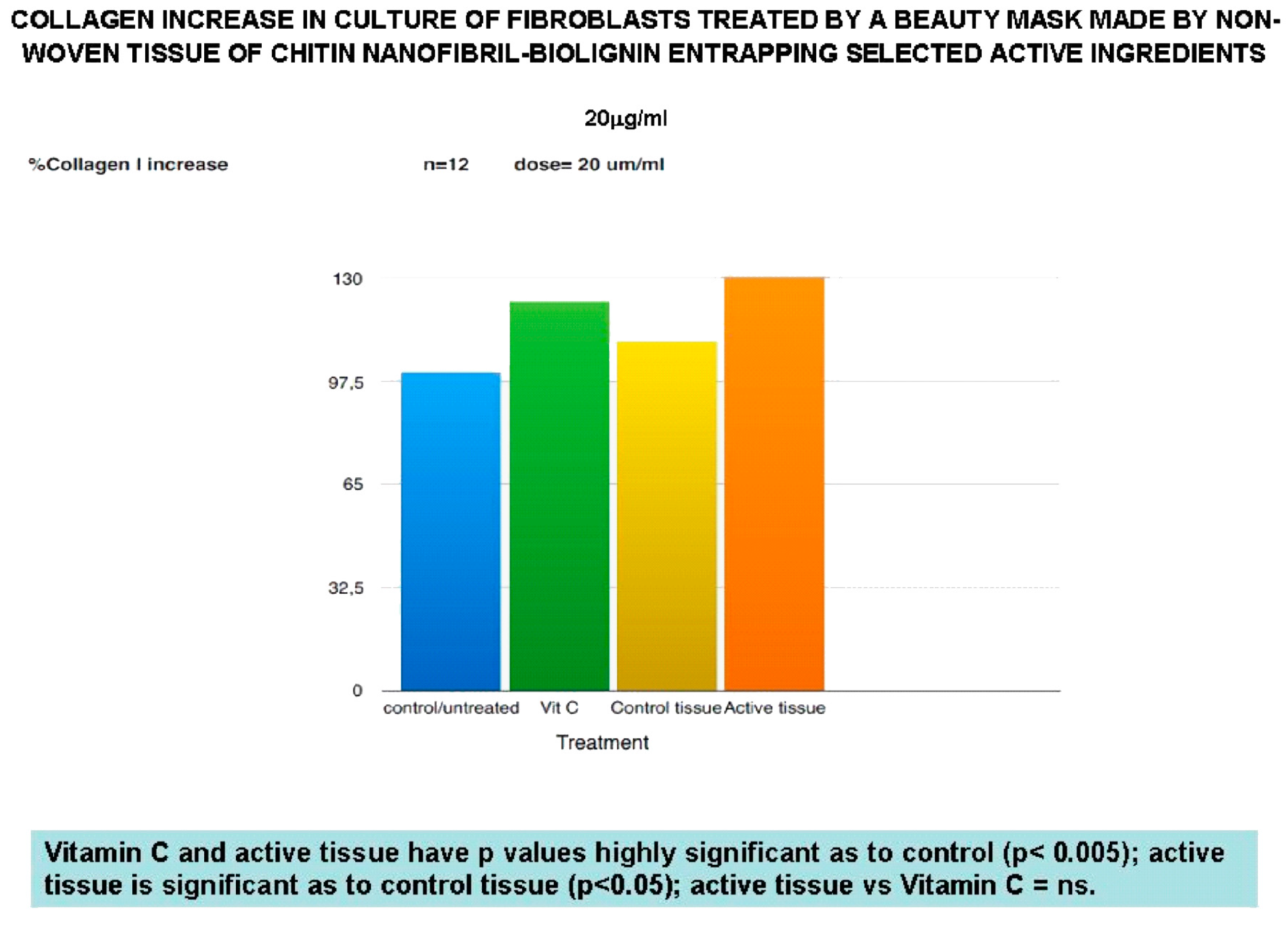
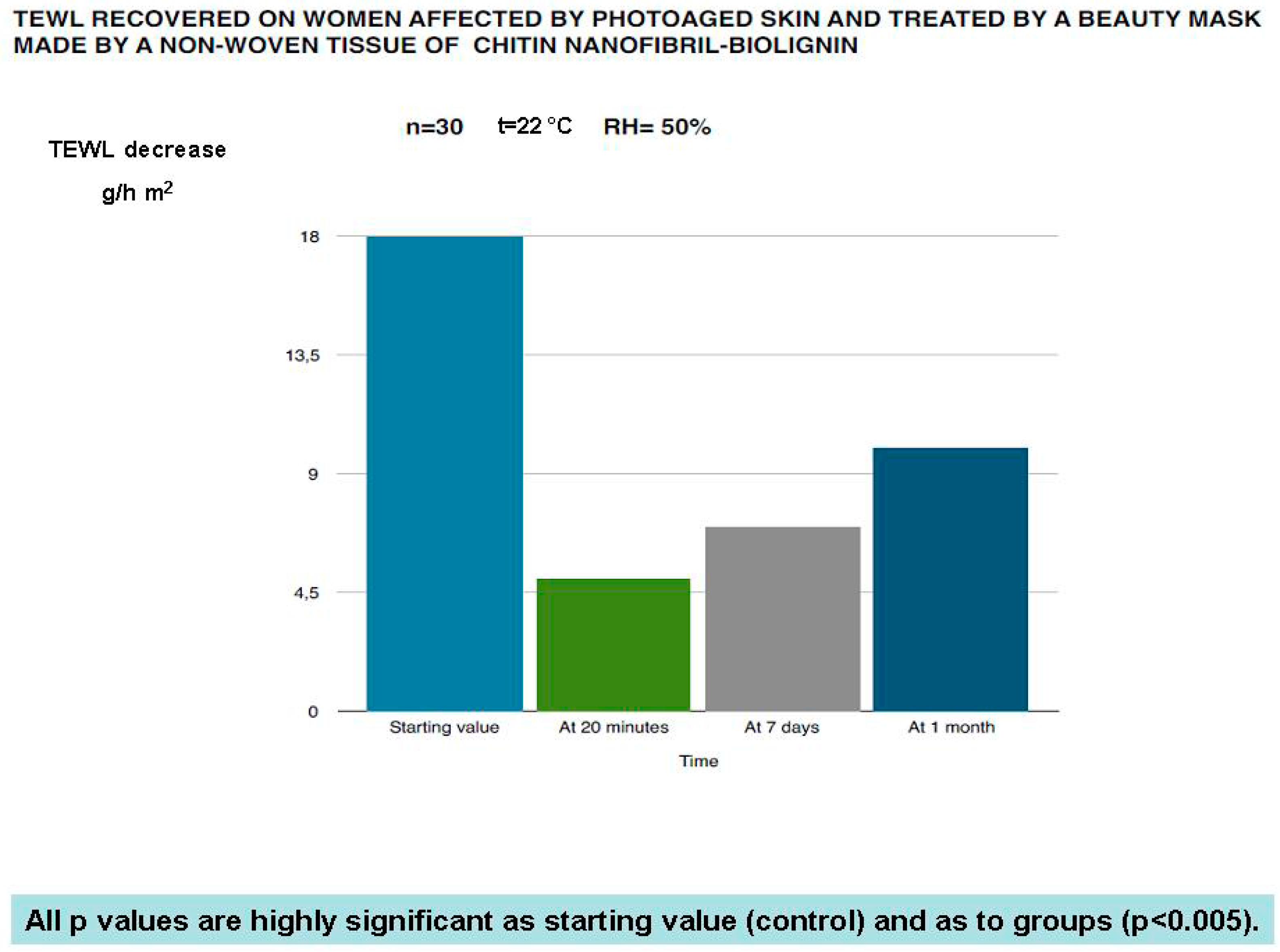
3. Results and Discussion
3.1. In Vitro
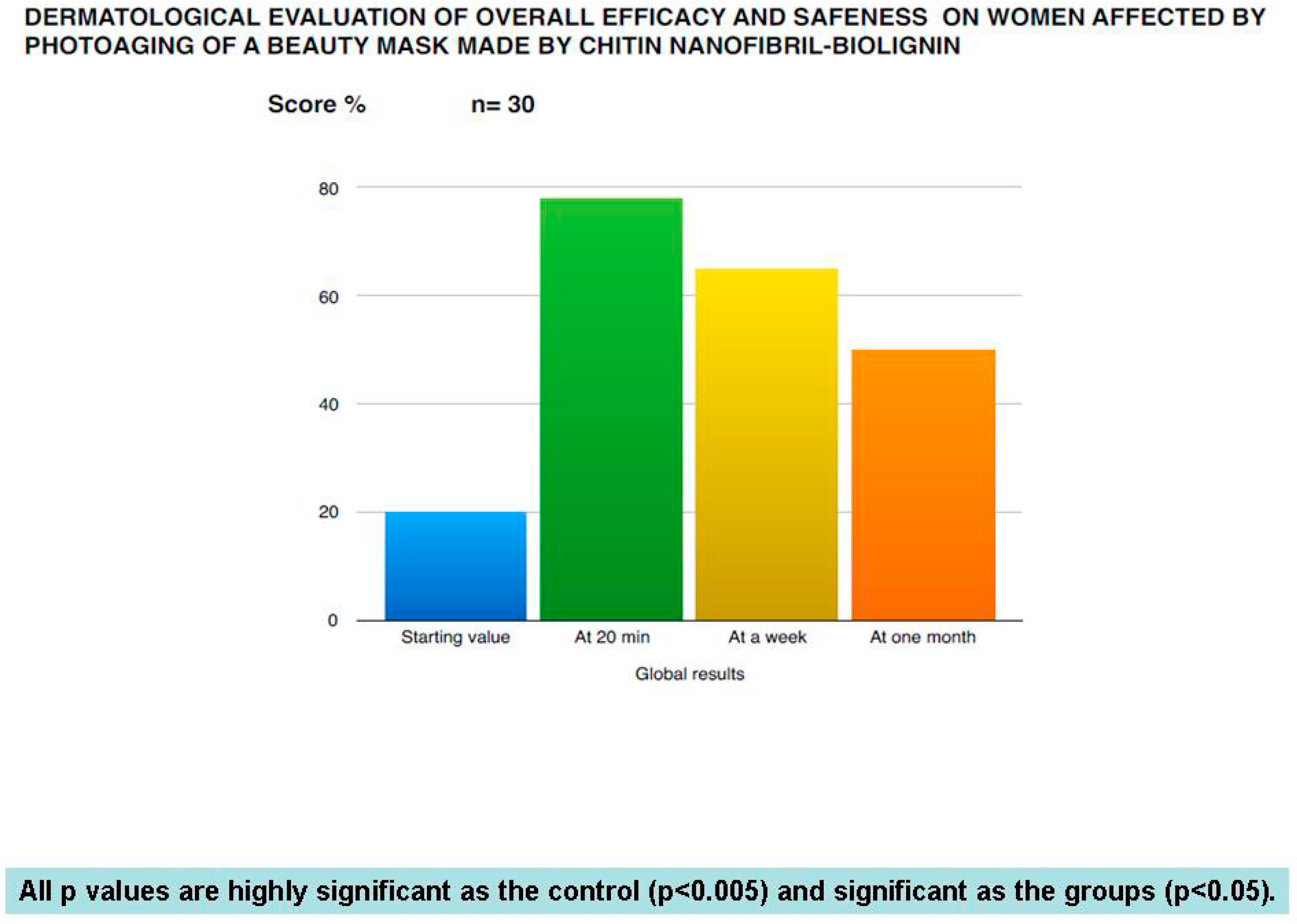
3.2. In Vivo
4. Conclusions
Acknowledgments
Author Contributions
Conflicts of Interest
References and Notes
- Roco, M.C.; Mirkin, C.A.; Hersan, M.C. WTEC Panel Report on Nanotechnology Research Direction for Societal Needs in 2020: Retrospective and Outlook. Available online: http://www.wtec.org/nano2/Nanotechnology_Research_Directions_to_2020 (accessed on 15 November 2016).
- National Science Foundation, FY 2015 Strategic Sustainability Performance Plan. Available online: https://www.nsf.gov/pubs/2016/nsf16025/nsf16025 (accessed on 15 November 2016).
- Nellmann, C.; MacDevette, M.; Manders, T.; Eickhout, B.; Svihus, B.; Prins, A.G.; Kaltenborn, B.P. The Environmental Food Crisis; UNEP, Birkeland Trykkeri AG: Oslo, Norway, 2009. [Google Scholar]
- The United Nations Environment Programme. Converting Waste Agricultural Biomass into a Resource; UNEP: Osaka, Japan; Shiga, Japan, 2009. [Google Scholar]
- Lipinski, B.; Hanson, C.; Lomax, J.; Kitinoja, L. Creating Food Loss and Waste; UNEP Working Paper, June; UNEP: Osaka, Japan; Shiga, Japan, 2013. [Google Scholar]
- The United Nations Environment Programme. Human Health and the Environment. Post Note 3. 2015. Available online: www.unep.org/post (accessed on 15 November 2016).
- European Commission. General Union Environment. Living Well, within the Limits of Our Planet; Publications Office of the European Union: Luxemburg, 2014. [Google Scholar]
- SOER. The European Environment-State and Outlook 2015-European Briefings—Green Economy; European Environment Agency: Copenhagen, Denmark, 2015. [Google Scholar]
- The United Nations Environment Programme. Towards a Green Economy: Pathways to Sustainable Development and Poverty Eradication; United Environment Programme: New York, NY, USA, 2011. [Google Scholar]
- Organisation for Economic Co-operation and Development. Towards Green Growth; Organization for Economic Cooperation and Development: Paris, France, 2011. [Google Scholar]
- Santis, M.R.E.; Fonseca, A.C.; Mendonca, P.V.; Branco, R.; Serra, A.C.; Morais, P.V.; Coelho, J.F.J. Recent Developments in Antimicrobial Polymers: A Review. Materials 2016, 9, 599. [Google Scholar] [CrossRef]
- Morganti, P. New Horizon in Cosmetic Dermatology. J. Appl. Cosmetol. 2016, 34, 9–18. [Google Scholar]
- Morganti, P.; Muzzarelli, C. Spray-Dried Chitin Nanofibrils, Method for Production Uses Thereof. U.S. Patent 8,552,164, 8 October 2013. [Google Scholar]
- Muzzarelli, C.; Morganti, P. Preparation of Chitin and Derivatives Thereof for Cosmetic and Therapeutic Use. U.S. Patent 8,383,157, 26 February 2013. [Google Scholar]
- Morganti, P.; Muzzarelli, R.A.A.; Muzzarelli, C. Multifunctional use of Innovative Chitin Nanofibrils for Skin Care. J. Appl. Cosmetol. 2006, 24, 105–114. [Google Scholar]
- Morganti, P.; Del Ciotto, P.; Fabrizi, G.; Guarneri, F.; Cardillo, A.; Palombo, M.; Morganti, G. Safety and Tolerability of Chitin Nanofibrils-Hyaluronic acid nanoparticles entrapping Lutein. Note 1: Nanoparticles Characterization and Bioavailability. SOFW J. 2013, 139, 12–23. [Google Scholar]
- Morganti, P.; Tishchenko, G.; Palombo, M.; Kelnar, L.; Brozova, L.; Spirkova, M.; Pavlova, E.; Kobera, L.; Carezzi, F. Chitin nanofibrils for biomimetic products: Nanoparticles and nanocomposite chitosan films in health-care. In Marine Biomaterials: Isolation, Characterization and Application; Kim, S.-K., Ed.; CRC-Press: New York, NY, USA, 2013; pp. 681–715. [Google Scholar]
- Morganti, P.; Chen, H.D.; Gao, X.H.; Del Ciotto, P.; Carezzi, F.; Morganti, G. Nanoparticles of Chitin Nanofibril Hyaluronan block polymer entrapping Lutein as UVA protective compound. In Carotenoids: Food Source, Production and Health benefits; Yamaguchi, M., Ed.; Nova Science Publishers, Inc.: New York, NY, USA, 2013; pp. 237–259. [Google Scholar]
- Morganti, P.; Carezzi, F.; Morganti, G.; Fabrizi, G. Chitin-Hyaluronan Nanoparticles to Deliver Anti-aging Ingredients through the Skin. Cosmetics 2014, 1, 140–158. [Google Scholar] [CrossRef]
- Morganti, P.; Carezzi, F.; Del Ciotto, P.; Tishchenco, G.; Chianese, A. A Green Multifunctional Polymer from Discarded Material: Chitin Nanofibril. Br. J. Appl. Sci. Technol. 2014, 4, 4175–4190. [Google Scholar] [CrossRef]
- Morganti, P.; Del Ciotto, P.; Carezzi, F.; Guarneri, F.; Yeo, Y.J. Skin Lightening Efficacy of New Formulations Enhanced by Chitin Nanoparticles Delivery System. Note 1. J. Appl. Cosmetol. 2014, 32, 57–71. [Google Scholar]
- Morganti, P. The meaning of nanodimension involving the Cosmetic Chemist from lab to the Industrial Process. J. Sci. Res. Rep. 2014, 4, 79–100. [Google Scholar]
- Eide, K.B.; Norberg, A.L.; Heggset, E.B.; Lindbom, A.R.; Varum, K.M.; Eijsink, V.G.H.; Sorlie, M. Human Chitotriosidase-Catalyzed Hydrolysis of Chitosan. Biochemistry 2012, 51, 487–495. [Google Scholar] [CrossRef] [PubMed]
- Morganti, P. Green economy and bionanotechnology to transform waste materials in useful goods: Results of EU projects. Eurocosmetics 2015, 22, 12–16. [Google Scholar]
- Mikesiva, J.; Haseka, J.; Tishchenko, G.; Morganti, P. Rheological study of Chitosan acetate solutions containing chitin nanofibrils. Carbohydr. Polym. 2014, 112, 753–757. [Google Scholar] [CrossRef] [PubMed]
- Vanholme, R.; Denedts, B.; Morreel, K.; Ralph, J.; Boerjan, W. Lignin biosynthesis and structure. Plant Physiol. 2010, 153, 895–905. [Google Scholar] [CrossRef] [PubMed]
- Ten, E.; Vermerris, W. Recent Developments in polymers derived from industrial lignin. J. Appl. Polym. Sci. 2015, 132, 1–13. [Google Scholar] [CrossRef]
- Èspinosa-Acosta, J.L.; Torres-Chavez, P.I.; Ramirez-Wong, B.; Lopez-Saiz, C.M.; Montano-Leyva, B. Antioxidant, Antimicrobial, and Antimutagenic Properties of Technical Lignins and Their Applications. BioResources 2016, 11, 1–30. [Google Scholar]
- Morganti, P. Green Ingredients in Cosmetic Dermatology. Molecular Aspects of Ingredients and Carriers. J. Appl. Cosmetol. 2016, 34, 65–79. [Google Scholar]
- Morganti, P. Innovative and Natural Beauty Masks Eco-compatible and Skin-friendly. Report presented at University of Shenyang and University of Jilin, China, 14 January 2015. [Google Scholar]
- Matts, P.J.; Rawlings, A.V. The effects of niacinamide-containing moisturizers. In Skin Moisturization, 2nd ed.; Rawlings, A.V., Leyden, J.J., Eds.; Informa: New York, NY, USA, 2009; pp. 232–333. [Google Scholar]
- Surjana, D.; Halliday, G.M.; Damian, D.L. Role of nicotinamide in DNA damage, mutagenesis, and DNA repair. J. Nucleic Acids 2010. [Google Scholar] [CrossRef] [PubMed]
- Susja, A.D.; Halliday, G.M.; Damian, D.L. Nicotinamide enhances repair of ultraviolet radiation-induced DNA damage in human keratinocytes and ex vivo skin. Carcinogenesis 2013, 34, 1144–1149. [Google Scholar]
- Klesriczynski, K.; Fischer, T.W. Melatonin and human skin aging. Dermatoendocrinology 2012, 4, 245–252. [Google Scholar] [CrossRef] [PubMed]
- Cardillo, A.; Morganti, P. Fast and non-invasive method for assessing skin hydration. J. Appl. Cosmetol. 1994, 12, 11–16. [Google Scholar]
- Morganti, P.; Fabrizi, G.; Palombo, P.; Palombo, M.; Ruocco, E.; Cardillo, A.; Morganti, G. Chitin-Nanofibrils: A New Active Cosmetic Carrier. J. Appl. Cosmetol. 2008, 26, 113–128. [Google Scholar]
- Morganti, P.; Del Ciotto, P.; Stoller, M.; Chianese, A. Antibacterial and Anti-inflammatory Green Nanocomposites. Chem. Eng. Trans. 2016, 47, 61–66. [Google Scholar]
- Modann, T. Rapid colorimetric assay for cellular growth and survival: Application to proliferation and citotoxicity assays. J. Immunol. Methods 1983, 65, 55–63. [Google Scholar]
- Morganti, P.; Palombo, M.; Fabrizi, G.; Guarneri, G.; Slovacchia, F.; Cardillo, A.; Del Ciotto, P.; Carezzi, F.; Morganti, G. New Insight on Anti-aging Activity of Chitin Nanofibril-Hyaluronan Block Copolymers Entrapping Active Ingredients: In vitro and in vivo study. J. Appl. Cosmetol. 2013, 31, 1–29. [Google Scholar]
- Krutmann, J.; Schroeder, P. Role of mitochondria in photoaging of human skin-the defective powerhouse model. J. Invest. Dermatol. Symp. Proc. 2009, 14, 44–49. [Google Scholar] [CrossRef] [PubMed]
- Larnier, C.; Ortonne, J.P.; Venot, A.; Faivre, B.; Beani, J.C.; Thomas, P.; Brown, T.C.; Sendagorta, E. Evaluation of Cutaneous photodamages using a photographic scale. Br. J. Dermatol. 1994, 130, 167–173. [Google Scholar] [CrossRef] [PubMed]
- Curfs, J.H.A.J.; Meis, J.F.G.M.; Hoogkamp-Kostanje, J.A.A. A primer on cytokines: Sources, receptors, effects and inducers. Clin. Microbiol. Rev. 1997, 10, 742–780. [Google Scholar] [PubMed]
- Walter, S.L.; Young, A.R. An action spectrum (299–320 nm) for TNA-alpha protein in human skin in vivo suggests that basal layer Epidermal DNA is the chromophore. PNAS 2007, 104, 19051–19054. [Google Scholar]
- Young, A.R.; Chadwick, C.A.; Harrison, G.I.; Nikaido, O.; Ramsden, J.; Potter, C.S. The similarity of action spectra of thymine dimers in human epidermis and erythema suggests that DNA is the chromophore of erythema. J. Invest. Dermatol. 1998, 111, 982–988. [Google Scholar] [CrossRef] [PubMed]
- Schottelius, A.J.G.; Moldawer, L.L.; Dinarello, C.A.; Asadullah, K.; Sterry, W.; Edwards, C.K. Biology of tumour necrosis Factor-alpha-implications of psoriasis. Exp. Dermatol. 2004, 13, 193–222. [Google Scholar] [CrossRef] [PubMed]
- Da Bara, A.; Sekido, N.; Akahoshi, T.; Wada, T.; Mukaida, N.; Matsushima, K. Essential involvement of interleukine-8 in acute inflammation. J. Leukoc. Biol. 1994, 56, 559–564. [Google Scholar]
- Rijken, F.; Bruijnzeel, P.L.B. The pathogenesis of photoaging: The role of neutrophils and neutrophil-derived enzymes. J. Invest. Dermatol. Symp. Proc. 2009, 14, 67–72. [Google Scholar] [CrossRef] [PubMed]
- Quan, T.; Qin, Z.; Xia, W.; Shao, Y.; Voorthees, J.J.; Fisher, G.J. Matrix-degrading metalloproteinases in photoaging. J. Invest. Dermatol. Symp. Proc. 2009, 14, 20–24. [Google Scholar] [CrossRef] [PubMed]
- Pillai, S.; Oresajo, C.; Hayward, J. Ultraviolet radiation and skin aging: Roles of reactive oxygen species, inflammation and protease activation, and strategies for prevention of inflammation-induced matrix degradation-a review. Int. J. Cosmet. Sci. 2005, 27, 17–34. [Google Scholar] [CrossRef] [PubMed]
- Antonicelli, F.; Bellon, G.; Debelle, L.; Homebeck, W. Elastin-elastase and inflamm-aging. Rev. Curr. Top Dev. Biol. 2007, 79, 99–155. [Google Scholar]
- Morganti, P.; Del Ciotto, P.; Fabien-Soule, V. Application of chitin nanofibrils and collagen of marine origin as bioactive ingredients. In Marine Cosmeceuticals: Latest Trends and Prospects; Kim, S.K., Ed.; CRC Press: New York, NY, USA, 2011; pp. 267–290. [Google Scholar]
- Morganti, P. Reflections on Cosmetics, Cosmeceuticals, and Nutraceuticals. Clin. Dermatol. 2008, 26, 318–320. [Google Scholar] [CrossRef] [PubMed]
- Morganti, P.; Palombo, P.; Palombo, M.; Fabrizi, G.; Cardillo, A.; Carezzi, F.; Morganti, G.; Ruocco, E.; Dziergowski, S. Cosmetic in skin aging: Achieving the efficacy by the chitin nano-structured crystallites. SOFW J. 2010, 136, 14–25. [Google Scholar]
- Morganti, P.; Fabrizi, G.; Palombo, P.; Palombo, M.; Guarneri, F.; Cardillo, A.; Morganti, G. New Chitin Complexes and their Anti-Aging Activity from Inside Out. JNHA 2012, 16, 242–245. [Google Scholar] [CrossRef]
- Morganti, P. Use of Chitin Nanofibrils from biomass for an Innovative Bioeconomy. In Nanofabrication Using Nanomaterials; Ebothe, J., Ahmed, W., Eds.; One Central Press: London, UK, 2016; pp. 1–22. [Google Scholar]
- Quian, Y.; Qiu, X.; Zhu, S. Lignin: A nature-inspired sun blocker for broad-spectrum sunscreens. Green Chem. 2015, 17, 320–324. [Google Scholar] [CrossRef]
- Morganti, P. The Easy Biodegradability of an Innovative Non-woven Tissue. Unpublished Data. 2016. [Google Scholar]


© 2016 by the authors; licensee MDPI, Basel, Switzerland. This article is an open access article distributed under the terms and conditions of the Creative Commons Attribution (CC-BY) license (http://creativecommons.org/licenses/by/4.0/).
Share and Cite
Morganti, P.; Palombo, M.; Carezzi, F.; Nunziata, M.L.; Morganti, G.; Cardillo, M.; Chianese, A. Green Nanotechnology Serving the Bioeconomy: Natural Beauty Masks to Save the Environment. Cosmetics 2016, 3, 41. https://doi.org/10.3390/cosmetics3040041
Morganti P, Palombo M, Carezzi F, Nunziata ML, Morganti G, Cardillo M, Chianese A. Green Nanotechnology Serving the Bioeconomy: Natural Beauty Masks to Save the Environment. Cosmetics. 2016; 3(4):41. https://doi.org/10.3390/cosmetics3040041
Chicago/Turabian StyleMorganti, Pierfrancesco, Marco Palombo, Francesco Carezzi, Maria Luisa Nunziata, Gianluca Morganti, Maria Cardillo, and Angelo Chianese. 2016. "Green Nanotechnology Serving the Bioeconomy: Natural Beauty Masks to Save the Environment" Cosmetics 3, no. 4: 41. https://doi.org/10.3390/cosmetics3040041