Article

GC-MS Analysis of the Phytochemical Constituents, Safety Assessment, Wound Healing and Anti-Inflammatory Activities of Cucurbita pepo Leaf Extract in Rats

by
Emmanuel Iroha Akubugwo
1,†,
Okezie Emmanuel
1,†,
Celestine Nwabu Ekweogu
2,†,
Ositadinma Chinyere Ugbogu
3,†,
Tochukwu Remigius Onuorah
1,†,
Ozioma Glory Egeduzu
1,† and
Eziuche Amadike Ugbogu
1,*
1
Department of Biochemistry, Abia State University, Uturu PMB 2000, Abia State, Nigeria
2
Department of Medical Biochemistry, Imo State University, Owerri PMB 2000, Imo State, Nigeria
3
Department of Microbiology, Abia State University, Uturu PMB 2000, Abia State, Nigeria
*
Author to whom correspondence should be addressed.
These authors contributed equally to this work.
Sci. Pharm. 2022, 90(4), 64; https://doi.org/10.3390/scipharm90040064
Submission received: 18 May 2022 / Revised: 21 June 2022 / Accepted: 22 June 2022 / Published: 14 October 2022

Abstract

:
In traditional medicine, Cucurbita pepo L. is used for the treatment of rheumatism, diabetes, inflammations, and wound injuries. This study was conducted to evaluate the phytochemical constituents, safety profile, wound healing, and anti-inflammatory activities of Cucurbita pepo leaf extract in rats. The phytochemical analysis of C. pepo extract was carried out using gas chromatography-mass spectrometry (GC-MS). In acute toxicity tests, the rats orally received a single dose of 5 g/kg extract of C. pepo. In a subacute toxicity study, the rats received 200, 400, and 800 mg/kg of the C. pepo extract via daily gavage for 14 days. Bioactive compounds 1-octen-3-ol, nonanal, trans-β-ionone, phytol, trans-farnesol, and squalene were identified. There were no toxic effects detected in any of the evaluated parameters, namely liver, kidney, haematological, lipid, and antioxidant enzymes. In wound healing, C. pepo extract showed greater % wound contraction and tensile strength, as well as reduced wound healing time (12 days) and epithelialization when compared to the control (normal saline) and povidone-iodine treated groups. Rats treated with C. pepo extract elicit anti-inflammatory activity. The findings of this study revealed that the C. pepo extract has wound healing and anti-inflammatory properties with a wide margin of safety.

1. Introduction

In recent times, the global use of medicinal plants or herbal medicines as either alternative or supportive medicine to conventional medicine has increased tremendously [1]. Today, medicinal plants and their bioactive components are widely accepted as an emerging source of alternative therapy for the management and treatment of various human diseases [2,3]. It is estimated that over 80% of the world population in one way or the other uses medicinal plants and their phytotherapeutic compounds as alternative primary healthcare due to their bioavailability, accessibility, efficacy, affordability, and low associated side effects [4,5]. They are not only used for healthcare remedies but can also serve as novel sources of unlimited raw materials for the discovery of new drugs. In ethnomedicine, many medicinal plants are effectively used for wound healing or wound management because they create a suitable moist environment, demonstrate the shortest healing time, and cause less pain or discomfort to the patients [1]. Wounds (acute, burns, or chronic wounds) are physical injuries that lead to the disruption of normal structures and functions of the tissues and skin [6]. They are traumatic and devastating types of injuries and therefore one of the leading global public concerns. Wound healing is a dynamic biochemical and cellular event involving the repair of damaged tissues and skins [7]. Bioactive components in medicinal plants or herbal medicines have great potential to reduce oxidative stress and inflammatory processes [8], inhibit infections, and promote blood clotting, as well as accelerate wound healing processes by repairing tissues and regenerating damaged skins [9]. Many researchers have reported that medicinal plants are successfully used in the management and treatment of different types of wounds [6,10,11,12], and one such plant is Cucurbita pepo.
Cucurbita pepo L. is a green vegetable that is globally used as functional food and medicine. It belongs to Cucurbitaceae. It is a rich source of carotenoids, liposoluble vitamins, and polyunsaturated fatty acids [13]. It has high contents of flavonoids, terpenoids, alkaloids, proteins, lipids, carbohydrates, minerals [14], and Δ5-, Δ7-, and Δ8-phytosterols [15], which are likely responsible for its medicinal properties. The extract of different parts of C. pepo (leaves, seeds, flowers, and fruits) has shown to exhibit various pharmacological activities such as antioxidants, anti-inflammatory [16], anti-proliferative [17], anti-depressant, anti-ulcer, inhibition of lipid peroxidation [18], anti-diabetic [19], anti-bacterial, anti-viral, anti-hypercholesterolemic, and sedative-hypnotic effects [20]. In traditional medicine, the leaves are used as an analgesic or blood booster [21], to remove biliousness, and for treatments of rheumatism, benign prostatic hyperplasia, burns, and wound injuries [4,14]. Despite the acclaimed ethnopharmacological use of C. pepo in the treatment of many diseases, there are few or no scientific data from experimental studies as regards its toxicity profile, wound healing, and anti-inflammatory effects. This present study was conducted to evaluate the toxicity profile, wound healing, and anti-inflammatory properties of phytochemical characterized C. pepo leaf extract in rats.

2. Materials and Methods

2.1. Plant Collection and Authentication

Fresh leaves of Cucurbita pepo were obtained in June 2021 from Ugbogu’s farmland in Amaku Nvosi, Isiala Ngwa South, a Local Government Area of Abia State, Nigeria, and authenticated by a taxonomist at the University of Nigeria Nsukka, Enugu State. The plant name Cucurbita pepo with voucher number UNH No 336 was deposited in the herbarium.

2.2. Extract Preparation

The fresh leaves of C. pepo were removed from the stem and the leaves thoroughly washed with distilled water. The extract was prepared by weighing 2 kg of the fresh C. pepo leaves in 2 L of water and squeezing for one (1) hour. The resulting juice was filtered using a sterile sieve and Whatmann No 1 filter paper. The filtrate was freeze-dried and preserved in the refrigerator at 4 °C as C. pepo extract until time of use.

2.3. Gas Chromatography-Mass Spectrometric (GC-MS) Analysis

GC-MS analysis of C. pepo extract was performed using GC/MS-QP2010 ultra (Shimadzu Co., Kyoto, Japan) with an autosampler. A 5 MS/HP-column (30 m × 0.25 mm × 0.25 µm) with ultra-pure helium as the carrier gas was used. The helium flows at a flow rate of 1.12 mL/min and a linear velocity of 39 cm/s. The oven temperature was fixed at 110 °C to 280 °C at the rate of 10 °C/min. The injector temperature was set at 250 °C and injection volume as 0.5 µL were made in the split-less mode with a split ratio of 10:1. The MS transfer line and ion source temperatures were maintained at 250 °C and 200 °C, respectively, and the mass spectra detector volt was set at 0.94 kV. The full-scan mass spectra were recorded at 10,000 u/s over the mass range (m/z) 85–500. Peaks and chemical constituents were identified by direct searching in the National Institute of Standards and Technology (NIST) library.

2.4. Animals

Healthy male unused albino rats (200–240 g), purchased from the Laboratory Animal Production Units of Veterinary Medicine College, Michael Okpara University of Agriculture, Umudike, Abia State, Nigeria, were used in the experiment. Prior to the experiments, the rats were allowed to acclimatize for two weeks, maintained under normal standard laboratory conditions of 12 h light/12 h dark cycle at the temperature of 25 ± 3 °C and relative humidity of 50%–70%. The rats were fed with standard commercial growers feed produced by Flour Mills of Nigeria and allowed free access to water. All the animal handling and experimental procedures were strictly adhered to throughout the study as specified by international standards and ethical principles established by Abia State University. This study was approved by Research Ethical Clearance Committee of Abia State University (ABSU/REC/BMR/0117).

2.4.1. Acute Toxicity Study

The method established by The Organization for Economic Cooperation and Development (OECD) guideline 423 [22,23] with some modifications were adopted for this study. Seventy (70) matured male and female rats were randomly divided into 7 groups of 10 rats (5 males and 5 females (kept in different cages) in each group). Group 1 served as a normal control and received 10 mL/kg distilled water whereas Groups 2–7 received graded single dose of 500, 1000, 2000, 3000, 4000, and 5000 mg/kg of C. pepo extract, respectively, by oral gavage. The rats were allowed free access to water and feed and were monitored for signs of toxicity and death within and after a period of 24 h.

2.4.2. Subacute Toxicity Study

The method of established by OECD [22,24] guideline 407 and 425 with some modifications was adopted for this study. Twenty-four (24) albino rats were randomly divided into four (4) groups of six rats. Group 1 served as a normal control and received 0 mg/kg C. pepo extract, whereas groups 2, 3, and 4 received 200, 400, and 800 mg/kg of C. pepo extract, respectively, daily for 14 days.

2.4.3. Blood Sample and Organ Collection

After two weeks (14 days) of extract treatment, the rats were starved of food but without restriction to water for about 12 hr and sacrificed on the 15th day by intraperitoneally administration of a combination of 50 mg/kg of ketamine and 5 mg/kg of xylazine. Then, the blood samples were collected via cardiac puncture with a needle and syringe. The blood samples for serum biochemistry and haematological analysis were dispensed into plain and ethylenediaminetetraacetic acid (EDTA) bottles, respectively. The vital organs, spleen, lungs, liver, kidneys, testes and heart, were harvested, and their respective weights recorded. The liver and kidney were fixed into 10% v/v formalin saline prior to histology evaluation.

2.4.4. Blood Analysis

The complete haematological parameters were performed using an Automated Haematology Analyser (model BC-2800 Mindray Company, Hyderabad, Telangana, India). The haematological parameters evaluated include haemoglobin (Hb) concentration, packed cell volume (PCV), white blood cell (WBC) count, platelets and red blood cell (RBC) indices (mean corpuscular haemoglobin (MCH), mean corpuscular haemoglobin concentration (MCHC), and mean corpuscular volume (MCV).

2.4.5. Clinical Biochemical Analysis

Renal function parameters (creatinine, urea, sodium, potassium, bicarbonate, and chloride), liver enzymes parameters (alanine aminotransferase, alkaline phosphatase aspartate aminotransferase, bilirubin, total protein, globulin, and albumin), and lipid profile parameters (total cholesterol, triacylglycerol, high-density lipoprotein cholesterol, low density lipoprotein cholesterol, and very low-density lipoprotein cholesterol) levels were evaluated spectrophotometrically using standard laboratory kits (Randox Laboratory Ltd., Co., Antrim, UK).
Oxidative stress markers were evaluated using the methods previously described by the following authors: superoxide dismutase [25], catalase [26], malondialdehyde [27], and glutathione [28].

2.4.6. Histopathology

The histopathological studies were performed on the liver and kidney. The liver and kidney were fixed in 10% formalin and routinely processed and embedded in paraffin wax. Thereafter, the tissues were sectioned (5 μm thickness), stained with haematoxylin and eosin (H and E) dyes, and then mounted and viewed under a light microscope at 400× g magnification.

2.5. Wound Healing Experiments

Excision and incision wound models were used to evaluate the wound healing potential of aqueous extract of C. pepo.

2.5.1. Excision Wound Models

Eighteen (18) healthy male rats (200–240 g) were divided into three (3) groups of six (6) rats each as follows. Group 1 was a negative control (treated with normal saline), while groups 2 and 3 were treated with 5% w/w povidone iodine (PI) and 10% w/w C. pepo extract, respectively, for 16 days. The rats were anaesthetized with 50 mg/kg of ketamine hydrochloride via intraperitoneal route [29,30]. The dorsal surface of the rats was shaved with a rechargeable clipper. After 30 min, an excisional wound of approximately 500 mm2 and 2 mm depth was created on the shaved back of the rats. Treatment of the wounds started immediately after creation of the wounds by daily application of the ointments as described above. Day 0 was considered as wound day. Prior to epithelialization and complete wound closure, the wounds were observed daily, whereas the dimensions of the excision wounds were measured every 4 days with transparent paper and thereafter estimated on a 1 mm2 graph sheet. Percentage wound contraction [6,31] was calculated using the formula:
%   Wound   contraction   =   wound   area   on   day   0 wound   area   on   specific   day(s)   wound   area   on   day   0   × 100
where s = number of days (4th, 8th, 12th, 16th, and 20th day).
The period of epithelialization was calculated as described in [32,33].

2.5.2. Wound Incision Model

Eighteen (18) healthy male rats (200–240 g) were divided into three (3) groups of six (6) rats each. The rats were anaesthetized with 50 mg/kg of ketamine hydrochloride via intraperitoneal route [29,30]. The dorsal surface of the rats was shaved with a rechargeable clipper. After 30 min, a sterile scalpel blade was used to make long paravertebral incision wounds of about 5 cm length and 1.5 cm from the middle of vertebral column on the shaved back [34]. Afterward, the incised wounds were closed by stitching 0.5 cm apart using sterile surgical thread and a curve needle no. 11 [35]. The rats were treated with normal saline, 5% w/w PI, and 10% w/w C. pepo extract for groups 1, 2, and 3, respectively, for 7 days. On the 8th day post wounding, the sutures were removed, and wound breaking strength was measured on the 10th day post-wounding.

2.5.3. Wound Breaking Strength Measurement

The wound breaking strength was determined using Lee’s constant water flow method [36]. The sedated rats were fastened on the laboratory bench on the 10th day after wounding, and a 3.5 mm line was drawn away from either side of the paravertebral long incision wounds, then two forceps were firmly applied to the line with both ends facing each other. The other forceps were attached to a freely suspended light weight graduated polypropylene container by a string flowing over to a pulley, and water was permitted to flow slowly and steadily into the graduated polypropylene container from the reservoir, while one of the forceps was fixed. The tensile strength of the healed wound was assessed by gradually raising the water levels drop by drop until the wound broke or opened, then stopping the water flow and measuring and recording the weight of the water in the container.
The following formula was used to compute the tensile strength:
Tensile   strength   =   Breaking   strength   (g) Cross   section   area   of   skin   ( mm 2 )

2.6. Anti-Inflammatory

In the anti-inflammatory experiment, the method described by Orieke et al. [37] was adopted for the study. Twenty-five (25) sets of male albino rats (200–240 g) were randomly divided into 5 groups of 5 rats each (n = 5/group). Group 1 served as a normal control and was orally administered 0.2 mL/kg of normal saline. Group 2 received 100 mg/kg of aspirin, while groups 3, 4, and 5 orally received 200, 400, and 800 mg/kg of C. pepo extract, respectively. Thirty minutes (30 min) after oral treatments, paw edema was induced by sub-plantar injection of 0.1 mL egg albumin to each rat the right hind paw. After the induction, paw circumferences of the treated rats and control were measured and recorded at 0 min, 30 min, 60 min, and 120 min time intervals. The degree of edema was obtained by subtracting the initial paw circumference from the final paw circumference.
%   Rise   in   paw   circumference   (c)   at   induction   =   C   at   induction C   before   induction C   before   induction × 100
%   Fall   in   paw   circumference   (c)   at   intervals   = C   at   induction C   at   interval C   at   induction × 100
%   Anti - inflammatory   activity   at   120   min   = Mean   fall   in   Test Mean   fall   in   control Mean   fall   in   control × 100

2.7. Data Analysis

Calculations and statistics analysis were performed using Microsoft excel, R™ Statistic software, version 3.0.3 (https://cran.r-project.org/src/base/R-3/, accessed on 17 November 2021) and GraphPad Prism version 5.0 (San Diego, CA, USA). Results were expressed as mean ± standard deviation. One-way analysis of variance (ANOVA) with a Tukey post hoc test was used to establish statistical significance at p-value of ≤0.05.

3. Results

3.1. Identification of Chemical Compounds Present in Methanol Extract of C. Pepo by GC-MS

GC-MS analysis of methanol extract of C. pepo leaf (Table 1A) revealed the presence of fifteen (15) chemical compounds, namely 1-octen-3-ol, hexane, 4-ethyl-2-methyl-, nonanal, 1-cyclohexene-1-carboxaldehyde, 2,6,6-trimethyl-,6,6-trimethyl-7-(3-oxobut-1-enyl)- 3,8-dioxatricyclo [5.1.0.0(2,4)]octan-5-one, cyclohexene, 2-ethenyl-1,3,3-trimethyl-, trans-β-ionone, cyclohexane, 1,1′-(1,2-dimethyl-1, 2-ethanediyl)bis-,2-pentadecanone, 6,10,14-trimethyl, 4-tetradecene, (Z)-, 1-heneicosanol, phytol, trans-farnesol, 1,5,9-undecatriene, 2,6,10-trimethyl-, (Z)-, and squalene. Of the compounds identified, 1-heneicosanol (12.32) was the most abundant, whereas squalene (1.73) was the least abundant. In addition, out of 15 chemical compounds identified, only six (6) compounds are bioactive compounds with pharmacological activities. The six (6) bioactive compounds are 1-octen-3-ol, nonanal, trans-β-ionone, phytol, trans-farnesol, and squalene (Table 1B). The chromatogram of the GC-MS spectral analysis of C. pepo reflecting the peaks of the distinct compounds, their elution, as well as their retention time are shown in Figure 1A,B.

3.2. Acute Oral Toxicity

Table 2 shows the acute toxicity study of C. pepo in albino rats. The study was carried out via oral administration of C. pepo aqueous leaf extract at the single doses of 500, 1000, 2000, 3000, 4000, and 5000 mg/kg. There were no deaths, behavioural changes, or adverse clinical and toxicity signs recorded within 24 h and thereafter for 14 days of C. pepo extract treatments.

3.3. Subacute Toxicity

The experimental animals did not lose weight throughout the study (Figure 2). Following the body weight increase, there was no significant difference (p > 0.05) observed in the final body weight (Day 14) of male rats treated with various doses of C. pepo compared to the control. The effect of C. pepo did not distort the organ weight of the male rats, and there was no significant difference (p > 0.05) observed in the extract treated groups compared to the control group (Table 3).

3.4. Effect on Biochemical and Hematological Parameters

There was significant decrease following a dose-dependent manner in the values of aspartate transaminase (AST) of the C. pepo groups. A significant (p < 0.05) decrease in ALT content was observed in 200 and 400 mg/kg body weight C. pepo treated rats when compared to the control group. However, other liver biomarkers, including total protein, alkaline phosphatase (ALP), albumin, globulin, and bilirubin, were not significantly affected (p > 0.05) when compared to the control group (Figure 3).
Administration of C. pepo did not reveal any statistical difference (p > 0.05) in the renal functional biomarkers (urea, creatinine, Na+, K+, Cl, and HCO3) among the C. pepo treated groups and the control group (Figure 4). The extract revealed a significant decrease in the values of TC, TAG, and LDL compared to the control, while HDL and VLDL did not reveal any statistical difference (p > 0.05) compared to the control (Figure 5). Administration of C. pepo increased all the analysed enzymatic antioxidant parameters (GSH, SOD, and CAT) with statistical increase (p < 0.05) recorded in the values of GSH in the treated groups compared to the control (Figure 6).
The effects of aqueous leaf extract of C. pepo administration after 14 days treatment on haematological parameters is shown in Table 4. The parameters of RBC, PCV, HB, WBC, MCH, platelet, monocyte, eosinophil, and basophil did not significantly differ (p > 0.05) from the control group, whereas there was a significant (p < 0.05) dose-dependent decrease in the values of MCV, MCHC, neutrophil, and lymphocyte when compared to the control group. The histopathological analysis of the liver and kidney of the C. pepo-treated rats and the control group did not reveal any alteration or detrimental changes in the architecture of the liver and kidney (Figure 7 and Figure 8).

3.5. Wound Healing Activity

Table 5 represents the effects of aqueous leaf extract of C. pepo on circular excision % wound contraction and epithelialization time. In this study, application of C. pepo extract on the excision wound rats resulted in a significantly (p < 0.05) higher rate of wound healing in comparison to the control group. The rats treated with C. pepo extract had a significant (p < 0.05) % WC on days 4, 8, 12, and 16 compared to the 5% w/w PI treated rats and control group. On day 16, 100% wound contraction was recorded for the C. pepo extract-treated group, whereas 94.06% and 65.88% were recorded for 5% w/w PI-treated rats and control groups, respectively. A complete and better healing closure was observed in C. pepo-treated rats (12–16 days) compared to the 5% w/w PI-treated rats and control group (12–16 days) (Figure 9). In addition, C. pepo extract-treated group had the shortest epithelialization period of 16 days compared to 5% w/w PI-treated rats (20 days) and control group (22 days).
In the measurement of tensile strength, the C. pepo extract-treated group had a significant (p < 0.05) increase in tensile strength (334.03 g) compared to 5% w/w PI-treated rats (240.33 g) and control group (192.83 g) (Table 6).

3.6. Effect of C. pepo Extract on Egg Albumin-Induced Paw Inflammation

All doses of orally administered C. pepo extract significantly inhibited egg albumin induced paw inflammations in experimental rats when compared with control (p < 0.05), with the degrees of anti-inflammatory activities produced by 200, 400, and 800 mg/kg of the extract comparing favourably with that of 100 mg/kg body weight of aspirin (Table 7).
Although at induction only the lowest dose (200 mg/kg) group had a significantly lower percentage rise in paw circumference when compared with control, the anti-inflammatory effects of all doses of the extract and that of aspirin became visible after 30 min of induction as the degrees of inflammations steadily dropped in the extract and aspirin groups but continued to rise in control (Table 8).
By the end of 120 min, the percentage fall in paw circumference in control was −6.27 ± 12.38 (still indicating steady rise) while in the 200, 400, and 800 mg/kg treated groups, percentage fall in paw circumferences were 11.66 ± 2.49%, 15.36 ± 2.52%, and 14.95 ± 8.58%, respectively, comparing favourably with the aspirin group where the fall was 25.74 ± 5.14% (Table 8).
Percentage anti-inflammatory activities for 200, 400, and 800 mg/kg treatment levels of C. pepo extract were 175.95 ± 75.93%, 217.14 ± 105.79%, and 215.00 ± 176.57%, respectively and compared well with 314.52 ± 193.20% obtained for aspirin (Table 9).

4. Discussion

There are several acclaimed therapeutic potentials of C. pepo as alternative medicine in the management and treatment of various diseases such as diabetes, benign prostatic hyperplasia, and rheumatism as well as inflammations, burns, and wound injuries. However, no scientific study has documented the toxicity profile or anti-inflammatory and wound healing potentials of C. pepo. Therefore, it is pertinent to investigate the safety profile, wound healing, and anti-inflammatory properties of phytochemical characterized aqueous leaf extract of C. pepo in rats.
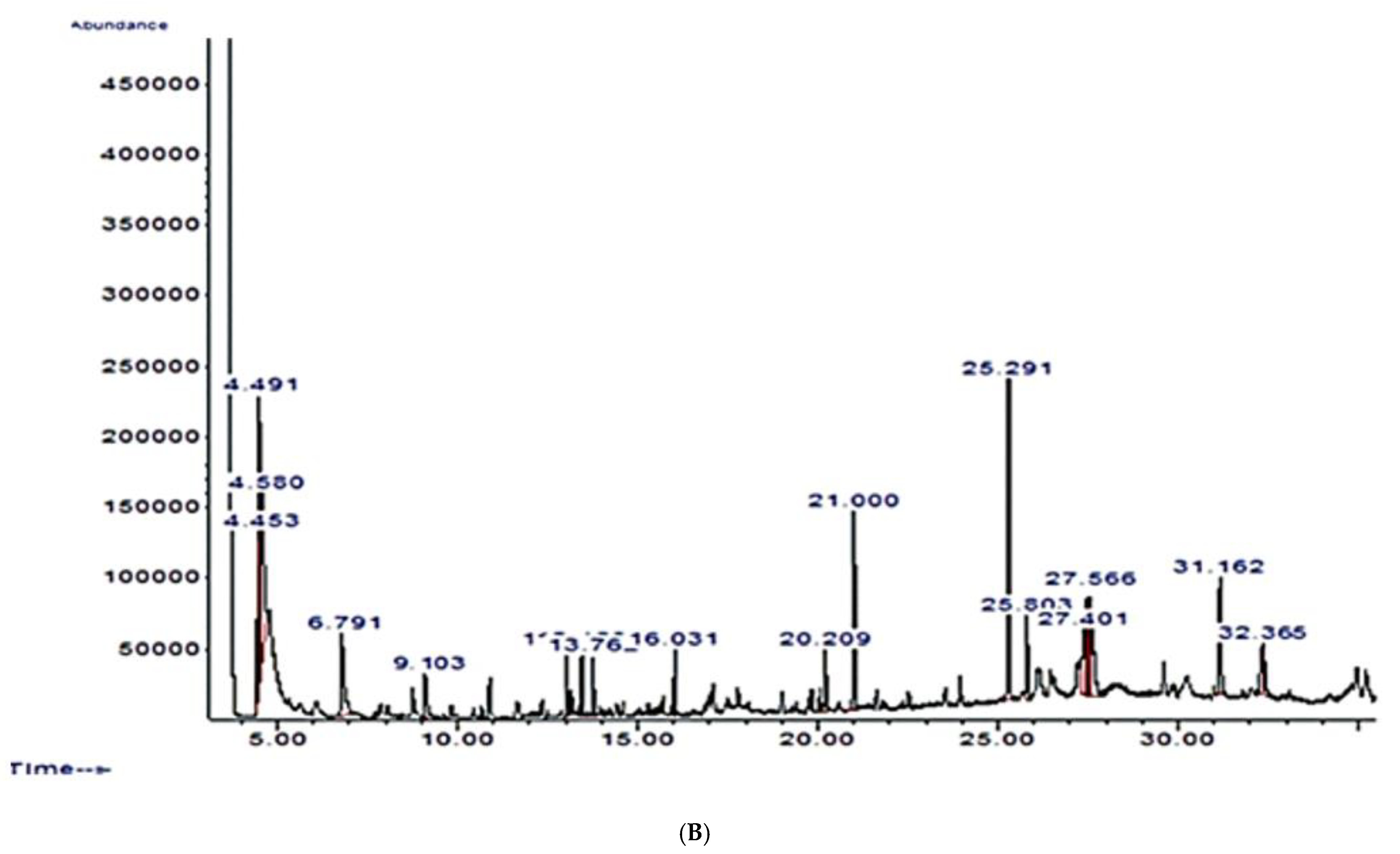
The use of gas chromatography-mass spectrometry (GC-MS) for a very detailed evaluation of bioactive chemicals in plant-based compounds is a well-accepted approach [45]. The GC-MS analysis of C. pepo showed the presence of fifteen (15) chemical compounds with 1-heneicosanol (12.32%) and squalene (1.73%) as the most abundant and least abundant, respectively. Six out of the fifteen identified compounds are known to have some pharmacological potential. These include 1-Octen-3-ol, noanal, trans-β-lonone, phytol, trans-farnesol, and squalene. Xiong et al. [38] reported the antimicrobial properties of 1-Octen-3-ol, while Zhang et al. [39] and Li et al. [40] revealed the antifungal properties of noanal. In other studies, trans-β-lonone has been shown to possess anti-inflammatory, cancer-preventing, antibacterial, antifungal, and anti-leishmanial activities [41], while phytol, trans-farnesol, and squalene have biological activities including anti-inflammatory, antinociceptive, antioxidant, anti-cancer, and antimicrobial properties [42,43,44].
In acute and subacute toxicity studies, appropriate animal models such as rats or mice can be commonly used to evaluate the health risk of any plant extracts or chemical substances [46]. In an acute toxicity study, single-dose oral administration of aqueous leaf extract of C. pepo up to 5000 mg/kg did not cause any death or toxicity signs. Based on the result, the LD50 of C. pepo is higher than 5000 mg/kg, and following OECD [23] guidelines C. pepo can be classified as a class 5 substance and therefore considered a safe and non-toxic substance.
In the subacute toxicity study, there was no change in body weight after 14 days of C. pepo treatment, indicating that it is safe at all dose levels. Adverse effects of medications and chemicals are identified by changes in body weight [47]. Organ weight changes have long been known as a sensitive sign of chemical-induced organ toxicity, such as organ hypertrophy [48]. As a result, relative organ weight is an important measure in toxicity investigations. When compared to normal control animals, no significant change (p > 0.05) in relative organ weight was found after treatment with C. pepo at all dose levels.
Hepatic and renal function tests are used as fingerprint markers to determine the inherent toxicity and pathological states that are tracked to determine organ toxicity or dysfunction caused by hazardous substances [49]. Electrolytes, urea, and creatinine can all be used to determine renal toxicity [50]. Cirrhosis, fibrosis, and liver destruction are all symptoms of hepatotoxicity, which is caused by exposure to toxic substances. Hepatic enzymes such as AST, ALT, and ALP were measured to determine the amount of liver necrosis [51]. In humans, elevated plasma bilirubin levels suggest haemolytic anaemia [52]. The C. pepo administration reduced AST levels but did not affect total protein, ALP, albumin, globulin, or bilirubin levels when compared to the control group. Electrolytes are necessary for the body’s homeostatic balance to be maintained. Sodium keeps the body’s acid–base balance in check and protects it from excessive fluid loss [53]. Pathological conditions can be caused by changes in plasma electrolyte balance. When compared to the normal control group, there was no significant change (p > 0.05) in plasma electrolyte levels (Na+, K+, Cl, and HCO3) after treatment with C. pepo, indicating that there was no harmful effect on electrolyte levels. Furthermore, there was no statistical difference (p < 0.05) in the values of urea and creatinine after administration of C. pepo, indicating no detrimental effect on the functionality of the renal system.
Toxic substances primarily influence the haematopoietic system, which is a very sensitive and significant indicator of physiological and pathological conditions in humans and animals [54]. Toxicants change haematological parameters, resulting in disorders such as anaemia, leukopenia, thrombocytopenia, and bone marrow depression, among others [52]. C. pepo leaf extract did not have a deleterious effect on WBC, RBC, or platelets, nor did it interfere with the haematopoiesis and leucopoiesis processes according to the findings. When compared to the normal control, no toxic changes in all of the studied haematological parameters were seen after 14 days of treatment with C. pepo, indicating no haematotoxic effect and safety at all dose levels.
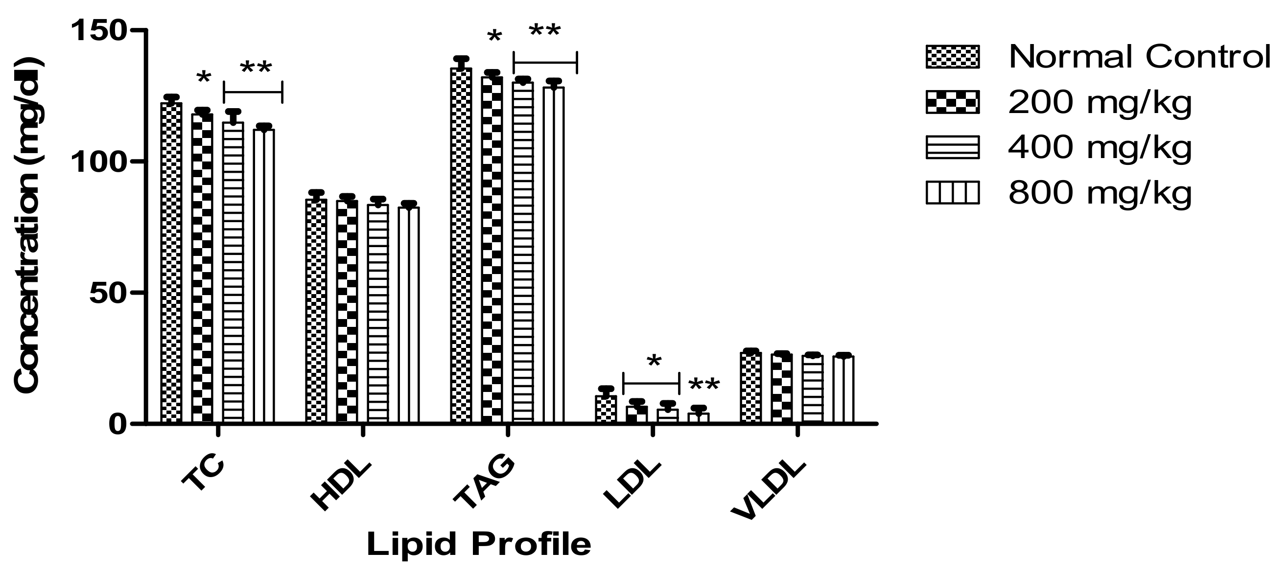
When compared to the control, C. pepo administration reduced atherogenic lipid markers including TC, TAG, and LDL considerably, while HDL and VLDL showed no statistical change (p > 0.05). This suggests that C. pepo may have hypolipidemic properties.
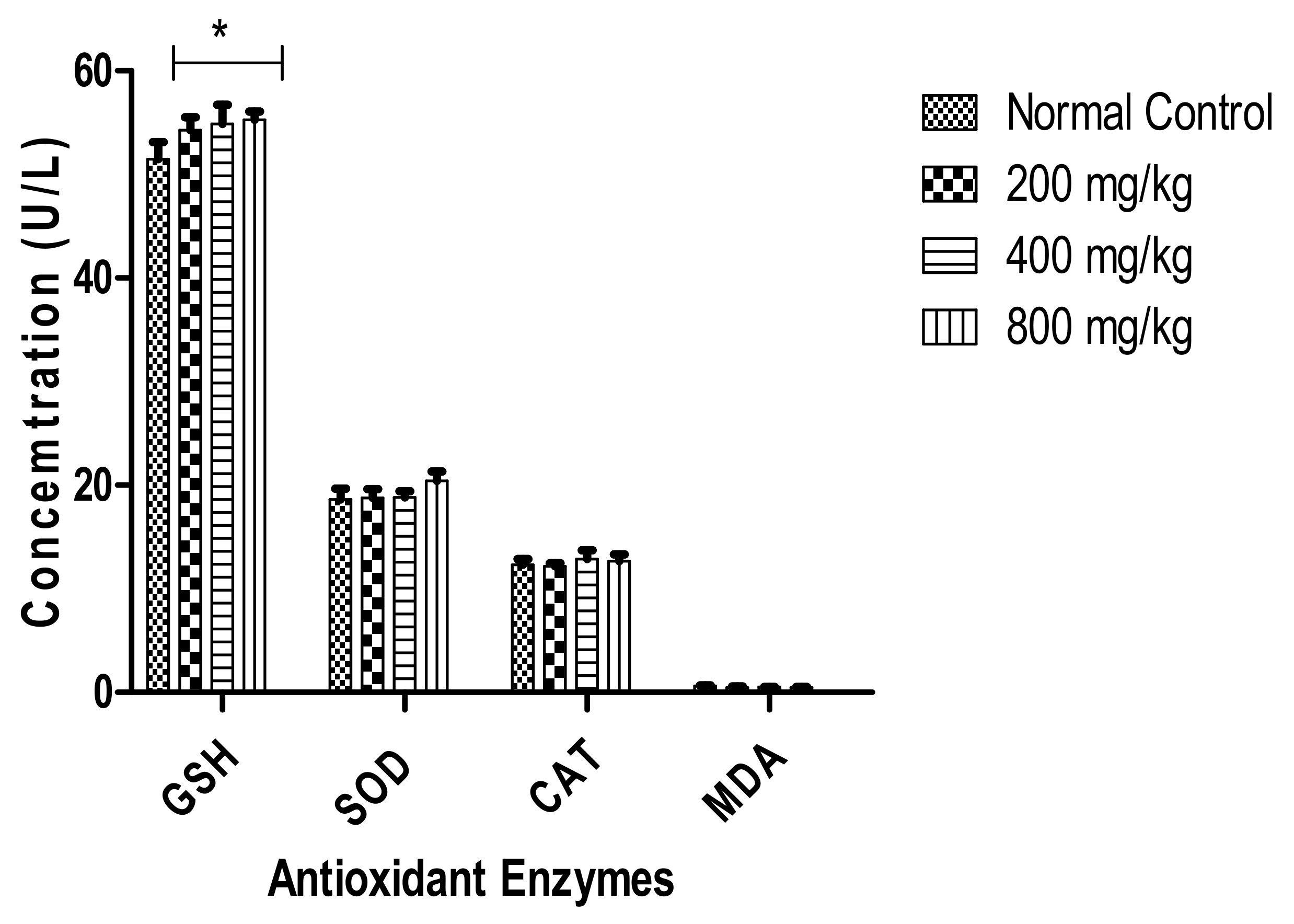
C. pepo administration slightly increased the enzymatic antioxidant parameters GSH, SOD, and CAT with a statistically significant increase (p < 0.05) in GSH levels in the treated groups compared to the control group. Other studies have shown that intake of toxic chemicals affects antioxidant enzymes and elevates MDA levels [55]. MDA is a lipid peroxidation biomarker that may be elevated as a result of toxin ingestion or other diseases [55]. Membrane polyunsaturated fatty acids are reduced by lipid peroxidation, which produces aldehydes including MDA, 4-hydroxynonenal, and acrolein, disrupting and changing the functions of the membrane [56,57]. Lipid peroxidation has been considered an indication of oxidative stress [58,59]. It alters the membrane’s structure by causing changes in fluidity and permeability [60]. Reduced MDA levels and higher GSH, SOD, and CAT levels seen in this study have been reported to protect against oxidative stress [61]. The results supported that C. pepo possesses antioxidant properties. Nkosi et al. [62] reported the antioxidative activity of C. pepo against carbon tetrachloride-induced hepatotoxicity in rats fed with a low protein diet.
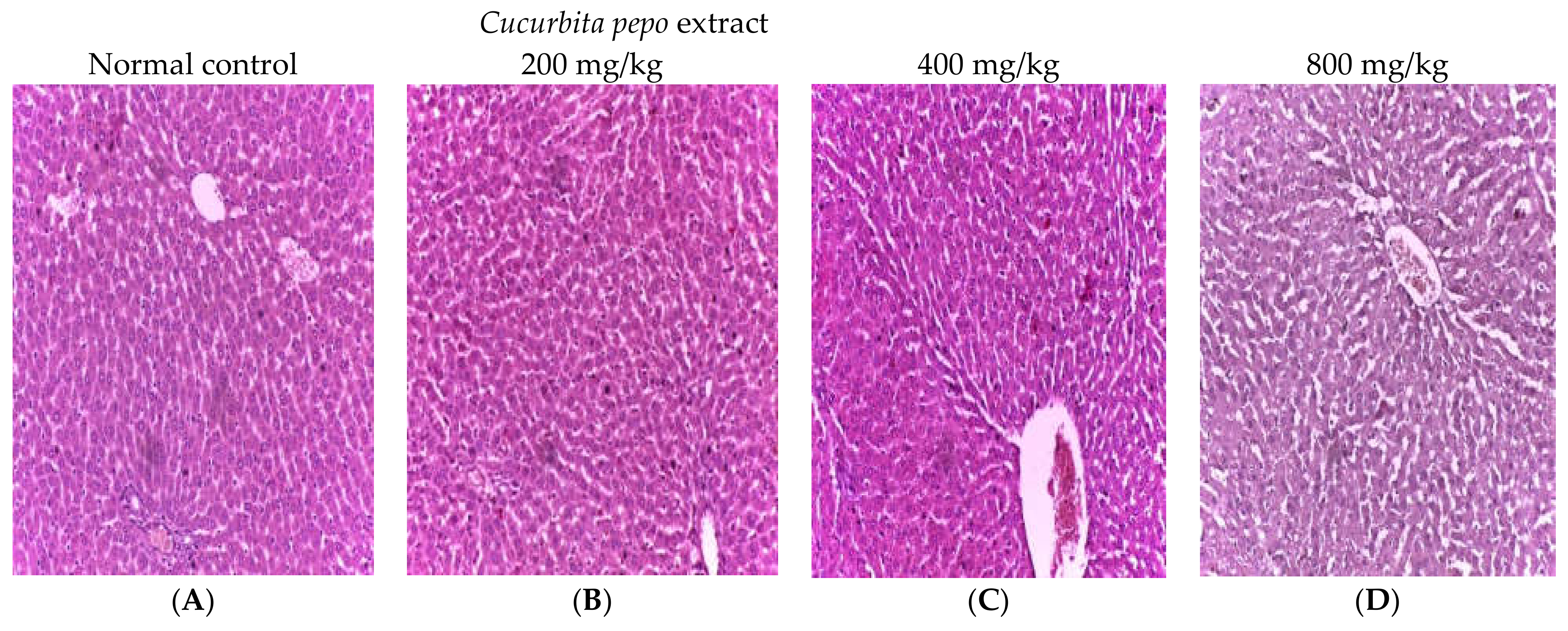
In humans and animals, exposure to various hazardous chemicals raises oxidant levels and induces oxidative DNA damage, which leads to cellular damage and tissue necrosis [63]. A histopathological examination can assist us to determine whether organs have been damaged at the cellular level as a result of a harmful chemical action. The liver and kidneys of rats given varying doses of C. pepo showed no signs of pathological damage. These findings backed up the results of biochemical tests, as well as the safety of C. pepo leaf extract in rats.
The wound healing process aids in the recovery of injured tissue layers and cellular structure. The wound area shrinks as the fibroblast stage begins [64]. Contraction, granulation, epithelization, and collagenation are all part of this complicated process [65,66]. Microbial infections, the presence of free radicals, and metabolic disruptions can all cause the multifactorial process of wound healing to be delayed or interrupted [67]. Wound closure, wound contraction, and functional barrier reorganization are significant markers in the wound healing process [68]. Again, because the type of injury has an impact on wound healing, healing phases of wound models, including excision and incision wound models and dead space, are all affected differently [69]. The fundamental goal of wound management is to heal the damage as quickly as possible while causing the least amount of pain and discomfort to the patient. In the current study, the effect of C. pepo leaf extract on tissue regeneration was investigated utilizing experimental rat models. Rats were continuously observed and treated topically with prepared ointment from C. pepo leaf extract as well as a reference drug after various types of wounds were created. When compared to the control group animals, the C. pepo leaf treatment groups and the reference drug-treated group (povidone-iodine) in the excision and incision wounds showed significant (p < 0.05) reductions in the wound area, rapid epithelization, and rapid rate of wound closure.
It has been reported that, at wound sites, high amounts of reactive oxygen species such as oxygen singlet, hydroxyl radicals, superoxide anion, and hydrogen peroxide stimulate collagen breakdown and hence extracellular matrix degradation (ECM). When the ECM is damaged, processes such as angiogenesis and re-epithelization that are necessary for wound healing are reduced [70,71]. The photograph section of the post-excision wound contraction is shown in Figure 9. From the result, there was a great tissue restoration in the groups that were treated with C. pepo, and this wound restoration manifested as the day progressed, significantly showed on day 4, and completely healed on day 16. Interestingly, the improvement in the excision wound healing compared favourably to the group that was treated with 5% w/w PI (standard drug). On day 16, the extract-treated group revealed a stronger healing effect compared to the untreated group. Our study showed that C. pepo increased % WC and tensile strength and reduced epithelialization days which are evidence of potent wound healing capacity. A phytochemical enriched antioxidant such as squalene may boost the production of antioxidants such as superoxide dismutase and catalase in excised wounds and lower the activity of malondialdehyde [42,72]. The findings from the antioxidant activities as well as the bioactive constituents of C. pepo leaf extract may have synergistically aided in the elicited wound healing potential of the resource plant.
This study also investigated the anti-inflammatory activity of C. pepo extract in egg albumin-induced paw inflammation in rats with results indicating significant inhibition of paw inflammations in all test rats pre-treated with the extract before induction. Paw circumferences of these treated rats were also found to be significantly lower than those of the control rats and compared favourably with those of rats pretreated with aspirin, a standard anti-inflammatory agent. The entire results, therefore, suggest that C. pepo extract may contain substances with anti-inflammatory activities and may have achieved this effect by truncating the established physiologic inflammatory pathway of egg albumin-induced inflammation. Usually, egg albumin induces edema due to its ability to trigger the release of histamine and serotonin and eventual prostaglandin release, leading to inflammation [73]. Another inflammatory pathway is activating the release of cyclo-oxygenase (COX) which also leads to prostaglandins release and eventual onset of pain, swelling (inflammation) and fever [37]. The extract at all administered doses progressively reduced the increased paw circumference (oedema) induced by egg albumin in the test rats and may have achieved this effect by inhibiting/reducing the release of histamine and serotonin due to egg albumin injection and by inhibiting COX activity, having produced activity like that of aspirin, a non-steroidal anti-inflammatory agent known for its anti-inflammatory activity via COX activity inhibition [37].

5. Conclusions

The present study demonstrated that C. pepo leaf has relevant bioactive compounds with known pharmacological activities. This study also showed that C. pepo leaf extract is non-toxic and could be used in ethnomedicine for the management and treatment of various diseases and as a source of nutrients without any detrimental effect. This is demonstrated by the extract’s non-distortive influence on the haematological and biochemical parameters of the treated rats. The therapeutic potential of aqueous leaf extract of C. pepo in wound healing was established in this study.
The topical ointment made from the aqueous extract of C. pepo leaf extract has a wide range of healing qualities for both the incision and excision wound models. The results demonstrated a remarkable improvement in percentage wound contraction. It also clearly exhibits enhanced wound tensile strength and significantly reduced epithelization days in rats treated with C. pepo leaf extract. In addition, the results obtained from this study also established the anti-inflammatory activity of C. pepo extract against egg albumin-induced paw oedema in rats. Based on the findings of this present study, C. pepo could be considered a wound healing and anti-inflammatory agent with a wide margin of safety. However, additional studies on the chronic toxicity of C. pepo and histological examination of the healed wound sites treated with C. pepo extract should be investigated.

Author Contributions

E.I.A., E.A.U. and O.E. designed the study; E.A.U., C.N.E., O.C.U., O.G.E. and T.R.O. conducted the experiment. E.A.U. and O.E. analysed the data. All the authors participated in data interpretation and writing of the manuscript. All authors have read and agreed to the published version of the manuscript.

Funding

This research was funded by Tertiary Education Trust Fund Institutional Base Research, grant number TETFUND/ABSU/RP/2018.

Institutional Review Board Statement

The animal study protocol was approved by the Institutional Ethics Committee of Abia State University (ABSU/REC/BMR/0117, 11 May 2021).

Informed Consent Statement

Not applicable.

Data Availability Statement

Not applicable.

Acknowledgments

All the authors wish to thank Solomon N. Ijioma and our undergraduate Biochemistry project students (2018 and 2019 academic session) who participated in the preliminary phases of this study.

Conflicts of Interest

The authors declare no conflict of interest. The funders had no role in the design of the study; in the collection, analyses, or interpretation of data; in the writing of the manuscript, or in the decision to publish the results.

Abbreviations

ALT—Alanine transaminase, AST—aspartate transaminase, ALP—alanine phosphatase, CAT—catalase, Cl—chloride ion, COX—cyclooxygenase, ECM—extracellular matrix, EDTA—ethylenediaminetetraacetic acid, EIN—Epithelialization, HA—Healed area, HB—haemoglobin, HCO3—bicarbonate, HDL—high density lipoprotein, GSH—glutathione, K+—potassium ion, LDL—low density lipoprotein, MCV—mean corpuscular volume, MCH—mean corpuscular haemoglobin, MCHC—mean corpuscular haemoglobin concentration, MDA—malondialdehyde, Na+—sodium ion, PCV—packed cell volume, PI—povidone—iodine, RBC—red blood cell, ROS—reactive oxygen species, SOD—superoxide dismutase, TC—total cholesterol, VLDL—very low density lipoprotein, WBC—white blood cell, and WC—wound contraction.

References

  1. Saleem, U.; Khalid, S.; Zaib, S.; Anwar, F.; Ahmad, B.; Ullah, I.; Zeb, A.; Ayaz, M. Phytochemical analysis and wound healing studies on ethnomedicinally important plant Malva neglecta Wallr. J. Ethnopharmacol. 2020, 249, 112401. [Google Scholar] [CrossRef] [PubMed]
  2. Ovais, M.; Zia, N.; Ahmad, I.; Khalil, A.T.; Raza, A.; Ayaz, M.; Sadiq, A.; Ullah, F.; Shinwari, Z.K. Phyto-therapeutic and nanomedicinal approach to cure Alzheimer disease: Present status and future opportunities. Front. Aging Neurosci. 2018, 10, 284. [Google Scholar] [CrossRef] [PubMed] [Green Version]
  3. Ayaz, M.; Ullah, F.; Sadiq, A.; Ullah, F.; Ovais, M.; Ahmed, J.; Devkota, H.P. Synergistic interactions of phytochemicals with antimicrobial agents: Potential strategy to counteract drug resistance. Chem. Biol. Interact. 2019, 308, 294–303. [Google Scholar] [CrossRef]
  4. Damiano, R.; Cai, T.; Fornara, P.; Franzese, C.A.; Leonardi, R.; Mirone, V. The role of Cucurbita pepo in the management of patients affected by lower urinary tract symptoms due to benign prostatic hyperplasia: A narrative review. Arch. Ital. Urol. Androl. 2016, 88, 136–143. [Google Scholar] [CrossRef] [PubMed] [Green Version]
  5. Ugbogu, E.A.; Ude, V.C.; Elekwa, I.; Arunsi, U.O.; Uche-Ikonne, C.; Nwakanma, C. Toxicological profile of the aqueous-fermented extract of Musa paradisiaca in rats. Avicenna J. Phytomed. 2018, 8, 478–487. [Google Scholar] [PubMed]
  6. Lei, Z.Y.; Chen, J.J.; Cao, Z.J.; Ao, M.Z.; Yu, L.J. Efficacy of Aeschynomene indica L. leaves for wound healing and isolation of active constituent. J. Ethnopharmacol. 2019, 228, 156–163. [Google Scholar] [CrossRef]
  7. Esimone, C.; Nworu, C.; Jackson, C. Cutaneous wound healing activity of herbal ointment containing the leaf extract of Jatropha curcas L. (Euphorbiaceae). Int. J. Appl. Res. Nat. Prod. 2008, 1, 1–4. [Google Scholar]
  8. Ghuman, S.; Ncube, B.; Finnie, J.F.; McGaw, L.J.; Mfotie Njoya, E.; Coopoosamy, R.M.; van Staden, J. Antioxidant, anti-inflammatory and wound healing properties of medicinal plant extracts used to treat wounds and dermatological disorders. S. Afr. J. Bot. 2019, 126, 232–240. [Google Scholar] [CrossRef]
  9. Bowler, P.G.; Duerden, B.I.; Armstrong, D.G. Wound microbiology and associated approaches to wound management. Clin. Microbiol. Rev. 2001, 14, 244–269. [Google Scholar] [CrossRef] [Green Version]
  10. Meguellati, H.; Ouafi, S.; Saad, S.; Djemouai, N. Evaluation of acute, subacute oral toxicity and wound healing activity of mother plant and callus of Teucrium polium L. subsp. geyrii Maire from Algeria. S. Afr. J. Bot. 2019, 127, 25–34. [Google Scholar] [CrossRef]
  11. Chabane, S.; Boudjelal, A.; Keller, M.; Doubakh, S.; Potterat, O. Teucrium polium—Wound healing potential, toxicity and polyphenolic profile. S. Afr. J. Bot. 2021, 137, 228–235. [Google Scholar] [CrossRef]
  12. Okur, M.E.; Karadağ, A.E.; Özhan, Y.; Sipahi, H.; Ayla, Ş.; Daylan, B.; Kültür, Ş.; Demirci, B.; Demirci, F. Anti-inflammatory, analgesic and in vivo-in vitro wound healing potential of the Phlomis rigida Labill. extract. J. Ethnopharmacol. 2021, 266, 113408. [Google Scholar] [CrossRef] [PubMed]
  13. Procida, G.; Stancher, B.; Cateni, F.; Zacchigna, M. Chemical composition and functional characterisation of commercial pumpkin seed oil. J. Sci. Food Agric. 2013, 93, 1035–1041. [Google Scholar] [CrossRef] [PubMed]
  14. Adnan, M.; Gul, S.; Batool, S.; Fatima, B.; Rehman, A.; Yaqoob, S.; Shabir, H.; Yousaf, T.; Mussarat, S.; Ali, N.; et al. A review on the ethnobotany, phytochemistry, pharmacology and nutritional composition of Cucurbita pepo L. J. Phytopharmacol. 2017, 6, 133–139. [Google Scholar]
  15. Müller, C.; Bracher, F. Determination by GC-IT/MS of phytosterols in herbal medicinal products for the treatment of lower urinary tract symptoms and food products marketed in Europe. Planta Med. 2015, 81, 613–620. [Google Scholar] [CrossRef] [PubMed] [Green Version]
  16. Jafarian, A.; Zolfaghari, B.; Parnianifard, M. The effects of methanolic, chloroform, and ethylacetate extracts of the Cucurbita pepo L. on the delay type hypersensitivity and antibody production. Res. Pharm. Sci. 2012, 7, 217–224. [Google Scholar] [PubMed]
  17. Sharma, P.; Kaur, G.; Kehinde, B.A.; Chhikara, N.; Panghal, A.; Kaur, H. Pharmacological and biomedical uses of extracts of pumpkin and its relatives and applications in the food industry: A review. Int. J. Veg. Sci. 2020, 26, 79–95. [Google Scholar] [CrossRef]
  18. Xanthopoulou, M.N.; Nomikos, T.; Fragopoulou, E.; Antonopoulou, S. Antioxidant and lipoxygenase inhibitory activities of pumpkin seed extracts. Food Res. Int. 2009, 42, 641–646. [Google Scholar] [CrossRef]
  19. Badr, S.E.; Shaaban, M.; Elkholy, Y.M.; Helal, M.H.; Hamza, A.S.; Masoud, M.S.; El Safty, M.M. Chemical composition and biological activity of ripe pumpkin fruits (Cucurbita pepo L.) cultivated in Egyptian habitats. Nat. Prod. Res. 2011, 25, 1524–1539. [Google Scholar] [CrossRef] [PubMed]
  20. Rahimi, V.B.; Askari, V.R.; Tajani, A.S.; Hosseini, A.; Rakhshandeh, H. Evaluation of the sleep-prolonging effect of Lagenaria vulgaris and Cucurbita pepo extracts on pentobarbital-induced sleep and possible mechanisms of action. Medicina 2018, 54, 55. [Google Scholar] [CrossRef] [Green Version]
  21. Asowata-Ayodele, A.M.; Afolayan, A.J.; Otunola, G.A. Ethnobotanical survey of culinary herbs and spices used in the traditional medicinal system of Nkonkobe Municipality, Eastern Cape, South Africa. S. Afr. J. Bot. 2016, 104, 69–75. [Google Scholar] [CrossRef]
  22. OECD. Guidance document on acute oral toxicity testing. In OECD Series on Testing and Assessment; OECD: Paris, France, 2001; Available online: https://www.oecd.org/officialdocuments/publicdisplaydocumentpdf/?doclanguage=en&cote=env/jm/mono(2007)10 (accessed on 20 June 2022).
  23. OECD. Test No. 425: Acute Oral Toxicity: Up-And-Down Procedure. In Guidelines for the Testing of Chemicals; OECD: Paris, France, 2008. [Google Scholar]
  24. OECD. Test No. 407: Repeated Dose 28-Day Oral Toxicity Study in Rodents. In Guidelines for the Testing of Chemicals; OECD: Paris, France, 2008. [Google Scholar] [CrossRef]
  25. Sun, M.; Zigma, S. An improved spectrophotometer assay of superoxide dismutase based on epinephrine antioxidation. Anal. Biochem. 1978, 90, 81–89. [Google Scholar] [CrossRef]
  26. Aebi, H. Catalase in vitro. In Method in Enzymology, 105; Academic Press: New York, NY, USA, 1984; pp. 121–126. [Google Scholar] [CrossRef]
  27. Buege, J.A.; Aust, S.D. Microsomal lipid peroxidation. Methods Enzymol. 1978, 52, 302–310. [Google Scholar] [CrossRef]
  28. Sedlak, J.; Lindsay, R.H. Estimation of total, protein-bound, and nonprotein sulfhydryl groups in tissue with Ellman’s reagent. Anal. Biochem. 1968, 25, 1192–11205. [Google Scholar] [CrossRef]
  29. Gautam, M.K.; Purohit, V.; Agarwal, M.; Singh, A.; Goel, R.K. In vivo healing potential of aegle marmelos in excision, incision, and dead space wound models. Sci. World J. 2014, 2014, 740107. [Google Scholar] [CrossRef] [Green Version]
  30. Adewumi, S.S.; Akinpelu, B.A.; Akinpelu, D.A.; Aiyegoro, O.A.; Alayande, K.A.; Agunbiade, M.O. Studies on wound healing potentials of the leaf extract of Terminalia avicennioides (Guill. & parr.) on Wistar rats. S. Afr. J. Bot. 2020, 133, 285–297. [Google Scholar] [CrossRef]
  31. Kumar, M.S.; Kirubanandan, S.; Sripriya, R.; Sehgal, P.K. Triphala promotes healing of infected full-thickness dermal wound. J. Surg. Res. 2008, 144, 94–101. [Google Scholar] [CrossRef] [PubMed]
  32. Nayak, B.S.; Anderson, M.; Pinto Pereira, L.M. Evaluation of wound-healing potential of Catharanthus roseus leaf extract in rats. Fitoterapia 2007, 78, 540–544. [Google Scholar] [CrossRef] [PubMed]
  33. Singh, H.; Ali, S.S.; Khan, N.A.; Mishra, A.; Mishra, A.K. Wound healing potential of Cleome viscosa Linn. seeds extract and isolation of active constituent. S. Afr. J. Bot. 2017, 112, 460–465. [Google Scholar] [CrossRef]
  34. Agarwal, P.K.; Singh, A.; Gaurav, K.; Goel, S.; Khanna, H.D.; Goel, R.K. Evaluation of wound healing activity of extracts of plantain banana (Musa sapientum var. paradisiaca) in rats. Indian J. Exp. Biol. 2009, 47, 32–40. [Google Scholar]
  35. Kumar, B.; Vinaykumar, M.; Govindarajan, R.; Pushpangadan, P. Ethanopharmacological approaches to wound healing exploring medicinal plants of India. J. Ethnopharmacol. 2007, 114, 103–113. [Google Scholar] [CrossRef] [PubMed]
  36. Lee, K.H. Studies on mechanism of action of salicylates. Retardation of wound healing by aspirin. J. Pharm. Sci. 1968, 51, 1042–1043. [Google Scholar] [CrossRef] [PubMed]
  37. Orieke, D.; Ohaeri, O.C.; Ijeh, I.I.; Ijioma, S.N. Gastrointestinal and uterine smooth muscles relaxant and anti-inflammatory effects of Corchorus olitorius leaf extract in laboratory animal models. J. Ethnopharmacol. 2020, 247, 112224. [Google Scholar] [CrossRef]
  38. Xiong, C.; Li, Q.; Li, S.; Chen, C.; Chen, Z.; Huang, W. In vitro antimicrobial activities and mechanism of 1-octen-3-ol against food-related bacteria and pathogenic fungi. J. Oleo Sci. 2017, 66, 1041–1049. [Google Scholar] [CrossRef] [PubMed] [Green Version]
  39. Zhang, J.H.; Sun, H.L.; Chen, S.Y.; Zeng, L.; Wang, T.T. Anti-fungal activity, mechanism studies on α-phellandrene and nonanal against Penicillium cyclopium. Bot. Stud. 2017, 58, 13. [Google Scholar] [CrossRef] [Green Version]
  40. Li, Q.; Zhu, X.; Xie, Y.; Liang, J. Antifungal properties and mechanisms of three volatile aldehydes (octanal, nonanal and decanal) on Aspergillus flavus. Grain Oil Sci. Technol. 2021, 4, 131–140. [Google Scholar] [CrossRef]
  41. Paparella, A.; Shaltiel-Harpaza, L.; Ibdah, M. β-Ionone: Its occurrence and biological function and metabolic engineering. Plants 2021, 10, 754. [Google Scholar] [CrossRef]
  42. De Moraes, J.; de Oliveira, R.N.; Costa, J.P.; Junior, A.L.; de Sousa, D.P.; Freitas, R.M.; Allegretti, S.M.; Pinto, P.L. Phytol, a diterpene alcohol from chlorophyll, as a drug against neglected tropical disease Schistosomiasis mansoni. PLoS Negl. Trop. Dis. 2014, 8, e2617. [Google Scholar] [CrossRef] [Green Version]
  43. Jung, Y.Y.; Hwang, S.T.; Sethi, G.; Fan, L.; Arfuso, F.; Ahn, K.S. Potential anti-inflammatory and anti-cancer properties of farnesol. Molecules 2018, 23, 2827. [Google Scholar] [CrossRef] [Green Version]
  44. Kim, S.K.; Karadeniz, F. Biological importance and applications of squalene and squalane. Adv. Food Nutr. Res. 2012, 65, 223–233. [Google Scholar] [CrossRef]
  45. Ugbogu, E.A.; Nwoku, C.D.; Ude, V.C.; Emmanuel, O. Evaluating bioactive constituents and toxicological effect of aqueous extract of fermented Pentaclethra macrophylla seeds in rats. Avicenna J. Phytomed. 2020, 10, 101–113. [Google Scholar]
  46. Bello, I.; Bakkouri, A.S.; Tabana, Y.M.; Al-Hindi, B.; Al-Mansoub, M.A.; Mahmud, R.; Asmawi, M.Z. Acute and sub-acute toxicity evaluation of the methanolic extract of Alstonia scholaris stem bark. Med. Sci. 2016, 4, 4. [Google Scholar] [CrossRef]
  47. Teo, S.; Stirling, D.; Thomas, S.; Hobeerman, A.; Kiorpes, A.; Khetani, V. A 90-day oral gavage toxicity study of D-methylphenidate and D, L-methylphenidate in Sprague-Dawley rats. Toxicology 2002, 179, 183–196. [Google Scholar] [CrossRef]
  48. Michael, B.; Yano, B.; Sellers, R.S.; Perry, R.; Morton, D.; Roome, N.; Johnson, J.K.; Schafer, K.; Pitsch, S. Evaluation of organ weights for rodent and non-rodent toxicity studies: A review of regulatory guidelines and a survey of current practices. Toxicol. Pathol. 2007, 35, 742–750. [Google Scholar] [CrossRef] [PubMed]
  49. James, R.W. The relevance of clinical pathology to toxicology studies. Comp. Haematol. Int. 1993, 3, 190–195. [Google Scholar] [CrossRef]
  50. Emmanuel, O.; Okezie, U.M.; Iweala, E.J.; Ugbogu, E.A. Pretreatment of red palm oil extracted from palm fruit (Elaeis guineensis) attenuates carbon tetrachloride induced toxicity in Wistar rats. Phytomed. Plus 2021, 1, 100079. [Google Scholar] [CrossRef]
  51. Nagata, S.; Nagase, H.; Kawane, K.; Mukae, N.; Fukuyama, H. Degradation of chromosomal DNA during apoptosis. Cell Death Differ. 2003, 10, 108–116. [Google Scholar] [CrossRef] [Green Version]
  52. Auti, S.; Kulkarni, Y. Acute and 28-day repeated dose oral toxicity study of caraway oil in rats. Drug Metab. Pers. Ther. 2019, 34, 20190011. [Google Scholar] [CrossRef]
  53. Thounaojam, M.C.; Jadeja, R.N.; Sankhari, J.M.; Devkar, R.V.; Ramachandran, A.V. Safety evaluations on ethanolic extract of red cabbage (Brassica oleracea L.) in mice. J. Food Sci. 2011, 76, 35–39. [Google Scholar] [CrossRef]
  54. Mukinda, J.T.; Syce, J.A. Acute and chronic toxicity of the aqueous extract of Artemisia afra in rodents. J. Ethnopharmacol. 2007, 112, 138–144. [Google Scholar] [CrossRef]
  55. Ismail, M.D.; Hossain-Faruk, M.D.; Tanu, R.A.; Shekhar, H.U. Effect of spirulina intervention on oxidative stress, antioxidant status, and lipid profile in chronic obstructive pulmonary disease patients. Biomed. Res. Int. 2015, 2015, 486120. [Google Scholar] [CrossRef] [PubMed] [Green Version]
  56. Buffone, M.G.; Verstraeten, S.V.; Calamera, J.C.; Doncel, G.F. High cholesterol content and decreased membrane fluidity in human spermatozoa are associated with protein tyrosine phosphorylation and functional deficiencies. J. Androl. 2009, 30, 552–558. [Google Scholar] [CrossRef] [PubMed]
  57. Erukainure, O.L.; Ajiboye, J.A.; Adejobi, R.O.; Okafor, O.Y.; Kosoko, S.B.; Owolabi, F.O. Effect of pineapple peel extract on total phospholipids and lipid peroxidation in brain tissues of rats. Asian Pac. J. Trop. Med. 2011, 4, 182–184. [Google Scholar] [CrossRef] [Green Version]
  58. Onyema, O.O.; Farombi, E.O.; Emerole, G.O.; Ukoha, A.I.; Onyeze, G.O. Effect of vitamin E on monosodium glutamate induced hepatoxicity and oxidative stress in rats. Indian J. Biochem. Biophys. 2005, 43, 20–24. [Google Scholar]
  59. Okafor, O.Y.; Erukainure, O.L.; Ajiboye, J.A.; Adejobi, R.O.; Owolabi, F.O.; Kosoko, S.B. Pineapple peel extract modulates lipid peroxidation, catalase activity and hepatic biomarker levels in blood plasma of alcohol-induced oxidative stressed rats. Asian Pac. J. Trop. Biomed. 2011, 1, 12–14. [Google Scholar] [CrossRef] [Green Version]
  60. Nigam, S.; Schewe, T. Phospholipase A2s and lipid peroxidation. Biochimica Biophysica Acta 2000, 1488, 167–181. [Google Scholar] [CrossRef]
  61. Okereke, G.; Emmanuel, O.; Ude, V.C.; Ekweogu, C.N.; Ikpeazu, V.O.; Ugbogu, E.A. Physicochemical characteristics, acute and subacute toxicity of cashew nut shell oil in Wistar rats. Sci. Afr. 2020, 8, e00391. [Google Scholar] [CrossRef]
  62. Nkosi, C.Z.; Opoku, A.R.; Terblanche, S.E. Antioxidative effects of pumpkin seed (Cucurbita pepo) protein isolate in CCl4- induced liver injury in low-protein fed rats. Phytother. Res. 2006, 20, 935–940. [Google Scholar] [CrossRef]
  63. Lynn, S.; Gurr, J.R.; Lai, H.T.; Jan, K.Y. NADH oxidase activation is involved in arsenite-induced oxidative DNA damage in human vascular smooth muscle cells. Circ. Res. 2000, 86, 514–519. [Google Scholar] [CrossRef] [Green Version]
  64. Chitra, S.; Patil, M.B.; Ravi, K. Wound healing activity of Hyptis suaveolens (L) poit (Laminiaceae). Int. J. Pharmtech. Res. 2009, 1, 737–744. [Google Scholar]
  65. Ayyanar, M.; Ignacimuthu, S. Herbal medicines for wound healing among tribal people in Southern India: Ethnobotanical and scientific evidences. Int. J. Appl. Res. Nat. Prod. 2009, 2, 29–42. [Google Scholar]
  66. Wild, T.; Rahbarnia, A.; Kellner, M.; Sobotka, L.; Eberlein, T. Basics in nutrition and wound healing. Nutrition 2010, 26, 862–866. [Google Scholar] [CrossRef] [PubMed]
  67. Frangez, I.; Cankar, K.; Frangez, H.B.; Smrke, D.M. The effect of LED on blood microcirculation during chronic wound healing in diabetic and on diabetic patients—a prospective, double-blind randomized study. Lasers Med. Sci. 2017, 32, 887–894. [Google Scholar] [CrossRef]
  68. Ozay, Y.; Guzel, S.; Erdogdu, I.H.; Yildirim, Z.; Pehlivanoglu, B.; Aydın Turk, B.; Darcan, S. Evaluation of the wound healing properties of luteolin ointments on excision and incision wound models in diabetic and non-diabetic rats. Records Nat. Prod. 2018, 12, 350–366. [Google Scholar] [CrossRef]
  69. Ananth, K.; Asad, M.; Kumar, N.P.; Asdaq, M.B.; Rao, G.S. Evaluation of wound healing potential of Bauhinia purpurea leaf extracts in rats. Indian J. Pharm. Sci. 2010, 72, 122–127. [Google Scholar]
  70. Siwik, D.A.; Pagano, P.J.; Colucci, W.S. Oxidative stress regulates collagen synthesis and matrix metalloproteinase activity in cardiac fibroblasts. Am. J. Physiol. 2001, 280, 53–60. [Google Scholar] [CrossRef]
  71. Rodriguez, P.G.; Felix, F.N.; Woodley, D.T.; Shim, E.K. The role of oxygen in wound healing: A review of the literature. Dermatol. Surg. 2008, 34, 1159–1169. [Google Scholar] [CrossRef] [PubMed]
  72. Boakye, Y.D.; Agyare, C.; Hensel, A. Anti-infective properties and time-kill kinetics of Phyllanthus muellerianus and its major constituent, geraniin. Med. Chem. 2016, 6, 95–104. [Google Scholar] [CrossRef]
  73. Akindele, J.A.; Oladimeji-Salami, J.A.; Usuwah, B.A. Antinociceptive and anti-inflammatory activities of Telfairia occidentalis hydroethanolic leaf extract (Cucurbitaceae). J. Med. Food 2015, 18, 1157–1163. [Google Scholar] [CrossRef]
Figure 1. (A). Chromatograms of methanol extract of C. pepo by Gas Chromatography-Mass Spectrometry. (B): Chromatograms of methanol extract of C. pepo by Gas Chromatography-Mass Spectrometry.
Scipharm 90 00064 g001aScipharm 90 00064 g001b
Figure 2. Body weight measurements of male rats treated with aqueous leaf extract of C. pepo for 14 days.
Scipharm 90 00064 g002
Figure 3. Effect of aqueous C. pepo leaf extract on liver function parameters of male rats. Values represent the mean ± SD for n = 6. * indicates significance at (p < 0.05) and double asterisk (**) indicates significance at p < 0.01. TP, Total Protein; AST, Aspartate Aminotransferase; ALT, Alanine Transaminase; and ALP, Alkaline Phosphatase.
Scipharm 90 00064 g003
Figure 4. Effect of aqueous C. pepo leaf extract on kidney function parameters of male rats. Values represent the mean ± SD for n = 6. Cl, Chloride ion; Na+, Sodium ion; K+, Potassium ion; and HCO3, Bicarbonate.
Scipharm 90 00064 g004
Figure 5. Effect of aqueous C. pepo leaf extract on lipid profile of male rats. Values represent the mean ± SD for n = 6. * indicates significance at (p < 0.05) and ** indicates significance at p < 0.01. TC Total Cholesterol; HDL-C, High-Density Lipoprotein Cholesterol; TAG, Triglyceride; LDL-C, Low-Density Lipoprotein Cholesterol; and VLDL-C, Very Low-Density Lipoprotein Cholesterol.
Scipharm 90 00064 g005
Figure 6. Effect of aqueous extract of C. pepo on antioxidant enzyme on male rats. Values represent the mean ± SD for n = 6. * indicates significance at (p < 0.05). Group of bars without asterisks are not significant at p < 0.05. GSH, Glutathione; SOD, Superoxide Dismutase; CAT, Catalase; and MDA, Malondialdehyde.
Scipharm 90 00064 g006
Figure 7. Micrographs of the liver sections from untreated (control) and treated albino rats with various doses of aqueous leaf extract of C. pepo. Haematoxylin and eosin staining (H and E), magnification (400× g). (A), control; (B), Albino rats treated with 200 mg/kg aqueous leaf extract of C. pepo; (C), Albino rats treated with 400 mg/kg aqueous leaf extract of C. pepo; and (D), Albino rats treated with 800 mg/kg aqueous leaf extract of C. pepo.
Scipharm 90 00064 g007
Figure 8. Micrographs of the kidney sections from untreated (control) and treated albino rats with various doses of aqueous leaf extract of C. pepo. Haematoxylin and eosin staining (H and E), magnification (400× g). (A), control; (B), Albino rats treated with 200 mg/kg aqueous leaf extract of C. pepo; (C), Albino rats treated with 400 mg/kg aqueous leaf extract of C. pepo; and (D), Albino rats treated with 800 mg/kg aqueous leaf extract of C. pepo.
Scipharm 90 00064 g008
Figure 9. Photographs of post-excision wound contraction of control, povidone (PI) iodine and C. pepo extract treated rats.
Scipharm 90 00064 g009
Table 1. A. Compounds identified from methanol extract of C. pepo leaf. B. Structural and pharmacological potentials of major bioactive compounds identified from methanol extract of C. pepo.
A
Peak No.CompoundRetention TimeQuantity (%)
11-Octen-3-ol4.452.71
2Hexane, 4-ethyl-2-methyl-4.5808.42
3Nonanal6.796.18
41-Cyclohexene-1-carboxaldehyde, 2, 6,6-trimethyl-9.102.32
51,6,6-Trimethyl-7-(3-oxobut-1-enyl)-3,8-dioxatricyclo[5.1.0.0(2,4)]octan-5-one13.041.87
6Cyclohexene, 2-ethenyl-1,3,3-trimethyl-13.462.01
7Trans-β-Ionone13.761.84
8Cyclohexane, 1,1’-(1,2-dimethyl-1, 2-ethanediyl)bis-16.031.86
92-Pentadecanone, 6,10,14-trimethyl20.202.24
104-Tetradecene, (Z)-21.006.99
111-Heneicosanol25.2912.32
12Phytol25.803.09
13Trans-Farnesol27.393.96
141,5,9-Undecatriene, 2,6,10-trimethyl-, (Z)-27.496.33
15Squalene27.531.73
B
Compound NameStructureMedicinal UsesReference
1-Octen-3-olScipharm 90 00064 i001Antimicrobial properties[38]
NonanalScipharm 90 00064 i002Antifungal activity[39,40]
trans-β-IononeScipharm 90 00064 i003Anti-inflammatory, cancer-preventing, antibacterial, antifungal, and antileishmanial[41]
PhytolScipharm 90 00064 i004Anti-inflammatory, antinociceptive and antioxidant effects[42]
trans-FarnesolScipharm 90 00064 i005Anti-Inflammatory, antimicrobial and anti-Cancer properties[43]
SqualeneScipharm 90 00064 i006Anticancer and antioxidant activities [44]
Table 2. Acute (oral) toxicity study of albino rats after 24 h oral administration of aqueous C. pepo leaf extract.
Death/Number of Animals
Dose (mg/kg)MaleFemaleDeath/Total Number of Rats
00/50/50/10
5000/50/50/10
10000/50/50/10
20000/50/50/10
30000/50/50/10
40000/50/50/10
50000/50/50/10
Table 3. Effect of aqueous C. pepo leaf extract on organ weights of male rats.
Cucarbita pepo Extract (mg/kg)
OrgansNormal Control200 mg/kg400 mg/kg800 mg/kg
Kidney1.17 ± 0.351.01 ± 0.060.98 ± 0.150.96 ± 0.06
Lungs1.17 ± 0.351.05 ± 0.120.98 ± 0.120.96 ± 0.29
Spleen0.97 ± 0.250.81 ± 0.170.76 ± 0.200.77 ± 0.10
Liver6.53 ± 0.585.69 ± 0.125.46 ± 0.475.19 ± 0.26
Heart0.57 ± 0.060.49 ± 0.120.49 ± 0.060.50 ± 0.06
Testes2.37 ± 0.062.07 ± 0.121.99 ± 0.201.97 ± 0.12
Values represent the mean ± SD for n = 6.
Table 4. Effect of aqueous C. pepo leaf extract on haematological parameters of male rats.
Cucarbita pepo Leaf Extract (mg/kg)
ParametersNormal Control200 mg/kg400 mg/kg800 mg/kg
RBC (×1012/L)6.19 ± 0.23 a5.91 ± 0.39 a5.82 ± 0.14 a5.78 ± 0.75 a
PCV (%)40.75 ± 0.96 a38.37 ± 2.22 a37.52 ± 0.82 a36.98 ± 5.20 a
HB (g/dL)12.41 ± 0.19 a11.86 ± 1.00 a11.62 ± 0.24 a11.54 ± 0.52 a
WBC (×109/L)12.99 ± 4.60 a11.08 ± 0.32 a10.34 ± 0.87 a9.97 ± 1.02 a
Platelet (×109/L)307.25 ± 26.22 a282.15 ± 18.39 a279.19 ± 18.63 a280.54 ± 35.00 a
MCV (fl)65.91 ± 1.24 a59.18 ± 2.15 b56.67 ± 1.75 b54.25 ± 9.82 b
MCH (pg)20.07 ± 0.50 a18.14 ± 0.48 a17.40 ± 0.41 a17.12 ± 0.73 a
MCHC (g/L)30.29 ± 0.59 a27.79 ± 0.75 b26.88 ± 0.33 b26.63 ± 1.16 b
Neutrophil (%)57.25 ± 2.22 a50.35 ± 1.71 b48.85 ± 1.29 b47.80 ± 2.16 b
Lymphocyte (%)34.50 ± 2.38 b32.49 ± 1.73 b31.13 ± 2.22 b30.66 ± 2.06 b
Monocyte (%)5.00 ± 0.82 a4.48 ± 0.50 a4.42 ± 0.50 a4.44 ± 0.58 a
Eosinophil (%)3.00 ± 0.20 a2.60 ± 0.96 a2.53 ± 0.50 a2.53 ± 0.82 a
Basophil (%)0.25 ± 0.50 a0.48 ± 0.50 a0.55 ± 0.50 a0.58 ± 0.50 a
Values represent the mean ± SD for n = 6. Values in the same row bearing the same letter of the alphabet are not significantly different from each other (p > 0.05). RBC, Red Blood Cells; PCV, Packed Cell Volume; HB, Haemoglobin; MCV, Mean Corpuscular Volume; MCH, Mean Corpuscular Haemoglobin; MCHC, Mean Corpuscular Haemoglobin Concentration; and WBC, White Blood Cell.
Table 5. Effect of C. pepo extract on circular excision wound model.
GroupDay 0Day 4Day 8Day 12Day 16Day 20EIN (Days)
C (HA)440.00 ± 10.00350.00 ± 20.00280.00 ± 26.45243.33 ± 15.28150.00 ± 10.0060.00 ± 10.00 b22
% WC-20.46 ± 2.23 a36.25 ± 6.52 a44.64 ± 4.46 a65.88 ± 2.71 a86.39 ± 1.97 b
PI (HA)416.00 ± 6.56270.00 ± 10.00160.00 ± 10.0090.00 ± 10.0040.00 ± 10.000.00 ± 0.0020
% WC-35.10 ± 2.23 b61.54 ± 2.25 b78.34 ± 2.74 b94.06 ± 4.61 b100 ± 0.00
C. pepo extract (HA)412.33 ± 2.52211.67 ± 7.64126.67 ± 20.8240.00 ± 10.000.00 ± 0.000.00 ± 0.0016
% WC-48.66 ± 1.81 c65.93 ± 4.46 b90.05 ± 2.83 c100 ± 0.00100 ± 0.00
C, Control; PI, Povidone iodine; HA, Healed area; WC, % of wound contraction; and EIN: Epithelialization. Data indicate mean ± SD, n = 6, p < 0.05. Values in the same row bearing the same letter of the alphabet are not significantly different from each other (p > 0.05).
Table 6. Effect of applying Povidone iodine and C. pepo extract on tensile strength of rats’ incision wounds.
GroupTensile Strength (g)
Control192.83 ± 7.15 a
Providone iodine240.33 ± 6.50 b
C. pepo extract334.03 ± 12.48 c
Data indicates mean ± SD, n = 6, p < 0.05. Values in the same column bearing the same letter of the alphabets are not significantly different from each other (p > 0.05).
Table 7. Effects of paw treatments on paw circumferences.
Cucarbita pepo Extract
ParametersControlAspirin
100 mg/kg
200 mg/kg400 mg/kg800 mg/kg
Pre induction paw circumference (mm)23.67 ± 2.62 a25.67 ± 1.89 a26.33 ± 0.94 a21.33 ± 2.62 a24.33 ± 0.47 a
Paw circumference at induction (mm)34.33 ± 1.70 a34.67 ± 1.25 a33.67 ± 0.47 a33.67 ± 0.47 a33.33 ± 1.89 a
Paw circumference, 30 min. post-induction (mm)38.67 ± 2.62 c31.67 ± 1.25 a33.67 ± 0.47 a33.00 ± 0.00 a35.00 ± 0.00 b
Paw circumference, 60 min. post-induction (mm)40.67 ± 0.47 b28.00 ± 2.16 a31.67 ± 1.25 a30.00 ± 0.82 a32.00 ± 0.00 a
Paw circumference, 120 min. post-induction (mm)37.33 ± 1.70 c24.33 ± 2.62 a30.00 ± 0.82 b28.33 ± 0.47 b30.00 ± 0.82 b
Data indicate mean ± SD, n = 6, p < 0.05. Values in the same row bearing the same letter of the alphabet are not significantly different from each other (p > 0.05).
Table 8. Effect of treatments on percentage increase in paw circumferences.
Cucarbita pepo Extract (mg/kg)
ParametersControlAspirin
100 mg/kg
200 mg/kg400 mg/kg800 mg/kg
% Rise in paw circumference at induction46.15 ± 9.81 c35.91 ± 12.36 b27.95 ± 2.86 a60.31 ± 20.35 d37.11 ± 9.37 b
% Fall in paw circumference, 30 min. post-induction−13.21 ± 12.64 a8.66 ± 0.32 d−0.03 ± 2.44 b1.96 ± 1.39 c−5.33 ± 5.73 b
% Fall in paw circumference, 60 min. post-induction−14.16 ± 12.79 a19.32 ± 3.90 d5.91 ± 4.14 b10.90 ± 1.48 c3.70 ± 5.24 b
% Fall in paw circumference, 120 min. post-induction−8.79 ± 2.43 a26.14 ± 5.09 d10.87 ± 2.72 b15.84 ± 1.29 c9.61 ± 7.07 b
Data indicate mean ± SD, n = 6, p < 0.05. Values in the same row bearing the same letter of the alphabet are not significantly different from each other (p > 0.05).
Table 9. Change in paw circumference and percentage anti-inflammatory activities after 120 min.
Cucarbita pepo Extract
ControlAspirin
100 mg/kg
200 mg/kg400 mg/kg800 mg/kg
Fall in paw circumference after 120 min (mm)−3.00 ± 0.82 a9.00 ± 1.41 c3.67 ± 0.94 b5.33 ± 0.47 b3.33 ± 2.62 b
Percentage anti- inflammatory activity0.00 ± 0.00318.65 ± 73.88 d225.00 ± 20.41 b288.89 ± 43.74 c163.89 ± 30.68 a
Data indicate mean ± SD, n = 6, p < 0.05. Values in the same row bearing the same letter of the alphabet are not significantly different from each other (p > 0.05).
Publisher’s Note: MDPI stays neutral with regard to jurisdictional claims in published maps and institutional affiliations.

