
Chitosan-Based Nanocarriers for Delivery of Remdesivir
by
 , and
, and
Viktoria Milkova
1,*,
Kamelia Kamburova
1,
Petar Martinov
1,
Neli Vilhelmova-Ilieva
2 and
Viktor Rashev
2 1
Institute of Physical Chemistry “Acad. R. Kaicshew”, Bulgarian Academy of Sciences, 1113 Sofia, Bulgaria
2
Stephan Angeloff Institute of Microbiology, Bulgarian Academy of Sciences, 1113 Sofia, Bulgaria
*
Author to whom correspondence should be addressed.
Sci. Pharm. 2023, 91(3), 37; https://doi.org/10.3390/scipharm91030037
Submission received: 2 July 2023
/
Revised: 21 July 2023
/
Accepted: 25 July 2023
/
Published: 9 August 2023
Abstract
:Stable multicomponent capsules for the delivery of remdesivir (Veklury®) are produced through subsequent electrostatic adsorption of oppositely charged components on oil emulsion droplets. For the first time, the encapsulation and release of the medicine Veklury® from polymer capsules was reported. In this study, the effect of the physicochemical properties of chitosan on the size and stability of the produced structures is investigated, on the loaded amount of drug and on the kinetics of drug release in conditions close to the physiological ones. Microbiological studies of the capsules and their constituents were performed via in vitro assays against HCT-8 cell lines and human coronavirus HCoV-OC43. A detailed analysis was performed on the influence of the properties of produced capsules on cytotoxicity against the chosen cell line, as well as their effect on the replication cycle of the virus, the virucidal activity of the samples against the viability of the extracellular virions, and their effect on viral adsorption on the cell membrane.
Keywords:
chitosan; remdesivir; Veklury®; aptamer; encapsulation; drug release; coronavirus HCoV-OC431. Introduction
Remdesivir (REM) is a prodrug of a monophosphoramidate nucleoside that is designed to easily pass the cell membrane and efficiently deliver its active metabolite [1]. Upon entering in the target cells, through intracellular metabolic activation, remdesivir monophosphate is rapidly converted into its active triphosphate form due to its ability to bypass the first inefficient and rate-limiting phosphorylation step [2]. The active triphosphate form can be incorporated into nascent viral RNA chains and interfere with RNA-dependent-RNA polymerase (RdRp), leading to the premature termination of viral RNA transcription and, finally, inhibition of the viral replication [3,4,5]. Recently, studies demonstrated the effectiveness of remdesivir (Veklury®, Gilead Science Inc., Foster City, CA, USA) as a more evident and promising drug for COVID-19 therapy [6,7]. It was the first medicament approved for the treatment of the disease. According to Clinical Drug Information [8], remdesivir is a broad-spectrum antiviral substance with pronounced in vitro and in vivo activity against a wide spectrum of RNA viruses, including coronaviruses [9]. Moreover, the drug completed phase III of a clinical trial for the treatment of Ebola virus infection and the pharmacokinetics and safety for the human cells or tissues have relatively complete data [10].
The low water solubility of REM (0.028 mg/mL at room temperature [11]) restricts the direct intravenous infusion. Therefore, the application of surfactants, polymers, or co-solvents is necessary to ensure the formation of stable and clear aqueous solutions suitable for intravenous administration. In the product Veklury® (Gilead Science Inc.), the cyclodextrin derivate (sulfobutylether-beta-cyclodextrin sodium, SBECD) is used to improve the water solubility of REM.
SBECD is highly soluble in water compounds that show typical behavior for cyclodextrin derivates hydrophobic/hydrophilic. The physicochemical properties of SBECD provide ideal conditions for the solubilization of hydrophobic drugs. The chiral molecule is composed of 7 α-D glucopyranose units, derivative variably substituted on 2-, 3-, and 6-positions of β-cyclodextrin. The molecule is suitable for molecular inclusion and one molecule of the active compound can be trapped in the cavity of the host molecule of SBECD [12,13,14]. However, it was reported that the direct entry of large amounts of SBECD into the bloodstream was limited for patients with renal insufficiently, and the drug was also not able to exert a fully therapeutic effect in the lung because of poor accumulation after intravenous administration and low expression of the enzymes necessary for activation [15]. Moreover, due to the high liver metabolism and low oral bioavailability, REM cannot be administered orally [16]. Therefore, the inhalation administration of REM can be an appropriate strategy to overcome these limitations [17]. Recently, studies reported the encapsulation of pure remdesivir in liposomes [17], lipid nanocarriers [18], polymer-drug conjugates [19], or hyper-branched dendritic nanocarriers [20].
The present study addressed the development of a procedure for the formation and characterization of stable chitosan-based oil-core capsules for encapsulation and target delivery of Veklury®. Veklury® was chosen instead of pure remdesivir because of its high water solubility. The cores of the capsules were oil droplets stabilized by thick lecithin-chitosan membrane resulting from the strong electrosteric interactions between chitosan monomers and hydrophilic/hydrophobic domains of lecithin [21,22]. The drug was loaded into the chitosan layer due to the electrostatic attraction interaction between the positively charged chitosan monomers and negatively charged SBECD-REM complexes (the model investigation of the interaction of the produced carriers with a lipid layer, as a model of a biological membrane, was a subject of the next study [23]).
A major component of the produced capsules is chitosan. It is a well-studied cationic polysaccharide with many applications in the area of drug and gene delivery nanomaterials, etc., including for the pulmonary system. Based on its structure, the polymer is capable of participating in electrostatic, hydrophobic, and hydrogen types of bonding. Moreover, the variations in the molecular structure of chitosan through control on the degree of acetylation (DA) or molecular weight (Mw) may entail different charge distribution, physicochemical properties of molecules, and biological activity. In order to evaluate the effect of the properties of chitosan on the properties of the produced capsules, chitosan samples with different physicochemical characteristics were used in the investigation.
To achieve a target delivery of the capsules, an aptamer with high binding affinity to viral spike glycoproteins of human coronavirus HCoV-O43 is involved in the formulations. The spike glycoproteins protruding from the viral surface play an important role in the infection process from the virus to the host cell. Since the function of the receptor-binding domain (RBD) of the protein is indispensable, the surface structure of the protein is particularly important and responsible for determining the range of the host, the specificity of the virus, and the mortality rate. This makes RBD of the spike glycoprotein a key target for diagnosis, treatment, and vaccination.
2. Materials and Methods
2.1. Materials
Chitosans (Sigma Aldrich, Taufkirchen, Germany) with low (50–190 kDa) CS-L and medium (190–310 kDa) CS-M molecular weight and degree of deacetylation (DDA) 75–85% were chosen for this study (product numbers 448869 and 448877). The stock polymer solutions were prepared with a concentration of 1 mg/mL in hydrochloric acid (HCl) and were filtered through a 0.45 µm filter (Minisart®, Sartorius, Gottingen, Germany) to remove the possible aggregates.
The stock solution of Veklury® (Gilead Science Inc. Ireland UC, Ireland) with a concentration of 150 mg/mL was prepared in double distilled water. The concentration of remdesivir in stock solution was estimated at 8.3 × 10⁻³ M.
Betadex sulfobutyl ether sodium (SBECD) was purchased from Sigma Aldrich (product number PHR2923).
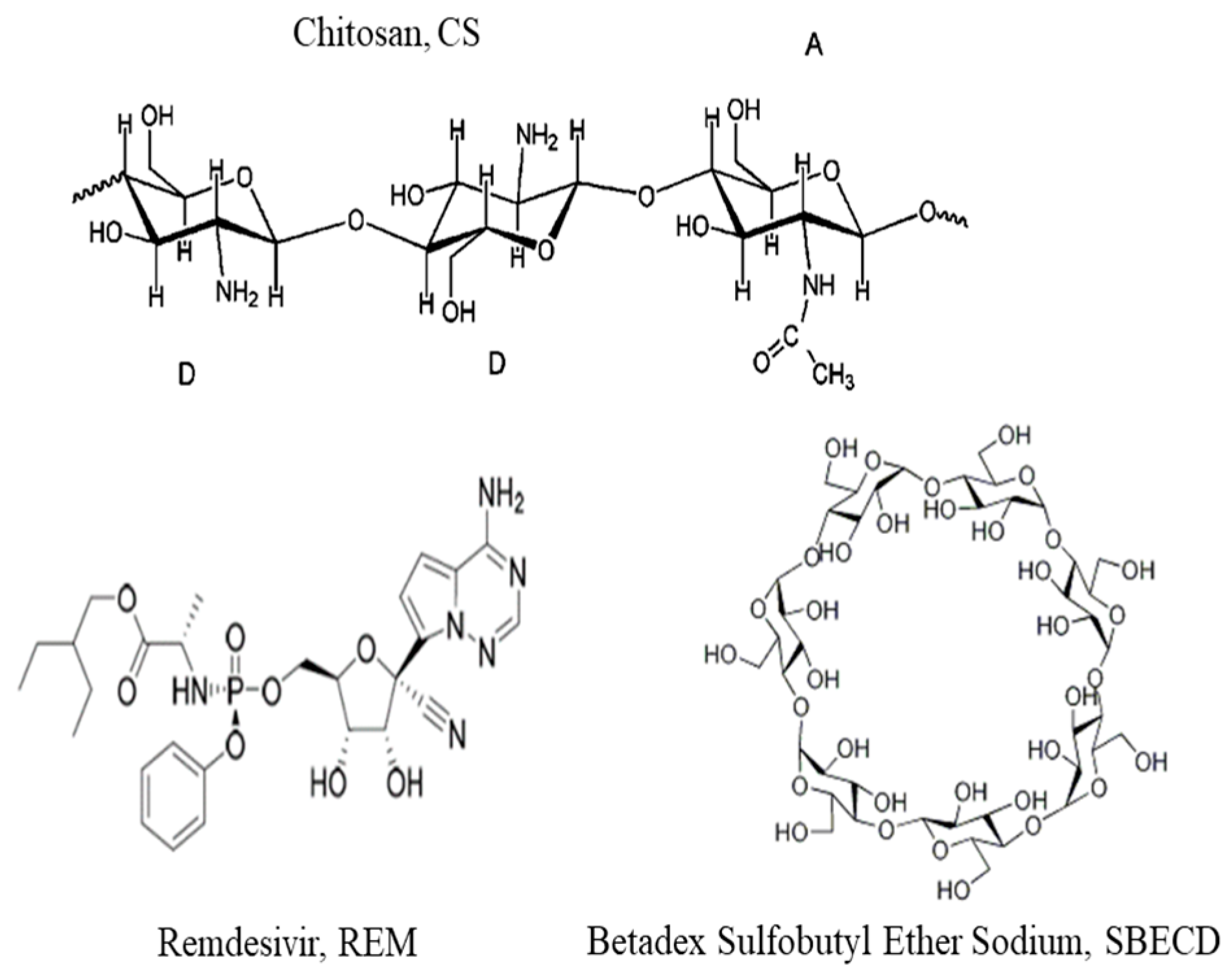
For the formation of the core of the capsules, soybean lecithin, PC 40% (Avanti, Germany, 341602G), was used. Miglyol 812 N® was kindly provided by Sasol Germany GmbH, Witten, Germany. The molecular structures of used compounds are presented in Figure 1.
The aptamer selection was performed by using a web server called PRIdictor (Protein–RNA Interaction predictor), which can predict mutual binding sites in oligonucleotide sequence in RNA molecules and protein at the nucleotide- and residue-level resolutions from their sequences [23]. PRIdictor was used as a web-based application [24]. The selected oligonucleotide sequence of aptamer (5′-AAA CAU UGC AC-3′) was synthesized from Biomers (Ulm, Germany). According to the product information, the molecular weight was 3457 g/mol. The sample was dissolved in double distilled water and the aptamer concentration in the stock solution was 60.76 μM.
2.2. Preparation of the Chitosan-Stabilized Oil-Core Capsules and Remdesivir Encapsulation
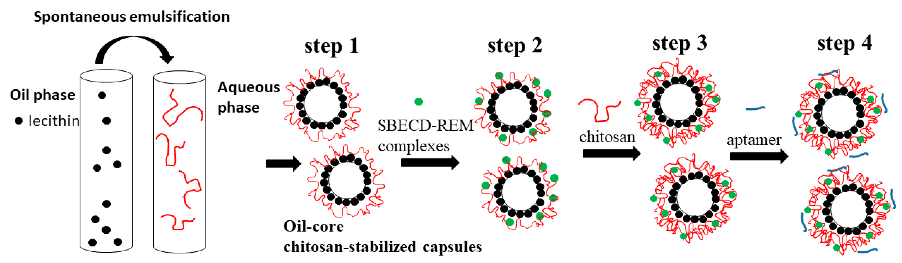
The subsequent steps in the experimental procedure used for the formation of the capsules are presented in Figure 2.
Step 1. The cores of the capsules were oil droplets stabilized by lecithin/chitosan membrane produced according to the procedure originally described by Calvo et al. [25]. Briefly, the oil phase was prepared from 0.5 mL of a 40 mg/mL ethanolic lecithin solution mixed with 0.125 mL Miglyol 812 N® and 9.5 mL ethanol, and the aqueous phase was chitosan solution (20 mL, 0.5 mg/mL). After mixing, the organic solvent was evaporated at 40 °C. The produced droplets were positively charged because of the deposition of a thick chitosan layer on their surface. For comparison, following the same procedure, an oil-in-water nanoemulsion was prepared by replacing the chitosan solution with a solution of hydrochloric acid (pH 4.04). In order to prevent a possible aggregation in the subsequent steps, the stock dispersion was diluted 1:19 with a solution of HCl (pH~4.04). The dispersion was sonicated for 15 min before the deposition of the drug (the estimated concentration was 7.25 × 10¹⁰ droplets/mL) (Figure 3).
Step 2. The negatively charged drug was impregnated into the chitosan layer of the produced structures. The diluted dispersion of oil-core capsules (20 mL) was added to a high enough concentration of the drug (0.9 mg/mL, Veklury®) and stirred for 20 min at room temperature (24 °C). The estimated final concentration of REM in dispersion was 50 μM (the amount of the drug was high enough to ensure the formation of stable structures and high encapsulation efficiency at almost constant experimental conditions of the dispersion (ionic strength and pH) (Table 1)). The excess non-adsorbed drug was removed by centrifugation (15,000 rpm, 21,382 g, at 15 °C for 60 min) using a laboratory centrifuge (PW-352R, Poland). The extracted cream was diluted to the initial volume with a solution of HCl (pH~4.04) and sonicated in an ultrasonic bath for 10 min.
Step 3. In order to improve the stability of the produced structures, a chitosan layer was adsorbed on the capsules’ surface. The polymer concentration (0.2 mg/mL) was high enough to ensure an overcompensation of the surface charge and re-stabilization of the dispersion. (The suitable concentrations of chitosan and SBECD-REM used in the procedure were determined also by additional investigations.)
In the subsequent step (step 4), the aptamer molecules (5′-AAA CAU UGC AC-3′) were adsorbed on the surface (5 μM). The dispersion (1836 µL) and aptamer solution (164 µL) were mixed using Vortex for a minute and the produced dispersion was then placed to rest without stirring for 20 min at room temperature (24 °C).
2.3. Determination of the Amount of Encapsulated Remdesivir
The concentration of REM loaded into the structures was determined by the difference between the initial concentration of the compound added to the dispersion and the concentration in the supernatant after centrifugation (after step 2, Figure 1) by monitoring with T60 UV-Visible Spectrophotometer (PG Instruments Limited, UK).
The drug was detected at a wavelength of 245 nm corresponding to the maximum absorbance peak of SBECD-REM complexes and the amount of free drug in solution was calculated by using an appropriate calibration curve. The encapsulation efficiency (EE%) was estimated by using
where Ctotal is the initial concentration of REM added to the dispersion (0.03 mg/mL) and Cfree is the calculated concentration of the compound in the supernatant.
EE% = (Ctotal − Cfree).100/Ctotal
The drug loading capacity (in %) was calculated with the following the equation
where mencapsulated is the amount of drug successfully loaded into the capsules (in mg/mL) and mcapsules is the amount of capsules (0.24 mg/mL).
LC% = mencapsulated.100/mcapsules
2.4. Determination of the Surface Charge and Size of the Capsules
The surface charge and hydrodynamic diameter of the produced structures were evaluated after each deposition step by using dynamic light scattering with non-invasive backscattering (DLS-NIBS, measuring angle 173°). The hydrodynamic thickness after each deposition step was determined by comparing the capsule size before and after adsorption. For estimation of the film thickness, it was assumed that the regular polymer adsorption was achieved on the surface and the spherical shape of the formulation did not change. The measurements were carried out using Zatasizer Pro (Malvern Panalytical Ltd., Malvern, UK) equipped with a He-Ne laser with a maximum power of 10 mW operating at a wavelength of 633 nm with a fixed scattering light angle of 173°. All measurements were performed at 24.0 ± 0.1° After five measurements, the average value was taken as the surface charge and size of the capsules.
2.5. Release of the Drug in Conditions Close to the Physiological Ones
The released amount of drug from the capsules was estimated by using two different experimental procedures. First, after the adsorption of aptamer on the capsule surface, a small volume of the dispersion (2 mL) was centrifuged (15,000 rpm, 21,382 g, at 15 °C for 90 min). The cream was extracted and diluted in a physiological solution (Unolab Manufacturing, S.L., Madrid, Spain) to the same volume as before centrifugation. The first sample was taken 15 h after the preparation of the dispersion. The solution of capsules was centrifuged and the cream was re-dispersed again in a physiological solution (2 mL). The procedure was repeated after 24 h. The free drug concentration in samples was estimated by UV-vis spectroscopy using appropriate calibration curves.
According to the second procedure (dialysis method), after adsorption of aptamer, an aliquot from the dispersion (1 mL) was added into a dialysis tube (D-Tube™ Dialyser Midi, MWCO 3.5 kDa, Sigma Aldrich) and incubated with 25 mL buffer pH 7.00 (Chem-Lab NV, Belgium) at room temperature (24 °C) and 37 °C with a stirring speed 50 rpm. Aliquots (2 mL) were drawn at predetermined time points and the medium was immediately replenished with fresh buffer. The concentration of free drug in samples was estimated by UV-vis spectroscopy.
2.6. Transmission Electron Microscopy (TEM)
The produced capsules were visualized by TEM. The samples for the TEM studies were prepared by dropping and drying a drop of suspension of capsules on formvar-covered TEM grids. The images were captured using High-Resolution Scanning Transmission electron microscope HR STEM JEOL JEM 2100 for investigations of surface morphology.
2.7. Microbiology Studies
2.7.1. Host Cell Culture
Human colon carcinoma cells (HCT-8) were obtained from the American Type Culture Collection (ATCC). Permanent HCT-8 [HRT-18] (ATCC-CCL-244, LGC Standards) were maintained at 37 °C and 5% CO2 using sterile RPMI 1640 (Roswell Park Memorial Institute Medium, ATCC-30-2001) supplemented with 0.3 mg/mL L-glutamine (Sigma-Aldrich, Darmstadt, Germany), 10% horse serum (ATCC-30-2021), 100 UI penicillin, and 0.1 mg streptomycin/mL (both purchased from Sigma-Aldrich).
2.7.2. Viruses
Human Coronavirus OC43 (HCoV-OC43) (ATCC: VR-1558) strain was propagated in HCT-8 cells in RPMI 1640 supplemented with 2% horse serum, 100 U/mL penicillin, and 100 μg/mL streptomycin. Cells were lysed 5 days after infection by 2 freeze and thaw cycles and the virus was titrated according to the Reed and Muench formula. Virus and mock aliquots were stored at −80 °C.
2.7.3. Cytotoxicity Assay
Confluent monolayer cell culture in a 96-well plate (Costar®, Corning Inc., Kennebunk, ME, USA) was treated with 0.1 mL/well containing a maintenance medium that did not contain/or contained decreasing concentrations of test substances. The cells were incubated at 37 °C and 5% CO2 for 5 days. After microscopic evaluation, the medium containing the test compound was removed, and the cells were washed and incubated with neutral red, at 37 °C for 3 h. After incubation, the neutral red dye was removed and the cells were washed with PBS and 0.15 mL/well desorbing solution (1% glacial acetic acid and 49% ethanol in distilled water) was added. The optical density (OD) of each well was registered at 540 nm in a microplate reader (Biotek Organon, West Chester, PA, USA). Then, 50% cytotoxic concentration (CC50) was defined as the concentration of the material that reduces cell viability by 50% compared to untreated controls. Each sample was tested in triplicate with four wells for cell culture on a test sample.
The maximum tolerable concentration (MTC) of the extracts was also determined, which was the concentration at which they did not affect the cell monolayer and in the sample, it looks like the cells in the control sample (untreated with compounds).
2.7.4. Antiviral Activity Assay
The cytopathic effect (CPE) inhibition test was used to assess the tested compounds’ antiviral activity. Confluent cell monolayer in 96-well plates was infected with 100 cell culture infectious dose 50% (CCID50) in 0.1 mL (coronavirus OC43 strain). After 120 min of virus adsorption, the tested compound was added in various concentrations and cells were incubated for 5 days at 33 °C and 5% CO2. The cytopathic effect was estimated using a neutral red dye uptake assay and the percentage of CPE inhibition was calculated:
where ODtest sample is the mean value of the ODs of the wells inoculated with a virus and treated with the test sample in the respective concentration, ODvirus control is the mean value of the ODs of the virus control wells (with no compound in the medium), and ODtoxicity control is the mean value of the ODs of the wells not inoculated with a virus but treated with the corresponding concentration of the test compound. The concentration of the test substance that inhibited 50% of viral replication when compared to the virus control is labelled as IC50. The selectivity index (SI) was calculated from the ratio CC50/IC50.
% CPE = (ODtest sample − ODvirus control).100/(ODtoxicity control − ODvirus control)
2.7.5. Virucidal Assay
Samples of 1 mL containing HCoV (CCID 50), and samples in their maximal tolerable concentration (MTC) were mixed in a 1:1 ratio and, subsequently, stored at room temperature for different time intervals. The residual infectious virus content in each sample was determined by the end-point dilution method of Reed and Muench [24] and Δlgs as compared to the untreated controls were evaluated.
2.7.6. Effect on the Viral Adsorption
Twenty-four-well plates containing HCT-8 cell monolayer were pre-cooled to 4 °C and inoculated with CCID50 of HCoV. In parallel, they were treated with tested samples at their MTC and incubated at 4 °C for the time of virus adsorption. At various time intervals (15, 30, 45, and 60 min), the cells were washed with PBS to remove both the compound and the unattached virus, and the cells were then covered with a support medium and incubated at 37 °C and 5% CO2 for 24 h. After freezing and thawing three times, the infectious viral titer of each sample was determined by the final dilution method. Δlgs was determined compared to the viral control (untreated with the compounds). Each sample was prepared in four replicates.
2.7.7. Statistical Analysis
Data on cytotoxicity and antiviral effects were analyzed statistically. The values of CC50 and IC50 were presented as means ± SD.
3. Results and Discussion
3.1. Characterization of the Produced Capsules
3.1.1. Electrokinetic ζ-Potential and Hydrodynamic
Figure 4 shows the comparison in the evolution of the hydrodynamic diameter (DH) and ζ-potential of produced composite capsules after each deposition step formed with different chitosan samples.
The cores of the structures were positively charged oil-core capsules (produced in step 1, Figure 2). The charge of the capsules resulted from the formation of a thick chitosan layer on the lecithin-stabilized emulsion droplets [25]. The comparison between the size of the capsules and emulsion droplets indicates that the thickness of the polymer layer was ca. 24 nm and 170 nm for the capsules formed from chitosan CS-L and CS-M, respectively.
According to the experimental results, the adsorption of the drug (SBECD-REM complexes) led to a decrease in the size of the capsules and achievement of high overcompensation of electrokinetic charge. The electrokinetic measurements showed that the complex of SBECD-REM was negatively charged (ζ-potential is ca. −6 mV at pH 4) with a diameter of ca. 1 nm. Therefore, we supposed that the complexes were adsorbed on the surface but also can penetrate into the chitosan layer. The saturation of the polysaccharide film with negatively charged complexes led to the shrinking of the film. This observation was similar to the effect of the addition of low molecular electrolyte on the structure of polyelectrolyte film. An approximate estimation showed that the ratio between the number of capsules and SBECD-REM complexes was ca. 1: 7 × 10⁷. (For comparison, if the complex is regarded as a sphere, the complete coverage of the surface of one capsule is achieved by adsorption of ca. 2.5 × 10⁵ SBECD-REM complexes.)
The subsequent adsorption of chitosan aimed to improve the stability of the structures and to ensure a positive charge in the outer layer of the composite capsules for the next adsorption of the negatively charged aptamer. The adsorption of a very low concentration of aptamer (5 μM) resulted in a slightly decreasing in the electrokinetic potential and did not influence the stability of the capsules. (An approximate estimation showed that the number of aptamer molecules per capsule was ca. 4 × 10⁷).
The experimental results clearly indicate the difference in the hydrodynamic size of structures produced from chitosans with different molecular weights (the degrees of acetylation of chitosans were similar). The higher values of the size and charge were registered for capsules (step 1) formed from chitosan with higher molecular weight (CS-M), which is in line with our previous studies [26,27,28].
The chitosan adsorption in step 3 also indicated an increase in the size of the structures and the formation of a thicker polymer layer from a polymer with higher molecular weight (ca. 30 nm for CS-L and ca. 200 nm for CS-M). The visualization of the produced capsules and comparison between their hydrodynamic and real size is presented in Figure 5.
3.1.2. Encapsulation of Remdesivir
The key step in capsule formation was the impregnation of the drug (SBECD-REM complexes) into the thick chitosan layer. In order to investigate the character of the interactions responsible for the impregnation of complexes into the capsules, it is important to analyze the structure and properties of the complex. For the first time, Szente et al. [11] reported the detailed characterization of cyclodextrin-enabled REM structures and the medicine Veklury®.
Remdesivir is poorly soluble in water compound (ca. 0.028 mg/mL) [11]. Therefore, the application of surfactants, polymers, or co-solvents was necessary to ensure the formation of stable and clear aqueous solutions suitable for intravenous administration. According to the product information, the medicine Veklury® contains REM, SBECD, NaCl, and HCl.
The experimental results presented by Szente et al. [11] proved that the non-covalent and host–guest type interaction between REM and SBECD is responsible for the incorporation of the drug into the complexes. The 1NMR titration experiments suggested the formation of a single SBECD-REM complex with 1:1 stoichiometry. Moreover, their computer docking experiments indicated that, at pH~7, no ionized REM molecules penetrate into the cyclodextrin matrix with its pyrrolotriazine-amine ring filling the central cavity of SBECD. The polar side chins of SBECD formed hydrogen bonds with the hydroxyl group of tetrahydrofuranyl with the amino group of REM.
It is known that SBECD has no buffering capacity and the presence of a suitable (unknown) amount of NaOH and HCl in each vial of Veklury® is responsible for adjusting the pH of the solution for infusion. “…the complex structure at such a low pH can be drastically different as compared to the structure of the host-guest complex at neutral pH, due to the ionization of REM and the change in water structure. At acidic pH, the aliphatic chain was found to penetrate into the cyclodextrin cavity rather than the ionized N-heterocycle of REM…” [11]. Moreover, Szente et al. showed that, in acidic conditions, the drug resides inside the cavity of the SBE matrix with its ethyl-butyl sidechain, while no inclusion phenomenon occurs between the positively charged heterocyclic moiety and SRE. Additionally, the formation of hydrogen bonds and participation of electrostatic interactions also support the formation of a stable SBECD-REM complex.
In order to evaluate the effect of the presence of SBECD on the REM encapsulation, the adsorption behavior of pure SBECD in the presence of chitosan-stabilized capsules (step 1) was investigated. According to the presented results, the overcompensation of the surface charge was registered in the presence of 0.6 M SBECD (ζ-potential is ca. −32.2 ± 1.2 mV). Consequently, the encapsulation of negatively charged SBECD-REM complexes in the capsule object of the present consideration was due to the electrostatic interaction between the negatively charged SBECD and positively charged monomers of chitosan molecules on the outer layer of the capsule surface.
In the present study, the stock solution of Veklury® (pH~3.96) was prepared in pure water according to the pharmaceutical protocol. The concentration of REM (50 μM) in dispersion was calculated from the product information.
The concentration of REM loaded into the capsules was estimated by using UV-vis spectroscopy and the estimated encapsulation efficiency was ca. 23% (6.90 µg/mL) and 45% (13.50 µg/mL) for capsules produced from CS-M and CS-L, respectively (Figure 6). Consequently, the estimated loading capacity of capsules was ca. 2.9% and 5.6% for capsules produced from CS-M and CS-L, respectively (Equation (2)).
According to the information from the literature, the encapsulation efficiency (EE%) of pure REM in different kinds of (nano)formulations depends on the morphology of the structures, the type of the components, and the procedure for formation. In juxtaposition, in liposomes (diameter ca. 120–130 nm, ζ-potential ca. −7 mV), the EE% was in the range of 62–89% (or LC ca. 2.7–11.0%) depending on the ratio between the drug and lipid [15]. In polymer–drug conjugates (diameter ca. 600–7100 nm, ζ-potential ca. –26 mV), the EE% (in the range of 38–69%) depends on the stirring time in the procedure [16]. Moreover, the encapsulation of REM in hyper-branched dendritic nano-scaffolds showed the EE% to be 82% (LC 14%) [17].
3.1.3. Release of REM from the Produced Capsules
The concentration of free drug in the samples was estimated by UV-vis spectroscopy by using appropriate calibration curves. Figure 7 presents the estimated released amount of REM from the capsules in a physiological solution and in buffer pH 7.0.
According to the results, the amount of drug released from the capsules formed from CS-L strongly depends on the pH and temperature. The release process was fast in phosphate buffer at 37 °C and, after 30 h, was registered as almost 100% released REM in solution, whereas at room temperature, the amount slightly increased up to 80% for 70 h. Similarly, in a physiological solution at room temperature (24 °C), the concentration of free REM in the release medium slightly increased (up to 60%). The different initial slope of the curves indicates the dependence of the velocity of release at the initial stage of the process on specific experimental conditions: 2.89 μg/h (pH7, 37 °C), 0.51 μg/h (pH7, 24 °C), and 0.26 μg/h (physiological solution, 24 °C), (per ml dispersion).
The experimental results showed that the free amount of drug from capsules formed from CS-M almost did not depend on the pH (ca. 60% for 70 h).
3.2. Microbiology Studies
3.2.1. Cytotoxicity Assay
The capsules and their components were tested for cytotoxic effects on HCT-8 cells. All tested substances showed low or no toxicity. According to the experimental results, chitosan solution and capsules loaded with the drug produced with CS-L showed lower cytotoxicity (Table 2).
3.2.2. Influence on the Replication Cycle of Human Coronavirus Strain OC-43
Chitosans, capsules, and Miglyol 812 N® were also investigated for their effect on the intracellular replication cycle of human coronavirus strain OC43. The experimental results indicated that when REM was administered during human coronavirus replication, the selectivity index was ca. 200. Drug-loaded capsules formed from chitosan CS-L showed low activity, whereas capsules with chitosan CS-M had stronger inhibitory activity against HCoV-OC43 replication compared to the pure drug (Table 2).
3.2.3. Effect on Extracellular Virions of Human Coronavirus Strain OC43
The virucidal activity of the samples against the viability of the extracellular HCoV-OC43 virions was studied. The chitosan solutions showed weak inhibition (most likely non-specific) and the effect of lower molecular chitosan was slightly higher (Δlg = 1.66). However, according to the experimental results, the influence of chitosan-stabilized capsules was significantly lower compared to the pure polysaccharide solutions in spite of the presence of chitosan in their structure (moreover, there was no difference in Δlg for the loaded and unloaded capsules). We supposed that the reason for the observed behavior of the capsules on the viability of the extracellular HCoV-OC43 virions was due to the lower amount of chitosan in samples (ca. 2 μg/mL) compared to the pure polymer solution (320.0 µg/mL and 100.0 µg/mL, respectively, for chitosan CS-L and CS-M) (Table 3).
The results showed that the pure drug did not affect the extracellular virions. This result was expected because REM has a pronounced in vitro and in vivo activity against coronaviruses that results from the inhibitor activity of the drug regarding RNA-dependent RNA polymerases (RdRps). RdRps is responsible for viral RNA synthesis and replication in the host cells.
3.2.4. Effect on Viral Adsorption
It was examined whether the samples affected the stage of coronavirus adsorption on cells. Experiments showed that the chitosan solutions inhibit the adsorption of the virus. The registered inhibition was weak at the first time interval (15 min). After 30 min, the effect was slightly enhanced, becoming significant after 60 min of exposure (Δlg = 1.75). The results indicated that after 120 min, the inhibition was increased to some extent, being more pronounced in CS-L (Δlg = 2.25). The effect was noticeably time-dependent—as the exposure time increased, the amount of attached coronavirus virions decreased (Table 4). Moreover, it was found that the samples of emulsion, unloaded capsules, and the pure drug did not affect the adsorption step of HCoV-OC43 on cells.
4. Conclusions
The present study addressed the design and characterization of model chitosan-based formulations suitable for encapsulation and target delivery of remdesivir in a potential therapy of coronavirus infection.
The reproducible procedure was developed for the formation of stable drug-loaded structures by subsequent (predominantly) electrostatic adsorption of oppositely charged components (chitosan, SBECD-REM complexes, and aptamer) on oil-core capsules. The influence of the molecular weight of chitosan on the properties of the produced structures was studied. Variation in the hydrodynamic size and surface charge of the structures was registered after each deposition step.
The complexes of SBECD-REM were loaded into the chitosan layer of the capsules because of the electrostatic attraction with the oppositely charged chitosan monomers. The encapsulation efficiencies of the drug were estimated at ca. 23% and 45% for capsules produced from chitosan with medium and low molecular weight (CS-M and CS-L), respectively.
The released amount of REM from the capsules was investigated in a physiological solution and buffer pH 7.0. The experimental results indicated that the released amount of REM from capsules formed from CS-L strongly depended on the pH and temperature, whereas the released amount of drug from capsules of CSM almost did not depend on the variation in the experimental conditions. Complete release of the drug from the capsules was achieved in a dispersion of capsules formed from CS-L incubated in phosphate buffer at 37 °C (after 30 h).
The cytotoxic assay indicated that all tested substances were not toxic towards HCT-8 cells. According to the experimental results, the solution of chitosan CS-L showed lower cytotoxicity compared to chitosan CS-M. Moreover, the toxicities of drug-loaded and unloaded capsules on the cells were close.
The investigations on the influence of the produced capsules on the replication cycle of HCoV-OC43 showed that the drug-loaded capsules formed from chitosan CS-L showed low activity, whereas capsules with chitosan CS-M had stronger inhibitory activity against virus replication compared to pure drug.
The analysis of the virucidal activity of the samples against the viability of the extracellular HCoV-OC43 virions indicated that the pure chitosan solutions had a weak inhibition effect. However, in spite of the presence of chitosan in the structure of capsules, the results showed that their influence on the virions was negligible. Moreover, there was no difference between the behavior of the loaded and unloaded capsules.
The investigation of the effect of the substances and capsules on viral adsorption on the cell membrane indicated that the chitosan solutions inhibit the adsorption of the virus. Moreover, unloaded capsules, loaded capsules, and the pure drug did not affect the adsorption of HCoV-OC43 on HCT-8 cells.
The main conclusion from the study was that the physicochemical and antiviral properties of the produced composite capsules strongly depend on the properties of the chitosan in their structure, and we supposed that the capsules formed from CS-L were more suitable for the potential pharmacological application.
Author Contributions
Conceptualization, V.M. and N.V.-I.; methodology, V.M., K.K. and N.V.-I.; investigation V.M., K.K., P.M., N.V.-I. and V.R.; resources, V.M.; writing—original draft preparation, V.M. and N.V.-I.; project administration, V.M. All authors have read and agreed to the published version of the manuscript.
Funding
This research was funded by the National Science Fund, contract No KП-06-ДK1/3. Miglyol 812 N® was kindly provided by Sasol Germany GmbH, Witten, Germany.
Acknowledgments
Research equipment of the Distributed Research Infrastructure INFRAMAT, part of the Bulgarian National Roadmap for Research Infrastructures, supported by the Bulgarian Ministry of Education and Science, was used for some investigations in the present study. The authors acknowledge the support of European Regional Development Fund within the OP Science and Education for Smart Growth 2014–2020, Project CoE National centre for mechatronics and clean technologies, No. BG05M2OP001-1.001-0008. The authors thank D. Karashanova for visualization analysis of the produced structures by Transmission Electron Microscopy and V. Lutov for support in the spectrophotometric experiments.
Conflicts of Interest
The authors declare no conflict of interest.
References
- Jordheim, L.P.; Durantel, D.; Zoulim, F.; Dumontet, C. Advances in the development of nucleoside and nucleotide analogues for cancer and viral diseases. Nat. Rev. Drug Discov. 2013, 12, 447–464. [Google Scholar] [CrossRef] [PubMed]
- Murakami, E.; Niu, C.; Bao, H.; Micolochick Steuer, H.M.; Whitaker, T.; Nachman, T.; Sofia, M.A.; Wang, P.; Otto, M.J.; Furman, P.A. The mechanism of action of beta-D-2′-deoxy-2′-fluoro-2′-C-methylcytidine involves a second metabolic pathway leading to beta-D-2′-deoxy-2′-fluoro-2′-Cmethyluridine 5′-triphosphate, a potent inhibitor of the hepatitis C virus RNA-dependent-RNA polymerase. Antimicrob. Agents Chemother. 2008, 52, 458–464. [Google Scholar] [CrossRef] [Green Version]
- Sheahan, T.P.; Sims, A.C.; Graham, R.L.; Menachery, V.D.; Gralinski, L.E.; Case, J.B. Broad-spectrum antiviral GS-5734 inhibits both epidemic and zoonotic coronaviruses. Sci. Transl. Med. 2017, 9, 396. [Google Scholar] [CrossRef] [Green Version]
- Grein, J.; Ohmagari, N.; Shin, D.; Diaz, G.; Flanigan, T. Compassionate use of remdesivir for patients with serve COVD-19. N. Engl. J. Med. 2020, 382, 2327–2336. [Google Scholar] [CrossRef]
- Koichiro, K.; Honma, T.; Fukuzawa, K. Intermolecular interaction among Remdesivir, RNA and RNA-dependent RNA polymerase of SARS-CoV-2 analyzed by fragment molecular orbital calculation. J. Mol. Graph. Model. 2020, 100, 107605. [Google Scholar]
- Grundeis, F.; Ansems, K.; Dahms, K.; Thieme, V.; Metzendorf, M.I.; Skoetz, N.; Benstoem, C.; Mikolajewska, A.; Griesel, M.; Fichtner, F.; et al. Remdesivir for the treatment of COVID-19. Cochrane Database Syst. Rev. 2023, 1, CD014962. [Google Scholar]
- Gracelin Princy Zacchaeus, N.; Samuel, P.; Jasmine, S.; Devasahayam, J.C.; Pichamuthu, K.; Rupali, P. Evaluation of Remdesivir to the outcomes of hospitalized patients with COVID-19 infection in a tertiary-care hospital in southern India. J. Infect. Dev. Ctries. 2023, 17, 304–310. [Google Scholar]
- Simonis, A.; Theobald, S.J.; Fätkenheuer, G.; Rybniker, J.; Malin, J.J. A comparative analysis of remdesivir and other repurposed antivirals against SARS-CoV-2. EMBO Mol. Med. 2021, 13, e13105. [Google Scholar] [CrossRef]
- Available online: https://www.drugs.com/history/remdesivir.html (accessed on 12 July 2023).
- Available online: https://www.clinicaltrials.gov (accessed on 30 June 2023).
- Szente, L.; Puskas, I.; Sohajda, T.; Varga, E.; Vass, P.; Nagy, Z.K.; Farkas, A.; Varnai, B.; Beni, S.; Hazai, E. Sulfobutylether-beta-cyclodextrin-enabled antiviral remdesivir: Characterisation of electrospun- and lyophilized formulations. Carbohydr. Polym. 2021, 264, 118011. [Google Scholar] [CrossRef]
- Okimoto, K.; Rajweski, R.A.; Uekama, K.; Jona, J.A.; Stella, V.J. The interaction of charged and uncharged drugs, with neutral (HP-β-CD) and anionically charged (SBE7-β-CD) β-cyclodextrin. Pharm. Res. 1996, 13, 256–264. [Google Scholar] [CrossRef]
- Luke, D.R.; Tomaszewski, K.; Damle, B.; Schlamm, H.T. Review of the basic and clinical pharmacology of sulfobutylether-β-cyclodextrin (SBECD). J. Pharm. Sci. 2010, 99, 3291–3301. [Google Scholar] [CrossRef] [PubMed]
- Zarandona, I.; Barba, C.; Guerrero, P.; de la Caba, K.; Mate, J. Development of chitosan films containing β-cyclodextrin inclusion complex for controlled release of bioacitves. Food Hydrocoll. 2020, 104, 105720. [Google Scholar] [CrossRef]
- Sun, D. Remdesivir for treatment of COVID-19: Combination of pulmonary and iv administration may offer additional benefit. AAPS J. 2020, 4, 77. [Google Scholar] [CrossRef]
- Cao, Y.; Deng, X.; Dai, S. Remdesivir for severe acute respiratory syndrome coronavirus 2 causing COVID-19: An evaluation of the evidence. Travel Med. Infect. Dis. 2020, 35, 101647. [Google Scholar] [CrossRef] [PubMed]
- Li, J.; Zhang, K.; Wu, D.; Ren, L.; Chu, X.; Qin, C.; Han, X.; Hang, T.; Xu, Y.; Yang, L.; et al. Liposomal remdesivir inhalation solution for targeted lung delivery as a novel therapeutic approach for COVID-19. Asian J. Pharm. Sci. 2022, 16, 772–783. [Google Scholar] [CrossRef]
- Jeon, W.J.; Lee, H.K.; Na, Y.G.; Jung, M.; Han, S.C.; Hwang, J.H.; Jung, E.; Hwang, D.; Shin, J.S.; Cho, C.W. Antiviral Lipid Nanocarrier Loaded with Remdesivir Effective Against SARS-CoV-2 in vitro Model. Int. J. Nanomed. 2023, 18, 1561–1575. [Google Scholar] [CrossRef]
- Qudsiani, K.S.; Rahmasari, R. Polyamidoamine-Remdesivir conjugate: Physical stability and cellular uptake enhancement. Biomed. Pharmacol. J. 2021, 14, 2073–2083. [Google Scholar] [CrossRef]
- Halevas, E.; Mavroidi, B.; Kokotidou, C.; Moschona, A.; Sagnou, M.; Mitraki, A.; Litsardakis, G.; Pelecanou, M. Remdesivir-loaded bis-MPA hyperbranched dendritic nanocarriers for pulmonary delivery. J. Drug Deliv. Sci. Technol. 2022, 75, 103635. [Google Scholar] [CrossRef]
- Quemeneur, F.; Rinaudo, M.; Maret, G.; Pepin-Donat, B. Decoration of lipid vesicles by polyelectrolytes: Mechanism and structure. Soft Matter 2010, 6, 4471–4481. [Google Scholar] [CrossRef]
- Tuvshinjargal, N.; Lee, W.; Park, B.; Han, K. PRIdictor: Protein–RNA Interaction predictor. BioSystems 2016, 139, 17–22. [Google Scholar] [CrossRef]
- Guyrova, A.; Milkova, V.; Kamburova, K.; Dimitrov, I. Interaction of remdesivir-loaded nanocarriers with spike protein of HCV-OC43 and model lipid membranes. 2023; in preparation. [Google Scholar]
- PRIdictor. Available online: http://bclab.inha.ac.kr/predictor (accessed on 24 July 2023).
- Calvo, P.; Remufifin-Lopez, C.; Vila-Jato, J.L.; Alonso, M.J. Development of positively charged colloidal drug carriers: Chitosan-coated polyester nanocapsules and submicron-emulsions. Colloid Polym. Sci. 1997, 275, 46–53. [Google Scholar]
- Goycoolea, F.M.; Milkova, V. Electrokinetic behavior of chitosan adsorbed on o/w nanoemulsion droplets. Colloids Surf. A Physicochem. Eng. Asp. 2017, 519, 205–211. [Google Scholar] [CrossRef]
- Milkova, V.; Goycoolea, F.M. Encapsulation of caffeine in polysaccharide oil-core nanocapsules. Colloid Polym. Sci. 2020, 298, 1035–1041. [Google Scholar] [CrossRef]
- Milkova, V. Electrosteric stabilization of oil/water emulsions by adsorption of chitosan oligosaccharides—An electrokinetic study. Carbohydr. Polym. 2021, 265, 118072. [Google Scholar]
Figure 1.
Schematic molecular structure of chitosan (CS) with completely acetylated monomers (A) and deacetylated monomers (D), remdesivir (REM), and betadex sulfobutyl ether sodium (SBECD).
Figure 1.
Schematic molecular structure of chitosan (CS) with completely acetylated monomers (A) and deacetylated monomers (D), remdesivir (REM), and betadex sulfobutyl ether sodium (SBECD).

Figure 2.
Deposition steps in the experimental procedure for preparation of capsules stabilized by lecithin/chitosan membrane and subsequent electrostatic adsorption of the oppositely charged drug (SBECD-REM complexes), chitosan, and aptamer.
Figure 2.
Deposition steps in the experimental procedure for preparation of capsules stabilized by lecithin/chitosan membrane and subsequent electrostatic adsorption of the oppositely charged drug (SBECD-REM complexes), chitosan, and aptamer.

Figure 3.
Dispersion of capsules produced according to the procedure described by Calvo at al. [25] (A) and diluted dispersion (ratio 1:19) used in the next adsorption steps (B).
Figure 3.
Dispersion of capsules produced according to the procedure described by Calvo at al. [25] (A) and diluted dispersion (ratio 1:19) used in the next adsorption steps (B).

Figure 4.
Dependence of the hydrodynamic size (diameter) (A) and electrokinetic potential (B) for capsules formed from chitosans with different molecular weights after each deposition step: CS-L (●) and CS-M (○).
Figure 4.
Dependence of the hydrodynamic size (diameter) (A) and electrokinetic potential (B) for capsules formed from chitosans with different molecular weights after each deposition step: CS-L (●) and CS-M (○).

Figure 5.
Representative TEM images of capsules loaded by SBECD-REM, stabilized by adsorption of chitosan, and functionalized by deposition of aptamer: CS-M (A) and CS-L (B).
Figure 5.
Representative TEM images of capsules loaded by SBECD-REM, stabilized by adsorption of chitosan, and functionalized by deposition of aptamer: CS-M (A) and CS-L (B).

Figure 6.
UV-vis spectra of supernatant after centrifugation of the dispersion (step 2 in Figure 2): CS-M (solid line) and CS-L (dash line). Inset: Calibration curve of SBECD-REM at pH~4.04.
Figure 6.
UV-vis spectra of supernatant after centrifugation of the dispersion (step 2 in Figure 2): CS-M (solid line) and CS-L (dash line). Inset: Calibration curve of SBECD-REM at pH~4.04.

Figure 7.
Kinetics of drug release from capsules formed from CS-L (A) and CS-M (B) in buffer pH7.0 at 24 °C (●) and 37 °C (○), and in physiological solution at 24 °C (■).
Figure 7.
Kinetics of drug release from capsules formed from CS-L (A) and CS-M (B) in buffer pH7.0 at 24 °C (●) and 37 °C (○), and in physiological solution at 24 °C (■).


Table 1.
Hydrodynamic size (diameter), ζ-potential, and conductivity, χ, of water dispersion of oil-core capsules stabilized by adsorption of CS-L in the presence of different concentrations of Veklury® or REM, respectively.
Table 1.
Hydrodynamic size (diameter), ζ-potential, and conductivity, χ, of water dispersion of oil-core capsules stabilized by adsorption of CS-L in the presence of different concentrations of Veklury® or REM, respectively.
| Veklury® [mg/mL] | REM [μM] | Size [nm] | ζ-Potential [mV] | Χ [mS/cm] |
|---|---|---|---|---|
| 0 | 0 | 329.8 ± 1.7 | 63.0 ± 0.1 | 0.018 |
| 0.18 | 10 | 448.8 ± 4.2 | −18.3 ± 0.4 | 0.019 |
| 0.9 | 50 | 275.8 ± 4.1 | −31.9 ± 0.5 | 0.093 |
| 1.8 | 100 | 286.5 ± 3.3 | −35.5 ± 0.7 | 0.143 |
| 9 | 500 | 409.6 ± 10.9 | −32.3 ± 0.4 | 0.525 |
| 18 | 1000 | 744.5 ± 9.4 | −19.6 ± 1.3 | 1.005 |
Table 2.
Cytotoxicity on HCT-8 cell lines and antiviral activity against human coronavirus (strain OC43): CS-L and CS-M are pure polymer solutions, capsules-L-REM and capsules-M-REM are samples of capsules loaded with the drug formed with chitosan with low and medium molecular weight, respectively.
Table 2.
Cytotoxicity on HCT-8 cell lines and antiviral activity against human coronavirus (strain OC43): CS-L and CS-M are pure polymer solutions, capsules-L-REM and capsules-M-REM are samples of capsules loaded with the drug formed with chitosan with low and medium molecular weight, respectively.
| Sample | Cytotoxicity | Antiviral Activity | ||
|---|---|---|---|---|
| CC50 * Mean ± SD ** [µg/mL] | MTC *** [µg/mL] | IC50 * Mean ± SD ** [µg/mL] | SI *** | |
| CS-L | 768.70 ± 11.7 | 320.00 | - | - |
| CS-M | 540.40 ± 9.3 | 100.00 | - | - |
| aptamer | - | ˃210 | - | - |
| Miglyol 812 N® pure substance | - | ˃100.0% | - | - |
| Veklury® | 2500.00 ± 4.30 | 1000.00 | 12.500 ± 0.900 | 200.0 |
| Capsules-L-REM # | 9.72 ± 0.49 | 1.35 | 0.081 ± 0.003 | 120.0 |
| Capsules-M-REM # | 4.55 ± 0.15 | 0.69 | 0.017 ± 0.001 | 268.0 |
* CC50—cytotoxic concentrations 50%; ** SD—standard deviation; *** MTC—maximum tolerable concentration; *** SI—selectivity index, calculated from the ratio CC50/IC50. # relative to the concentration of a drug loaded in the capsules.
Table 3.
Virucidal activity of the studied samples against human coronavirus virions (strain OC43). The sample’s name is the same as in Table 2.
Table 3.
Virucidal activity of the studied samples against human coronavirus virions (strain OC43). The sample’s name is the same as in Table 2.
| Sample | Δlg | ||||
|---|---|---|---|---|---|
| 15 min | 30 min | 60 min | 90 min | 120 min | |
| CS-L | 1.66 | 1.66 | 1.66 | 1.66 | 1.66 |
| CS-M | 0.66 | 1.33 | 1.33 | 1.33 | 1.33 |
| Veklury® | 0.00 | 0.00 | 0.00 | 0.00 | 0.00 |
| Capsules-L-REM | 0.25 | 0.25 | 0.25 | 0.25 | 0.25 |
| Capsules-M-REM | 0.25 | 0.25 | 0.25 | 0.25 | 0.25 |
Table 4.
Effect of the studied samples on the human coronavirus (strain OC43) adsorption on the cells (HCT-8). The sample’s name is the same as in Table 2.
Table 4.
Effect of the studied samples on the human coronavirus (strain OC43) adsorption on the cells (HCT-8). The sample’s name is the same as in Table 2.
| Sample | Δlg | ||||
|---|---|---|---|---|---|
| 15 min | 30 min | 60 min | 90 min | 120 min | |
| CS-L | 1.33 | 1.50 | 1.75 | 2.0 | 2.25 |
| CS-M | 1.00 | 1.25 | 1.75 | 1.75 | 2.00 |
| Veklury® | 0.00 | 0.00 | 0.00 | 0.00 | 0.00 |
| Capsules-L-REM | 0.25 | 0.25 | 0.33 | 0.33 | 0.50 |
| Capsules-M-REM | 0.25 | 0.25 | 0.33 | 0.33 | 0.50 |
Disclaimer/Publisher’s Note: The statements, opinions and data contained in all publications are solely those of the individual author(s) and contributor(s) and not of MDPI and/or the editor(s). MDPI and/or the editor(s) disclaim responsibility for any injury to people or property resulting from any ideas, methods, instructions or products referred to in the content. |
© 2023 by the authors. Licensee MDPI, Basel, Switzerland. This article is an open access article distributed under the terms and conditions of the Creative Commons Attribution (CC BY) license (https://creativecommons.org/licenses/by/4.0/).
Share and Cite
MDPI and ACS Style
Milkova, V.; Kamburova, K.; Martinov, P.; Vilhelmova-Ilieva, N.; Rashev, V. Chitosan-Based Nanocarriers for Delivery of Remdesivir. Sci. Pharm. 2023, 91, 37. https://doi.org/10.3390/scipharm91030037
AMA Style
Milkova V, Kamburova K, Martinov P, Vilhelmova-Ilieva N, Rashev V. Chitosan-Based Nanocarriers for Delivery of Remdesivir. Scientia Pharmaceutica. 2023; 91(3):37. https://doi.org/10.3390/scipharm91030037
Chicago/Turabian StyleMilkova, Viktoria, Kamelia Kamburova, Petar Martinov, Neli Vilhelmova-Ilieva, and Viktor Rashev. 2023. "Chitosan-Based Nanocarriers for Delivery of Remdesivir" Scientia Pharmaceutica 91, no. 3: 37. https://doi.org/10.3390/scipharm91030037