
The Genetic Background of Abnormalities in Metabolic Pathways of Phosphoinositides and Their Linkage with the Myotubular Myopathies, Neurodegenerative Disorders, and Carcinogenesis
 , and
, and
Abstract
:1. Introduction
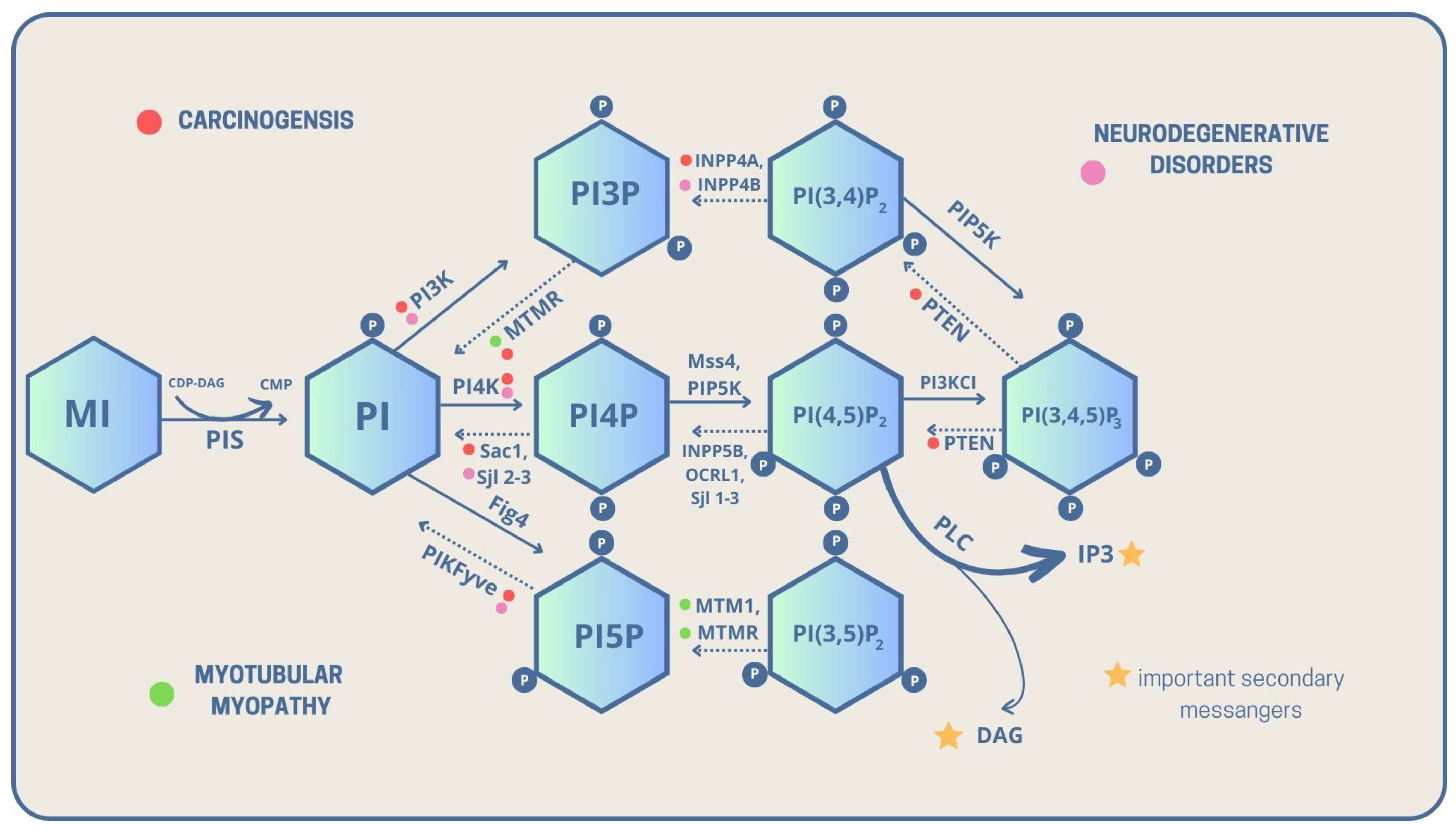
2. The Family of Phosphoinositol and Phosphoinositides
3. Routes and Interconversions of PIs
4. Myotubular Myopathy
5. Neurodegenerative Diseases
6. Carcinogenesis
7. Conclusions
Author Contributions
Funding
Institutional Review Board Statement
Informed Consent Statement
Data Availability Statement
Conflicts of Interest
Abbreviations
| 3E10Fv-MTM1 | Prototypical targeted protein replacement agent |
| AD | Alzheimer’s disease |
| ALS | Amyotrophic lateral sclerosis |
| ArPIKfyve | Associated regulator of PIKfyve |
| BRRS | Bannayan-Riley-Ruvalcaba syndrome |
| BIN1 | Bridging Integrator 1 |
| CD | Cathepsin D |
| CDP-DAG | Cytidine diphosphate diacylglycerol |
| CMP | Cytidine monophosphate diacylglycerol |
| CMT | Charcot-Marie-Tooth disease |
| CMT4B | Charcot-Marie-Tooth disease type 4B |
| CNM | Centronuclear myopathy |
| CNS | Central nervous system |
| CS | Cowden syndrome |
| CTSS | Cathepsin S |
| Cvt | Cytoplasm to vacuole transport |
| DAG | Diacylglycerol |
| DNM2 | dynamin 2 |
| EGF | epidermal growth factor |
| EGFR | Epidermal growth factor receptor |
| FSH | Follicle stimulating hormone |
| GLUT 4 | Glucose transporter type 4 |
| GPI | Glycosyl-phosphatidylinositol |
| GSK-3β | Glycogen synthase kinase |
| INPP4A | Phosphatidylinositol 4,5-bisphosphate 5-phosphatase A |
| INPP4B | Phosphatidylinositol 4,5-bisphosphate 5-phosphatase B |
| Ins(1,4,5)P3 | Inositol 1,4,5-trisphosphate |
| IP | Inositol Phosphate |
| IP3 | Inositol trisphosphate |
| IPG | Inositol-phosphoglycan |
| IRF3 | Interferon regulatory transcription factor 3 |
| JNK | c-Jun N-terminal kinase |
| KCa3.1 | Calcium-activated potassium channel |
| KCNN4 | Potassium calcium-activated channel subfamily N member 4 |
| LH | Luteinizing hormone |
| MHC class II | Major histocompatibility complex class II |
| MI | Myo-inositol |
| MTM | Myotubular myopathy |
| MTM1 | Myotubularin 1 |
| MTMR1 | Myotubularin-related protein 1 |
| MTMR2 | Myotubularin-related protein 2 |
| MTMR3 | Myotubularin-related protein 3 |
| MTMR4 | Myotubularin-related protein 4 |
| MTMR6 | Myotubularin-related protein 6 |
| MTMR7 | Myotubularin-related protein 7 |
| MTMR8 | Myotubularin-related protein 8 |
| MTMR9 | Myotubularin-related protein 9 |
| MTMR12 | Myotubularin-related protein 12 |
| NCL | Neuronal ceroid lipofuscinosis |
| Nedd4 | Neural precursor cell expression protein 4 |
| NFT | Neurofibrillary tangles |
| NMDAR | N-methyl-D-aspartate-type glutamate receptor |
| NPC | Nieman-Pick type C |
| NPC 1 | Nieman-Pick type C gene 1 |
| NPC 2 | Nieman-Pick type C gene 2 |
| P | Phosphate group |
| PHTS | PTEN hamartoma tumor syndrome |
| PI(3,4)P2/PtdIns(3,4)P2 | Phosphatidylinositol 3,4-bisphosphate |
| PI(3,4,5)P3/PtdIns(3,4,5)P3 | Phosphatidylinositol 3,4,5-trisphosphate |
| PI(3,5)P2/PtdIns(3,5)P2 | Phosphatidylinositol 3,5-bisphosphate |
| PI(4,5)P2 /PtdIns(4,5)P2/PIP2 | Phosphatidylinositol 4,5-bisphosphate |
| PI/PtdIns | Phosphatidylinositol |
| PI3K | Phosphatidylinositol-3 kinase |
| PI3P | Phosphatidylinositol 3-phosphate |
| PI3P/PtdIns3P | Phosphatidylinositol 3-phosphate |
| PI4K | Phosphatidylinositol-4 kinase |
| PI4K2A | Phosphatidylinositol 4-Kinase Type 2 Alpha |
| PI4P/PtdIns4P | Phosphatidylinositol 4-phosphate |
| PI5P | Phosphatidylinositol 5-phosphate |
| PI5P/PtdIns5P | Phosphatidylinositol 5-phosphate |
| PI5P4K | Phosphatidylinositol 5-phosphate 4-kinase |
| PLC | Phospholipase C |
| PIP/PtdInsP | Phosphatidylinositol phosphate |
| PIP4K | Phosphatidylinositol 4 phosphate kinase |
| PIS1 | Phosphatidylinositol synthase 1 |
| PKB/Akt | Protein kinase B |
| PKC | Protein kinase C |
| PS | Proteus syndrome |
| rAAV8-cMTM1 | recombinant AAV8 vector restoring MTM1 expression |
| RABEPK | Rab9 effector protein with Kelch motifs |
| ROS | Reactive oxygene species |
| Ser | Serine |
| Sjl2-3 | Synaptojanin-like proteins 2-3 |
| SPAG9 | Sperm Associated Antigen 9 |
| TBK 1 | tank-binding kinase 1 |
| Thr | hreonine |
| TSH | Thyroid stimulating hormone |
| Tyr | Tyrosine |
| Vac 14 | Vacuole 14 protein homolog |
| XLMTM | X-linked myotubular myopathy |
| Ymr1 | Yeast myotubularin related 1 |
References
- Downes, C.P.; Macphee, C.H. myo-Inositol Metabolites as Cellular Signals. Eur. J. Biochem. 1990, 193, 1–18. [Google Scholar] [CrossRef] [PubMed]
- Derkaczew, M.; Martyniuk, P.; Osowski, A.; Wojtkiewicz, J. Cyclitols: From Basic Understanding to Their Association with Neurodegeneration. Nutrients 2023, 15, 2029. [Google Scholar] [CrossRef] [PubMed]
- Daniel, E.D.; Kew, J.N.; Maycox, P.R. Investigation of the H+– Myo -Inositol Transporter (HMIT) as a Neuronal Regulator of Phosphoinositide Signalling. Biochem. Soc. Trans. 2009, 37, 1139–1143. [Google Scholar] [CrossRef] [PubMed]
- Croze, M.L.; Soulage, C.O. Potential Role and Therapeutic Interests of Myo-Inositol in Metabolic Diseases. Biochimie 2013, 95, 1811–1827. [Google Scholar] [CrossRef]
- Schneider, S. Inositol Transport Proteins. FEBS Lett. 2015, 589, 1049–1058. [Google Scholar] [CrossRef]
- Bizzarri, M.; Fuso, A.; Dinicola, S.; Cucina, A.; Bevilacqua, A. Pharmacodynamics and Pharmacokinetics of Inositol(s) in Health and Disease. Expert Opin. Drug Metab. Toxicol. 2016, 12, 1181–1196. [Google Scholar] [CrossRef]
- Benvenga, S.; Antonelli, A. Inositol(s) in Thyroid Function, Growth and Autoimmunity. Rev. Endocr. Metab. Disord. 2016, 17, 471–484. [Google Scholar] [CrossRef]
- De Craene, J.-O.; Bertazzi, D.; Bär, S.; Friant, S. Phosphoinositides, Major Actors in Membrane Trafficking and Lipid Signaling Pathways. Int. J. Mol. Sci. 2017, 18, 634. [Google Scholar] [CrossRef]
- Elabbadi, N.; Ancelin, M.L.; Vial, H.J. Characterization of Phosphatidylinositol Synthase and Evidence of a Polyphosphoinositide Cycle in Plasmodium-Infected Erythrocytes. Mol. Biochem. Parasitol. 1994, 63, 179–192. [Google Scholar] [CrossRef]
- Cockcroft, S. Phosphatidylinositol Transfer Proteins Couple Lipid Transport to Phosphoinositide Synthesis. Semin. Cell Dev. Biol. 2001, 12, 183–191. [Google Scholar] [CrossRef]
- Devereaux, K.; Dall’Armi, C.; Alcazar-Roman, A.; Ogasawara, Y.; Zhou, X.; Wang, F.; Yamamoto, A.; De Camilli, P.; Di Paolo, G. Regulation of Mammalian Autophagy by Class II and III PI 3-Kinases through PI3P Synthesis. PLoS ONE 2013, 8, e76405. [Google Scholar] [CrossRef]
- Byfield, M.P.; Murray, J.T.; Backer, J.M. HVps34 Is a Nutrient-Regulated Lipid Kinase Required for Activation of P70 S6 Kinase. J. Biol. Chem. 2005, 280, 33076–33082. [Google Scholar] [CrossRef] [PubMed]
- Strahl, T.; Thorner, J. Synthesis and Function of Membrane Phosphoinositides in Budding Yeast, Saccharomyces Cerevisiae. Biochim. Biophys. Acta Mol. Cell Biol. Lipids 2007, 1771, 353–404. [Google Scholar] [CrossRef]
- Dickson, E.J.; Hille, B. Understanding Phosphoinositides: Rare, Dynamic, and Essential Membrane Phospholipids. Biochem. J. 2019, 476, 1–23. [Google Scholar] [CrossRef]
- Whitley, P.; Hinz, S.; Doughty, J. Arabidopsis FAB1/PIKfyve Proteins Are Essential for Development of Viable Pollen. Plant Physiol. 2009, 151, 1812–1822. [Google Scholar] [CrossRef] [PubMed]
- Buckley, C.M.; Heath, V.L.; Guého, A.; Bosmani, C.; Knobloch, P.; Sikakana, P.; Personnic, N.; Dove, S.K.; Michell, R.H.; Meier, R.; et al. PIKfyve/Fab1 Is Required for Efficient V-ATPase and Hydrolase Delivery to Phagosomes, Phagosomal Killing, and Restriction of Legionella Infection. PLoS Pathog. 2019, 15, e1007551. [Google Scholar] [CrossRef] [PubMed]
- Sbrissa, D.; Ikonomov, O.C.; Shisheva, A. PIKfyve, a Mammalian Ortholog of Yeast Fab1p Lipid Kinase, Synthesizes 5-Phosphoinositides. J. Biol. Chem. 1999, 274, 21589–21597. [Google Scholar] [CrossRef] [PubMed]
- Rajala, A.; Rajala, R.; Teel, K.; Rajala, R.V.S. Ribosomal Targeting Strategy and Nuclear Labeling to Analyze Photoreceptor Phosphoinositide Signatures. Biochim. Biophys. Acta Mol. Cell Biol. Lipids 2022, 1867, 159161. [Google Scholar] [CrossRef]
- Lu, N.; Shen, Q.; Mahoney, T.R.; Neukomm, L.J.; Wang, Y.; Zhou, Z. Two PI 3-Kinases and One PI 3-Phosphatase Together Establish the Cyclic Waves of Phagosomal PtdIns(3)P Critical for the Degradation of Apoptotic Cells. PLoS Biol. 2012, 10, e1001245. [Google Scholar] [CrossRef]
- York, H.M.; Joshi, K.; Wright, C.S.; Kreplin, L.Z.; Rodgers, S.J.; Moorthi, U.K.; Gandhi, H.; Patil, A.; Mitchell, C.A.; Iyer-Biswas, S.; et al. Deterministic Early Endosomal Maturations Emerge from a Stochastic Trigger-and-Convert Mechanism. Nat. Commun. 2023, 14, 4652. [Google Scholar] [CrossRef]
- Godi, A. Regulation and Molecular Composition of the Golgi-Associated Spectrin Skeleton. Ph.D. Thesis, Open University (United Kingdom), Milton Keynes, UK, 2002. [Google Scholar]
- Foti, M.; Audhya, A.; Emr, S.D. Sac1 Lipid Phosphatase and Stt4 Phosphatidylinositol 4-Kinase Regulate a Pool of Phosphatidylinositol 4-Phosphate That Functions in the Control of the Actin Cytoskeleton and Vacuole Morphology. Mol. Biol. Cell 2001, 12, 2396–2411. [Google Scholar] [CrossRef] [PubMed]
- Böttcher-Sehlmeyer, C. Subcellular Localization and Molecular Interactions of Phosphoinositide 5′-Phosphatases of the Yeast Synaptojanin-like Protein Family. Ph.D. Thesis, University of Stuttgart, Stuttgart, Germany, 2006. [Google Scholar]
- Ebrahimzadeh, Z. Exploring the Roles of Phosphoinositides in the Biology of the Malaria Parasite Plasmodium Falciparum. Ph.D. Thesis, Université Laval, Quebec City, QC, Canada, 2019. [Google Scholar]
- Lowe, M. Structure and Function of the Lowe Syndrome Protein OCRL1: Structure and Function of OCRL1. Traffic 2005, 6, 711–719. [Google Scholar] [CrossRef] [PubMed]
- Mehta, Z.B.; Pietka, G.; Lowe, M. The Cellular and Physiological Functions of the Lowe Syndrome Protein OCRL1. Traffic 2014, 15, 471–487. [Google Scholar] [CrossRef] [PubMed]
- Tariq, K.; Luikart, B.W. Striking a Balance: PIP2 and PIP3 Signaling in Neuronal Health and Disease. Explor. Neuroprot. Ther. 2021, 1, 86. [Google Scholar] [CrossRef]
- Antonietta De Matteis, M.; Di Campli, A.; Godi, A. The Role of the Phosphoinositides at the Golgi Complex. Biochim. Biophys. Acta Mol. Cell Res. 2005, 1744, 396–405. [Google Scholar] [CrossRef]
- Sasaki, J.; Kofuji, S.; Itoh, R.; Momiyama, T.; Takayama, K.; Murakami, H.; Chida, S.; Tsuya, Y.; Takasuga, S.; Eguchi, S.; et al. The PtdIns(3,4)P2 Phosphatase INPP4A Is a Suppressor of Excitotoxic Neuronal Death. Nature 2010, 465, 497–501. [Google Scholar] [CrossRef]
- Ivetac, I.; Munday, A.D.; Kisseleva, M.V.; Zhang, X.-M.; Luff, S.; Tiganis, T.; Whisstock, J.C.; Rowe, T.; Majerus, P.W.; Mitchell, C.A. The Type Iα Inositol Polyphosphate 4-Phosphatase Generates and Terminates Phosphoinositide 3-Kinase Signals on Endosomes and the Plasma Membrane. Mol. Biol. Cell 2005, 16, 2218–2233. [Google Scholar] [CrossRef] [PubMed]
- Chaudhuri, R.; Khanna, K.; Desiraju, K.; Pattnaik, B.; Vatsa, D.; Agrawal, A.; Ghosh, B. Novel Nuclear Translocation of Inositol Polyphosphate 4-Phosphatase Is Associated with Cell Cycle, Proliferation and Survival. Biochim. Biophys. Acta Mol. Cell Res. 2018, 1865, 1501–1514. [Google Scholar] [CrossRef]
- Wang, L.; Wang, Y.; Duan, C.; Yang, Q. Inositol Phosphatase INPP4A Inhibits the Apoptosis of in Vitro Neurons with Characteristic of Intractable Epilepsy by Reducing Intracellular Ca2+ Concentration. Int. J. Clin. Exp. Pathol. 2018, 11, 1999. [Google Scholar] [PubMed]
- Lopez, S.M.; Hodgson, M.C.; Packianathan, C.; Bingol-Ozakpinar, O.; Uras, F.; Rosen, B.P.; Agoulnik, I.U. Determinants of the Tumor Suppressor INPP4B Protein and Lipid Phosphatase Activities. Biochem. Biophys. Res. Commun. 2013, 440, 277–282. [Google Scholar] [CrossRef]
- Maekawa, M.; Terasaka, S.; Mochizuki, Y.; Kawai, K.; Ikeda, Y.; Araki, N.; Skolnik, E.Y.; Taguchi, T.; Arai, H. Sequential Breakdown of 3-Phosphorylated Phosphoinositides Is Essential for the Completion of Macropinocytosis. Proc. Natl. Acad. Sci. USA 2014, 111, E978–E987. [Google Scholar] [CrossRef]
- Gewinner, C.; Wang, Z.C.; Richardson, A.; Teruya-Feldstein, J.; Etemadmoghadam, D.; Bowtell, D.; Barretina, J.; Lin, W.M.; Rameh, L.; Salmena, L.; et al. Evidence That Inositol Polyphosphate 4-Phosphatase Type II Is a Tumor Suppressor That Inhibits PI3K Signaling. Cancer Cell 2009, 16, 115–125. [Google Scholar] [CrossRef]
- Guo, C.; Yang, M.; Jing, L.; Wang, J.; Yu, Y.; Li, Y.; Duan, J.; Zhou, X.; Li, Y.; Sun, Z. Amorphous Silica Nanoparticles Trigger Vascular Endothelial Cell Injury through Apoptosis and Autophagy via Reactive Oxygen Species-Mediated MAPK/Bcl-2 and PI3K/Akt/MTOR Signaling. Int. J. Nanomed. 2016, 11, 5257–5276. [Google Scholar] [CrossRef] [PubMed]
- Zhai, S.; Liu, Y.; Lu, X.; Qian, H.; Tang, X.; Cheng, X.; Wang, Y.; Shi, Y.; Deng, X. INPP4B As A Prognostic and Diagnostic Marker Regulates Cell Growth of Pancreatic Cancer Via Activating AKT. OncoTargets Ther. 2019, 12, 8287–8299. [Google Scholar] [CrossRef]
- Jefferson, A.B.; Majerus, P.W. Properties of Type II Inositol Polyphosphate 5-Phosphatase. J. Biol. Chem. 1995, 270, 9370–9377. [Google Scholar] [CrossRef]
- Bothwell, S.P.; Farber, L.W.; Hoagland, A.; Nussbaum, R.L. Species-Specific Difference in Expression and Splice-Site Choice in Inpp5b, an Inositol Polyphosphate 5-Phosphatase Paralogous to the Enzyme Deficient in Lowe Syndrome. Mamm. Genome 2010, 21, 458–466. [Google Scholar] [CrossRef]
- Blondeau, F.; Laporte, J.; Bodin, S.; Superti-Furga, G.; Payrastre, B.; Mandel, J.-L. Myotubularin, a Phosphatase Deficient in Myotubular Myopathy, Acts on Phosphatidylinositol 3-Kinase and Phosphatidylinositol 3-Phosphate Pathway. Hum. Mol. Genet. 2000, 9, 2223–2229. [Google Scholar] [CrossRef]
- Taylor, G.S.; Maehama, T.; Dixon, J.E. Myotubularin, a Protein Tyrosine Phosphatase Mutated in Myotubular Myopathy, Dephosphorylates the Lipid Second Messenger, Phosphatidylinositol 3-Phosphate. Proc. Natl. Acad. Sci. USA 2000, 97, 8910–8915. [Google Scholar] [CrossRef] [PubMed]
- Schaletzky, J.; Dove, S.K.; Short, B.; Lorenzo, O.; Clague, M.J.; Barr, F.A. Phosphatidylinositol-5-Phosphate Activation and Conserved Substrate Specificity of the Myotubularin Phosphatidylinositol 3-Phosphatases. Curr. Biol. 2003, 13, 504–509. [Google Scholar] [CrossRef]
- Tsujita, K.; Itoh, T.; Ijuin, T.; Yamamoto, A.; Shisheva, A.; Laporte, J.; Takenawa, T. Myotubularin Regulates the Function of the Late Endosome through the GRAM Domain-Phosphatidylinositol 3,5-Bisphosphate Interaction. J. Biol. Chem. 2004, 279, 13817–13824. [Google Scholar] [CrossRef] [PubMed]
- Gupta, V.A.; Hnia, K.; Smith, L.L.; Gundry, S.R.; McIntire, J.E.; Shimazu, J.; Bass, J.R.; Talbot, E.A.; Amoasii, L.; Goldman, N.E.; et al. Loss of Catalytically Inactive Lipid Phosphatase Myotubularin-Related Protein 12 Impairs Myotubularin Stability and Promotes Centronuclear Myopathy in Zebrafish. PLoS Genet. 2013, 9, e1003583. [Google Scholar] [CrossRef] [PubMed]
- Lawlor, M.W.; Dowling, J.J. X-Linked Myotubular Myopathy. Neuromuscul. Disord. 2021, 31, 1004–1012. [Google Scholar] [CrossRef] [PubMed]
- Kim, S.-A.; Taylor, G.S.; Torgersen, K.M.; Dixon, J.E. Myotubularin and MTMR2, Phosphatidylinositol 3-Phosphatases Mutated in Myotubular Myopathy and Type 4B Charcot-Marie-Tooth Disease. J. Biol. Chem. 2002, 277, 4526–4531. [Google Scholar] [CrossRef]
- Bong, S.M.; Son, K.; Yang, S.-W.; Park, J.-W.; Cho, J.-W.; Kim, K.-T.; Kim, H.; Kim, S.J.; Kim, Y.J.; Lee, B.I. Crystal Structure of Human Myotubularin-Related Protein 1 Provides Insight into the Structural Basis of Substrate Specificity. PLoS ONE 2016, 11, e0152611. [Google Scholar] [CrossRef]
- Walker, D.M.; Urbe, S.; Dove, S.K.; Tenza, D. Characterization of MTMR3: An Inositol Lipid 3-Phosphatase with Novel Substrate Specificity. Curr. Biol. 2001, 11, 1600–1605. [Google Scholar] [CrossRef] [PubMed]
- Zhao, R.; Qi, Y.; Chen, J.; Zhao, Z.J. FYVE-DSP2, a FYVE Domain-Containing Dual Specificity Protein Phosphatase That Dephosphorylates Phosphotidylinositol 3-Phosphate. Exp. Cell Res. 2001, 265, 329–338. [Google Scholar] [CrossRef]
- Lahiri, A.; Hedl, M.; Abraham, C. MTMR3 Risk Allele Enhances Innate Receptor-Induced Signaling and Cytokines by Decreasing Autophagy and Increasing Caspase-1 Activation. Proc. Natl. Acad. Sci. USA 2015, 112, 10461–10466. [Google Scholar] [CrossRef]
- Dewi Pamungkas Putri, D.; Kawasaki, T.; Murase, M.; Sueyoshi, T.; Deguchi, T.; Ori, D.; Suetsugu, S.; Kawai, T. PtdIns3P Phosphatases MTMR3 and MTMR4 Negatively Regulate Innate Immune Responses to DNA through Modulating STING Trafficking. J. Biol. Chem. 2019, 294, 8412–8423. [Google Scholar] [CrossRef]
- Plant, P.J.; Correa, J.; Goldenberg, N.; Bain, J.; Batt, J. The Inositol Phosphatase MTMR4 Is a Novel Target of the Ubiquitin Ligase Nedd4. Biochem. J. 2009, 419, 57–63. [Google Scholar] [CrossRef]
- Kumar, P.; Munnangi, P.; Chowdary, K.R.; Shah, V.J.; Shinde, S.R.; Kolli, N.R.; Halehalli, R.R.; Nagarajaram, H.A.; Maddika, S. A Human Tyrosine Phosphatase Interactome Mapped by Proteomic Profiling. J. Proteome Res. 2017, 16, 2789–2801. [Google Scholar] [CrossRef] [PubMed]
- Naughtin, M.J.; Sheffield, D.A.; Rahman, P.; Hughes, W.E.; Gurung, R.; Stow, J.L.; Nandurkar, H.H.; Dyson, J.M.; Mitchell, C.A. The Myotubularin Phosphatase MTMR4 Regulates Sorting from Early Endosomes. J. Cell Sci. 2010, 123, 3071–3083. [Google Scholar] [CrossRef]
- Zou, J.; Chang, S.-C.; Marjanovic, J.; Majerus, P.W. MTMR9 Increases MTMR6 Enzyme Activity, Stability, and Role in Apoptosis. J. Biol. Chem. 2009, 284, 2064–2071. [Google Scholar] [CrossRef]
- Zou, J.; Zhang, C.; Marjanovic, J.; Kisseleva, M.V.; Majerus, P.W.; Wilson, M.P. Myotubularin-Related Protein (MTMR) 9 Determines the Enzymatic Activity, Substrate Specificity, and Role in Autophagy of MTMR8. Proc. Natl. Acad. Sci. USA 2012, 109, 9539–9544. [Google Scholar] [CrossRef]
- Srivastava, S.; Li, Z.; Lin, L.; Liu, G.; Ko, K.; Coetzee, W.A.; Skolnik, E.Y. The Phosphatidylinositol 3-Phosphate Phosphatase Myotubularin- Related Protein 6 (MTMR6) Is a Negative Regulator of the Ca2+-Activated K+ Channel KCa3. Mol. Cell. Biol. 2005, 25, 3630–3638. [Google Scholar] [CrossRef]
- Mochizuki, Y.; Ohashi, R.; Kawamura, T.; Iwanari, H.; Kodama, T.; Naito, M.; Hamakubo, T. Phosphatidylinositol 3-Phosphatase Myotubularin-Related Protein 6 (MTMR6) Is Regulated by Small GTPase Rab1B in the Early Secretory and Autophagic Pathways. J. Biol. Chem. 2013, 288, 1009–1021. [Google Scholar] [CrossRef]
- Weidner, P.; Söhn, M.; Gutting, T.; Friedrich, T.; Gaiser, T.; Magdeburg, J.; Kienle, P.; Ruh, H.; Hopf, C.; Behrens, H.-M.; et al. Myotubularin-Related Protein 7 Inhibits Insulin Signaling in Colorectal Cancer. Oncotarget 2016, 7, 50490–50506. [Google Scholar] [CrossRef]
- Zhao, D.; Shen, C.; Gao, T.; Li, H.; Guo, Y.; Li, F.; Liu, C.; Liu, Y.; Chen, X.; Zhang, X.; et al. Myotubularin Related Protein 7 Is Essential for the Spermatogonial Stem Cell Homeostasis via PI3K/AKT Signaling. Cell Cycle 2019, 18, 2800–2813. [Google Scholar] [CrossRef] [PubMed]
- Sanchez-Juan, P.; Bishop, M.T.; Aulchenko, Y.S.; Brandel, J.-P.; Rivadeneira, F.; Struchalin, M.; Lambert, J.-C.; Amouyel, P.; Combarros, O.; Sainz, J.; et al. Genome-Wide Study Links MTMR7 Gene to Variant Creutzfeldt-Jakob Risk. Neurobiol. Aging 2012, 33, 1487.e21–1487.e28. [Google Scholar] [CrossRef] [PubMed]
- Weidner, P.; Söhn, M.; Schroeder, T.; Helm, L.; Hauber, V.; Gutting, T.; Betge, J.; Röcken, C.; Rohrbacher, F.N.; Pattabiraman, V.R.; et al. Myotubularin-Related Protein 7 Activates Peroxisome Proliferator-Activated Receptor-Gamma. Oncogenesis 2020, 9, 59. [Google Scholar] [CrossRef] [PubMed]
- Yoo, K.-Y.; Son, J.Y.; Lee, J.U.; Shin, W.; Im, D.-W.; Kim, S.J.; Ryu, S.E.; Heo, Y.-S. Structure of the Catalytic Phosphatase Domain of MTMR8: Implications for Dimerization, Membrane Association and Reversible Oxidation. Acta Crystallogr. D Biol. Crystallogr. 2015, 71, 1528–1539. [Google Scholar] [CrossRef]
- Gaudet, P.; Livstone, M.S.; Lewis, S.E.; Thomas, P.D. Phylogenetic-Based Propagation of Functional Annotations within the Gene Ontology Consortium. Brief. Bioinform. 2011, 12, 449–462. [Google Scholar] [CrossRef] [PubMed]
- De Matteis, M.A.; Staiano, L.; Emma, F.; Devuyst, O. The 5-Phosphatase OCRL in Lowe Syndrome and Dent Disease 2. Nat. Rev. Nephrol. 2017, 13, 455–470. [Google Scholar] [CrossRef] [PubMed]
- Zhang, B.; Nandakumar, R.; Reinert, L.S.; Huang, J.; Laustsen, A.; Gao, Z.; Sun, C.; Jensen, S.B.; Troldborg, A.; Assil, S.; et al. STEEP Mediates STING ER Exit and Activation of Signaling. Nat. Immunol. 2020, 21, 868–879. [Google Scholar] [CrossRef] [PubMed]
- Thoresen, S.B.; Pedersen, N.M.; Liestøl, K.; Stenmark, H. A Phosphatidylinositol 3-Kinase Class III Sub-Complex Containing VPS15, VPS34, Beclin 1, UVRAG and BIF-1 Regulates Cytokinesis and Degradative Endocytic Traffic. Exp. Cell Res. 2010, 316, 3368–3378. [Google Scholar] [CrossRef]
- Hu, H.; Dong, J.; Liang, D.; Gao, Z.; Bai, L.; Sun, R.; Hu, H.; Zhang, H.; Dong, Y.; Lan, K. Genome-Wide Mapping of the Binding Sites and Structural Analysis of Kaposi’s Sarcoma-Associated Herpesvirus Viral Interferon Regulatory Factor 2 Reveal That It Is a DNA-Binding Transcription Factor. J. Virol. 2016, 90, 1158–1168. [Google Scholar] [CrossRef]
- Fruman, D.A.; Chiu, H.; Hopkins, B.D.; Bagrodia, S.; Cantley, L.C.; Abraham, R.T. The PI3K Pathway in Human Disease. Cell 2017, 170, 605–635. [Google Scholar] [CrossRef]
- Fruman, D.A.; Cantley, L.C. Idelalisib—A PI3Kδ Inhibitor for B-Cell Cancers. N. Engl. J. Med. 2014, 370, 1061–1062. [Google Scholar] [CrossRef]
- Arcaro, A.; Guerreiro, A. The Phosphoinositide 3-Kinase Pathway in Human Cancer: Genetic Alterations and Therapeutic Implications. Curr. Genom. 2007, 8, 271–306. [Google Scholar] [CrossRef]
- Clayton, E.L.; Minogue, S.; Waugh, M.G. Phosphatidylinositol 4-Kinases and PI4P Metabolism in the Nervous System: Roles in Psychiatric and Neurological Diseases. Mol. Neurobiol. 2013, 47, 361–372. [Google Scholar] [CrossRef]
- Zhou, Q.; Li, J.; Yu, H.; Zhai, Y.; Gao, Z.; Liu, Y.; Pang, X.; Zhang, L.; Schulten, K.; Sun, F.; et al. Molecular Insights into the Membrane-Associated Phosphatidylinositol 4-Kinase IIα. Nat. Commun. 2014, 5, 3552. [Google Scholar] [CrossRef]
- Pataer, A.; Ozpolat, B.; Shao, R.; Cashman, N.R.; Plotkin, S.S.; Samuel, C.E.; Lin, S.H.; Kabil, N.N.; Wang, J.; Majidi, M.; et al. Therapeutic Targeting of the PI4K2A/PKR Lysosome Network Is Critical for Misfolded Protein Clearance and Survival in Cancer Cells. Oncogene 2020, 39, 801–813. [Google Scholar] [CrossRef] [PubMed]
- Gehrmann, T.; Gülkan, H.; Suer, S.; Herberg, F.W.; Balla, A.; Vereb, G.; Mayr, G.W.; Heilmeyer, L.M.G. Functional Expression and Characterisation of a New Human Phosphatidylinositol 4-Kinase PI4K230. Biochim. Biophys. Acta Mol. Cell Biol. Lipids 1999, 1437, 341–356. [Google Scholar] [CrossRef] [PubMed]
- Nakatsu, F.; Baskin, J.M.; Chung, J.; Tanner, L.B.; Shui, G.; Lee, S.Y.; Pirruccello, M.; Hao, M.; Ingolia, N.T.; Wenk, M.R.; et al. PtdIns4P Synthesis by PI4KIIIα at the Plasma Membrane and Its Impact on Plasma Membrane Identity. J. Cell Biol. 2012, 199, 1003–1016. [Google Scholar] [CrossRef] [PubMed]
- Pagnamenta, A.T.; Howard, M.F.; Wisniewski, E.; Popitsch, N.; Knight, S.J.L.; Keays, D.A.; Quaghebeur, G.; Cox, H.; Cox, P.; Balla, T.; et al. Germline Recessive Mutations in PI4KA Are Associated with Perisylvian Polymicrogyria, Cerebellar Hypoplasia and Arthrogryposis. Hum. Mol. Genet. 2015, 24, 3732–3741. [Google Scholar] [CrossRef]
- Shisheva, A. PIKfyve and Its Lipid Products in Health and in Sickness. In Phosphoinositides and Disease; Falasca, M., Ed.; Current Topics in Microbiology and Immunology; Springer: Dordrecht, Netherlands, 2012; Volume 362, pp. 127–162. ISBN 978-94-007-5024-1. [Google Scholar]
- Kim, J.; Jahng, W.J.; Di Vizio, D.; Lee, J.S.; Jhaveri, R.; Rubin, M.A.; Shisheva, A.; Freeman, M.R. The Phosphoinositide Kinase PIKfyve Mediates Epidermal Growth Factor Receptor Trafficking to the Nucleus. Cancer Res. 2007, 67, 9229–9237. [Google Scholar] [CrossRef]
- Sbrissa, D.; Ikonomov, O.C.; Fu, Z.; Ijuin, T.; Gruenberg, J.; Takenawa, T.; Shisheva, A. Core Protein Machinery for Mammalian Phosphatidylinositol 3,5-Bisphosphate Synthesis and Turnover That Regulates the Progression of Endosomal Transport. J. Biol. Chem. 2007, 282, 23878–23891. [Google Scholar] [CrossRef]
- Sbrissa, D.; Ikonomov, O.C.; Filios, C.; Delvecchio, K.; Shisheva, A. Functional Dissociation between PIKfyve-Synthesized PtdIns5P and PtdIns(3,5)P2 by Means of the PIKfyve Inhibitor YM201636. Am. J. Physiol. Cell Physiol. 2012, 303, C436–C446. [Google Scholar] [CrossRef]
- Krishna, S.; Palm, W.; Lee, Y.; Yang, W.; Bandyopadhyay, U.; Xu, H.; Florey, O.; Thompson, C.B.; Overholtzer, M. PIKfyve Regulates Vacuole Maturation and Nutrient Recovery Following Engulfment. Dev. Cell 2016, 38, 536–547. [Google Scholar] [CrossRef]
- Dayam, R.M.; Sun, C.X.; Choy, C.H.; Mancuso, G.; Glogauer, M.; Botelho, R.J. The Lipid Kinase PIKfyve Coordinates the Neutrophil Immune Response through the Activation of the Rac GTPase. J. Immunol. 2017, 199, 2096–2105. [Google Scholar] [CrossRef]
- Liggins, M.C.; Flesher, J.L.; Jahid, S.; Vasudeva, P.; Eby, V.; Takasuga, S.; Sasaki, J.; Sasaki, T.; Boissy, R.E.; Ganesan, A.K. PIKfyve Regulates Melanosome Biogenesis. PLoS Genet. 2018, 14, e1007290. [Google Scholar] [CrossRef]
- Baranov, M.V.; Bianchi, F.; Schirmacher, A.; Van Aart, M.A.C.; Maassen, S.; Muntjewerff, E.M.; Dingjan, I.; Ter Beest, M.; Verdoes, M.; Keyser, S.G.L.; et al. The Phosphoinositide Kinase PIKfyve Promotes Cathepsin-S-Mediated Major Histocompatibility Complex Class II Antigen Presentation. iScience 2019, 11, 160–177. [Google Scholar] [CrossRef]
- De Campos, C.B.; Zhu, Y.X.; Sepetov, N.; Romanov, S.; Bruins, L.A.; Shi, C.-X.; Stein, C.K.; Petit, J.L.; Polito, A.N.; Sharik, M.E.; et al. Identification of PIKfyve Kinase as a Target in Multiple Myeloma. Haematologica 2020, 105, 1641–1649. [Google Scholar] [CrossRef] [PubMed]
- Iijima, M.; Devreotes, P. Tumor Suppressor PTEN Mediates Sensing of Chemoattractant Gradients. Cell 2002, 109, 599–610. [Google Scholar] [CrossRef]
- Funamoto, S.; Meili, R.; Lee, S.; Parry, L.; Firtel, R.A. Spatial and Temporal Regulation of 3-Phosphoinositides by PI 3-Kinase and PTEN Mediates Chemotaxis. Cell 2002, 109, 611–623. [Google Scholar] [CrossRef]
- Wessels, D.; Lusche, D.F.; Kuhl, S.; Heid, P.; Soll, D.R. PTEN Plays a Role in the Suppression of Lateral Pseudopod Formation during Dictyostelium Motility and Chemotaxis. J. Cell Sci. 2007, 120, 2517–2531. [Google Scholar] [CrossRef] [PubMed]
- McMains, V.C.; Liao, X.-H.; Kimmel, A.R. Oscillatory Signaling and Network Responses during the Development of Dictyostelium Discoideum. Ageing Res. Rev. 2008, 7, 234–248. [Google Scholar] [CrossRef] [PubMed]
- Gruver, J.S.; Wikswo, J.P.; Chung, C.Y. 3′-Phosphoinositides Regulate the Coordination of Speed and Accuracy during Chemotaxis. Biophys. J. 2008, 95, 4057–4067. [Google Scholar] [CrossRef]
- Maeda, Y.T.; Inose, J.; Matsuo, M.Y.; Iwaya, S.; Sano, M. Ordered Patterns of Cell Shape and Orientational Correlation during Spontaneous Cell Migration. PLoS ONE 2008, 3, e3734. [Google Scholar] [CrossRef]
- Yehia, L.; Eng, C. PTEN Hamartoma Tumor Syndrome. Genet. Med. 2009, 11, 687–694. [Google Scholar]
- Zhong, R.; Burk, D.H.; Nairn, C.J.; Wood-Jones, A.; Morrison, W.H.; Ye, Z.-H. Mutation of SAC1, an Arabidopsis SAC Domain Phosphoinositide Phosphatase, Causes Alterations in Cell Morphogenesis, Cell Wall Synthesis, and Actin Organization. Plant Cell 2005, 17, 1449–1466. [Google Scholar] [CrossRef]
- Del Bel, L.M.; Brill, J.A. Sac1, a Lipid Phosphatase at the Interface of Vesicular and Nonvesicular Transport. Traffic 2018, 19, 301–318. [Google Scholar] [CrossRef]
- Ghaemmaghami, S.; Huh, W.-K.; Bower, K.; Howson, R.W.; Belle, A.; Dephoure, N.; O’Shea, E.K.; Weissman, J.S. Global Analysis of Protein Expression in Yeast. Nature 2003, 425, 737–741. [Google Scholar] [CrossRef]
- Hardies, K.; Cai, Y.; Jardel, C.; Jansen, A.C.; Cao, M.; May, P.; Djémié, T.; Hachon Le Camus, C.; Keymolen, K.; Deconinck, T.; et al. Loss of SYNJ1 Dual Phosphatase Activity Leads to Early Onset Refractory Seizures and Progressive Neurological Decline. Brain 2016, 139, 2420–2430. [Google Scholar] [CrossRef]
- Quadri, M.; Fang, M.; Picillo, M.; Olgiati, S.; Breedveld, G.J.; Graafland, J.; Wu, B.; Xu, F.; Erro, R.; Amboni, M.; et al. Mutation in the SYNJ1 Gene Associated with Autosomal Recessive, Early-Onset Parkinsonism. Hum. Mutat. 2013, 34, 1208–1215. [Google Scholar] [CrossRef] [PubMed]
- Walker, S.M.; Downes, C.P.; Leslie, N.R. TPIP: A Novel Phosphoinositide 3-Phosphatase. Biochem. J. 2001, 360, 277–283. [Google Scholar] [CrossRef] [PubMed]
- Jungbluth, H.; Wallgren-Pettersson, C.; Laporte, J. Centronuclear (Myotubular) Myopathy. Orphanet J. Rare Dis. 2008, 3, 26. [Google Scholar] [CrossRef] [PubMed]
- Ross, J.A.; Tasfaout, H.; Levy, Y.; Morgan, J.; Cowling, B.S.; Laporte, J.; Zanoteli, E.; Romero, N.B.; Lowe, D.A.; Jungbluth, H.; et al. RAAV-Related Therapy Fully Rescues Myonuclear and Myofilament Function in X-Linked Myotubular Myopathy. Acta Neuropathol. Commun. 2020, 8, 167. [Google Scholar] [CrossRef] [PubMed]
- Childers, M.K.; Joubert, R.; Poulard, K.; Moal, C.; Grange, R.W.; Doering, J.A.; Lawlor, M.W.; Rider, B.E.; Jamet, T.; Danièle, N.; et al. Gene Therapy Prolongs Survival and Restores Function in Murine and Canine Models of Myotubular Myopathy. Sci. Transl. Med. 2014, 6, 220ra10. [Google Scholar] [CrossRef] [PubMed]
- Lawlor, M.W.; Armstrong, D.; Viola, M.G.; Widrick, J.J.; Meng, H.; Grange, R.W.; Childers, M.K.; Hsu, C.P.; O’Callaghan, M.; Pierson, C.R.; et al. Enzyme Replacement Therapy Rescues Weakness and Improves Muscle Pathology in Mice with X-Linked Myotubular Myopathy. Hum. Mol. Genet. 2013, 22, 1525–1538. [Google Scholar] [CrossRef]
- Massana-Muñoz, X.; Goret, M.; Nattarayan, V.; Reiss, D.; Kretz, C.; Chicanne, G.; Payrastre, B.; Vanhaesebroeck, B.; Laporte, J. Inactivating the Lipid Kinase Activity of PI3KC2β Is Sufficient to Rescue Myotubular Myopathy in Mice. JCI Insight 2023, 8, e151933. [Google Scholar] [CrossRef]
- Cowling, B.S.; Chevremont, T.; Prokic, I.; Kretz, C.; Ferry, A.; Coirault, C.; Koutsopoulos, O.; Laugel, V.; Romero, N.B.; Laporte, J. Reducing Dynamin 2 Expression Rescues X-Linked Centronuclear Myopathy. J. Clin. Investig. 2014, 124, 1350–1363. [Google Scholar] [CrossRef]
- Lionello, V.M.; Nicot, A.-S.; Sartori, M.; Kretz, C.; Kessler, P.; Buono, S.; Djerroud, S.; Messaddeq, N.; Koebel, P.; Prokic, I.; et al. Amphiphysin 2 Modulation Rescues Myotubular Myopathy and Prevents Focal Adhesion Defects in Mice. Sci. Transl. Med. 2019, 11, eaav1866. [Google Scholar] [CrossRef]
- Previtali, S.C.; Quattrini, A.; Bolino, A. Charcot–Marie–Tooth Type 4B Demyelinating Neuropathy: Deciphering the Role of MTMR Phosphatases. Expert Rev. Mol. Med. 2007, 9, 1–16. [Google Scholar] [CrossRef]
- Romani, M.; Mehawej, C.; Mazza, T.; Mégarbané, A.; Valente, E.M. “Fork and Bracket” Syndrome Expands the Spectrum of SBF1 -Related Sensory Motor Polyneuropathies. Neurol. Genet. 2016, 2, e61. [Google Scholar] [CrossRef] [PubMed]
- Barisic, N.; Claeys, K.G.; Sirotković-Skerlev, M.; Löfgren, A.; Nelis, E.; De Jonghe, P.; Timmerman, V. Charcot-Marie-Tooth Disease: A Clinico-Genetic Confrontation: Charcot-Marie-Tooth Disease. Ann. Hum. Genet. 2008, 72, 416–441. [Google Scholar] [CrossRef] [PubMed]
- Dugger, B.N.; Dickson, D.W. Pathology of Neurodegenerative Diseases. Cold Spring Harb. Perspect. Biol. 2017, 9, a028035. [Google Scholar] [CrossRef]
- Voet, S.; Srinivasan, S.; Lamkanfi, M.; Van Loo, G. Inflammasomes in Neuroinflammatory and Neurodegenerative Diseases. EMBO Mol. Med. 2019, 11, e10248. [Google Scholar] [CrossRef] [PubMed]
- Carmel, L.; Efroni, S.; White, P.D.; Aslakson, E.; Vollmer-Conna, U.; Rajeevan, M.S. Gene Expression Profile of Empirically Delineated Classes of Unexplained Chronic Fatigue. Pharmacogenomics 2006, 7, 375–386. [Google Scholar] [CrossRef] [PubMed]
- Fogarty, K.; Kashem, M.; Bauer, A.; Bernardino, A.; Brennan, D.; Cook, B.; Farrow, N.; Molinaro, T.; Nelson, R. Development of Three Orthogonal Assays Suitable for the Identification and Qualification of PIKfyve Inhibitors. ASSAY Drug Dev. Technol. 2017, 15, 210–219. [Google Scholar] [CrossRef]
- Zhang, Y.; Zolov, S.N.; Chow, C.Y.; Slutsky, S.G.; Richardson, S.C.; Piper, R.C.; Yang, B.; Nau, J.J.; Westrick, R.J.; Morrison, S.J.; et al. Loss of Vac14, a Regulator of the Signaling Lipid Phosphatidylinositol 3,5-Bisphosphate, Results in Neurodegeneration in Mice. Proc. Natl. Acad. Sci. USA 2007, 104, 17518–17523. [Google Scholar] [CrossRef]
- McCartney, A.J.; Zhang, Y.; Weisman, L.S. Phosphatidylinositol 3,5-Bisphosphate: Low Abundance, High Significance: Prospects & Overviews. BioEssays 2014, 36, 52–64. [Google Scholar] [CrossRef]
- Bi, X.; Liu, J.; Yao, Y.; Baudry, M.; Lynch, G. Deregulation of the Phosphatidylinositol-3 Kinase Signaling Cascade Is Associated with Neurodegeneration in Npc1−/− Mouse Brain. Am. J. Pathol. 2005, 167, 1081–1092. [Google Scholar] [CrossRef] [PubMed]
- Walls, K.C.; Klocke, B.J.; Saftig, P.; Shibata, M.; Uchiyama, Y.; Roth, K.A.; Shacka, J.J. Altered Regulation of Phosphatidylinositol 3-Kinase Signaling in Cathepsin D-Deficient Brain. Autophagy 2007, 3, 222–229. [Google Scholar] [CrossRef] [PubMed]
- Boffey, H.K.; Rooney, T.P.C.; Willems, H.M.G.; Edwards, S.; Green, C.; Howard, T.; Ogg, D.; Romero, T.; Scott, D.E.; Winpenny, D.; et al. Development of Selective Phosphatidylinositol 5-Phosphate 4-Kinase γ Inhibitors with a Non-ATP-Competitive, Allosteric Binding Mode. J. Med. Chem. 2022, 65, 3359–3370. [Google Scholar] [CrossRef] [PubMed]
- Ji, W.; Wang, E.S.; Manz, T.D.; Jiang, J.; Donovan, K.A.; Abulaiti, X.; Fischer, E.S.; Cantley, L.C.; Zhang, T.; Gray, N.S. Development of Potent and Selective Degraders of PI5P4Kγ. Eur. J. Med. Chem. 2023, 247, 115027. [Google Scholar] [CrossRef]
- Nishikawa, T.; Takahashi, T.; Nakamori, M.; Yamazaki, Y.; Kurashige, T.; Nagano, Y.; Nishida, Y.; Izumi, Y.; Matsumoto, M. Phosphatidylinositol-4,5-bisphosphate Is Enriched in Granulovacuolar Degeneration Bodies and Neurofibrillary Tangles. Neuropathol. Appl. Neurobiol. 2014, 40, 489–501. [Google Scholar] [CrossRef]
- Landman, N.; Jeong, S.Y.; Shin, S.Y.; Voronov, S.V.; Serban, G.; Kang, M.S.; Park, M.K.; Paolo, G.D.; Chung, S.; Kim, T.-W. Presenilin Mutations Linked to Familial Alzheimer’s Disease Cause an Imbalance in Phosphatidylinositol 4,5-Bisphosphate Metabolism. Proc. Natl. Acad. Sci. USA 2006, 103, 19524–19529. [Google Scholar] [CrossRef]
- Desale, S.E.; Chinnathambi, S. Phosphoinositides Signaling Modulates Microglial Actin Remodeling and Phagocytosis in Alzheimer’s Disease. Cell Commun. Signal 2021, 19, 28. [Google Scholar] [CrossRef]
- Peters, J.M.; Gonzalez, F.J. The Evolution of Carcinogenesis. Toxicol. Sci. 2018, 165, 272–276. [Google Scholar] [CrossRef]
- Bertram, J.S. The Molecular Biology of Cancer. Mol. Asp. Med. 2001, 21, 167–223. [Google Scholar] [CrossRef]
- Fattahi, S.; Amjadi-Moheb, F.; Tabaripour, R.; Ashrafi, G.H.; Akhavan-Niaki, H. PI3K/AKT/MTOR Signaling in Gastric Cancer: Epigenetics and Beyond. Life Sci. 2020, 262, 118513. [Google Scholar] [CrossRef] [PubMed]
- Ediriweera, M.K.; Tennekoon, K.H.; Samarakoon, S.R. Role of the PI3K/AKT/MTOR Signaling Pathway in Ovarian Cancer: Biological and Therapeutic Significance. Semin. Cancer Biol. 2019, 59, 147–160. [Google Scholar] [CrossRef] [PubMed]
- Levine, B.; Kroemer, G. Biological Functions of Autophagy Genes: A Disease Perspective. Cell 2019, 176, 11–42. [Google Scholar] [CrossRef]
- Magagnoli, M.; Carlo-Stella, C.; Santoro, A. Copanlisib for the Treatment of Adults with Relapsed Follicular Lymphoma. Expert Rev. Clin. Pharmacol. 2020, 13, 813–823. [Google Scholar] [CrossRef]
- Patnaik, A.; Appleman, L.J.; Tolcher, A.W.; Papadopoulos, K.P.; Beeram, M.; Rasco, D.W.; Weiss, G.J.; Sachdev, J.C.; Chadha, M.; Fulk, M.; et al. First-in-Human Phase I Study of Copanlisib (BAY 80-6946), an Intravenous Pan-Class I Phosphatidylinositol 3-Kinase Inhibitor, in Patients with Advanced Solid Tumors and Non-Hodgkin’s Lymphomas. Ann. Oncol. 2016, 27, 1928–1940. [Google Scholar] [CrossRef] [PubMed]
- Dunbar, J.; Nevejans, J.; McKee, C.; Faia, K.; Van Lier, J.J.; Pruim, P.; Stegehuis, J.; Zhao, S.; Kahl, B.S.; Horwitz, S.M.; et al. Pharmacokinetics and Pharmacodynamics of IPI-145, a Potent Inhibitor of Phosphoinositide-3-Kinase-δ,γ, Following Single- and Multiple-Dose Administration in Healthy Subjects and Patients with Advanced Hematologic Malignancies. Blood 2012, 120, 4853. [Google Scholar] [CrossRef]
- Janku, F.; Yap, T.A.; Meric-Bernstam, F. Targeting the PI3K Pathway in Cancer: Are We Making Headway? Nat. Rev. Clin. Oncol. 2018, 15, 273–291. [Google Scholar] [CrossRef]
- Matasar, M.J.; Capra, M.; Özcan, M.; Lv, F.; Li, W.; Yañez, E.; Sapunarova, K.; Lin, T.; Jin, J.; Jurczak, W.; et al. Copanlisib plus Rituximab versus Placebo plus Rituximab in Patients with Relapsed Indolent Non-Hodgkin Lymphoma (CHRONOS-3): A Double-Blind, Randomised, Placebo-Controlled, Phase 3 Trial. Lancet Oncol. 2021, 22, 678–689. [Google Scholar] [CrossRef]
- Maehama, T.; Dixon, J.E. The Tumor Suppressor, PTEN/MMAC1, Dephosphorylates the Lipid Second Messenger, Phosphatidylinositol 3,4,5-Trisphosphate. J. Biol. Chem. 1998, 273, 13375–13378. [Google Scholar] [CrossRef]
- Chung, J.-H.; Ginn-Pease, M.E.; Eng, C. Phosphatase and Tensin Homologue Deleted on Chromosome 10 (PTEN) Has Nuclear Localization Signal–Like Sequences for Nuclear Import Mediated by Major Vault Protein. Cancer Res. 2005, 65, 4108–4116. [Google Scholar] [CrossRef]
- Li, D.-M.; Sun, H. TEP1, Encoded by a Candidate Tumor Suppressor Locus, Is a Novel Protein Tyrosine Phosphatase Regulated by Transforming Growth Factor. Cancer Res. 1997, 57, 2124–2129. [Google Scholar] [PubMed]
- Davidson, L.; Maccario, H.; Perera, N.M.; Yang, X.; Spinelli, L.; Tibarewal, P.; Glancy, B.; Gray, A.; Weijer, C.J.; Downes, C.P.; et al. Suppression of Cellular Proliferation and Invasion by the Concerted Lipid and Protein Phosphatase Activities of PTEN. Oncogene 2010, 29, 687–697. [Google Scholar] [CrossRef] [PubMed]
- Zhang, J.; Zhang, P.; Wei, Y.; Piao, H.; Wang, W.; Maddika, S.; Wang, M.; Chen, D.; Sun, Y.; Hung, M.-C.; et al. Deubiquitylation and Stabilization of PTEN by USP13. Nat. Cell Biol. 2013, 15, 1486–1494. [Google Scholar] [CrossRef]
- Yuan, L.; Lv, Y.; Li, H.; Gao, H.; Song, S.; Zhang, Y.; Xing, G.; Kong, X.; Wang, L.; Li, Y.; et al. Deubiquitylase OTUD3 Regulates PTEN Stability and Suppresses Tumorigenesis. Nat. Cell Biol. 2015, 17, 1169–1181. [Google Scholar] [CrossRef] [PubMed]
- Mishra, R.R.; Chaudhary, J.K.; Rath, P.C. Cell Cycle Arrest and Apoptosis by Expression of a Novel TPIP (TPIP-C2) CDNA Encoding a C2-Domain in HEK-293 Cells. Mol. Biol. Rep. 2012, 39, 7389–7402. [Google Scholar] [CrossRef]
- Zhai, Y.; Liu, D.; Jiang, Y.; Chen, X.; Shao, L.; Li, J.; Sheng, K.; Zhang, X.; Song, H. Near-Infrared-Light-Triggered Photoelectrochemical Biosensor for Detection of Alpha-Fetoprotein Based on Upconversion Nanophosphors. Sens. Actuators B Chem. 2019, 286, 468–475. [Google Scholar] [CrossRef]
- Tokuda, E.; Itoh, T.; Hasegawa, J.; Ijuin, T.; Takeuchi, Y.; Irino, Y.; Fukumoto, M.; Takenawa, T. Phosphatidylinositol 4-Phosphate in the Golgi Apparatus Regulates Cell–Cell Adhesion and Invasive Cell Migration in Human Breast Cancer. Cancer Res. 2014, 74, 3054–3066. [Google Scholar] [CrossRef]
- Shisheva, A. PIKfyve: Partners, Significance, Debates and Paradoxes. Cell Biol. Int. 2008, 32, 591–604. [Google Scholar] [CrossRef]
- Hasegawa, J.; Strunk, B.S.; Weisman, L.S. PI5P and PI(3,5)P2: Minor, but Essential Phosphoinositides. Cell Struct. Funct. 2017, 42, 49–60. [Google Scholar] [CrossRef]
- Lodovichi, S.; Mercatanti, A.; Cervelli, T.; Galli, A. Computational Analysis of Data from a Genome-Wide Screening Identifies New PARP1 Functional Interactors as Potential Therapeutic Targets. Oncotarget 2019, 10, 2722–2737. [Google Scholar] [CrossRef]
- Gayle, S.; Landrette, S.; Beeharry, N.; Conrad, C.; Hernandez, M.; Beckett, P.; Ferguson, S.M.; Mandelkern, T.; Zheng, M.; Xu, T.; et al. Identification of Apilimod as a First-in-Class PIKfyve Kinase Inhibitor for Treatment of B-Cell Non-Hodgkin Lymphoma. Blood 2017, 129, 1768–1778. [Google Scholar] [CrossRef] [PubMed]
- Maltese, W.A.; Overmeyer, J.H. Methuosis. Am. J. Pathol. 2014, 184, 1630–1642. [Google Scholar] [CrossRef] [PubMed]

| PI Phosphatidylinositol | 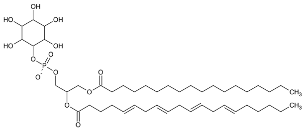 |
| PI3P Phosphatidylinositol 3-phosphate |  |
| PI(3,5)P2 Phosphatidylinositol 3,5-bisphosphate |  |
| PI4P Phosphatidylinositol 5-phosphate |  |
| PI(3,4)P2 Phosphatidylinositol 3,4-bisphosphate | 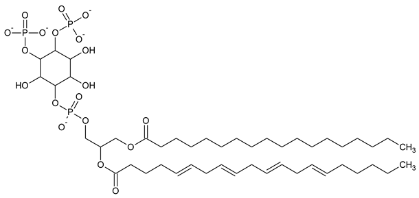 |
| PI(3,4,5)P3 Phosphatidylinositol 3,4,5-trisphosphate | 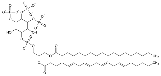 |
| PI5P Phosphatidylinositol 5-phosphate |  |
| PI(4,5)P2 Phosphatidylinositol 4,5-bisphosphate | 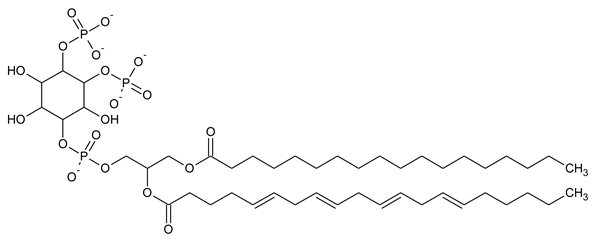 |
| Gene | Protein | Gene ID | Protein Function | Protein Function Literature | Associated Illness or Defect | Associated Illness or Defect Literature |
|---|---|---|---|---|---|---|
| INPP4A | Inositol polyphosphate-4-phosphatase type I A | 3631 |
| Junko Sasaki et al., 2010 [29] Ivan Ivetac et al., 2005 [30] Rituparna Chaudhuri et al., 2018 [31] |
| Junko Sasaki et al., 2010 [29] Li Wang et al., 2018 [32] |
| INPP4B | Inositol polyphosphate 4-phosphatase type II | 8821 |
| Sandra M. Lopez et al., 2013 [33] Masashi Maekawa et al., 2014 [34] Christina Gewinner et al., 2009 [35] |
and breast cancer | ST Guo et al., 2015 [36] Shuyu Zhai et al., 2019 [37] |
| INPP5B | Type II inositol 1,4,5-trisphosphate 5-phosphatase | 3633 |
| Jefferson et al., 1995 [38] |
| Susan P. Bothwell et al., 2010 [39] |
| MTM1 | Myotubularin 1 | 4534 |
| Blondeau et al., 2000 [40] Taylor et al., 2000 [41] Schaletzky et al., 2003 [42] Tsujita et al., 2004 [43] Gupta et al., 2013 [44] |
| M’elanie Annoussamy et al., 2021 [45] |
| MTMR1 | Myotubularin-related protein 1 | 8776 |
| Soo-A Kim et al., 2002 [46] Seoung Min Bong et al., 2016 [47] |
| Soo-A Kim et al., 2001 [46] |
| MTMR2 | Myotubularin-related protein 2 | 8898 |
| Soo-A Kim et al., 2002 [46] |
| Soo-A Kim et al., 2002 [46] |
| MTMR3 | Myotubularin-related protein 3 | 8897 |
| Donna M. Walker, 2001 [48] Runxiang Zhao, 2001 [49] |
| Amit Lahiri et al., 2015 [50] |
| MTMR4 | Myotubularin-related protein 4 | 9110 |
| Dyaningtyas Dewi Pamungkas Putri et al., 2019 [51] Pamela J. Plant et al., 2009 [52] Kumar et al., 2017 [53] |
| Monica J. Naughtin et al., 2010 [54] |
| MTMR6 | Myotubularin-related protein 6 | 9107 |
| Jun Zou et al., 2009 [55] Jun Zou et al., 2012 [56] Masashi Maekawa et al., 2014 [34] Shekhar Srivastava et al., 2005 [57] |
| Yasuhiro Mochizuki et al., 2013 [58] |
| MTMR7 | Myotubularin-related protein 7 | 9108 |
| Philip Weidner et al., 2016 [59] |
| Dan Zhao et al., 2019 [60] Philip Weidner et al., 2016 [59] Pascual Sanchez-Juan et al., 2012 [61] Philip Weidner et al., 2020 [62] |
| MTMR8 | Myotubularin-related protein 8 | 55613 |
| Jun Zou et al., 2012 [56] Ki-Young Yoo et al., 2015 [63] |
| Jun Zou et al., 2012 [56] |
| OCRL-1 | OCRL inositol polyphosphate-5-phosphatase | 4952 |
| Pascale Gaudet et al., 2011 [64] |
| Maria Antonietta De Matteis et al., 2017 [65] |
| PIK3C3 | Phosphatidylinositol 3-kinase catalytic subunit type 3 | 5289 |
| Bao-cun Zhang et al., 2020 [66] Sigrid B. Thoresen et al., 2010 [67] |
| Haidai Hu et al., 2015 [68] |
| PI3K | Phosphatidylinositol 3-kinase | 39089293 |
| David A. Fruman et al., 2017 [69] |
| David A. Fruman et al., 2014 [70] Alexandre Arcaro et al., 2007 [71] |
| PI4K2A | Phosphatidylinositol 4-kinase type 2 alpha | 55361 |
| Emma L. Clayton et al., 2013 [72] Qiangjun Zhou et al., 2014 [73] |
| Apar Pataer et al., 2020 [74] |
| PI4KA | Phosphatidylinositol 4-kinase alpha | 5297 |
| T Gehrmann et al., 1999 [75] Fubito Nakatsu et al., 2012 [76] |
| Alistair T. Pagnamenta et al., 2015 [77] |
| PIKFYVE | 1-phosphatidylinositol 3-phosphate 5-kinase | 200576 |
| Shisheva et al., 2012 [78] Kim et al., 2007 [79] Sbrissa et al., 2007 [80] Sbrissa et al., 2012 [81] Krishna et al., 2016 [82] Dayam et al., 2017 [83] Liggins et al., 2018 [84] Baranov et al., 2019 [85] |
| Cecilia Bonolo de Campos et al., 2020 [86] |
| PTEN | Phosphatidylinositol 3,4,5-trisphosphate 3-phosphatase and dual-specificity protein phosphatase PTEN | 5728 |
| Iijima et al., 2002 [87] Funamoto et al., 2002 [88] Wessels et al., 2007 [89] McMains et al., 2008 [90] Gruver et al., 2008 [91] Maeda et al., 2008 [92] |
| Yehia and Eng, 2021 [93] |
| SAC1 | Phosphatidylinositol-3-phosphatase SAC1 | 22908 |
| Zhong et al., 2005 [94] |
| Del Bel and Brill, 2018 [95] |
| STT4 | Phosphatidylinositol 4-kinase STT4 | 851014 |
| Ghaemmaghami et al., 2003 [96] |
| Ghaemmaghami et al., 2003 [96] |
| SYNJ1 | Synaptojanin-1 | 8867 |
| Hardies et al., 2016 [97] |
| Quadri et al., 2013 [98] |
| TPIP α | TPTE and PTEN homologous inositol lipid phosphatase | 93492 |
| Walker et al., 2001 [99] |
| Walker et al., 2001 [99] |
Disclaimer/Publisher’s Note: The statements, opinions and data contained in all publications are solely those of the individual author(s) and contributor(s) and not of MDPI and/or the editor(s). MDPI and/or the editor(s) disclaim responsibility for any injury to people or property resulting from any ideas, methods, instructions or products referred to in the content. |
© 2023 by the authors. Licensee MDPI, Basel, Switzerland. This article is an open access article distributed under the terms and conditions of the Creative Commons Attribution (CC BY) license (https://creativecommons.org/licenses/by/4.0/).
Share and Cite
Derkaczew, M.; Martyniuk, P.; Hofman, R.; Rutkowski, K.; Osowski, A.; Wojtkiewicz, J. The Genetic Background of Abnormalities in Metabolic Pathways of Phosphoinositides and Their Linkage with the Myotubular Myopathies, Neurodegenerative Disorders, and Carcinogenesis. Biomolecules 2023, 13, 1550. https://doi.org/10.3390/biom13101550
Derkaczew M, Martyniuk P, Hofman R, Rutkowski K, Osowski A, Wojtkiewicz J. The Genetic Background of Abnormalities in Metabolic Pathways of Phosphoinositides and Their Linkage with the Myotubular Myopathies, Neurodegenerative Disorders, and Carcinogenesis. Biomolecules. 2023; 13(10):1550. https://doi.org/10.3390/biom13101550
Chicago/Turabian StyleDerkaczew, Maria, Piotr Martyniuk, Robert Hofman, Krzysztof Rutkowski, Adam Osowski, and Joanna Wojtkiewicz. 2023. "The Genetic Background of Abnormalities in Metabolic Pathways of Phosphoinositides and Their Linkage with the Myotubular Myopathies, Neurodegenerative Disorders, and Carcinogenesis" Biomolecules 13, no. 10: 1550. https://doi.org/10.3390/biom13101550