Article

Postweaning Development Influences Endogenous VPAC1 Modulation of LTP Induced by Theta-Burst Stimulation: A Link to Maturation of the Hippocampal GABAergic System

1
BioISI—Biosystems & Integrative Sciences Institute, Faculdade de Ciências, Universidade de Lisboa, 1749-016 Lisbon, Portugal
2
Unidade de Neurociências, Instituto de Medicina Molecular João Lobo Antunes, Faculdade de Medicina, Universidade de Lisboa, Av. Prof. Egas Moniz, 1649-028 Lisbon, Portugal
3
Instituto de Farmacologia e Neurociências, Faculdade de Medicina, Universidade de Lisboa, Av. Prof. Egas Moniz, 1649-028 Lisbon, Portugal
4
Departamento de Química e Bioquímica, Faculdade de Ciências, Universidade de Lisboa, Campo Grande, 1749-016 Lisboa, Portugal
*
Author to whom correspondence should be addressed.
Current address: Departamento de Biologia Vegetal, Faculdade de Ciências, Universidade de Lisboa, Campo Grande, 1749-016 Lisboa, Portugal.
Biomolecules 2024, 14(3), 379; https://doi.org/10.3390/biom14030379
Submission received: 23 February 2024 / Revised: 15 March 2024 / Accepted: 18 March 2024 / Published: 20 March 2024
(This article belongs to the Special Issue Advances in Brain Development and Disease)

Abstract

:
Long-term potentiation (LTP) induced by theta-burst stimulation (TBS) undergoes postweaning developmental changes partially linked to GABAergic circuit maturation. Endogenous vasoactive intestinal peptide (VIP) acting on its VPAC1 receptor strongly influences LTP induced by theta-burst stimulation (TBS), an effect dependent on GABAergic transmission. Although VPAC1 receptor levels are developmentally regulated during embryogenesis, their variation along postweaning development is unknown, as is the VPAC1 modulation of LTP or its relation to hippocampal GABAergic circuit maturation. As such, we investigated how VPAC1 modulation of LTP adjusts from weaning to adulthood along with GABAergic circuit maturation. As described, LTP induced by mild TBS (5 bursts, 4 pulses delivered at 100 Hz) was increasingly greater from weaning to adulthood. The influence of the VPAC1 receptor antagonist PG 97-269 (100 nM) on TBS-induced LTP was much larger in juvenile (3-week-old) than in young adult (6–7-week-old) or adult (12-week-old) rats. This effect was not associated with a developmental decrease in synaptic VPAC1 receptor levels. However, an increase in pre and post-synaptic GABAergic synaptic markers suggests an increase in the number of GABAergic synaptic contacts that is more prominent than the one observed in glutamatergic connections during this period. Conversely, endogenous VPAC2 receptor activation did not significantly influence TBS-induced LTP. VPAC2 receptor levels enhance pronouncedly during postweaning development, but not at synaptic sites. Given the involvement of VIP interneurons in several aspects of hippocampal-dependent learning, neurodevelopmental disorders, and epilepsy, this could provide important insights into the role of VIP modulation of hippocampal synaptic plasticity during normal and altered brain development potentially contributing to epileptogenesis.

1. Introduction

Vasoactive intestinal peptide (VIP), a neuropeptide present exclusively in hippocampal interneurons [1], is a modulator of hippocampal synaptic transmission and excitability [2]. Its endogenous actions are believed to be most relevant in conditions eliciting high-frequency firing such as occurring during hippocampal synaptic plasticity and seizures [2,3,4]. Its actions are mediated by hippocampal VPAC1 and VPAC2 receptors that bind with similar affinity to the neuropeptide pituitary adenylate cyclase-activating polypeptide (PACAP). These integrate the VIP/PACAP family of G protein-coupled receptors [2] that includes yet another receptor, PAC1, that has a significantly higher affinity for PACAP than for VIP.
The hippocampal actions of VIP are largely dependent on GABAergic transmission [2,5,6] and involve multiple cellular and molecular targets, as expected from the expression of VIP in different interneuron populations with different target selectivity [2,6] and from the opposing actions of VIP on exocytotic GABA release when activating VPAC1 or VPAC2 receptors [7]. Nevertheless, VIP enhancement of synaptic transmission to CA1 pyramidal cell dendrites is mostly mediated by VPAC1 receptors located in the strata oriens and radiatum [2,8,9]. This, in turn, triggers disinhibition of pyramidal cell dendrites by inhibiting dendritic-targeting GABAergic interneurons [6]. Notwithstanding, VIP also enhances pyramidal cell excitability in the absence of GABAergic transmission [6,10], an effect believed to occur through the activation of VPAC2 receptors in pyramidal cell bodies [11].
Long-term potentiation (LTP) as induced by theta-burst stimulation (TBS), a form of activity-dependent synaptic plasticity, is a key cellular mechanism for memory storage [12,13]. TBS is a sequence of electrical stimuli that mimics CA3 and CA1 pyramidal complex-spike cell discharges perceived during the hippocampal theta rhythm (3–7 Hz). This EEG pattern is linked to hippocampal spatial memory formation believed to work as a ‘tag’ for short-term memory processing [13,14]. TBS triggers an early LTP, acting through suppression of feedforward inhibition by a priming burst/single pulse. This allows for enough depolarization to trigger NMDA receptors [15,16], through an indirect mechanism that involves activation of GABAB autoreceptors, thus suppressing GABA release from feedforward interneurons [17] and synaptic GABA availability.
Endogenous VIP, acting on VPAC1 receptors, is an important endogenous modulator of NMDA-dependent TBS-induced LTP as well as LTD and depotentiation in the CA1 area of the hippocampus [3,4]. This is a prospective indirect effect through modulation of disinhibition since it is fully dependent on GABAergic transmission. Yet this, together with previous reports that VIP directly enhances NMDA currents in CA1 pyramidal cells, an effect mimicked by VPAC2 and to a lesser extent by VPAC1 selective agonists [18], highlights the diversity and importance of VIP mechanisms in the regulation of hippocampal synaptic plasticity phenomena. Accordingly, VIP is a crucial endogenous modulator of several hippocampal-dependent learning tasks [19,20,21,22,23]. Furthermore, VIP-deficient mice fail to develop hippocampal-dependent learning skills like reversal learning [24] while the VIP knockout is unviable. VIP interneuron dysfunction is also implicated in three prominent neurodevelopmental disorders, Rett syndrome, Dravet Syndrome, and Down’s syndrome [25,26,27], and may contribute to enhanced seizure susceptibility and epileptogenesis associated with altered synaptic plasticity in these developmental disorders as in other acquired epilepsies [2,28,29,30].
The activity of hippocampal GABAergic interneurons influences not only the balance between excitation and inhibition but also a complex network of circuits regulating feedforward, feedback, and disinhibitory mechanisms [31]. Synaptic inhibition at pyramidal cell dendrites is crucial for selective Ca2+-dependent input selectivity and precision of LTP induction [32] yet the long-lasting plasticity responses of interneurons to mild TBS remain largely unknown. Accumulated evidence suggests that interneurons serve as mediators of normal circuit development by regulating critical period plasticity and shaping the formation of sensory maps [31]. Furthermore, although interneurons are present in the hippocampus from early postnatal development, a few of these, like parvalbumin-expressing interneurons only fully develop and set in human hippocampal circuits until adulthood [33] This can dramatically influence the dynamics of hippocampal inhibition from weaning to adulthood, and its control of synaptic plasticity phenomena. We showed recently that TBS-induced LTP undergoes post-weaning developmental maturation until adulthood [34], and preliminary data from our lab suggests this may be related to postweaning maturation of GABAergic circuits [35].
In this paper, we investigated the postweaning developmental changes in VPAC1 receptor modulation of hippocampal LTP induced by mild TBS by endogenous VIP and their correlation with developmental changes in the expression of VIP and VIP receptors, GABAergic and glutamatergic pre and postsynaptic markers and overall synaptic density.

2. Material and Methods

The experiments were performed in juvenile (3 weeks old), young adult (6–7 weeks old), and adult (12 weeks old) male outbred Wistar rats (Charles River, Barcelona, Spain) essentially as previously described [3,34] and all protocols and procedures were performed according to ARRIVE guidelines for experimental design, analysis, and their reporting. Animal housing and handling was performed in accordance with Portuguese law (DL 113/2013) and European Community guidelines (86/609/EEC and 63/2010/CE). The animals were anesthetized with fluothane, decapitated, and the right hippocampus dissected free in ice-cold artificial cerebrospinal fluid (aCSF) of the following composition in mM: NaCl 124, KCl 3, NaH2PO4 1.25, NaHCO3 26, MgSO4 1.5, CaCl2 2, glucose 10, and gassed with a 95% O2–5% CO2 mixture.

2.1. LTP Experiments

Hippocampal slices (400 µm thick) were cut perpendicularly to the long axis of the hippocampus with a McIlwain tissue chopper (Campden Instruments, Loughborough, UK), then they were kept in a resting chamber in gassed aCSF at room temperature 22 °C–25 °C for at least 1 h to allow energetic and functional recovery. Each slice was transferred at a time to a submerged recording chamber of 1 mL capacity and continuously superfused at a rate of 3 mL/min with gassed aCSF at 30.5 °C. To obtain electrophysiological recordings slices were stimulated (rectangular pulses of 0.1 ms) through bipolar concentric wire electrodes placed in two separate sets (S1 and S2) of the Schaffer collateral/commissural fibres in the stratum radiatum (Figure S1). Responses were evoked alternately on the two pathways every 10 s and thus each pathway was stimulated every 20 s (0.05 Hz). Evoked field excitatory post-synaptic potentials (fEPSPs) were recorded extracellularly from CA1 stratum radiatum using aCSF-filled micropipettes. The initial stimulus intensity was chosen to elicit a field excitatory post-synaptic potential (fEPSP) of 600–900 μV amplitude (about 50% of the maximal response) while minimizing contamination by the population spike, and of similar magnitude in both pathways. The averages of six consecutive fEPSP responses from each pathway were obtained, measured, graphically plotted, and recorded for further analysis with a personal computer using the WinLTP software (version 2.96, WinLTP Ltd., Bristol, UK) [36]. The slope of the initial phase of the potential was used to quantify fEPSPs intensity. At the end of the experiments, the independence of the two pathways was tested by studying paired-pulse facilitation (PPF) across both pathways, with less than 10% facilitation being usually observed. PPF was elicited by stimulating the two Schaffer pathways with a 50 ms interval between pulses. The ratio P2/P1 between the fEPSP slopes elicited by the second P2 and the first P1 stimuli was used to quantify synaptic facilitation.
When a stable fEPSP slope baseline was observed for at least 20 min, LTP was induced either by mild TBS (five trains of 100 Hz, 4 stimuli, separated by 200 ms) or a moderate TBS (fifteen trains of 100 Hz, 4 stimuli, separated by 200 ms). The intensity of the stimulus was not changed during these stimulation protocols. LTP was taken as the % change in the average slope of fEPSPs observed from 50 to 60 min after the induction protocol, in relation to one measured during the 10 min that preceded TBS. Control and test conditions were tested in independent pathways in the same slice. In all experiments, S1 always refers to the first pathway (left or right, randomly assigned) to which TBS was applied. Test drugs were added to the perfusion solution 20 min before TBS stimulation of the test pathway (S2) and were present until the end of the experiment.

2.2. Western Blot Analysis of Synaptic Proteins

The hippocampi of 3 week-old, 6–7-week-old, and 12–14 week-old rats were collected in sucrose solution (320 mM Sucrose, 1 mg/mL BSA, 10 mM HEPES e 1 mM EDTA, pH 7.4) containing protease (complete, mini, EDTA-free Protease Inhibitor Cocktail, Sigma, St. Louis, MO, USA) and phosphatase (1 mM PMSF, 2 mM Na3VO4, and 10 mM NaF) inhibitors, homogenized with a Potter–Elvejham apparatus (Labbox, Barcelona, Spain) and either total hippocampal membranes or hippocampal synaptosomes were isolated as described [6,34]. For isolation of total hippocampal membranes, the hippocampal homogenates were centrifuged at 1500× g for 10 min. The supernatant was collected and further centrifuged at 14,000× g for 12 min. The pellet was washed twice with modified aCSF (20 mM HEPES, 1 mM MgCl2, 1.2 mM NaH2PO4, 120 mM NaCl; 2.7 mM KCl, 1.2 mM CaCl2, 10 mM glucose, pH 7.4) also containing protease and phosphatase inhibitors and resuspended in 300 µL modified aCSF per hippocampus. For isolation of hippocampal synaptosomes, homogenates were centrifuged at 3000× g for 10 min at 4 °C; the supernatant was then centrifuged at 14,000× g for 12 min at 4 °C and the pellet resuspended in 3 mL of a Percoll 45% (v/v) in modified aCSF. The top layer (synaptosomal fraction) obtained after centrifugation at 14,000× g for 2 min at 4 °C was washed twice with aCSF and resuspended in 200 µL modified aCSF per hippocampus. Aliquots of these suspensions of hippocampal membranes/synaptosomes were snap-frozen in liquid nitrogen and stored at −80 °C until use.
For Western blot, samples incubated at 95 °C for 5 min with Laemmli buffer (125 mM Tris-BASE, 4% SDS, 50% glycerol, 0.02% Bromophenol Blue, 10% β-mercaptoethanol), were run on standard 12% sodium dodecyl sulfate polyacrylamide gel electrophoresis (SDS-PAGE) and transferred to PVDF membranes (pore size 0.45 μm, GE Healthcare Life Sciences, Marlborough, MA, USA). These were then blocked for 1 h with either a 3% BSA solution or 5% milk solution in Tris-buffered saline (20 mM Tris, 150 mM NaCl) containing 0.1% Tween-20 (TBST), and incubated overnight at 4 °C with and either rabbit polyclonal anti-VPAC1 (1:600, Alomone Labs (Jerusalem, IL, USA) #AVR-001, RRID: AB_2341081), rabbit polyclonal anti-VPAC2 (1:500, Alomone Labs #AVR-002, RRID: RRID: AB_2341082), rabbit polyclonal anti VIP (1:300, Proteintech Europe (Manchester, UK), # 16233-1-AP, RRID: AB_2878233), mouse monoclonal anti-gephyrin (1:3000, #147011, Synaptic Systems (Göttingen, Germany), AB_2810214), rabbit polyclonal anti-PSD-95 (1:750, #CST-2507, Cell Signalling Tech. (Danvers, MA, USA), AB_561221), rabbit polyclonal anti-synaptophysin (1:7500, Synaptic Systems #101002, RRID:AB_887905), rabbit polyclonal anti-VGAT (1:2500, Synaptic Systems #131002, RRID: AB_887871), rabbit polyclonal anti-VGlut1 (1:3000, Synaptic Systems #135302, RRID: AB_887877), and either mouse monoclonal anti-β-actin (1:5000, Proteintech #60008-1-Ig, RRID: AB_2289225) or rabbit polyclonal anti-alpha-tubulin (1:5000, Proteintech #11224-1-AP; RRID: AB_2210206) primary antibodies. After washing the membranes were incubated for 1 h with anti-rabbit or anti-mouse IgG secondary antibody both conjugated with horseradish peroxidase (HRP) (Proteintech) at room temperature. HRP activity was visualized by enhanced chemiluminescence with Clarity ECL Western Blotting Detection System (Bio-Rad, Richmond, CA, USA). The intensity of the bands was evaluated with the Image J software (version 1.52a, public domain, NIH, USA). Alpha-tubulin band density was used as a loading control.

2.3. Materials

PG 97-269 and PG 99-465, (Phoenix peptides, Europe) were made up of 0.1 mM stock solution in CH3COOH 1% (v/v−1). The maximal CH3COOH concentration added to the slices, 0.001% (v/v) was devoid of effects on fEPSP slope (n = 4). Aliquots of the stock solutions were kept frozen at −20 °C until use. In each experiment, one aliquot was thawed and diluted in aCSF.

2.4. Statistics

LTP values are depicted as the mean ± S.E.M of n experiments. Each n represents a single experiment performed in slices obtained from one different animal for LTP experiments and experiments performed in duplicate for each single animal in Western blot studies. The significance of the differences between the means was calculated using the paired Student’s t-test when comparing control and test conditions for LTP expression in electrophysiological studies, or with repeated measures ANOVA with Tukey’s post-hoc test (when F was significant) when comparing the levels of synaptic proteins in different age groups. p values of 0.05 or less were considered to represent statistically significant differences.

3. Results

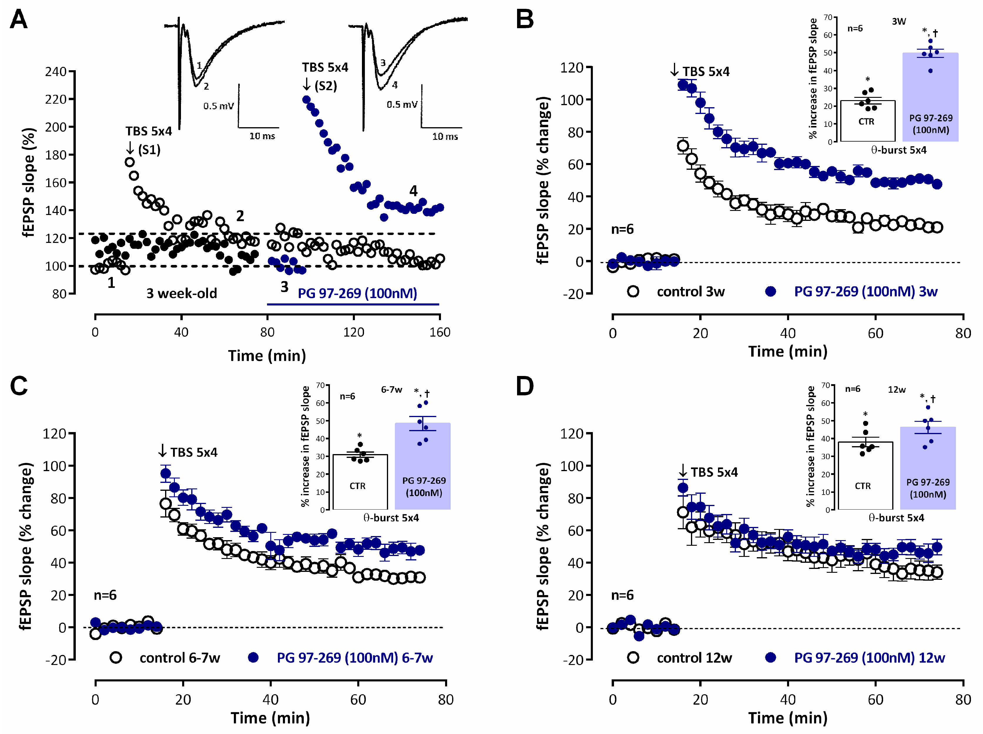
Recordings of fEPSP obtained under basal stimulation conditions (40–60% of the maximal response in each slice) in hippocampal slices from juvenile (3-week-old) rats (Figure 1A, raw data from a single experiment) showed an average slope of 0.579 ± 0.036 mV/ms (n = 12). When mild TBS (5 × 4 pulses with a 200 ms interval) was applied to the control pathway (S1) an LTP was induced, corresponding to a 25.6 ± 1.9% increase in fEPSP slope (n = 12, p < 0.05) observed 50–60 min after TBS. Application of a second mild TBS train in the test pathway (S2) in the absence of drugs always resulted in an LTP of similar magnitude (% increase in fEPSP slope of 26.1 ± 3.4%, n = 4) to the one observed in the control pathway (S1), i.e., LTP obtained under these experimental conditions was similar in S1 and S2. When mild TBS of the test pathway (S2) was delivered in the presence of the VPAC1 receptor antagonist PG 97-269 (100 nM) LTP was almost twice as large, since the enhancement of fEPSP slope observed 1 h later was 49.6 ± 2.3%, (n = 6), suggesting a strong inhibition of LTP by endogenous VIP acting on VPAC1 receptors. This effect was larger than the one previously observed in young adult rats (6–7 weeks old) [4]. In this study, fEPSPs in young adult rats had an average slope of 0.553 ± 0.022 mV/ms (n = 17). Induction of LTP with a mild TBS caused a larger potentiation of the fEPSP slope (% increase in fEPSP slope 30.0 ± 1.2%, n= 12, p < 0.05, Figure 1C) than the one observed in juvenile rats as previously described [34]. When mild TBS was delivered to S2 in the presence of the VPAC1 receptor antagonist PG 97-269 (100 nM), this resulted in an enhancement of LTP (% increase in fEPSP slope of 48.4 ± 3.9%, n = 6, Figure 1C) that was less pronounced than the one observed in juvenile rats (Figure 1B). In adult rats (12 weeks old) the average fEPSP slope was 0.582 ± 0.017 mV/ms (n = 16). The same mild TBS paradigm caused an even larger LTP since mild TBS now induced a potentiation of the fEPSP slope of 38.1 ± 2.7%, n = 6, p < 0.05, Figure 1D). When LTP was induced in the presence of the VPAC1 receptor antagonist PG 97-269 (100 nM) the enhancement of LTP was even more reduced, since mild TBS induced an enhancement of 46.3 ± 3.4% (n = 6, Figure 1D) in fEPSP slope. PG 97-269 (100 nM) did not significantly change basal synaptic transmission when added to the hippocampal slices.
Our previous studies did not show any influence of endogenous VIP on LTP induced by mild TBS through activation of VPAC2 receptors in young adult rats [4]. However, there could be post-weaning developmental changes in this respect. As shown in Figure 2, when LTP was induced by mild TBS in the presence of the selective VPAC2 receptor antagonist PG 99-465 (100 nM) there was a mild but significant enhancement of LTP in juvenile rats for which an enhancement of 34.2 ± 3.4% (n = 5, Figure 2A) in fEPSP slope was observed upon mild TBS stimulation. An even milder tendency to an increase in LTP by VPAC2 receptor blockage was observed in young adult and adult rats, but it was not consistently observed in all animals (Figure 2B,C), and this effect did not reach statistical significance. When added to the hippocampal slices PG 99-465 (100 nM) did not significantly change basal synaptic transmission.
Increasing the number of bursts to 15 (moderate TBS) enhanced the resulting potentiation evaluated 50–60 min after TBS, leading to a 46.2 ± 1.8% (n = 5, p < 0.05) enhancement in fEPSP slope (Figure 3A) in young adult rats and to a 63.6 ± 3.3% (n = 5, p < 0.05) enhancement in fEPSP slope (Figure 3B) in adult rats, as previously described [34]. Using this stronger LTP induction paradigm the influence of endogenous VIP, acting on VPAC1 receptors, on LTP expression was altered since the potentiation induced in fEPSP slope by moderate TBS in the presence of the selective VPAC1 receptor antagonist PG 97-269 (100 nM) was of 59.9 ± 3.1% (n = 5, p < 0.05, Figure 3A) in young adult rats and of a 56.5 ± 4.4% (n = 5, p < 0.05, Figure 3B) in adult rats, thus displaying a much lower potentiation of LTP by the VPAC1 receptor antagonist in young adult rats and a mild but non-significant reversal of the effect in adult rats, suggesting that a developmental reshaping of GABAergic synapses, VIPergic interneurons or its activation pathways may be occurring or that VIP receptor expression and its role in hippocampal GABAergic synapses may be altered.
To elucidate the changes in VIP, VIP receptors, GABAergic, and glutamatergic synapses during postweaning development in the hippocampus we studied the levels of VIP VPAC1 and VPAC2 receptors and synaptic glutamatergic and GABAergic markers by Western blot in total hippocampal membranes. The synaptic changes in glutamatergic and GABAergic transmission were inferred from the evolution of GABAergic and glutamatergic pre and postsynaptic markers and its relation to broad-spectrum presynaptic marker synaptophysin, which was taken as an estimator of the overall synaptic density in total hippocampal membranes. The levels of the synaptic vesicle integral protein synaptophysin enhanced from weaning to adulthood by 63.9 ± 14.9% (n = 5, p < 0.05, Figure 4I). The levels of both pre- and post-synaptic glutamatergic synaptic proteins VGlutT-1 and PSD-95 were enhanced by 29.9 ± 6.6% (n = 5, p < 0.05, Figure 4A,C) and 44.5 ± 11.0% (n = 5, p < 0.05, Figure 4A,B), respectively, in relation to the observed protein levels at 3 weeks of age. The increase in pre- and post-synaptic GABAergic synaptic proteins VGAT and gephyrin was more pronounced since VGAT and gephyrin levels enhanced by 71.1 ± 16.0% (n = 5, p < 0.05, Figure 4E,G) and 55.4 ± 10.9% (n = 5, p < 0.05, Figure 4E,F), respectively, in relation to the protein levels observed at 3 weeks, the age of weaning. By plotting the VGlut1/PSD-95 and VGAT/gephyrin ratios (Figure 4D,H) we could observe that the ratios do not significantly change along postweaning development, suggesting that the increase in these synaptic markers is indeed representative of an increase in the number of synapses. To estimate the rate of change of glutamatergic and GABAergic synapses vs global synaptic changes in hippocampal tissue we calculated the VGlut1/synaptophysin and the VGAT/synaptophysin immunoreactivity ratios (Figure 4K,L). The decrease in the hippocampal VGlut1/synaptophysin ratio from 3 to 12 weeks suggests that glutamatergic synapses do not accompany the global increase in new synapses while GABAergic synapses seem to contribute to this increase in hippocampal synaptic content, as estimated by a stable VGAT/synaptophysin ratio from 3 to 12 weeks.
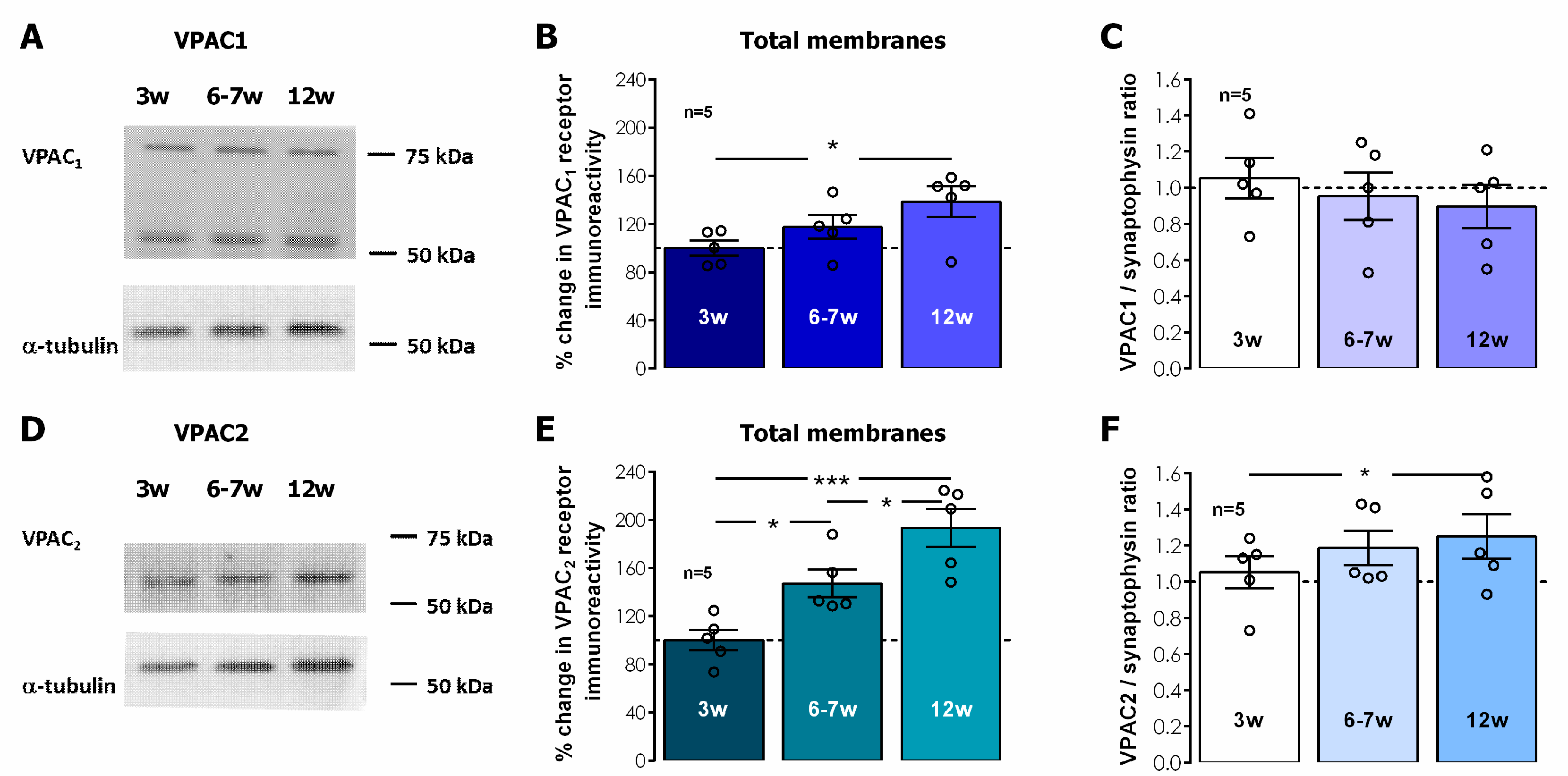
In total hippocampal membranes, VPAC1 receptor immunostaining was detected by two independent bands located at 55 and 75 KDa, respectively, likely reflecting two different N-glycosylation levels, a feature that is also relevant for VPAC1 membrane targeting, as previously described [37]. The levels of both VPAC1 and VPAC2 receptors in total hippocampal membranes displayed a gradual postweaning developmental enhancement, that reached 42.5 ± 8.9% enhancement in 12-week-old rats for VPAC1 receptors (Figure 5A,B, n = 5, p < 0.05) and was even more pronounced for VPAC2 receptors, for which the receptor levels nearly doubled (% increase 93.5 ± 15.7, Figure 5D,E, n = 5, p < 0.05) in 12-week-old rats when compared to 3-week-old animals. By plotting the VPAC1/ and VPAC2/synaptophysin ratios (Figure 5C,F) we were able to infer that the increase in VPAC1 receptors accompanied the overall gain in synaptophysin yet VPAC2 receptors were increasing even further, suggesting this increase was occurring also in non-synaptic locations.
To further clarify the cellular location of these receptors and their relevance for synaptic transmission and synaptic plasticity we performed also Western blot experiments to probe for VIP, VPAC1, and VPAC2 receptors in hippocampal synaptosomes, a preparation that is composed mainly of presynaptic nerve terminals, that often maintain attached part of the postsynaptic density [38]. In hippocampal synaptosomes, both VIP and to a lesser extent VPAC1 receptors increased along post-weaning development by 58.3 ± 15.9% and 43.7 ± 10.1% (n = 5, p < 0.05, Figure 6A,B,E,F) at 12 weeks of age, respectively, suggesting that VIP and VPAC1 containing nerve terminals enhance substantially along postweaning development. Conversely, VPAC2 receptors did not significantly change hippocampal synaptosomes from 3- to 12-week-old rats (% change at 12 weeks vs. 3 weeks 20.3 ± 11.7%, n = 5, p > 0.05, Figure 6C,D).

4. Discussion

The main findings of the present work are that: (1) VPAC1 receptor-mediated modulation of LTP induced by mild TBS in the hippocampal CA1 area by endogenous VIP is progressively weaker from weaning (3 week-old) through adulthood (12-week-old) in Wistar rats; (2) A stronger LTP-inducing train (moderate TBS) elicited an increased magnitude and stability of LTP in young-adult (6–7-week-old) and adult (12-week-old) Wistar rats, that was even less influenced by endogenous VIP acting on VPAC1 receptors; (3) The LTP elicited by mild TBS was only weakly dependent on endogenous VPAC2 receptor activation in juvenile (3-week-old) rats; (4) These observations were concurrent with an increase in both GABAergic and glutamatergic pre- and post-synaptic markers in total hippocampal membranes from weaning to adulthood, the enhancement in GABAergic being more prominent; (5) There was an increase in both VPAC1 and VPAC2 receptor levels in total hippocampal membranes during this developmental period while (6) VIP levels enhanced markedly, while only VPAC1, but not VPAC2, receptor levels were mildly enhanced in adult (12-week-old) vs juvenile (3-week-old) rats.
In the present study mild TBS (5 bursts, 4 pulses delivered at 100 Hz), an LTP-inducing stimulation pattern, elicited a long-lasting potentiation of fEPSP slope that was increasingly larger in 3-, 6–7-, and 12-week-old rats, as previously described by our group [34]. This is consistent with previous studies showing that during postnatal development there is a reinforcement of the cellular mechanisms leading to LTP expression and stability [13,39,40]. Stronger TBS, achieved by increasing the number of bursts to 15 (moderate TBS), enhanced proportionally the resulting potentiation for all age groups studied [34]. Mild TBS elicits an LTP that is fully dependent on NMDA and partially dependent on GABAB receptor activation for its induction in our model [34] and is of ideal magnitude for pharmacological studies aiming to improve LTP outcome, while moderate TBS elicits a near-maximal LTP for this stimulation pattern [13]. Suppression of feed-forward phasic inhibition, mediated by GABAB autoreceptor inhibition of GABA release, underlies induction of LTP by TBS in excitatory synapses of the hippocampus [13,41,42]. This allows for temporal summation of excitation and sustained depolarization ultimately activating NMDA receptors, a crucial mechanism for TBS LTP induction.
We previously described that LTP induced by mild TBS-induced LTP is inhibited by endogenous VIP acting on VPAC1 receptors in young adult rats [4]. This work further describes that VPAC1 receptor blockade with PG 97-269 elicited an enhancement of LTP and that effect was progressively smaller from weaning, at 3 weeks of age, to adulthood, at 12 weeks of age. In addition, the LTP obtained in the presence of PG 97-269 was very similar for all age groups (46.3–49.6%) while LTP induced by moderate TBS, an LTP that is near maximal for this stimulation pattern (56.5–59.9%) [13] could not be further enhanced by VPAC1 receptor blockade. Last, the levels of VPAC1 receptors in both total hippocampal membranes and hippocampal synaptosomes increased from weaning to adulthood. Altogether, this suggests that the decrease in endogenous VIP control of hippocampal CA1 LTP is not due to changes in VPAC1 receptor levels and that enhanced LTP afforded by VPAC1 receptor blockade reaches a near maximum value for mild TBS-induced LTP. This is also consistent with previous observations of a developmental reinforcement of the cellular mechanisms leading to LTP expression and stability [13,39,40].
Conversely, as previously described in young-adult rats [4], VPAC2 receptor blockade with PG 99-465 did not change LTP of synaptic transmission induced by mild TBS in the CA1 area of the hippocampus except for a mild increase in juvenile rats, suggesting that endogenous VIP controlling CA1 LTP does not significantly activate VPAC2 receptors. This does not preclude an important role for VPAC2 receptors in the control of other hippocampal plasticity of neuronal excitability mechanisms, such as modulation of pyramidal cell excitability, as inferred from the main localization of these receptors at the stratum pyramidale in the CA1 [8,11,18]. Nevertheless, the enhancement in VPAC2 receptor levels observed from weaning to adulthood in total hippocampal membranes was not reproduced in hippocampal synaptosomes, suggesting that it may be occurring either in non-neuronal cells or away from synaptic sites. Astrocytes are important players in neuronal communication and express functional VPAC2 receptors regulating glutamate uptake [43,44], suggesting a major influence in the control of hippocampal synaptic activity and plasticity by endogenous VIP. This should be further investigated but is beyond the scope of the current paper.
Feed-forward phasic inhibition to CA1 pyramidal cells is mediated by physiologically and morphologically distinct GABAergic interneurons [45,46] and its TBS-induced suppression, mediated by GABAB autoreceptors, is important, but not fundamental, for TBS-induced CA1 LTP [17,34], as mild TBS also activates postsynaptic mechanisms leading to a sustained potentiation of fast GABAA synaptic transmission to pyramidal cells [47]. The net result of all these mechanisms is potentiation of synaptic transmission by mild TBS, and blocking GABAA receptors using bicuculline elicits an enhancement of LTP in the CA1 area [34,48]. Altogether, this suggests that further excitatory and inhibitory pathways, like transient changes in tonic inhibition during LTP induction, may determine the outcome of LTP induced by TBS [13,49]. This is corroborated by observations that, both synaptic and non-synaptic inhibition can hinder spike backpropagation and Ca2+ spikes in CA1 pyramidal cell dendrites [32,49], an essential mechanism in TBS-induced LTP.
In this study, we observed a developmental decrease in VPAC1 receptor influence on hippocampal LTP, even though there was a small but significant synaptic enhancement in both VIP and VPAC1 receptors, which could be considered a paradox. Furthermore, VPAC1 receptors mediate modulation of hippocampal synaptic transmission and plasticity through mechanisms that are fully dependent on GABAergic transmission [2,3,4], VPAC1 receptors are important nerve terminal modulators of GABA release [7] and VIP is present in the hippocampus exclusively in three distinct interneuron populations with very distinct target selectivity [1,50]. In view of all this, and of the fact that developmental changes in tonic GABAergic inhibition at pyramidal cell dendrites were associated with developmental changes in LTP expression [51], with relevance for LTP induced by mild TBS at developmental stages addressed in this study, the distinct capacity of the VPAC1 receptor antagonist (and thus endogenous VIP) to influence the outcome of LTP induced by mild TBS may be conditioned by postweaning maturation of GABAergic circuits. In fact, we previously showed that VPAC1 receptor-mediated inhibition of CA1 LTP by endogenous VIP is majorly occurring by curtailing disinhibition impinging on phasic feed-forward inhibition [4]. As such, a developmental enhancement in the influence of tonic inhibition on LTP expression would reduce the influence of endogenous VIP acting on VPAC1 receptors to modulate the same LTP.
Given that VPAC1 receptors can also be activated by endogenous PACAP, postweaning alteration of PACAP levels could conceptually be the cause for altered endogenous modulation of LTP through VPAC1 receptors [2]. However, it is worth noticing that PACAP is mostly present in glutamatergic nerve terminals, and likely co-released with glutamate, while this study and our previous study in young adults show this effect to be mostly dependent on GABAergic transmission [4]. Our previous work also excluded the endogenous action of PACAP on VPAC1 receptors in what concerns the modulation of TBS-induced LTP [4]. It is thus not likely that PACAP is involved in the postweaning changes in VPAC1 receptor modulation of LTP here reported.
To further explore the possible influence of GABAergic system maturation during postweaning development on modulation of hippocampal LTP in our rat model, we also investigated the alterations in GABAergic and glutamatergic synapses. Changes in synaptic GABAergic and glutamatergic synaptic markers were studied in total hippocampal membranes, which allowed us to perceive the increase in the total number of synapses in the whole tissue, while analysis of the ratios between pre- and post-synaptic GABAergic or glutamatergic markers allowed us to ascribe these changes to synaptic rather than single-molecule alterations. In addition, synaptophysin levels were taken as a measure of the global increase in the number of synapses. With this strategy, we were able to demonstrate that the increase in the number of hippocampal GABAergic synapses is larger than the increase in the number of glutamatergic synapses during this post-weaning developmental period. Studies on the maturation of hippocampal GABAergic transmission and its relations to GABAergic synapse establishment and synaptic plasticity are more often focused on early preweaning brain development [52], but accumulated evidence suggests that the maturation of hippocampal circuits continues to progress into childhood and adolescence [53]. This involves not only the maturation of resting properties and the setting up of new or altered synaptic connections but also the migration and insertion of new neurons, and involved most prominently interneurons and their synaptic spines [33,53,54]. The observations in the present work are consistent with these studies.
The findings in this paper are of particular interest for the understanding of alterations of hippocampal synaptic plasticity in several developmental brain pathologies that have concomitantly been associated with alterations of VIP levels or VIP immunoreactive interneurons [31,55,56]. VIP and VPAC1 receptors are upregulated in mouse models of Down syndrome [56], a disease associated with excessive GABAergic inhibition, which also displays impaired synaptic plasticity specifically in TBS but not high-frequency stimulation-induced LTP in vitro [57]. In vivo, the deficits in LTP and learning and memory deficits in this model can be rescued by selective GABA antagonists and an α5-selective GABAA inverse agonist [58,59], a subunit associated with both tonic and synaptic inhibition by GABAA receptors in the hippocampus [60]. Enhanced VIP and VPAC1 receptor levels may also be involved in restraining disinhibition contributing to impaired LTP in this model. Furthermore, VIP interneuron impairment promotes in vivo circuit dysfunction in the cerebral cortex and autism-related behaviors in Dravet syndrome, a disease caused by mutation of the gene coding for voltage-gated sodium channel subunit Nav1.1, and in Rett syndrome, associated with mutations in the gene coding for methyl-CpG binding protein 2 (MECP2) located on the X chromosome, and both associated with recurrent seizures that ultimately lead to epilepsy [31]. Although the firing patterns of irregular spiking VIP expressing interneurons were altered in a mouse model of Dravet syndrome (Scn 1a+/−) [25], suggesting this could contribute to Dravet syndrome cognitive pathogenesis, the alterations in the hippocampus are likely secondary to the development of seizures but should be further investigated, especially in what regards the development of epilepsy, since it has been recently demonstrated that inhibition of VIP-expressing interneurons of the ventral subiculum was sufficient to reduce seizures in the intrahippocampal kainic acid model of epilepsy, thus suggesting a prominent role of these cells in seizure propagation and epileptogenesis [30].
In conclusion, this paper reveals the postweaning alterations in VPAC1 receptor modulation of TBS-induced LTP by endogenous VIP in the rat hippocampus. Besides providing us with important insights into VPAC1 receptors as putative drug targets aiming to improve synaptic plasticity and ultimately memory, this study also sheds light into the ongoing physiological adaptations of GABAergic transmission and in the control of synaptic plasticity during postweaning development, a developmental period that is often overlooked in brain development. Understanding these physiological adaptations may prove useful to also understand the pathophysiology of neurodevelopmental disorders affecting the hippocampus thus providing a better guide to the discovery of new therapeutic targets.

Supplementary Materials

The following supporting information can be downloaded at: https://www.mdpi.com/article/10.3390/biom14030379/s1. Figure S1—Electrophysiological recordings, LTP induction and drug testing. Original figures can be found in Supplementary Materials.

Author Contributions

M.G.: formal analysis and methodology; A.C.-R.: formal analysis and methodology; M.B.: formal analysis and methodology; N.C.R.: formal analysis and methodology; A.S.-C.: formal analysis and methodology; J.A.R.: resources, supervision, and writing—review and editing and D.C.-R.: formal analysis and methodology, resources, supervision, funding acquisition, project administration, and writing—original draft, review, and editing. All authors have read and agreed to the published version of the manuscript.

Funding

Work was supported by national and international funding managed by Fundação para a Ciência e a Tecnologia (FCT, IP), Portugal. Grants: UIDB/04046/2020 (DOI: 10.54499/UIDB/04046/2020) and UIDP/04046/2020 (DOI: 10.54499/UIDP/04046/2020) Centre grants to BioISI, and research grants PTDC/SAU-NEU/103639/2008 and FCT/POCTI (PTDC/SAUPUB/28311/2017) EPIRaft grant (to DC-R). Fellowships: SFRH/BPD/81358/2011 to DCR and Researcher contract: Norma Transitória–DL57/2016/CP1479/CT0044 to D.C.-R. (DOI: 10.54499/DL57/2016/CP1479/CT0044). Marta Gil was in receipt of an Erasmus+ Mobility Fellowship from the Faculty of Biological Sciences, University of Wrocław. Funding sources made no contribution to the writing, research plan, and decision to publish this paper.

Institutional Review Board Statement

All protocols and procedures were performed according to ARRIVE guidelines for experimental design, analysis, and their reporting. Animal housing and handing was performed in accordance with the Portuguese law (DL 113/2013) and European Community guidelines (86/609/EEC and 63/2010/CE). Experimental protocols were approved by the Ethics Committee of the Faculty of Medicine University of Lisbon (Comissão de ética para a saúde do CHLN/FMUL).

Informed Consent Statement

Not applicable.

Data Availability Statement

The data that support the findings of this study are readily available on request from the corresponding author. The data are not publicly available due to privacy or ethical restrictions.

Acknowledgments

We acknowledge the Institute of Physiology, FMUL, for animal housing facilities and Raquel Batista Dias for technical contribution to initial LTP experiments in juvenile rats.

Conflicts of Interest

The authors have no conflicts of interest in the publication of this paper.

References

  1. Acsády, L.; Arabadzisz, D.; Freund, T.F. Correlated Morphological and Neurochemical Features Identify Different Subsets of Vasoactive Intestinal Polypeptide-Immunoreactive Interneurons in Rat Hippocampus. Neuroscience 1996, 73, 299–315. [Google Scholar] [CrossRef]
  2. Cunha-Reis, D.; Caulino-Rocha, A.; Correia-de-Sá, P. VIPergic Neuroprotection in Epileptogenesis: Challenges and Opportunities. Pharmacol. Res. 2021, 164, 105356. [Google Scholar] [CrossRef]
  3. Cunha-Reis, D.; Aidil-Carvalho, F.; Ribeiro, J.A.; Aidil-Carvalho, M.D.F.; Ribeiro, J.A. Endogenous Inhibition of Hippocampal LTD and Depotentiation by Vasoactive Intestinal Peptide VPAC1 Receptors. Hippocampus 2014, 24, 1353–1363. [Google Scholar] [CrossRef]
  4. Caulino-Rocha, A.; Rodrigues, N.C.; Ribeiro, J.A.; Cunha-Reis, D. Endogenous VIP VPAC1 Receptor Activation Modulates Hippocampal Theta Burst Induced LTP: Transduction Pathways and GABAergic Mechanisms. Biology 2022, 11, 627. [Google Scholar] [CrossRef]
  5. Wang, H.L.; Li, A.; Wu, T. Vasoactive Intestinal Polypeptide Enhances the GABAergic Synaptic Transmission in Cultured Hippocampal Neurons. Brain Res. 1997, 746, 294–300. [Google Scholar] [CrossRef]
  6. Cunha-Reis, D.; Sebastião, A.M.; Wirkner, K.; Illes, P.; Ribeiro, J.A. VIP Enhances Both Pre- and Postsynaptic GABAergic Transmission to Hippocampal Interneurones Leading to Increased Excitatory Synaptic Transmission to CA1 Pyramidal Cells. Br. J. Pharmacol. 2004, 143, 733–744. [Google Scholar] [CrossRef]
  7. Cunha-Reis, D.; Ribeiro, J.A.; de Almeida, R.F.M.; Sebastião, A.M. VPAC1 and VPAC2 Receptor Activation on GABA Release from Hippocampal Nerve Terminals Involve Several Different Signalling Pathways. Br. J. Pharmacol. 2017, 174, 4725–4737. [Google Scholar] [CrossRef]
  8. Joo, K.M.; Chung, Y.H.; Kim, M.K.; Nam, R.H.; Lee, B.L.; Lee, K.H.; Cha, C.I. Distribution of Vasoactive Intestinal Peptide and Pituitary Adenylate Cyclase-Activating Polypeptide Receptors (VPAC1, VPAC2, and PAC1 Receptor) in the Rat Brain. J. Comp. Neurol. 2004, 476, 388–413. [Google Scholar] [CrossRef]
  9. Vertongen, P.; Schiffmann, S.N.; Gourlet, P.; Robberecht, P. Autoradiographic Visualization of the Receptor Subclasses for Vasoactive Intestinal Polypeptide (VIP) in Rat Brain. Peptides 1997, 18, 1547–1554. [Google Scholar] [CrossRef]
  10. Ciranna, L.; Cavallaro, S. Opposing Effects by Pituitary Adenylate Cyclase-Activating Polypeptide and Vasoactive Intestinal Peptide on Hippocampal Synaptic Transmission. Exp. Neurol. 2003, 184, 778–784. [Google Scholar] [CrossRef]
  11. Cunha-Reis, D.; Ribeiro, J.A.; Sebastião, A.M. VPAC2 Receptor Activation Mediates VIP Enhancement of Population Spikes in the CA1 Area of the Hippocampus. Ann. N. Y. Acad. Sci. 2006, 1070, 210–214. [Google Scholar] [CrossRef]
  12. Bliss, T.; Collingridge, G. Persistent Memories of Long-Term Potentiation and the N -Methyl-d-Aspartate Receptor. Brain Neurosci. Adv. 2019, 3, 2398212819848213. [Google Scholar] [CrossRef]
  13. Larson, J.; Munkácsy, E. Theta-Burst LTP. Brain Res. 2015, 1621, 38–50. [Google Scholar] [CrossRef]
  14. Vertes, R.P. Hippocampal Theta Rhythm: A Tag for Short-Term Memory. Hippocampus 2005, 15, 923–935. [Google Scholar] [CrossRef]
  15. Larson, J.; Lynch, G. Role of N-Methyl-D-Aspartate Receptors in the Induction of Synaptic Potentiation by Burst Stimulation Patterned after the Hippocampal Theta-Rhythm. Brain Res. 1988, 441, 111–118. [Google Scholar] [CrossRef]
  16. Davies, C.H.; Davies, S.N.; Collingridge, G.L. Paired-Pulse Depression of Monosynaptic GABA-Mediated Inhibitory Postsynaptic Responses in Rat Hippocampus. J. Physiol. 1990, 424, 513–531. [Google Scholar] [CrossRef]
  17. Cobb, S.R.; Manuel, N.A.; Morton, R.A.; Gill, C.H.; Collingridge, G.L.; Davies, C.H. Regulation of Depolarizing GABA(A) Receptor-Mediated Synaptic Potentials by Synaptic Activation of GABA(B) Autoreceptors in the Rat Hippocampus. Neuropharmacology 1999, 38, 1723–1732. [Google Scholar] [CrossRef]
  18. Yang, K.; Trepanier, C.H.; Li, H.; Beazely, M.A.; Lerner, E.A.; Jackson, M.F.; MacDonald, J.F. Vasoactive Intestinal Peptide Acts via Multiple Signal Pathways to Regulate Hippocampal NMDA Receptors and Synaptic Transmission. Hippocampus 2009, 19, 779–789. [Google Scholar] [CrossRef]
  19. Itoh, S.; Takashima, A.; Morimoto, T. Impaired Spatial Learning by Vasoactive Intestinal Peptide in Morris Water Maze Task in the Rat. Can. J. Physiol. Pharmacol. 1994, 72, 25–29. [Google Scholar] [CrossRef]
  20. Ivanova, M.; Ternianov, A.; Tashev, R.; Belcheva, S.; Belcheva, I. Lateralized Learning and Memory Effects of Vasoactive Intestinal Peptide Infused into the Rat Hippocampal CA1 Area. Regul. Pept. 2009, 156, 42–46. [Google Scholar] [CrossRef]
  21. Takashima, A.; Maeda, Y.; Itoh, S. Influence of Chronic Intracerebroventricular Infusion of Vasoactive Intestinal Peptide (VIP) on Memory Processes in Morris Water Pool Test in the Rat. Peptides 1993, 14, 1073–1078. [Google Scholar] [CrossRef]
  22. Takashima, A.; Maeda, Y.; Itoh, S. Vasoactive Intestinal Peptide (VIP) Causes Memory Impairment in Passive Avoidance Responding of the Rat. Peptides 1993, 14, 1067–1071. [Google Scholar] [CrossRef]
  23. Turi, G.F.; Li, W.K.; Chavlis, S.; Pandi, I.; O’Hare, J.; Priestley, J.B.; Grosmark, A.D.; Liao, Z.; Ladow, M.; Zhang, J.F.; et al. Vasoactive Intestinal Polypeptide-Expressing Interneurons in the Hippocampus Support Goal-Oriented Spatial Learning. Neuron 2019, 101, 1150–1165.e8. [Google Scholar] [CrossRef]
  24. Stack, C.M.; Lim, M.A.; Cuasay, K.; Stone, M.M.; Seibert, K.M.; Spivak-Pohis, I.; Crawley, J.N.; Waschek, J.A.; Hill, J.M. Deficits in Social Behavior and Reversal Learning Are More Prevalent in Male Offspring of VIP Deficient Female Mice. Exp. Neurol. 2008, 211, 67–84. [Google Scholar] [CrossRef]
  25. Goff, K.M.; Goldberg, E.M. Vasoactive Intestinal Peptide-Expressing Interneurons Are Impaired in a Mouse Model of Dravet Syndrome. eLife 2019, 8, e46846. [Google Scholar] [CrossRef]
  26. Hernández, S.; Gilabert-Juan, J.; Blasco-Ibáñez, J.M.; Crespo, C.; Nácher, J.; Varea, E. Altered Expression of Neuropeptides in the Primary Somatosensory Cortex of the Down Syndrome Model Ts65Dn. Neuropeptides 2012, 46, 29–37. [Google Scholar] [CrossRef]
  27. Mossner, J.M.; Batista-Brito, R.; Pant, R.; Cardin, J.A. Developmental Loss of MeCP2 from VIP Interneurons Impairs Cortical Function and Behavior. eLife 2020, 9, e55639. [Google Scholar] [CrossRef]
  28. David, L.S.; Topolnik, L. Target-Specific Alterations in the VIP Inhibitory Drive to Hippocampal GABAergic Cells after Status Epilepticus. Exp. Neurol. 2017, 292, 102–112. [Google Scholar] [CrossRef]
  29. de Lanerolle, N.C.; Gunel, M.; Sundaresan, S.; Shen, M.Y.; Brines, M.L.; Spencer, D.D. Vasoactive Intestinal Polypeptide and Its Receptor Changes in Human Temporal Lobe Epilepsy. Brain Res. 1995, 686, 182–193. [Google Scholar] [CrossRef]
  30. Rahimi, S.; Salami, P.; Matulewicz, P.; Schmuck, A.; Bukovac, A.; Ramos-Prats, A.; Tasan, R.O.; Drexel, M. The Role of Subicular VIP-Expressing Interneurons on Seizure Dynamics in the Intrahippocampal Kainic Acid Model of Temporal Lobe Epilepsy. Exp. Neurol. 2023, 370, 114580. [Google Scholar] [CrossRef]
  31. Goff, K.M.; Goldberg, E.M. A Role for Vasoactive Intestinal Peptide Interneurons in Neurodevelopmental Disorders. Dev. Neurosci. 2021, 43, 168–180. [Google Scholar] [CrossRef]
  32. Müllner, F.E.; Wierenga, C.J.; Bonhoeffer, T. Precision of Inhibition: Dendritic Inhibition by Individual GABAergic Synapses on Hippocampal Pyramidal Cells Is Confined in Space and Time. Neuron 2015, 87, 576–589. [Google Scholar] [CrossRef]
  33. Ábrahám, H.; Kojima, H.; Götzer, K.; Molnár, A.; Tornóczky, T.; Seress, L. Development of Parvalbumin-Immunoreactive Neurons in the Postnatal Human Hippocampal Formation. Front. Neuroanat. 2023, 17, 1058370. [Google Scholar] [CrossRef]
  34. Rodrigues, N.C.; Silva-Cruz, A.; Caulino-Rocha, A.; Bento-Oliveira, A.; Alexandre Ribeiro, J.; Cunha-Reis, D. Hippocampal CA1 Theta Burst-Induced LTP from Weaning to Adulthood: Cellular and Molecular Mechanisms in Young Male Rats Revisited. Eur. J. Neurosci. 2021, 54, 5272–5292. [Google Scholar] [CrossRef]
  35. Gil, M.; Bento, M.; Rodrigues, N.C.; Silva-Cruz, A.; Cunha-Reis, D. Postweaning Development Shifts Endogenous VPAC1 Modulation of LTP Induced by Theta-Burst Stimulation. Acta Physiol. 2022, 236, 795. [Google Scholar] [CrossRef]
  36. Anderson, W.W.; Collingridge, G.L. The LTP Program: A Data Acquisition Program for on-Line Analysis of Long-Term Potentiation and Other Synaptic Events. J. Neurosci. Methods 2001, 108, 71–83. [Google Scholar] [CrossRef]
  37. Langer, I.; Leroy, K.; Gaspard, N.; Brion, J.P.; Robberecht, P. Cell Surface Targeting of VPAC1 Receptors: Evidence for Implication of a Quality Control System and the Proteasome. Biochim. Biophys. Acta Mol. Cell Res. 2008, 1783, 1663–1672. [Google Scholar] [CrossRef]
  38. Breukel, A.I.; Besselsen, E.; Ghijsen, W.E. Synaptosomes. A Model System to Study Release of Multiple Classes of Neurotransmitters. Methods Mol. Biol. 1997, 72, 33–47. [Google Scholar] [CrossRef]
  39. Cao, G.; Harris, K.M. Developmental Regulation of the Late Phase of Long-Term Potentiation (L-LTP) and Metaplasticity in Hippocampal Area CA1 of the Rat. J. Neurophysiol. 2012, 107, 902–912. [Google Scholar] [CrossRef]
  40. Kramár, E.A.; Lynch, G. Developmental and Regional Differences in the Consolidation of Long-Term Potentiation. Neuroscience 2003, 118, 387–398. [Google Scholar] [CrossRef]
  41. Pacelli, G.J.; Su, W.; Kelso, S.R. Activity-Induced Depression of Synaptic Inhibition during LTP-Inducing Patterned Stimulation. Brain Res. 1989, 486, 26–32. [Google Scholar] [CrossRef]
  42. Grover, L.M.; Yan, C. Blockade of GABA(A) Receptors Facilitates Induction of NMDA Receptor- Independent Long-Term Potentiation. J. Neurophysiol. 1999, 81, 2814–2822. [Google Scholar] [CrossRef]
  43. Nishimoto, M.; Miyakawa, H.; Wada, K.; Furuta, A. Activation of the VIP/VPAC2 System Induces Reactive Astrocytosis Associated with Increased Expression of Glutamate Transporters. Brain Res. 2011, 1383, 43–53. [Google Scholar] [CrossRef]
  44. Goursaud, S.; Maloteaux, J.M.; Hermans, E. Activation of VIP/PACAP Type 2 Receptor by the Peptide Histidine Isoleucine in Astrocytes Influences GLAST-Mediated Glutamate Uptake. J. Neurochem. 2008, 105, 1165–1175. [Google Scholar] [CrossRef]
  45. Ferrante, M.; Ascoli, G.A. Distinct and Synergistic Feedforward Inhibition of Pyramidal Cells by Basket and Bistratified Interneurons. Front. Cell. Neurosci. 2015, 9, 439. [Google Scholar] [CrossRef]
  46. Elfant, D.; Pál, B.Z.; Emptage, N.; Capogna, M. Specific Inhibitory Synapses Shift the Balance from Feedforward to Feedback Inhibition of Hippocampal CA1 Pyramidal Cells. Eur. J. Neurosci. 2008, 27, 104–113. [Google Scholar] [CrossRef]
  47. Patenaude, C.; Chapman, C.A.; Bertrand, S.; Congar, P.; Lacaille, J.C. GABAB Receptor- and Metabotropic Glutamate Receptor-Dependent Cooperative Long-Term Potentiation of Rat Hippocampal GABAA Synaptic Transmission. J. Physiol. 2003, 553, 155–167. [Google Scholar] [CrossRef]
  48. Chapman, C.A.; Perez, Y.; Lacaille, J.C. Effects of GABA(A) Inhibition on the Expression of Long-Term Potentiation in CA1 Pyramidal Cells Are Dependent on Tetanization Parameters. Hippocampus 1998, 8, 289–298. [Google Scholar] [CrossRef]
  49. Tsubokawa, H.; Ross, W.N. IPSPs Modulate Spike Backpropagation and Associated [Ca2+](i) Changes in the Dendrites of Hippocampal CA1 Pyramidal Neurons. J. Neurophysiol. 1996, 76, 2896–2906. [Google Scholar] [CrossRef]
  50. Acsády, L.; Görcs, T.J.; Freund, T.F. Different Populations of Vasoactive Intestinal Polypeptide-Immunoreactive Interneurons Are Specialized to Control Pyramidal Cells or Interneurons in the Hippocampus. Neuroscience 1996, 73, 317–334. [Google Scholar] [CrossRef]
  51. Groen, M.R.; Paulsen, O.; Pérez-Garci, E.; Nevian, T.; Wortel, J.; Dekker, M.P.; Mansvelder, H.D.; van Ooyen, A.; Meredith, R.M. Development of Dendritic Tonic GABAergic Inhibition Regulates Excitability and Plasticity in CA1 Pyramidal Neurons. J. Neurophysiol. 2014, 112, 287–299. [Google Scholar] [CrossRef]
  52. Warm, D.; Schroer, J.; Sinning, A. Gabaergic Interneurons in Early Brain Development: Conducting and Orchestrated by Cortical Network Activity. Front. Mol. Neurosci. 2022, 14, 807969. [Google Scholar] [CrossRef]
  53. Cossart, R.; Khazipov, R. How Development Sculpts Hippocampal Circuits and Function. Physiol. Rev. 2022, 102, 343–378. [Google Scholar] [CrossRef]
  54. Georgiou, C.; Kehayas, V.; Lee, K.S.; Brandalise, F.; Sahlender, D.A.; Blanc, J.; Knott, G.; Holtmaat, A. A Subpopulation of Cortical VIP-Expressing Interneurons with Highly Dynamic Spines. Commun. Biol. 2022, 5, 352. [Google Scholar] [CrossRef]
  55. Hill, J.M.; Ades, A.M.; McCune, S.K.; Sahir, N.; Moody, E.M.; Abebe, D.T.; Crnic, L.S.; Brenneman, D.E. Vasoactive Intestinal Peptide in the Brain of a Mouse Model for Down Syndrome. Exp. Neurol. 2003, 183, 56–65. [Google Scholar] [CrossRef]
  56. Sahir, N.; Brenneman, D.E.; Hill, J.M. Neonatal Mice of the Down Syndrome Model, Ts65Dn, Exhibit Upregulated VIP Measures and Reduced Responsiveness of Cortical Astrocytes to VIP Stimulation. J. Mol. Neurosci. 2006, 30, 329–340. [Google Scholar] [CrossRef]
  57. Costa, A.C.; Grybko, M.J. Deficits in Hippocampal CA1 LTP Induced by TBS but Not HFS in the Ts65Dn Mouse: A Model of Down Syndrome. Neurosci. Lett. 2005, 382, 317–322. [Google Scholar] [CrossRef]
  58. Duchon, A.; Gruart, A.; Albac, C.; Delatour, B.; Zorrilla de San Martin, J.; Delgado-García, J.M.; Hérault, Y.; Potier, M.C. Long-Lasting Correction of in Vivo LTP and Cognitive Deficits of Mice Modelling Down Syndrome with an A5-Selective GABAA Inverse Agonist. Br. J. Pharmacol. 2020, 177, 1106–1118. [Google Scholar] [CrossRef]
  59. Braudeau, J.; Delatour, B.; Duchon, A.; Pereira, P.L.; Dauphinot, L.; De Chaumont, F.; Olivo-Marin, J.C.; Dodd, R.H.; Hérault, Y.; Potier, M.C. Specific Targeting of the GABA-A Receptor A5 Subtype by a Selective Inverse Agonist Restores Cognitive Deficits in Down Syndrome Mice. J. Psychopharmacol. 2011, 25, 1030–1042. [Google Scholar] [CrossRef]
  60. Jacob, T.C. Neurobiology and Therapeutic Potential of A5-GABA Type A Receptors. Front. Mol. Neurosci. 2019, 12, 476502. [Google Scholar] [CrossRef]
Figure 1. Endogenous inhibition of hippocampal CA1 long-term mild-TBS induced LTP elicited by VIP acting on VPAC1 receptors is attenuated from weaning to adulthood. (A) Time-course of changes in fEPSP slope caused by mild theta-burst stimulation (5 bursts at 5 Hz, each composed of four pulses at 100 Hz, mild TBS) in a typical experiment showing both the control pathway (–○–, S1, absence of added drugs) and the test pathway (––, S2) to which the VPAC1 receptor antagonist PG 97-269 (100 nM) was added 30 min before LTP induction, in the same slice obtained from a juvenile rat (3 weeks). Mild TBS was delivered after obtaining a stable baseline for at least 16 min. Inset: Traces of fEPSPs obtained in the same experiment before (time point 1) and 50–60 min after (time point 2) theta burst stimulation in control conditions and before (time point 3) and 50–60 min after (time point 4) theta burst stimulation in the presence of PG 97-269 (100 nM). Traces are the average of eight consecutive responses and are composed of the stimulus artifact, the presynaptic volley, and the fEPSP. Averaged time-course of changes in fEPSP slope caused by theta-burst stimulation (mild TBS) in the absence (–○–) and in the presence (––) of the selective VPAC1 receptor antagonist PG 97-269 (100 nM) in juvenile rats (3-week-old, (B)), young adult rats (6–7-week-old, (C)) and adult rats (12-week-old, (D)). Control and test conditions (absence and presence of added drugs) were evaluated in independent pathways in the same slice. Inset: LTP magnitude estimated from the averaged enhancement of fEPSP slope observed 50–60 min, after mild TBS in the absence of added drugs (–○–) and in the presence of PG 97-269 (––, 100 nM) in juvenile rats (B), young adult rats (C) and adult rats (D). Values (BD) are the mean ± S.E.M. * p < 0.05 (Student’s t-test) as compared to the null hypothesis. † p < 0.05 (paired Student’s t-test) as compared to the LTP obtained in control conditions (absence of added drugs) for the same age.
Biomolecules 14 00379 g001
Figure 2. Activation of VPAC2 receptors is not involved in the endogenous control of hippocampal CA1 long-term potentiation of synaptic transmission by VIP from weaning to adulthood. (A) Time-course of changes in fEPSP slope caused by theta-burst stimulation (5 bursts at 5 Hz, each composed of four pulses at 100 Hz, mild TBS) in a typical experiment showing both the control pathway (–○–, S1, absence of added drugs) and the test pathway (––, S2), to which the VPAC2 receptor antagonist PG 99-465 (100 nM) was added 30 min before LTP induction, in the same slice obtained from a juvenile rat (3 weeks). Mild TBS was delivered after obtaining a stable baseline for at least 16 min. Inset: Traces of fEPSPs obtained in the same experiment before (time point 1) and 50–60 min after (time point 2) theta burst stimulation in control conditions and before (time point 3) and 50–60 min after (time point 4) theta burst stimulation in the presence of PG 99-465 (100 nM). Traces are the average of eight consecutive responses and are composed of the stimulus artifact, the presynaptic volley, and the fEPSP. Averaged time-course of changes in fEPSP slope caused by theta-burst stimulation (mild TBS) in the absence (–○–) and in the presence (––) of the selective VPAC2 receptor antagonist PG 99-465 (100 nM) in juvenile rats (3-week-old, (B)), young adult rats (6–7-week-old, (C)) and adult rats (12-week-old, (D)). Control and test conditions (absence and presence of added drugs) were evaluated in independent pathways in the same slice. Inset: LTP magnitude estimated from the averaged enhancement of fEPSP slope observed 50–60 min, after mild TBS in the absence of added drugs (–○–) and in the presence of PG 99-465 (––, 100 nM) in juvenile rats (B), young adult rats (C), and adult rats (D). Values (BD) are the mean ± S.E.M. * p < 0.05 (Student’s t-test) as compared to the null hypothesis. † p < 0.05 (paired Student’s t-test) as compared to the LTP obtained in control conditions (absence of added drugs) for the same age.
Biomolecules 14 00379 g002
Figure 3. Endogenous inhibition of hippocampal CA1 strong-TBS induced LTP elicited by VIP acting on VPAC1 receptors is abolished reaching adulthood. (A) Time-course of changes in fEPSP slope caused by moderate theta-burst stimulation (5 bursts at 5 Hz, each composed of four pulses at 100 Hz, mild TBS in a typical experiment showing both the control pathway (–○–, S1, absence of added drugs) and the test pathway (––, S2), to which the VPAC1 receptor antagonist PG 97-269 (100 nM) was added 30 min before LTP induction, in the same slice obtained from a young adult rat (6–7 weeks). Moderate TBS was delivered after obtaining a stable baseline for at least 16 min. Inset: Traces of fEPSPs obtained in the same experiment before (time point 1) and 50–60 min after (time point 2) moderate theta burst stimulation in control conditions and before (time point 3) and 50–60 min after (time point 4) moderate theta burst stimulation in the presence of PG 97-269 (100 nM). Traces are the average of eight consecutive responses and are composed of the stimulus artifact, the presynaptic volley, and the fEPSP. Averaged time-course of changes in fEPSP slope caused by theta-burst stimulation (moderate TBS) in the absence (–○–) and in the presence (––) of the selective VPAC1 receptor antagonist PG 97-269 (100 nM) in young-adult rats (6–7-week-old, (B)) and adult rats (12-week-old, (C)). Control and test conditions (absence and presence of added drugs) were evaluated in independent pathways in the same slice. Inset: LTP magnitude estimated from the averaged enhancement of fEPSP slope observed 50–60 min, after moderate TBS in the absence of added drugs (–○–) and in the presence of PG 97-269 (––, 100 nM) in young-adult rats (B) and adult rats (C). Values (A,B) are the mean ± S.E.M. * p < 0.05 (Student’s t-test) as compared to the null hypothesis. † p < 0.05 (paired Student’s t-test) as compared to the LTP obtained in control conditions (absence of added drugs) for the same age.
Biomolecules 14 00379 g003
Figure 4. Impact of post-weaning development into adulthood on the levels of structural, GABAergic, and glutamatergic synaptic proteins. (A,E,I). Western-blot immunodetection of PSD-95, VGlut1, Gephyrin, VGAT, and synaptophysin as obtained in one individual experiment in which total hippocampal membranes were subjected to WB analysis. Averaged total PSD-95 (B), VGlut1 (C), Gephyrin (F), VGAT (G), and synaptophysin (J) immunoreactivities. VGlut1/PSD-95 (D) ratio and VGAT/Gephyrin ratio (H) for each animal were also plotted as an estimate of synaptic growth. VGlut1/synaptophysin (K) ratio and VGAT/Synaptophysin ratio (L) are also depicted and were used as an estimate of GABAergic vs glutamatergic synaptic enhancement. Values are mean ± S.E.M of five independent experiments performed in duplicate and were normalized to the immunoreactivity of α-tubulin, used as a loading control. A total of 100%—averaged target immunoreactivity was obtained for 3-week-old animals. * Represents p < 0.05 ** represents p < 0.01 (ANOVA, Tukey’s multiple comparison test) as compared to protein levels in 3-week-old animals or as otherwise indicated. Original figures can be found in Supplementary Materials.
Biomolecules 14 00379 g004
Figure 5. Influence of post-weaning development into adulthood on the global hippocampal levels VPAC1 and VPAC2 receptors. Western-blot immunodetection of VIP VPAC1 (A) and VPAC2 (D) receptors as obtained in one individual experiment in which hippocampal synaptosomes were subjected to WB analysis. Averaged total VPAC1 (B) and VPAC2 (E) immunoreactivity. Values are mean ± S.E.M of five independent experiments performed in duplicate and were normalized to the immunoreactivity of α-tubulin, used as a loading control. 100%–averaged VPAC1 or VPAC2 immunoreactivity obtained for 3-week-old animals. VPAC1/synaptophysin (C) ratio and VPAC2/Synaptophysin ratio (F) are also depicted. * Represents p < 0.05 and *** represents p < 0.001 (ANOVA, Tukey’s multiple comparison test) as compared to protein levels in 3-week-old animals or as otherwise indicated. Original figures can be found in Supplementary Materials.
Biomolecules 14 00379 g005
Figure 6. Influence of post-weaning development into adulthood on the VIP synaptic content and synaptic VPAC1 and VPAC2 receptors. (A,C,E). Western-blot immunodetection of VIP, VPAC1 and VPAC2 receptors as obtained in one individual experiment in which hippocampal synaptosomes were subjected to WB analysis. Averaged total VPAC1 (B), VPAC2 (D), and VIP (F) immunoreactivities. Values are mean ± S.E.M of five independent experiments performed in duplicate and were normalized to the immunoreactivity of α-tubulin, used as a loading control. 100%–averaged VIP, VPAC1 or VPAC2 immunoreactivity obtained for 3-week-old animals. * Represents p < 0.05 (ANOVA, Tukey’s multiple comparison test) as compared to protein levels in 3-week-old animals. Original figures can be found in Supplementary Materials.
Biomolecules 14 00379 g006
Disclaimer/Publisher’s Note: The statements, opinions and data contained in all publications are solely those of the individual author(s) and contributor(s) and not of MDPI and/or the editor(s). MDPI and/or the editor(s) disclaim responsibility for any injury to people or property resulting from any ideas, methods, instructions or products referred to in the content.

